Modeling oxidative injury response in human kidney organoids
←
→
Page content transcription
If your browser does not render page correctly, please read the page content below

Modeling oxidative injury response in human kidney organoids Aneta Przepiorski University of Pittsburgh School of Medicine Thitinee Vanichapol University of Auckland Eugenel B. Espiritu University of Pittsburgh School of Medicine Amanda E. Crunk University of Pittsburgh School of Medicine Emily Parasky University of Pittsburgh School of Medicine Michael D. McDaniels University of Pittsburgh School of Medicine Dave R. Emlet University of Pittsburgh School of Medicine Ryan Salisbury University of Pittsburgh School of Medicine Cassandra L. Happ University of Pittsburgh School of Medicine Lawrence A. Vernetti University of Pittsburgh School of Medicine Matthew L. MacDonald University of Pittsburgh School of Medicine John A. Kellum University of Pittsburgh School of Medicine Thomas R. Kleyman University of Pittsburgh School of Medicine Catherine J. Baty University of Pittsburgh School of Medicine Alan J. Davidson University of Auckland Neil A. Hukriede ( hukriede@pitt.edu ) University of Pittsburgh School of Medicine https://orcid.org/0000-0002-9655-9030
Research Article Keywords: Kidney organoids, iPSCs, AKI, hemin, mitochondria, Cytochrome C Posted Date: August 24th, 2021 DOI: https://doi.org/10.21203/rs.3.rs-826877/v1 License: This work is licensed under a Creative Commons Attribution 4.0 International License. Read Full License

Modeling oxidative injury response in human kidney organoids

Aneta Przepiorski1, Thitinee Vanichapol2, Eugenel B. Espiritu1, Amanda E. Crunk1,

Emily Parasky1, Michael D. McDaniels1, Dave R. Emlet3, Ryan Salisbury4, Cassandra L.

Happ4, Lawrence A. Vernetti5, Matthew L. MacDonald4, John A. Kellum3, Thomas R.

Kleyman6, Catherine J. Baty6, Alan J. Davidson2, and Neil A. Hukriede1, #

1
    Department of Developmental Biology, 3Center for Critical Care Nephrology,
4
    Department of Psychiatry, 5Department of Computational and Systems Biology,
6
    Renal-Electrolyte Division, Department of Medicine, University of Pittsburgh, School of

Medicine, 3501 5th Ave., 5061 BST3, Pittsburgh, PA 15213, USA; 2Department of

Molecular Medicine and Pathology, School of Medical Sciences, University of Auckland,

Auckland, New Zealand

#Corresponding author: Neil Hukriede Ph.D., Department of Developmental Biology,

University of Pittsburgh, 3501 5th Ave. 5061 BST3, Pittsburgh, PA 15213. Telephone:

412-648-9918. Email: hukriede@pitt.edu

                                                                                              1
Abstract

Background

Persistent acute kidney injury (AKI) leads to tubular atrophy, kidney fibrosis, and, if severe

enough, chronic kidney disease (CKD). A common feature of AKI is the generation of

excessive reactive oxygen species (ROS) which damage cells and induce inflammation.

Methods

Human kidney organoids were treated with hemin, an iron-containing porphyrin derived

from lysed red blood cells, that generates ROS in disease settings such as

rhabdomyolysis, sepsis and ischemia reperfusion leading to AKI.             In addition, we

developed an induced pluripotent stem cell line expressing the biosensor, CytochromeC-

GFP (CytoC-GFP), which provides a real-time readout of mitochondrial morphology,

health, and early apoptotic events.

Results

We found that hemin-treated kidney organoids show oxidative damage, increased

expression of injury markers, impaired functionality of organic anion and cation transport

and undergo fibrosis. Tubule injury could be detected in live CytoC-GFP organoids by

cytoplasmic localization of fluorescence. Finally, we show that 4-(phenylthio)butanoic

acid, an HDAC inhibitor with anti-fibrotic effects in vivo, reduces hemin-induced human

kidney organoid fibrosis.

Conclusion

Together this work establishes a hemin-induced model of kidney organoid injury and

fibrosis as a new model to study renal repair and a human platform for developing AKI

therapeutics.

                                                                                            2
Keywords

Kidney organoids, iPSCs, AKI, hemin, mitochondria, Cytochrome C.

Background

Intravascular hemolysis occurs in many disease settings such as rhabdomyolysis, sepsis,

ischemia reperfusion injury (IRI), sickle cell anemia or cardiac bypass [1, 2]. Hemolytic

events can lead to acute kidney injury (AKI), and recurrent hemolysis can eventually lead

to chronic kidney disease (CKD). Under healthy conditions, cell-free hemoglobin is

present in circulation at very small amounts due to scavenging proteins such as

haptoglobin or hemopexin [3]. Under pathological conditions, when hemolysis occurs,

cell-free hemoglobin is released in significant amounts into the circulation [4]. The sudden

release of cell-free hemoglobin, also releasing heme and iron, can overwhelm the

mechanisms in place that neutralize the impact, and as a result, lead to cellular injury.

Iron levels are also tightly regulated in the body and are normally supplied via absorption

in the gut or recycling of senescent red blood cells. Under healthy conditions ferrous iron

(Fe2+) is bound with heme. However, if it becomes released into the circulation, it is very

quickly oxidized to Fe3+ and Fe4+. Cell-free hemoglobin and heme are highly reactive and

will facilitate formation of other reactive oxygen, nitrogen, and lipid molecules. Hemin is

the Fe3+ oxidized form of heme which under serum or protein-free conditions can be used

in cell culture to study cell responses [5].

Renal injury, due to hemoglobin and heme, appears to be multifactorial including cellular

membrane damage, oxidative stress imbalance, reaction with other proteins and lipids,

                                                                                          3
and activation of immune responses. Due the hydrophobic nature of heme, it readily binds
to cellular and intracellular membranes and proteins leading to lipid peroxidation and
altered protein function [6-8]. Once heme is internalized by the cells, it is catabolized by
heme oxygenase 1 (HMOX1) enzyme. This reaction is rate limiting and the breakdown
of heme produces biliverdin, iron, and carbon monoxide, which are released into the
cytosol [9]. Even though carbon monoxide and biliverdin have anti-inflammatory effects,
release of iron can trigger Fenton reactions, which generates increases in hydroxyl
radicals and oxidative stress that can overwhelm the cellular antioxidant capacity leading
to injury [6, 10].
Reactive oxygen species (ROS) are generated as part of normal metabolic functions [11].
Mitochondria are the most metabolically active organelles in the cell and generate the
greatest amounts of ROS, releasing most of it in the form of hydroxyl. Since cell-free
heme and iron are highly reactive with hydroxyl radicals, mitochondria are vulnerable to
injury. Studies conducted in rats using glycerol to cause rhabdomyolysis induced AKI,
show that mitochondria readily accumulate heme within hours of injury, leading to a rapid
rise in oxygen consumption and corresponding increase in ROS. This immediate effect is
followed by a reduction in both oxygen consumption and transmembrane potential leading
to mitochondrial dysfunction [12]. Another consequence of increased ROS is reaction of
hydroxyl radicals with nitric oxide (NO) to form peroxynitrite. Peroxynitrite modifies
tyrosine residues on proteins to 3-nitrotyrosine [13], which leads to altered protein and
enzymatic function, lipid peroxidation, and cellular damage [14]. To study ROS-mediated
injury in real-time, we developed a novel biosensor induced pluripotent stem cells (iPSC)
4line, using CytochromeC-GFP (CytoC-GFP) [15]. CytoC is a transmembrane protein that
is localized to the inner mitochondrial membrane and is part of the electron transport
chain. In healthy cells CytoC acts as an electron carrier to reduce the levels of ROS
produced by the mitochondria [16]. However, rapid increases in ROS (such as found in
cellular injury) results in CytoC translocating to the cytoplasm where it activates the
caspase cascade in the apoptotic pathway [17].
Developing targeted AKI therapeutics has resulted in no approved drugs, with the likely
culprit being the inability to translate findings from model systems to the human kidney.
One way to address this shortcoming is to utilize human kidney organoids. Many
protocols have now been developed to generate kidney organoids to study kidney injury
and disease pathways in vitro. However, few models of injury have been developed, and
to date, most have focused on nephrotoxin (cisplatin, gentamicin) mediated injury [18-
24]. Using a simple bioreactor-based method for generating human kidney organoids
from iPSCs [25, 26], we hypothesized hemin-induced ROS-mediated injury and fibrosis
could be a valuable tool for evaluating potential therapeutics. Here, we show how this
fibrotic model can be used to test the efficacy of promising anti-fibrotic agents such as 4-
(phenylthio)butanoic acid (PTBA), which we have previously shown can ameliorate injury
and fibrosis in mouse models of AKI [27-31].
In this study, we developed a human in vitro hemolytic AKI model that can be utilized to
study acute tubular injury, mitochondrial dysfunction, and fibrosis. We chose to use
hemin to induce AKI. Hemin is a potent ROS inducer, is a central player in renal damage
5in AKI and mimics the hemolytic response seen in many diseases leading to renal injury
[3]. As such, we hypothesize this model can be used to extrapolate common disease
pathways and focus on therapeutic outcomes that will be widely applicable for a multi-
factorial disease, such as AKI. This work, for the first time, establishes a hemin-induced
model of human kidney organoid injury and fibrosis as a tool to study renal regeneration
and provides a human platform for developing targeted renal therapeutics.
Methods
iPSC culture
All work was performed in compliance with institutional guidelines (IBC201600244), and
was carried out in a Class II biosafety hood with appropriate personal protective
equipment. iPSCs were maintained on 10 cm cell culture dishes coated with Geltrex
(Thermo Fisher) and mTeSR1 (Stemcell Technologies) medium, and passaged every 3-
4 days. Experiments were performed with MANZ-2-2 (female) and MANZ4-37 (male)
iPSC lines [32].
Kidney organoid assays and multi-well magnetic spinner
Kidney organoid assays and setting up of magnetic stir-plate were performed as
described previously [26].
Hemin treatment
Hemin (Millipore-Sigma) was resuspended in 0.1M NaOH, sterile filtered and prepared
fresh for every experiment. Day 14 organoids were washed thrice with DMEM-low
6glucose, then transferred into protein-free medium (1:1 ratio of DMEM-low glucose and
Hams F-12 Nurtrient mixture, 1x HEPES (to stabilize pH) 1% penicillin/streptomycin
(Gibco), and 2.5 ug/mL Plasmocin) containing hemin in a 6-well ultra-low attachment
(ULA) plate. The assay was then placed on a magnetic stir plate (2mag-USA) at 120 rpm,
25% power. Except where stated, hemin concentration was at 25 µM. Control well
contained equivalent volume of 0.1M NaOH as a vehicle control. All treatments were
maintained for 48 hours, and thereafter washed thrice with Stage II medium (DMEM-low
glucose, 10% knock-out serum replacement, 1% penicillin/streptomycin (Gibco), 1%
Glutamax (Gibco), 1% HEPES, 1% MEM non-essential amino acids, 0.5% polyvinyl
alcohol, 2.5 ug/mL Plasmocin) before proceeding to compound treatment.
Compound treatments
Day 16 kidney organoids (post hemin treatment) were treated with UPHD25 compound
[29, 30]. Stage II medium supplemented with 0.3% DMSO (Stage II-DMSO) was
prepared. Firstly, a 2x stock solution of compound was prepared in Stage II-DMSO, and
a calculated amount was added to each well to make up 1x working solution in a total of
3 mL volume, per well of a 6-well ULA plate. The plates were maintained on the magnetic
stirrer at 25% power and 120 revolutions until fixation at day 26.
RNA extraction, cDNA synthesis and qPCR
Organoids were washed in PBS and homogenized in TRIzol (Thermo Fisher). Total RNA
was extracted first using Phase Separation Reagent (Molecular Research Center) and
using Qiagen RNeasy kit. cDNA was synthesized using qScript cDNA SuperMix (Quanta).
7qPCR was performed using the Power SYBR Green reagent (Thermo Fisher) on a QuantStudio 12 Flex Real-Time PCR machine. Gene expression was calculated using the dCt method using HPRT1 for normalization [33]. Error bars represent standard deviation of triplicate measurements. All qPCR analyses were performed in organoids derived from three independent kidney organoid assays and representative results are shown. RNA-seq and analysis Total RNA from quadruplicate samples of control and hemin treated kidney organoids (~100 organoids/sample) was prepared using TRIzol and Phase Separation Reagent, purified using Ambion PureLink RNA Mini Kit with in-column RNase-Free DNase I (Qiagen) treatment. All samples contained > 1 µg total RNA. All samples sequenced had a RIN value of ≥ 9.4. Library preparation was done using TruSeq Stranded mRNA (PolyA+) kit, and sequencing on Illumina Sequencing using NextSeq500. Quality control, library preparation and sequencing were performed by Health Sciences Sequencing Core, UPMC Children’s Hospital of Pittsburgh. Reads were mapped on the human genome, GRCh38.p13 using STAR [34] and counted using featureCounts [35]. Differential expression analysis was carried out using edgeR [36] and limma [37] with a threshold of log fold change = 1 and adjusted P value of
CytoC-GFP iPSC line development and Imaging

CytoC-GFP iPSC lines were generated using AAVS1 Safe Harbor Site Targeting 2.0

Complete Kit 2.0, with an all-purpose HR donor vector (System Biosciences). The CytoC-

GFP construct generated in Douglas Green’s lab (Addgene #41182) [42] was cloned into

the AAVS1-SA-puro-EF1α-MCS donor vector using Cold Fusion Cloning kit (System

Biosciences). The donor vector was transfected into the MANZ2-2 cell line using

Lipofectamine stem reagent according to manufacturer’s specifications (ThermoFisher).

When the plated cells reached 70% confluency puromycin was added at 0.5 µg/mL to

select positive cells that underwent homologous recombination and integration of the

donor construct. After 10 days of daily treatment with puromycin, stable colonies positive

for EF1A-CytoC-GFP emerged.           Colonies which had the strongest and most

homogeneous expression of CytoC-GFP in the mitochondria were manually isolated and

expanded to generate individual cell lines. Three cell lines were selected for further

validation and experiments. Live organoids treated with hemin were imaged on a Leica

Sp8 confocal in coverglass bottom 30 mm dish (Cellvis) in protein free medium using an

OKO environmental chamber at 40x water objective 1.1 NA, 0.42 µm step, 3z.

MitoTracker Red CMXRos staining

Stock concentration of 1 mM MitoTracker Red CMXRos was prepared in DMSO

according to manufacturer’s instructions. CytoC-GFP iPSC lines were washed with

DPBS, and then incubated for 30 min in a working solution of 500 nM MitoTracker Red

                                                                                        9
CMXRos in mTeSR1. Cell were then washed in DPBS and imaged in FluoroBrite™
DMEM (Gibco) on the Zeiss LSM700 confocal microscope.
Methods of Functional Transport Assay
Organoids were loaded in 1 mM 6-carboxyfluorescein (6CF) diluted in OptiMem for 35
min at 37°C and then 10 mM of ethidium bromide was added to mixture for remaining 10
min. Organoids were then washed prior to imaging in OptiMem in a 30 mm coverglass
bottom dish. A Leica SP8 confocal microscope with Okolab environmental chamber (37°C
and 5% CO2), motorized stage, and 25x 0.95 N.A water immersion objective was used
for live cell imaging. Conventional settings for fluorescein (488 nm laser line) and ethidium
bromide (552 laser line) were used with sequential between line acquisition to avoid bleed
through, bidirectional imaging for speed and 2 mm step size. At least ten well organized
kidney organoids were imaged for each treatment group.
Histochemistry and analysis
Kidney organoids fixed in 4% paraformaldehyde and embedded in paraffin as previously
described [25]. Briefly, 6 µm thick sections were deparaffinized and heat-induced antigen
retrieval performed using sodium citrate pH 6.0 buffer. Primary antibodies used were as
follows; KIM-1 (R&D Systems, AF1750), gH2AX (ThermoFisher, 50-194-123), HMOX-1
(Santa Cruz, sc-136960), nitrotyrosine (Novus, NB110-96877). Collagen hybridizing
peptide (CHP-Cy3, 3Helix) staining was performed after deparaffinization, according to
manufacturer’s instructions. Fluorescently stained sections were imaged on a Zeiss
LSM700 confocal microscope. All CHP imaging was done under the same settings and
10analyzed using ImageJ. For analysis at least 3 assays were examined with >10 individual
organoid sections per condition.
Proteomic analysis
Approximately 100 organoids per sample (25 µg of total protein) were washed in cold
PBS three times and flash frozen -80 °C until processing. Samples were thawed on ice
and resuspended in 1X SDS solubilization buffer (5% SDS in 50mM TEAB), reduced by
the addition of dithiothreitol (DTT) at a final concentration of 20mM and heated to 95oc
for 10 minutes. Alkylation was performed by the addition of iodoacetamide (IAA) to a final
concentration of 40mM followed by incubation in the dark for 30 minutes at room
temperature. The SDS lysate was acidified with 12% aqueous phosphoric acid, 1:10 (v/v)
to a final concentration of 1.2%. The solution was then diluted with the S-Trap protein
binding buffer (90% methanol, 0.1 M TEAB, pH 7.55), 6:1 (v/v, S-Trap: total vol.). The
mixture was transferred to the S-Trapä micro spin column, centrifuged at 4,000 g for 20 s
and processed according to manufacturer’s instructions. The three eluents were pooled
and dried with vacuum centrifuging at 4 °C (Labconco, Kansas City, MO, USA). Peptides
were re-suspended with 20 μL of 0.1% formic acid (FA) for fractionation. Fractionation
was performed as described in the manufacturer’s manual (Cat No. 84868, Thermo
Fisher Scientific). Desalted peptides were reconstituted in 0.1% FA and peptide
concentration was determined using the PierceTM BCA Protein Assay Kit. Samples were
normalized to 0.5 µg/µL . Peptides were TMT-labeled as described previously [43]. TMT-
tagged peptides were diluted to 300 μL with 0.1% trifluoroacetic acid. The samples were
11run on a Thermo Scientific™ Orbitrap™ Tribrid™ mass spectrometer and analyzed with
Proteome Discoverer™ Software.
Statistical analysis
Statistical significance was determined using one-way ANOVA with Dunnett’s multiple
comparisons test in Prism (GraphPad). P-values of < 0.05 were considered to be
statistically significant.
Results
Hemin-induced injury in kidney organoids models kidney injury
Before initiating hemin treatments, we needed to choose a treatment day. Based on a
previous publication, on which our organoid induction method is modeled, we rationalized
day 14 would be the ideal treatment day, as the organoids are well formed at this stage
and proliferation required to establish the nephrons begins to diminish [25]. Focusing on
day 14 organoids, we wanted to confirm if heme binding, and iron transporters were
present to assay functional outcomes of hemin injured kidney organoids, therefore we
performed both proteomic and RNA-seq studies. We found day 14 kidney organoids
expressed many heme pathway proteins necessary for homeostasis and hemolytic injury,
including heme binding proteins (HEBP1, HEBP2), enzymes involved in catabolism of
heme (BLVRA, BLVRB), iron transport (ACO1, LRP1, TFRC) and heme biosynthesis
(ALAD, HMBS, UROD) (Supplemental Figure 1). We also examined lower abundance,
membrane bound heme and iron transporters required for cellular heme and iron handling
12by RNA-seq, and found expression of LRP2, SLC40A1, HRG, SLC39A14, SLC11A2,
SLC39A8 at day 14 (Supplemental Figure 2) [44-46].
Previous studies in cell culture monolayers have reported hemin dose treatments ranging
between 10 and 30 µM lead to an increase in apoptosis and oxidative stress [47, 48]. To
investigate whether hemin could be used to reproducibly induce injury in kidney organoids
and recapitulate hemolytic events, we first tested a range of hemin concentrations. Day
14 kidney organoids were treated with 6.25 µM to 100 µM hemin in a protein free medium
for 48 hours. Following treatment, the extent of injury was examined by quantitative PCR
for key inflammatory markers known to be upregulated during heme induced injury [49].
Induction of HMOX1, an enzyme that catabolizes hemin and is induced by ROS, showed
a concentration dependent increase in gene expression (Figure 1A) indicative of a
successful response to hemin. Expression of HAVCR1 (a kidney injury marker, also
known as KIM1) increased 3.7 fold with 25 µM of hemin, and more than a sixfold in
concentrations of 50 µM and above, suggesting that proximal tubules in the kidney
organoids were injured (Figure 1A). A similar response was observed with the
inflammatory markers IL6 (17.4 fold at 25 µM, 113.3 fold at 50 µM, and 350 fold at 100
µM) and CXCL8 (5.7 fold at 25 µM, 23.2 fold at 50 µM, and 51 fold at 100 µM). Based on
these results we selected the 25 µM dose for further characterization as this was the
lowest dose where a reproducible increase in all the injury/inflammatory markers was
observed.
13We next performed a time-course analysis for HMOX1, HAVCR1, IL6 and CXCL8 from
day 15 (24 hours post hemin addition) to day 20 of organoid culture (Figure 1B).
Consistent with HMOX1 being induced by ROS, its expression is maximal at day 15
(143.6 fold over control), and then sharply drops off once hemin is removed at day 16
(37.5 fold on day 16 and then low levels until day 20; Figure 1B). This profile is roughly
paralleled by IL6 and CXCL8 with a more gradual decline to day 20 (Figure 1B).
Expression of HAVCR1 shows a more complex profile with peaks of high expression
relative to controls at day 15 and 20 (Figure 1B). Immunofluorescence staining showed
that hemin-treated organoids display high levels of HAVCR1 in the proximal tubules
(Lotus tetragonolobus lectin, LTL) compared to the controls (Figure 1C). We found that
staining for gH2AX, a marker of double stranded DNA breaks, which is induced by high
levels of ROS, and caspase 3 labeling apoptotic cells (Casp3) is also higher in the hemin-
treated organoids compared to controls, and was largely found in podocyte clusters
(Figure 1C). We next examined by immunofluorescence whether 3-nitrotyrosine was
increased in the hemin treated organoids. We examined organoids treated at day 16, 18
and 20, and found virtually no expression of 3-nitrotyrosine in the control organoids.
However, 3-nitrotyrosine was readily detected in hemin-treated organoids through to day
20, where it appeared as a punctate staining throughout the tissue, particularly in the
stroma (Figure 1D).
Transcriptional analysis of hemin injury
While qPCR analysis of known injury markers demonstrates an injury response in hemin
treated organoids, we wanted a global analysis of the transcriptional changes and altered
14pathways during the initial stages of hemin treatment. We performed RNA-seq on
samples at days 15 and 16 (24 and 48 hrs, respectively) post-hemin treatment (Figure 2).
Hallmark pathway analysis [50, 51] was performed to gain insight into the relevant
biological processes being altered. The top pathways significantly upregulated at day 15
and 16 included those related to inflammation, apoptosis, and cellular stress (p53
pathway, hypoxia, UV response, unfolded protein response, reactive oxygen species;
Supplemental Figure 3). One major pathway that is induced in response to heme-induced
stress is the protein degradation/ubiquitination pathway [52]. We found a number of
proteasomal genes (HSPA1B, HSBP1, HSPA1A, HSP90AB1) and UCHL5, UBQLN1,
USP14 which are involved in the ubiquitination pathway [53], were significantly
upregulated in the hemin-treated organoids on day 15 with lower, but still elevated, levels
on day 16 (Figure 2A). As expected, reactive oxygen species responsive genes (SOD2,
SOD1, NQO1; Figure 2B), and inflammatory markers IL6, CXCL8, NFKB1, NFKBIA,
(Supplement Figure 4A) were also highly upregulated at day 15. We also examined
differences between day 15 and 16 hemin treated samples to determine if EMT and
collagen induction was taking place. The major pathways that were overrepresented at
day 16 were associated with cell cycle and EMT in hemin treated organoids (Figure 2,
Supplement Figure 4B). In addition, a number of collagen and fibrosis related genes
(COL16A1, FBN1, COL5A1, COL12A1, ACTA2, COL1A1, COL6A3) were upregulated at
day 16 (Figure 2D) suggesting initiation of early stages of fibrosis [54].
Hemin injury leads to compromised function in the kidney organoids
15Since kidney organoids lack adequate vascularization and perfusion [24], transport
throughout the nephron in the organoids is limited. However, we wanted to determine
whether we could apply functional assays [55, 56] specifically adapted (C.J. Baty personal
communication) to test live kidney organoids and examine whether hemin-induced injury
has an impact on organic anion and cation transport by the OAT and OCT transporters in
the proximal tubule. We used a combined functional assay for proximal tubule transport
of a fluorescent prototypical cation (ethidium bromide (EB)) and anion (6-
carboxyfluorescein (6CF)) detected by fluorescence intensity and localization (e.g.,
luminal secretion or tubular uptake) (Figure 3A). Kidney organoids were tested on day
16, after being treated for 48 hrs with hemin. We tested hemin concentrations of 12.5 µM,
25 µM, and 50 µM to confirm there was a concentration-dependent difference in function
similar to what we observed with gene expression analysis (Figure 3A, Figure 1A). OAT
function, as measured with 6CF, decreased with increasing concentrations of hemin, with
essentially no detectable uptake or secretion at 50 µM hemin (Figure 3A). OCT function,
as measured with EB, showed decreased tubular fluorescence at 50 µM hemin, and an
increase in punctate fluorescence, marking apoptotic cells, throughout the kidney
organoids at all doses (Figure 3A) [57]. RNA-seq analysis of specific basolateral
(SLC22A2, SLC22A6, SLC22A8) and apical transporters (SLC47A1-A2, ABCC2)
associated with organic anion and cation transport showed that they were all
downregulated in the hemin-treated samples, except for ABCC2, (coding for a multidrug
resistant protein 2 – MRP2) which was higher in the hemin-treated samples. These results
suggest that overall reduction in transport is associated with reduced gene expression of
16proximal tubule transporters, likely because of hemin-induced cellular injury. Additionally,
the increase in ABCC2 may suggest a greater cellular detoxification effort [58].
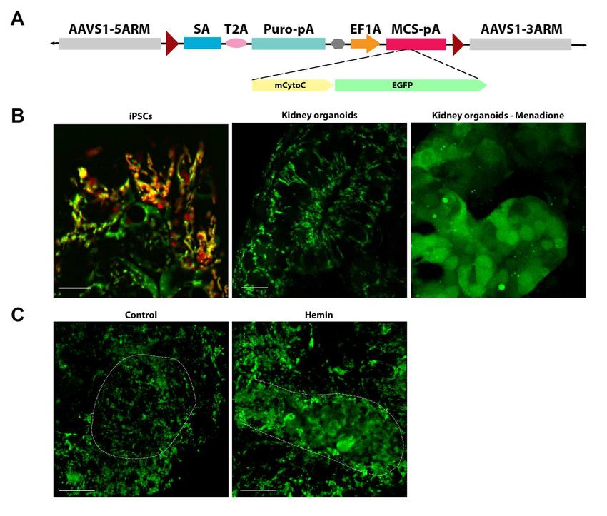
Cytochrome-C-GFP biosensor to model injury in real-time
We generated iPSC reporter lines that express CytochromeC-GFP (CytoC-GFP), a
biosensor that is released from mitochondria upon cellular injury to assess apoptosis via
non-invasive, real-time monitoring [42]. In healthy cells, CytoC-GFP is associated with
mitochondria and shows a punctate or network-like fluorescence pattern. Upon cellular
injury or increase in ROS, the fluorescence pattern rapidly shifts to being cytoplasmic.
We confirmed that the CytoC-GFP signal in iPSCs was colocalized to the mitochondria
by live co-immunofluorescence with MitoTracker Red CMXRos, a mitochondrial
membrane potential dependent dye (Figure 4B, Supplemental Figure 5). Next, we
generated kidney organoids from the CytoC-GFP iPSCs and treated them at day 14 with
menadione, known to depolarize the mitochondrial membrane and induce apoptosis [59].
Menadione treated organoids exhibited diffuse cytoplasmic staining of CytoC-GFP in
comparison to the untreated control (Figure 4B, Supplemental Figure 4). To examine the
effect of hemin treatment, CytoC-GFP kidney organoids were monitored over a 48-hour
treatment window post-hemin treatment. While control organoids showed CytoC-GFP
signal in discrete elongated and interconnected tubular structures, consistent with
mitochondria, hemin treatment induced foci of cells within the tubules and podocyte
clusters with diffuse CytoC-GFP signal (Figure 4C). These results suggest hemin-
induced ROS is driving apoptotic events, which are leading to mitochondrial dysfunction.
17Hemin-injured organoids show reduced fibrosis upon treatment with UPHD25.
ROS induces inflammation in the kidney and leads to an upregulation of pro-inflammatory
and pro-fibrotic cytokines, such as MCP1 and TGF- β1, resulting in interstitial fibrosis [49].
We therefore tested if hemin treatment increased deposition of collagen in kidney
organoids. Following a 48-hour hemin treatment, hemin was washed out and kidney
organoids were cultured to day 26 based on previously published data [25]. To determine
the extent of the fibrotic response, we stained kidney organoid sections with collagen
hybridizing peptide (CHP) which stains collagen undergoing remodeling or degradation
seen in fibrotic diseases [60]. We found that there was a significant increase in CHP
staining in hemin-treated kidney organoids compared to controls (Figure 5A, B). To
determine if the hemin injury model is responsive to therapeutic intervention, we
examined whether the fibrotic response could be pharmacologically inhibited. In prior
work, we have shown that 4-(phenylthio)butanoic acid (PTBA) displays promising anti-
fibrotic activity when delivered as a prodrug (UPHD25 or UPHD186) [28, 30]. We
therefore tested the effect of the methyl ester PTBA prodrug UPHD25 in hemin-injured
kidney organoids. First, we used the CytoC-GFP organoids to assess the toxicity of
UPHD25. No change in CytoC-GFP signal was seen over a dose range of 1-27 µM (data
not shown) and saw no increase in the appearance of cellular foci with diffuse CytoC-
GFP signal. These results demonstrate that UPHD25 is relatively non-toxic over a broad
concentration range. We therefore tested non-toxic doses of 1, 3 and 9 µM of UPHD25
in the hemin injury assay by delivering daily treatments from days 16-26 following hemin
treatment from days 14-16. We tested two independent cell lines (MANZ2-2 and MANZ4-
37) and found that UPHD25 significantly decreased CHP staining at a concentration of 1
18µM in MANZ2-2 cell line, and 3 and 9 µM in MANZ4-37 (Figure 5B). Treatment with 3 µM
UPHD25, 48 hours post injury (day 18) also reduced expression of pro-inflammatory
markers (IL6, CXCL8 and MCP1) (Figure 5C). These results suggest both the fibrotic
response and corresponding inflammatory cytokine expression can be reduce with
therapeutic intervention.
Discussion
Kidney organoids can be generated in large quantities, genetically manipulated, and have
been shown to be a biologically relevant model for human disease [24]. Producing kidney
organoids in bulk makes them well suited for testing therapeutic interventions [25]. While
shortcomings with kidney organoid maturity are well-documented, [22] they are a good
system to develop models for chronic organ diseases and fibrosis. Liver and lung
organoids, which also display fetal like states comparable to early trimester development,
have been used to study pulmonary and liver fibrosis [61].
Hemin is a potent inducer of ROS that mimics the hemolytic response seen in many
diseases leading to renal injury [3]. Time course analysis of hemin-treated organoids
revealed that the response to hemin injury was immediate and acute, with the
inflammatory response and HMOX1 induction being rapidly downregulated within a
couple of days of hemin removal. However, sustained HAVCR1 expression suggests that
the proximal tubule cells undergo enduring injury in response to the brief hemin exposure.
This is in accordance with other hemolytic models in mice which show a rapid decline of
inflammatory markers but persistent expression of HAVCR1 [62]. Unilateral urothelial
19obstruction (UUO) and IRI injury models in mice also show that continued HAVCR1
expression is associated with higher levels of fibrosis and a progression to CKD [63, 64].
Elevated perdurance of HAVCR1 also translates to the clinic as not only do patients
suffering from AKI show elevated levels of urinary HAVCR1, but patients suffering from
CKD show even higher levels of HAVCR1 [65].
High levels of ROS and nitrogen species are found during hemolytic events forming highly
reactive peroxynitrite, and lead to nitrosylation of proteins [14]. We found that 3-
nitrotyrosine (byproduct of nitration) was elevated in hemin treated organoids. In patients
with septic shock and CKD, high levels of nitrotyrosine as well as nitrite/nitrate (indicating
NO production) levels are found in plasma. The levels were significantly higher compared
to patients with CKD without septic shock, supporting the notion that high peroxynitrite
levels and nitric oxide play a critical role in hemolysis events and the subsequent injury
[66]. Rats loaded with iron also exhibited nitrotyrosine labeled tubules and interstitial cells.
In addition, they had elevated levels of NO synthase enzymes (responsible for NO
production), indicating increased levels of reactive nitrogen species due to high levels of
iron [67]. Another major cellular response to high oxidative stress is an increase in
ubiquitination and proteasomal degradation of proteins [68]. Our RNA-seq analysis
revealed the top DE genes were part of the ubiquitin-proteasome degradation network
and one of the overrepresented pathways was the unfolded protein response, suggesting
the hemin treated organoids were trying to eliminate unfolded or damaged proteins in
order to ameliorate cellular injury [69].
20Inhibition of the proteasome system leads to an increase in ubiquitination of mitochondrial
proteins and recruitment of heat shock, and autophagy related components [70].
Therefore, mitochondria need to be continuously monitored during ROS events for
potential dysfunction. We developed the CytoC-GFP iPSC line as a real-time biosensor,
which can be utilized to monitor mitochondrial health, morphology, and cellular response
to toxins [15]. Recent studies show that CytoC may have other functions beyond
apoptosis induction, which could further aid in understanding injury mechanisms. CytoC
has been shown to translocate to the nucleus before activating the apoptotic cascade,
where the protein plays a role in the DNA damage repair response [71, 72]. This suggests
that not all cells that release CytoC from the mitochondria undergo apoptosis. Dissecting
this mechanism further, may shed light on the repair pathways and potential future
treatments. Integration with new imaging technologies and other biosensors may further
our understanding of cellular injury particularly in PT cells which are rich in mitochondria
[73].
Finally, we show that our model of hemin mediated injury can be adapted to therapeutic
compound testing by showing a reduction in the fibrotic response. Previous studies have
shown that PTBA, delivered as a prodrug, can ameliorate injury and reduce fibrosis in
multiple models of renal injury [28-30]. As with most potential renal therapeutics, PTBA
has never been validated against a human proteome, especially in an injury setting.
Conclusions
21This work demonstrates we can reproducibly model aspects of renal injury such as
fibrosis and pro-inflammatory cytokine expression, and therapeutically intervene to
reduce the injury and possibly stimulate repair. For the scientific community to take full
advantage of kidney organoid systems, we need to further our understanding of the injury
processes that take place in organoids and how they compare to in vivo kidney injury.
This includes understanding the repair process that occurs after the initial insult and the
longer-term outcomes. However, the hemin injury model, displays many of the same
hallmarks as in vivo hemolytic injury, and will allow validation of the efficacy of targeted
renal therapeutics in a human model of renal injury.
List of abbreviations
6CF 6-carboxyfluorescein
ACO1 Aconitase 1
ALAD Aminolevulinate Dehydratase
AKI Acute kidney injury
BLVR Biliverdin Reductase
CHP Collagen hybridizing peptide
CKD Chronic kidney disease
COL_A_ Collagen Type _ Alpha _
CXCL8 C-X-C Motif Chemokine Ligand 8
CytoC-GFP Cytochrome C-GFP
DE Differential expression
EB Ethidium bromide
22FBN1 Fibrillin 1
FA Formic acid
HAVCR1 Hepatitis A Virus Cellular Receptor 1
HDAC Histone Deacetylase
HEBP Heme Binding Protein
HMBS Hydroxymethylbilane Synthase
HMOX1 Heme oxygenase 1
HRG Histidine Rich Glycoprotein
IL6 Interleukin 6
iPSCs Induced pluripotent stem cells
IRI Ischemia reperfusion injury
LRP LDL Receptor Related Protein
LTL Lotus tetragonolobus lectin
MCP1 Monocyte Chemoattractant Protein-1
MRP2 Multidrug resistant protein 2
NO Nitric oxide
NQO1 NAD(P)H Quinone Dehydrogenase 1
OAT Organic anion transporter
OCT Organic cation transporter
PTBA 4-(phenylthio) butanoic
ROS Reactive oxygen species
SLC_ Solute Carrier Family _ Member
SOD Superoxide Dismutase 1
23TFRC Transferrin Receptor
TGF- β1 Transforming Growth Factor Beta 1
UCHL5 Ubiquitin C-Terminal Hydrolase L5
UBQLN1 Ubiquilin 1
USP14 Ubiquitin Specific Peptidase 14
ULA Ultra-low attachment
UROD Uroporphyrinogen Decarboxylase
UUO Unilateral urothelial obstruction
Ethics approval
All work was performed in compliance with institutional guidelines (IBC201600244).
Consent for publication
Not applicable
Availability of data and materials
All relevant data have been uploaded as part of supplementary files and available on
National Center for Biotechnology Information’s GEO database.
Competing Interests
The authors have declared that no conflict of interest exists.
Funding
24This research was funded by the National Institutes of Health [2R01DK069403,
2UC2DK126122, to N.A.H., 1P30DK079307 to T,R.K., and T32DK061296 to E.B.E.]; the
U.S. Department of Defense [W81XWH-17-1-0610 to N.A.H.]; the American Society of
Nephrology Ben J. Lipps Award to A.P.
Author Contributions
A.P., T.V., E.B.E., A.E.C., D.R.E., L.A.V., M.L.M., J.A.K., T.R.K., C.J.B., A.J.D., N.A.H. -
designed research studies; A.P., E.B.E., A.E.C., E.P., M.D.M., R.S., C.L.H., C.J.B. -
conducted experiments; A.P., E.B.E., A.E.C., R.S., C.L.H., C.J.B. - acquired data; A.P.,
T.V., E.B.E., A.E.C., M.L.M., T.R.K., C.J.B., A.J.D., N.A.H. - analyzed data; and A.P.,
A.E.C., C.J.B., N.A.H. - wrote the manuscript.
Acknowledgments
We would like to thank Michele Mulkeen for histology sectioning.
25References
1. Haase M, Bellomo R, Haase-Fielitz A: Novel biomarkers, oxidative stress, and
the role of labile iron toxicity in cardiopulmonary bypass-associated acute
kidney injury. J Am Coll Cardiol 2010, 55(19):2024-2033.
2. Reiter CD, Wang X, Tanus-Santos JE, Hogg N, Cannon RO, 3rd, Schechter AN,
Gladwin MT: Cell-free hemoglobin limits nitric oxide bioavailability in sickle-
cell disease. Nat Med 2002, 8(12):1383-1389.
3. Rother RP, Bell L, Hillmen P, Gladwin MT: The clinical sequelae of
intravascular hemolysis and extracellular plasma hemoglobin: a novel
mechanism of human disease. JAMA 2005, 293(13):1653-1662.
4. Wang L, Vijayan V, Jang MS, Thorenz A, Greite R, Rong S, Chen R,
Shushakova N, Tudorache I, Derlin K et al: Labile Heme Aggravates Renal
Inflammation and Complement Activation After Ischemia Reperfusion
Injury. Front Immunol 2019, 10:2975.
5. Vallelian F, Schaer CA, Deuel JW, Ingoglia G, Humar R, Buehler PW, Schaer
DJ: Revisiting the putative role of heme as a trigger of inflammation.
Pharmacol Res Perspect 2018, 6(2):e00392.
6. Malle E, Woenckhaus C, Waeg G, Esterbauer H, Grone EF, Grone HJ:
Immunological evidence for hypochlorite-modified proteins in human
kidney. Am J Pathol 1997, 150(2):603-615.
7. Antolini F, Valente F, Ricciardi D, Baroni M, Fagugli RM: Principal component
analysis of some oxidative stress parameters and their relationships in
hemodialytic and transplanted patients. Clin Chim Acta 2005, 358(1-2):87-94.
8. Jeney V, Balla J, Yachie A, Varga Z, Vercellotti GM, Eaton JW, Balla G: Pro-
oxidant and cytotoxic effects of circulating heme. Blood 2002, 100(3):879-
887.
9. Raatikainen-Ahokas A, Hytönen M, Tenhunen A, Sainio K, Sariola H: BMP-4
affects the differentiation of metanephric mesenchyme and reveals an early
anterior-posterior axis of the embryonic kidney. Developmental dynamics :
an official publication of the American Association of Anatomists 2000,
217(2):146-158.
10. Gutteridge JMC: Iron promoters of the Fenton reaction and lipid
peroxidation can be released from haemoglobin by peroxides. FEBS Letters
1986, 201(2):291-295.
11. Ushio-Fukai M, Ash D, Nagarkoti S, Belin de Chantemele EJ, Fulton DJR, Fukai
T: Interplay Between Reactive Oxygen/Reactive Nitrogen Species and
Metabolism in Vascular Biology and Disease. Antioxid Redox Signal 2021,
34(16):1319-1354.
12. Nath KA, Grande JP, Croatt AJ, Likely S, Hebbel RP, Enright H: Intracellular
targets in heme protein-induced renal injury. Kidney Int 1998, 53(1):100-111.
13. Balabanli B, Kamisaki Y, Martin E, Murad F: Requirements for heme and thiols
for the nonenzymatic modification of nitrotyrosine. Proc Natl Acad Sci U S A
1999, 96(23):13136-13141.
14. MacMillan-Crow LA, Crow JP, Kerby JD, Beckman JS, Thompson JA: Nitration
and inactivation of manganese superoxide dismutase in chronic rejection
26of human renal allografts. Proc Natl Acad Sci U S A 1996, 93(21):11853-
11858.
15. Goldstein JC, Waterhouse NJ, Juin P, Evan GI, Green DR: The coordinate
release of cytochrome c during apoptosis is rapid, complete and kinetically
invariant. Nat Cell Biol 2000, 2(3):156-162.
16. Min L, Jian-xing X: Detoxifying function of cytochrome c against oxygen
toxicity. Mitochondrion 2007, 7(1-2):13-16.
17. Cai J, Yang J, Jones DP: Mitochondrial control of apoptosis: the role of
cytochrome c. Biochim Biophys Acta 1998, 1366(1-2):139-149.
18. Digby JLM, Vanichapol T, Przepiorski A, Davidson AJ, Sander V: Evaluation of
cisplatin-induced injury in human kidney organoids. Am J Physiol Renal
Physiol 2020, 318(4):F971-F978.
19. Higgins JW, Chambon A, Bishard K, Hartung A, Arndt D, Brugnano J, Er PX,
Lawlor KT, Vanslambrouck JM, Wilson S et al: Bioprinted pluripotent stem
cell-derived kidney organoids provide opportunities for high content
screening. bioRxiv 2018:505396.
20. Uchimura K, Wu H, Yoshimura Y, Humphreys BD: Human Pluripotent Stem
Cell-Derived Kidney Organoids with Improved Collecting Duct Maturation
and Injury Modeling. Cell Rep 2020, 33(11):108514.
21. Czerniecki SM, Cruz NM, Harder JL, Menon R, Annis J, Otto EA, Gulieva RE,
Islas LV, Kim YK, Tran LM et al: High-Throughput Screening Enhances
Kidney Organoid Differentiation from Human Pluripotent Stem Cells and
Enables Automated Multidimensional Phenotyping. Cell Stem Cell 2018,
22(6):929-940 e924.
22. Takasato M, Er PX, Chiu HS, Maier B, Baillie GJ, Ferguson C, Parton RG,
Wolvetang EJ, Roost MS, Lopes SM et al: Kidney organoids from human iPS
cells contain multiple lineages and model human nephrogenesis. Nature
2016, 536(7615):238.
23. Morizane R, Lam AQ, Freedman BS, Kishi S, Valerius MT, Bonventre JV:
Nephron organoids derived from human pluripotent stem cells model
kidney development and injury. Nat Biotechnol 2015, 33(11):1193-1200.
24. Przepiorski A, Crunk AE, Espiritu EB, Hukriede NA, Davidson AJ: The Utility of
Human Kidney Organoids in Modeling Kidney Disease. Semin Nephrol 2020,
40(2):188-198.
25. Przepiorski A, Sander V, Tran T, Hollywood JA, Sorrenson B, Shih JH,
Wolvetang EJ, McMahon AP, Holm TM, Davidson AJ: A Simple Bioreactor-
Based Method to Generate Kidney Organoids from Pluripotent Stem Cells.
Stem Cell Reports 2018, 11(2):470-484.
26. Przepiorski A, Crunk AE, Holm TM, Sander V, Davidson AJ, Hukriede NA: A
Simplified Method for Generating Kidney Organoids from Human
Pluripotent Stem Cells. J Vis Exp 2021(170).
27. Brilli Skvarca L, Han HI, Espiritu EB, Missinato MA, Rochon ER, McDaniels MD,
Bais AS, Roman BL, Waxman JS, Watkins SC et al: Enhancing regeneration
after acute kidney injury by promoting cellular dedifferentiation in
zebrafish. Dis Model Mech 2019, 12(4).
2728. Cianciolo Cosentino C, Skrypnyk NI, Brilli LL, Chiba T, Novitskaya T, Woods C,
West J, Korotchenko VN, McDermott L, Day BW et al: Histone deacetylase
inhibitor enhances recovery after AKI. J Am Soc Nephrol 2013, 24(6):943-
953.
29. Novitskaya T, McDermott L, Zhang KX, Chiba T, Paueksakon P, Hukriede NA,
de Caestecker MP: A PTBA small molecule enhances recovery and reduces
postinjury fibrosis after aristolochic acid-induced kidney injury. Am J
Physiol Renal Physiol 2014, 306(5):F496-504.
30. Skrypnyk NI, Sanker S, Skvarca LB, Novitskaya T, Woods C, Chiba T, Patel K,
Goldberg ND, McDermott L, Vinson PN et al: Delayed treatment with PTBA
analogs reduces postinjury renal fibrosis after kidney injury. Am J Physiol
Renal Physiol 2016, 310(8):F705-F716.
31. Wen X, Li S, Frank A, Chen X, Emlet D, Hukriede NA, Kellum JA: Time-
dependent effects of histone deacetylase inhibition in sepsis-associated
acute kidney injury. Intensive Care Med Exp 2020, 8(1):9.
32. Oh JK, Przepiorski A, Chang H-H, Dodd RC, Sander V, Sorrenson B, Shih J-H,
Hollywood JA, de Zoysa JR, Shepherd PR et al: Derivation of induced
pluripotent stem cell lines from New Zealand donors. Journal of the Royal
Society of New Zealand 2020:1-14.
33. Livak KJ, Schmittgen TD: Analysis of relative gene expression data using
real-time quantitative PCR and the 2(-Delta Delta C(T)) Method. Methods
2001, 25(4):402-408.
34. Dobin A, Davis CA, Schlesinger F, Drenkow J, Zaleski C, Jha S, Batut P,
Chaisson M, Gingeras TR: STAR: ultrafast universal RNA-seq aligner.
Bioinformatics 2013, 29(1):15-21.
35. Liao Y, Smyth GK, Shi W: featureCounts: an efficient general purpose
program for assigning sequence reads to genomic features. Bioinformatics
2014, 30(7):923-930.
36. Robinson MD, McCarthy DJ, Smyth GK: edgeR: a Bioconductor package for
differential expression analysis of digital gene expression data.
Bioinformatics 2010, 26(1):139-140.
37. Ritchie ME, Phipson B, Wu D, Hu Y, Law CW, Shi W, Smyth GK: limma powers
differential expression analyses for RNA-sequencing and microarray
studies. Nucleic Acids Res 2015, 43(7):e47.
38. Ashburner M, Ball CA, Blake JA, Botstein D, Butler H, Cherry JM, Davis AP,
Dolinski K, Dwight SS, Eppig JT et al: Gene ontology: tool for the unification
of biology. The Gene Ontology Consortium. Nat Genet 2000, 25(1):25-29.
39. Liberzon A, Birger C, Thorvaldsdottir H, Ghandi M, Mesirov JP, Tamayo P: The
Molecular Signatures Database (MSigDB) hallmark gene set collection. Cell
Syst 2015, 1(6):417-425.
40. Gene Ontology C: The Gene Ontology resource: enriching a GOld mine.
Nucleic Acids Res 2021, 49(D1):D325-D334.
41. Yu G, Wang LG, Han Y, He QY: clusterProfiler: an R package for comparing
biological themes among gene clusters. OMICS 2012, 16(5):284-287.
2842. Goldstein JC, Waterhouse NJ, Juin P, Evan GI, Green DR: The coordinate
release of cytochrome c during apoptosis is rapid, complete and kinetically
invariant. Nature cell biology 2000, 2(3):156-162.
43. Zecha J, Satpathy S, Kanashova T, Avanessian SC, Kane MH, Clauser KR,
Mertins P, Carr SA, Kuster B: TMT Labeling for the Masses: A Robust and
Cost-efficient, In-solution Labeling Approach. Mol Cell Proteomics 2019,
18(7):1468-1478.
44. Jiang B, Liu G, Zheng J, Chen M, Maimaitiming Z, Chen M, Liu S, Jiang R,
Fuqua BK, Dunaief JL et al: Hephaestin and ceruloplasmin facilitate iron
metabolism in the mouse kidney. Sci Rep 2016, 6:39470.
45. Bednarz A, Lipinski P, Starzynski RR, Tomczyk M, Nowak W, Mucha O, Ogorek
M, Pierzchala O, Jonczy A, Staron R et al: Role of the kidneys in the
redistribution of heme-derived iron during neonatal hemolysis in mice. Sci
Rep 2019, 9(1):11102.
46. van Raaij S, van Swelm R, Bouman K, Cliteur M, van den Heuvel MC, Pertijs J,
Patel D, Bass P, van Goor H, Unwin R et al: Tubular iron deposition and iron
handling proteins in human healthy kidney and chronic kidney disease. Sci
Rep 2018, 8(1):9353.
47. Gonzalez-Michaca L, Farrugia G, Croatt AJ, Alam J, Nath KA: Heme: a
determinant of life and death in renal tubular epithelial cells. Am J Physiol
Renal Physiol 2004, 286(2):F370-377.
48. Goldstein L, Teng ZP, Zeserson E, Patel M, Regan RF: Hemin induces an iron-
dependent, oxidative injury to human neuron-like cells. J Neurosci Res
2003, 73(1):113-121.
49. Van Avondt K, Nur E, Zeerleder S: Mechanisms of haemolysis-induced
kidney injury. Nat Rev Nephrol 2019, 15(11):671-692.
50. Subramanian A, Tamayo P, Mootha VK, Mukherjee S, Ebert BL, Gillette MA,
Paulovich A, Pomeroy SL, Golub TR, Lander ES et al: Gene set enrichment
analysis: a knowledge-based approach for interpreting genome-wide
expression profiles. Proc Natl Acad Sci U S A 2005, 102(43):15545-15550.
51. Mootha VK, Lindgren CM, Eriksson KF, Subramanian A, Sihag S, Lehar J,
Puigserver P, Carlsson E, Ridderstrale M, Laurila E et al: PGC-1alpha-
responsive genes involved in oxidative phosphorylation are coordinately
downregulated in human diabetes. Nat Genet 2003, 34(3):267-273.
52. Vasconcellos LR, Dutra FF, Siqueira MS, Paula-Neto HA, Dahan J, Kiarely E,
Carneiro LA, Bozza MT, Travassos LH: Protein aggregation as a cellular
response to oxidative stress induced by heme and iron. Proc Natl Acad Sci
U S A 2016, 113(47):E7474-E7482.
53. Whiteley AM, Prado MA, Peng I, Abbas AR, Haley B, Paulo JA, Reichelt M,
Katakam A, Sagolla M, Modrusan Z et al: Ubiquilin1 promotes antigen-
receptor mediated proliferation by eliminating mislocalized mitochondrial
proteins. Elife 2017, 6.
54. Canaud G, Brooks CR, Kishi S, Taguchi K, Nishimura K, Magassa S, Scott A,
Hsiao LL, Ichimura T, Terzi F et al: Cyclin G1 and TASCC regulate kidney
epithelial cell G2-M arrest and fibrotic maladaptive repair. Sci Transl Med
2019, 11(476).
2955. Lee WK, Reichold M, Edemir B, Ciarimboli G, Warth R, Koepsell H, Thevenod F:
Organic cation transporters OCT1, 2, and 3 mediate high-affinity transport
of the mutagenic vital dye ethidium in the kidney proximal tubule. Am J
Physiol Renal Physiol 2009, 296(6):F1504-1513.
56. Lawrence ML, Chang CH, Davies JA: Transport of organic anions and cations
in murine embryonic kidney development and in serially-reaggregated
engineered kidneys. Sci Rep 2015, 5:9092.
57. Ribble D, Goldstein NB, Norris DA, Shellman YG: A simple technique for
quantifying apoptosis in 96-well plates. BMC Biotechnol 2005, 5:12.
58. Faucher Q, Alarcan H, Marquet P, Barin-Le Guellec C: Effects of Ischemia-
Reperfusion on Tubular Cell Membrane Transporters and Consequences in
Kidney Transplantation. J Clin Med 2020, 9(8).
59. Gerasimenko JV, Gerasimenko OV, Palejwala A, Tepikin AV, Petersen OH,
Watson AJ: Menadione-induced apoptosis: roles of cytosolic Ca(2+)
elevations and the mitochondrial permeability transition pore. J Cell Sci
2002, 115(Pt 3):485-497.
60. Li Y, Ho D, Meng H, Chan TR, An B, Yu H, Brodsky B, Jun AS, Michael Yu S:
Direct detection of collagenous proteins by fluorescently labeled collagen
mimetic peptides. Bioconjug Chem 2013, 24(1):9-16.
61. Lee J, Kim JH, Hong SH, Yang SR: Organoid Model in Idiopathic Pulmonary
Fibrosis. Int J Stem Cells 2021, 14(1):1-8.
62. Merle NS, Grunenwald A, Figueres ML, Chauvet S, Daugan M, Knockaert S,
Robe-Rybkine T, Noe R, May O, Frimat M et al: Characterization of Renal
Injury and Inflammation in an Experimental Model of Intravascular
Hemolysis. Front Immunol 2018, 9:179.
63. Humphreys BD, Xu F, Sabbisetti V, Grgic I, Movahedi Naini S, Wang N, Chen G,
Xiao S, Patel D, Henderson JM et al: Chronic epithelial kidney injury
molecule-1 expression causes murine kidney fibrosis. J Clin Invest 2013,
123(9):4023-4035.
64. Ko GJ, Grigoryev DN, Linfert D, Jang HR, Watkins T, Cheadle C, Racusen L,
Rabb H: Transcriptional analysis of kidneys during repair from AKI reveals
possible roles for NGAL and KIM-1 as biomarkers of AKI-to-CKD transition.
Am J Physiol Renal Physiol 2010, 298(6):F1472-1483.
65. Tian L, Shao X, Xie Y, Wang Q, Che X, Zhang M, Xu W, Xu Y, Mou S, Ni Z:
Kidney Injury Molecule-1 is Elevated in Nephropathy and Mediates
Macrophage Activation via the Mapk Signalling Pathway. Cell Physiol
Biochem 2017, 41(2):769-783.
66. Fukuyama N, Takebayashi Y, Hida M, Ishida H, Ichimori K, Nakazawa H:
Clinical evidence of peroxynitrite formation in chronic renal failure patients
with septic shock. Free Radic Biol Med 1997, 22(5):771-774.
67. Zhou XJ, Laszik Z, Wang XQ, Silva FG, Vaziri ND: Association of renal injury
with increased oxygen free radical activity and altered nitric oxide
metabolism in chronic experimental hemosiderosis. Lab Invest 2000,
80(12):1905-1914.
68. Shang F, Taylor A: Ubiquitin-proteasome pathway and cellular responses to
oxidative stress. Free Radic Biol Med 2011, 51(1):5-16.
3069. Vallelian F, Deuel JW, Opitz L, Schaer CA, Puglia M, Lonn M, Engelsberger W,
Schauer S, Karnaukhova E, Spahn DR et al: Proteasome inhibition and
oxidative reactions disrupt cellular homeostasis during heme stress. Cell
Death Differ 2015, 22(4):597-611.
70. Sulkshane P, Duek I, Ram J, Thakur A, Reis N, Ziv T, Glickman MH: Inhibition
of proteasome reveals basal mitochondrial ubiquitination. J Proteomics
2020, 229:103949.
71. Diaz-Moreno I, Velazquez-Cruz A, Curran-French S, Diaz-Quintana A, De la
Rosa MA: Nuclear cytochrome c - a mitochondrial visitor regulating
damaged chromatin dynamics. FEBS Lett 2018, 592(2):172-178.
72. Rivero-Rodriguez F, Diaz-Quintana A, Velazquez-Cruz A, Gonzalez-Arzola K,
Gavilan MP, Velazquez-Campoy A, Rios RM, De la Rosa MA, Diaz-Moreno I:
Inhibition of the PP2A activity by the histone chaperone ANP32B is long-
range allosterically regulated by respiratory cytochrome c. Redox Biol 2021,
43:101967.
73. Okkelman IA, Papkovsky DB, Dmitriev RI: Estimation of the Mitochondrial
Membrane Potential Using Fluorescence Lifetime Imaging Microscopy.
Cytometry A 2020, 97(5):471-482.
3132
Figure 1 Hemin injury leads to induction of inflammation and nitric oxide mediated nitrosylation.

A) Representative quantitative PCR (qPCR) of kidney organoids treated with hemin at increasing

concentrations. Expression relative to the control (untreated) organoids. B) Time course qPCR of control

and hemin treated organoids from day 15 to day 20. Results show mean and standard deviation of 3

technical replicates. C) Immunofluorescence of kidney organoid paraffin sections of control and hemin

treated organoids, showing localization of kidney injury marker 1 (KIM1, red) to proximal tubules labelled

with Lotus tetragonolobus lectin (LTL, green), apoptotic marker activated caspase 3 (Casp3, red), DNA

damage marker ( gH2AX, green), and heme catabolizing enzyme heme oxygenase 1 (HMOX1, orange).

D) Immunofluorescence of kidney organoid paraffin sections at day 16, 18 and 20 showing 3-nitrotyrosine

staining (red) indicative of nitrositive stress. Scale bar = 200 µm.

                                                                                                       33
34
Figure 2 Transcriptome analysis of hemin injured kidney organoids.

Heatmaps showing z-score of differentially expressed genes between untreated control day 15 and 16 (_C)

and hemin treated kidney organoids (_H). A) GO Proteasomal ubiquitin dependent protein catabolic

process. B) Hallmark pathway analysis reactive oxygen species pathway. C) Dot plot of overrepresented

Hallmark pathways between days 16 and 15. D) Heatmap of Hallmark epithelial-to-mesenchymal transition

pathway showing differences between hemin-treated organoids day 15 and 16.

                                                                                                    35
36
Figure 3 Assessment of proximal tubule transport function in kidney organoids

A) Representative image of OAT (6CF) and OCT (EB) in the 12.5, 25 and 50 µM hemin treated kidney

organoids. Note, transport declines with increasing concentrations of hemin. Each row shows the same

corresponding organoid with 6CF, EB and overlay with transmitted light image. White arrow in 6CF control

points to luminal accumulation in tubule structure. Maximum projections shown. Scale bar 100 µm. B) Heat

map showing expression in day 16 (D16) hemin (_H) and control (_C) samples of the OAT (SLC22A6,

SLC22A8), OCT transporters (SLC22A2) and multi-drug resistant protein transporters (SLC47A1,

SLC47A2, ABCC2).

                                                                                                     37
Figure 4 CytoC-GFP biosensor localizes to the mitochondria
A) Schematic representation of the safe harbor AAVS1 donor vector with CytoC-GFP construct. B) Live
immunofluorescence showing CytoC-GFP expression is colocalized to MitoTracker Red CMXRos labelled
mitochondria in the iPSCs. Mitochondrial expression in the tubule of the kidney organoids, and diffuse
cytoplasmic staining in the menadione treated tubule. Scale bar = 10 µm C) Representative live images of
3D projections showing mitochondrial localization in the control and 25 µM hemin treated tubules of kidney
organoids. Note the diffuse CytoC-GFP localization in the hemin treated tubule (white arrow). Data from
isolated clone #8 CytoC-GFP iPSC line. Scale bars = 20 µm.
38You can also read