Lutein as a Modulator of Oxidative Stress-Mediated Inflammatory Diseases - MDPI
←
→
Page content transcription
If your browser does not render page correctly, please read the page content below

antioxidants

Review
Lutein as a Modulator of Oxidative Stress-Mediated
Inflammatory Diseases
Yu Jin Ahn and Hyeyoung Kim *

                                          Department of Food and Nutrition, BK21 FOUR, College of Human Ecology, Yonsei University,
                                          Seoul 03722, Korea; anyujin@gmail.com
                                          * Correspondence: kim626@yonsei.ac.kr; Tel.: +82-2-2123-3125; Fax: +82-2-364-5781

                                          Abstract: Lutein is a xanthophyll carotenoid obtained from various foods, such as dark green leafy
                                          vegetables and egg yolk. Lutein has antioxidant activity and scavenges reactive oxygen species
                                          such as singlet oxygen and lipid peroxy radicals. Oxidative stress activates inflammatory mediators,
                                          leading to the development of metabolic and inflammatory diseases. Thus, recent basic and clinical
                                          studies have investigated the anti-inflammatory effects of lutein based on its antioxidant activity
                                          and modulation of oxidant-sensitive inflammatory signaling pathways. Lutein suppresses activa-
                                          tion of nuclear factor-kB and signal transducer and activator of transcription 3, and induction of
                                          inflammatory cytokines (interleukin-1β, interleukin-6, monocyte chemoattratant protein-1, tumor
                                          necrosis factor-α) and inflammatory enzymes (cyclooxygenase-2, inducible nitric oxide synthase). It
                                          also maintains the content of endogenous antioxidant (glutathione) and activates nuclear factor ery-
                                          throid 2–related factor 2 (Nrf2) and Nrf2 signaling-related antioxidant enzymes (hemeoxygenase-1,
                                NAD(P)H: quinone oxidoreductase 1, glutathione-s-transferase, glutathione peroxidase, superox-

                                          ide dismutase, catalase). In this review, we have discussed the current knowledge regarding the
Citation: Ahn, Y.J.; Kim, H. Lutein as    anti-inflammatory function of lutein against inflammatory diseases in various organs, including neu-
a Modulator of Oxidative
                                          rodegenerative disorders, eye diseases, diabetic retinopathy, osteoporosis, cardiovascular diseases,
Stress-Mediated Inflammatory
                                          skin diseases, liver injury, obesity, and colon diseases.
Diseases. Antioxidants 2021, 10, 1448.
https://doi.org/10.3390/
                                          Keywords: inflammation; lutein; reactive oxygen species
antiox10091448

Academic Editors: Edward
E. Schmidt, Hozumi Motohashi and
Anna Kipp                                 1. Introduction
                                                Carotenoids are divided into two classes based on their chemical structure: the
Received: 7 August 2021                   carotenes (hydrocarbons, such as β-carotene and lycopene) and xanthophylls (polar com-
Accepted: 8 September 2021                pounds that contain oxygen atoms in their molecules, such as lutein and its stereoisomer
Published: 13 September 2021              zeaxanthin) [1]. Lutein is the second most prevalent carotenoid in human serum and is
                                          synthesized only by plants. It is abundantly present in eggs and dark green leafy vegetables
Publisher’s Note: MDPI stays neutral      such as kale and spinach [2–4].
with regard to jurisdictional claims in         Lutein acts as an antioxidant and protects plants from photo-induced free radical dam-
published maps and institutional affil-   age [5]. Xanthophyll carotenoids modulate oxidative stress and regulate redox-sensitive
iations.
                                          intracellular signaling [6]. Ozawa et al. [7] suggested that lutein inhibited oxidative stress-
                                          induced triggering of inflammatory signaling pathways such as the activated signal trans-
                                          ducer and activator of transcription 3 (STAT3) signaling pathway and IL-6 expression in
                                          the retina. Lutein preserves visual function by preventing degradation of the functional
Copyright: © 2021 by the authors.         proteins, rhodopsin (a visual pigment) and synaptophysin (a synaptic vesicle protein that
Licensee MDPI, Basel, Switzerland.        is altered in neurodegenerative diseases). Lutein treatment reduced the concentrations of
This article is an open access article    nitric oxide (NO), tumor necrosis factor (TNF)-α, interleukin (IL)-6, prostaglandin (PG)E2 ,
distributed under the terms and           and monocyte chemoattractant protein (MCP)-1 in aqueous humor of mice with endotoxin-
conditions of the Creative Commons
                                          induced uveitis [8]. Lutein treatment suppressed the development of choroidal neovascular-
Attribution (CC BY) license (https://
                                          ization, which plays a critical role in the pathogenesis of age-related macular degeneration
creativecommons.org/licenses/by/
                                          and inflammatory processes, including nuclear factor (NF)-κB activation and subsequent
4.0/).

Antioxidants 2021, 10, 1448. https://doi.org/10.3390/antiox10091448                                  https://www.mdpi.com/journal/antioxidants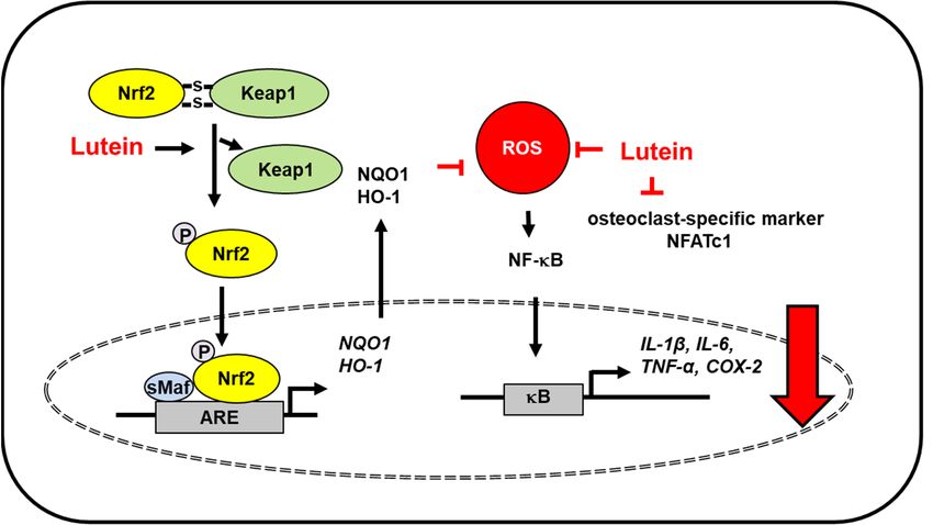
Antioxidants 2021, 10, 1448                                                                                              2 of 23

                              upregulation of inflammatory molecules such as MCP-1 in mice [9]. Horvath et al. [10]
                              found that lutein inhibited the activation of transient receptor potential ankyrin 1 and the
                              resultant inflammation of the mouse skin. This study showed that lutein decreased TRPA1
                              activation-induced neutrophil accumulation. Although these significant findings provide
                              new insights into the anti-inflammatory actions of lutein, the mechanism underlying these
                              observations need to be further investigated in humans.
                                    Lutein has a long carbon chain with alternating single and double carbon-carbon
                              bonds with attached methyl side groups. Due to the presence of a hydroxyl group at both
                              ends of the molecule, lutein has distinct characteristics compared to other carotenoids [4].
                              Anti-inflammatory and anti-oxidant effects of lutein are attributed to its unique structure,
                              particularly the presence of conjugated double bonds and hydroxyl groups [11]. The
                              conjugated double bond acts as a powerful antioxidant by donating the electrons and
                              reacting with free radicals to form a more stable product. This structural feature may also
                              affect its uptake efficiency via the modulation of carotenoid polarity and flexibility.
                                    Lutein is mainly delivered to the retina; therefore, most studies have focused on its
                              visual activity. Lutein has attracted attention in relation to human health due to its putative
                              role in protection against other inflammatory diseases, in addition to eye diseases. Dietary
                              guidance for lutein shows that it has antioxidant and anti-inflammatory effects [12]. This
                              review covers the current understanding of the protective effects of lutein against oxidative
                              stress-mediated inflammatory diseases.

                              1.1. Absorption and Transport of Lutein
                                    Lutein is accumulated in the eyes, liver, and lipophilic tissues, such as adipose tissue. It
                              is transported from the gut to various organs through the bloodstream via lipoproteins [13].
                              The polar and flexible structure of the lutein molecule increases the affinity for lipid
                              transporters and plasma membranes, leading to its increased absorption in the gut [14–16].
                              Lutein uptake occurs by both simple and facilitated diffusion and is mediated through
                              cholesterol membrane transporters such as scavenger receptor class B member 1 (SR-B1)
                              and a cluster of differentiation 36 (CD 36) [17,18]. When lutein is emulsified into small
                              lipid droplets or vesicles in the stomach, it is converted into mixed micelles by bile salts
                              with biliary phospholipids. Then, these mixed micelles are taken up by enterocytes with
                              SR-B1 [19].

                              1.2. Bioavailability and Metabolism of Lutein
                                    The bioavailability of lutein is affected by food source and matrix, fat content, pro-
                              cessing, cooking, and dietary fiber. Depending on solubilization in the digestive system,
                              the bioavailability of lutein is about 10–15% [20,21], which is very poor. Lutein has poor
                              oral absorption because the high hydrophobicity of the C40 isoprenoid carbon skeleton of
                              lutein makes it soluble in digestive fluids [22]. Lutein and its metabolites are found in the
                              liver, plasma, retina, and adipose tissue. The common metabolites of lutein are 30 -hydroxy-
                              ε,ε-caroten-3-one, 30 -hydroxy-ε,ε-caroten-3-one, and 3-hydroxy-β,ε-caroten-30 -one. In the
                              mice model, the first two were mostly found in the plasma, kidney, adipose tissue, and
                              liver with lutein. However, 3-hydroxy-β,ε-caroten-30 was the major metabolite of lutein in
                              human retina and plasma [22].

                              1.3. Toxicity and Safety of Lutein
                                   There have been no reports of adverse effects on the genotoxicity of lutein formulations.
                              The upper limit of safe lutein consumption has been set to 20 mg/day [23]. The daily
                              intake of lutein is 2 mg/kg body weight, which is equivalent to 120 mg/day for a 60 kg
                              person. Furthermore, long-term supplementation of dietary lutein has not been shown to
                              have any adverse effects in humans. These studies found higher doses of lutein (30 mg
                              and 40 mg/kg body weight) [24,25] to be safe. Similarly, lutein did not show any safety
                              concerns in rats and monkeys [26,27].
Antioxidants 2021, 10, 1448 3 of 23
2. Lutein in Inflammatory Diseases
2.1. Neurodegenerative Disorders
Xanthophylls, such as lutein and zeaxanthin, cross the blood-retina barrier to form the
macular pigment in the eye [28]. The lutein level in the macula was found to be significantly
correlated with its concentration in matched brain tissue. A significant correlation was
observed between macular pigment density and global cognitive function in healthy older
adults [28]. Lutein, zeaxanthin, and meso-zeaxanthin are collectively called the macular
pigment [29]. Adequate maternal intake of lutein couples with the placental transfer of
maternal lutein to support fetal brain and retina development [29]. Therefore, macular
pigment is used as a biomarker of lutein in brain tissues. A recent study suggested that
lutein preferentially accumulated in those regions in the brain that are related to visual
perception, cognition, and motor coordination [30].
Oxidative stress and inflammation of neural tissues induce age-related macular de-
generation and Alzheimer’s disease. The Irish longitudinal study demonstrated the rela-
tionship between lutein, a plasma antioxidant, and improved cognitive function in healthy
older adults [31]. Moreover, lutein depletion was observed in individuals with mild cogni-
tive impairment [32] and Alzheimer’s disease [33,34]. A study using data from the third
Nutrition and Health Examination Survey (NHANES III) database and the NHANES III
Linked Mortality File suggested that high levels of lutein reduce the risk of mortality due
to Alzheimer’s disease in older adults [35]. Severe traumatic brain injury is involved in
oxidative stress-induced inflammation and apoptosis [36]. Lutein protected against severe
traumatic brain injury by suppressing IL-1β, IL-6, MCP-1 expression and reducing serum
reactive oxygen species (ROS) levels in rats with severe traumatic brain injury [37]. Addi-
tionally, lutein attenuated neuroinflammation in lipopolysaccharide-activated microglia
by inhibiting inflammatory signaling such as NF-κB and expression of TNF-α, IL-1β, in-
ducible NO synthase (iNOS), and cyclooxygenase-2 (COX-2). Lutein promoted nuclear
factor erythroid 2–related factor 2 (Nrf2) activation and subsequent upregulation of heme
oxygenase (HO)-1 and NAD(P)H: quinone oxidoreductase 1 (NQO1). The effect of lutein
for Nrf2 activation was mediated with extracellular signal-regulated kinase (ERK) [38].
Antioxidants 2021, 10, x FOR PEER REVIEW 4 of 23
Mitogen activated protein kinases, including ERK phosphorylate Nrf2, in modulating the
Nrf2-dependent antioxidant response [39]. Therefore, lutein may activate ERK, which phos-
phorylates
caudalis and Nrf2
upperto translocate
cervical into neurons.
dorsal horn the nucleus
Theseand induces
regions the expression
relay information to of antioxidant
enzymes. Theseabout
higher pain centers results demonstrated
the location that
and intensity lutein
of pain induced
stimulus. This the
studyexpression
supports of Nrf2-target
lutein as(antioxidant
genes a potential therapeutic
enzymes) agent to reduce
and or prevent
reduced acute trigeminal
the levels inflammatory
of inflammatory mediators to protect
pain. Overall, dietary lutein may be beneficial in maintaining
against inflammation-related neurodegenerative disorders (Figure 1).cognitive health and pro-
tecting against inflammation-induced neurodegenerative diseases.
Figure 1. The proposed mechanism by which lutein inhibits oxidative stress-induced inflammatory
Figure
responses1.inThe proposed
the brain. mechanism
ROS levels increase inby which
severe lutein
traumatic inhibits
brain injury oxidative stress-induced inflammatory
and lipopolysaccharide-
responses in the brain.
activated microglia. ROS levels
Lutein reduces increase
ROS levels in severe
and inhibits traumatic
ROS-mediated brain injury
activation of NF-kB and
andlipopolysaccharide-
expression of inflammatory mediators (IL-1β, IL-6, MCP-1, TNF-α, COX-2, iNOS) [37]. In lipopoly-
activated microglia. Lutein reduces ROS levels and inhibits ROS-mediated activation of NF-kB
saccharide-activated microglia, lutein activates ERK, which phosphorylates Nrf2 and increases dis-
and expression
sociation of inflammatory
of Keap1 from mediators
the Nfr2/Keap1 complex. Thus,(IL-1β, IL-6,
it promotes MCP-1,
nuclear TNF-α,
translocation of COX-2,
Nrf2, iNOS) [37]. In
which forms a heterodimer with sMaf protein and binds to a regulatory region of DNA called
lipopolysaccharide-activated microglia, lutein activates ERK, which phosphorylates Nrf2 and increases ARE.
It induces the expression of Nrf2- target antioxidant genes (HO-1, NQO1). These antioxidant en-
zymes reduce intracellular ROS levels, which suppresses inflammatory responses [38]. Thus, lutein
prevents oxidative stress-mediated neuroinflammation. ARE, antioxidant response element; COX-
2, cyclooxygenase-2; ERK, extracellular signal-regulated kinase; HO-1, hemeoxygenase-1; iNOS, in-
ducible nitric oxide synthase; IL, interleukin; Keap1, kelch like ECH associated protein 1; MCP-1;
monocyte chemoattratant protein-1; NF−κB, nuclear factor-κB; Nrf2, nuclear factor erythroid 2–re-Antioxidants 2021, 10, 1448 4 of 23
dissociation of Keap1 from the Nfr2/Keap1 complex. Thus, it promotes nuclear translocation of
Nrf2, which forms a heterodimer with sMaf protein and binds to a regulatory region of DNA called
ARE. It induces the expression of Nrf2- target antioxidant genes (HO-1, NQO1). These antioxidant
enzymes reduce intracellular ROS levels, which suppresses inflammatory responses [38]. Thus,
lutein prevents oxidative stress-mediated neuroinflammation. ARE, antioxidant response element;
COX-2, cyclooxygenase-2; ERK, extracellular signal-regulated kinase; HO-1, hemeoxygenase-1; iNOS,
inducible nitric oxide synthase; IL, interleukin; Keap1, kelch like ECH associated protein 1; MCP-
1; monocyte chemoattratant protein-1; NF−κB, nuclear factor-κB; Nrf2, nuclear factor erythroid
2–related factor 2; NQO-1, NAD(P)H: quinone oxidoreductase 1; ROS, reactive oxygen species; sMaf,
small Maf; TNF-α, tumor necrosis factor-α.
There are two types of Nrf2 activators. Most Nrf2 inducers interact with cysteine
residues of kelch like ECH-associated protein 1 (Keap1) by utilizing the electrophilic nature
of the molecules and inactivating the Keap1 E3 ligase activity that targets Nrf2 for ubiquitin-
dependent degradation. The other type of Nrf2 inducer is nonelectrophilic inducers, which
interrupt the interaction between Keap1 and Nrf2 [40]. Lutein does not have electrophilic
groups. Thus, lutein metabolites that possess electrophilic groups may react with Keap1. In
another way, lutein may directly disturb the interaction between Keap1 and Nrf2. Further
study should be performed to determine whether lutein metabolites are electrophiles to
react with cysteine residues of Keap1.
Since ROS activate Nrf2 signaling and produce antioxidant enzyems as a defense
mechansim in some cells [41], further detailed study is necessary to determine the mecha-
nism of how lutein induces dissociation of Nrf2/Keap1 and increases nuclear translocation
of Nrf2.
Shimazu et al. [42] suggested that lutein attenuated acute inflammation-induced
nocifensive behavior and augmented nociceptive processing of spinal trigeminal nucleus
caudalis and upper cervical dorsal horn neurons. These regions relay information to higher
pain centers about the location and intensity of pain stimulus. This study supports lutein
as a potential therapeutic agent to reduce or prevent acute trigeminal inflammatory pain.
Overall, dietary lutein may be beneficial in maintaining cognitive health and protecting
against inflammation-induced neurodegenerative diseases.
2.2. Eye Diseases
Lutein, as a component of macular pigment, protects the macula from photo-oxidative
damage and enhances visual function [29]. Lutein is an ocular antioxidant that can quench
both singlet oxygen and lipid peroxy radicals [43]. In addition, lutein inhibits activation
of STAT3 and IL-6 expression in the retina [7]. Therefore, supplementation with lutein
has been very effective for restoring ocular antioxidants of age-related maculopathy and
AMD [44–46]
Oxidative stress is an important factor in the pathogenesis of age-related macular
degeneration; thus, anti-oxidative stress is a good marker for the prevention or treatment of
age-related macular degeneration. Lutein is a very effective quencher of singlet molecular
oxygen and lipid peroxy radicals. However, lutein gets oxidized to its corresponding radical
cations in the process. These cations must be reduced to regenerate the original carotenoids,
which thus, allows its use as an antioxidant [47]. Lutein reduced ROS levels and suppressed
apoptosis by reversing G2/M phase arrest through activation of cyclin-dependent kinase
1 and cell division cycle 25C in retinal pigment epithelial cells exposed to hydrogen
peroxide [48]. Bian et al. [49] showed that lutein suppressed lipopolysaccharide-stimulated
production of IL-6 and TNF-α in both retinal pigmental epithelial cells and macrophages
isolated from the peritoneum of age-related macular degeneration model mice.
Lutein treatment reduced the light-induced increase in local ROS levels and inhibited
tight junction disruption, determined by zona occludens-1 immunostaining, in mice [50].
Lutein intake increased macular pigment optical density and visual contrast sensitivityand macrophages isolated from the peritoneum of age-related macular degeneration
model mice.
Lutein treatment reduced the light-induced increase in local ROS levels and inhibited
Antioxidants 2021, 10, 1448 5 of 23
tight junction disruption, determined by zona occludens-1 immunostaining, in mice [50].
Lutein intake increased macular pigment optical density and visual contrast sensitivity in
90 patients with atrophic age-related macular degeneration [51], suggesting the lutein in-
take-mediated
in 90 patients with improvement in visual function.
atrophic age-related macular degeneration [51], suggesting the lutein
Human clinical
intake-mediated trials reported
improvement in visual thatfunction.
individuals receiving lutein/zeaxanthin supple-
ments experienced
Human clinicalless vision
trials loss than
reported the controls receiving
that individuals [52]. Ma etlutein/zeaxanthin
al. [53] showed that a 12-
supple-
week lutein
ments supplementation
experienced less vision improved
loss than the visual function
controls [52].inMa healthy
et al. subjects
[53] showedexposed
that toa
long-term
12-week computer
lutein display light.
supplementation These studies
improved show that
visual function a high intake
in healthy subjects of exposed
lutein may to
have beneficial
long-term computereffectsdisplay
on visual performance.
light. These studies show that a high intake of lutein may
have Cataracts
beneficial occur
effectsdueon to the loss
visual of lens transparency caused by the aggregation of lens
performance.
crystallins
Cataracts[54].occur
The risk
due factors attributed
to the loss of lens to the onset ofcaused
transparency cataractsby include aging, diabetes,
the aggregation of lens
exposure to
crystallins UVThe
[54]. light, hypertension,
risk factors attributed and oxidative
to the onsetstress
of [55]. ROSinclude
cataracts cause cross-linking
aging, diabetes, and
exposure
degradation to UV of light, hypertension,
lens proteins, thereby andinitiating
oxidativecataractogenesis
stress [55]. ROS cause cross-linking and
[56]. Padmanabha and
degradation
Vallikannan of [57]lens proteins,
showed thatthereby initiating cataractogenesis
eicosapentaenoic [56]. Padmanabha
acid and docosahexaenoic and
acid increased
Vallikannan [57] showed
the anti-cataract activitythat eicosapentaenoic
of lutein. Lutein decreasedacid and thedocosahexaenoic
serum and lens acid increased the
malondialdehyde
anti-cataract
levels, and the activity
serum of lutein. Lutein(PGE
eicosanoids decreased the serum
2, leukotriene B4and
, and lens malondialdehyde
leukotriene levels,
C4), C-reactive
and the serum
protein, eicosanoids
and cytokines (PGEIL1-β,
(TNF-α, 2 , leukotriene
and MCP-1), B4 , andbutleukotriene
increased the C4 ),activities
C-reactive of protein,
antioxi-
and
dantcytokines (TNF-α, superoxide
enzymes catalase, IL1-β, and dismutase
MCP-1), but increased
(SOD), the activities
and glutathione of antioxidant
peroxidase in rats.
enzymes catalase,
They suggested superoxide
that therapy with dismutase (SOD), and glutathione
lutein, eicosapentaenoic acid, andperoxidase in rats. They
docosahexaenoic acid
suggested
for regulation of oxidative stress and inflammation to counter cataracts may be morefor
that therapy with lutein, eicosapentaenoic acid, and docosahexaenoic acid ef-
regulation
fective. of oxidative stress and inflammation to counter cataracts may be more effective.
The
Thebiological
biologicalrole roleofoflutein
lutein inin thethe
retina
retina and lens
and hashas
lens notnotyet yet
beenbeen
wellwell
elucidated, but
elucidated,
these findings suggest that dietary lutein supplementation may
but these findings suggest that dietary lutein supplementation may be beneficial for pre- be beneficial for preventing
age-related macular macular
venting age-related degeneration and other
degeneration and eye diseases
other by reducing
eye diseases oxidative
by reducing stress.
oxidative
The proposed mechanism by which lutein inhibits oxidative stress-induced
stress. The proposed mechanism by which lutein inhibits oxidative stress-induced inflam- inflammatory
responses in the eye
matory responses inisthe
shown
eye isinshown
Figurein2.Figure 2.
Figure2.
Figure 2. The
The proposed
proposed mechanism
mechanism by
by which
which lutein
lutein inhibits
inhibits oxidative
oxidative stress-induced
stress-inducedinflammatory
inflammatory
responses in the eye. ROS levels increase in aged retina and lipopolysaccharide-stimulated retinal
responses in the eye. ROS levels increase in aged retina and lipopolysaccharide-stimulated retinal
pigment epithelial cells. Lutein reduces ROS levels and inhibits ROS-mediated activation of STAT3
pigment epithelial cells. Lutein reduces ROS levels and inhibits ROS-mediated activation of STAT3 [7]
[7] and the expression of inflammatory mediators (IL-1β, IL-6, MCP-1, TNF-α) [7,49,57]. Thus, it
and the expression
prevents of inflammatory
age-related mediators In
macular degeneration. (IL-1β, IL-6, MCP-1,
addition, TNF-α) [7,49,57].
lutein prevents oxidativeThus, it prevents
stress-mediated
age-related macular degeneration. In addition, lutein prevents oxidative stress-mediated G2/M
G2/M arrest and apoptosis in retinal pigmental epithelial cells [48] and cross-linking and degrada-
arrest and apoptosis in retinal pigmental epithelial cells [48] and cross-linking and degradation of
lens proteins which prevents cataractogenesis [56]. IL, interleukin; MCP-1; monocyte chemoattratant
protein-1; ROS, reactive oxygen species; STAT, signal transducer and activator of transcription;
TNF-α, tumor necrosis factor-α.from osteoporosis by reducing lipid peroxidation, inhibiting NF-κB activation, and reduc-
ing the levels of inflammatory cytokines (TNF-α, IL-6, IL-8) and osteoclast-specific marker
[nuclear factor of activated T cells 1 (NFATc1)]. Further, lutein upregulated Nrf2-driven
antioxidant gene expression (HO-1, NQO1) in ovariectomized rats [59] (Figure 3).
Antioxidants 2021, 10, 1448 Lutein increased the formation of mineralized bone nodules by upregulating bone 6 of 23
morphogenetic protein 2 expression and downregulating sclerostin expression in osteo-
blast cultures [60]. Bone morphogenetic protein 2 plays a critical role in osteoblast differ-
entiation and new bone formation. Sclerostin has anti-anabolic effects on bone formation.
IL-1-induced2.3. Osteoporosis
osteoclast differentiation and bone resorption were suppressed by lutein
Due to its anti-inflammatory
[61]. Four-week supplementation effects, lutein
with lutein increased is expected
the femoral bonetomass
haveinbone-protective
growing proper-
ties. Lutein
mice by stimulating bone treatment inhibited
formation inflammatorybone
and suppressing proteins (NF-κB,[61].
resorption COX-2) and pro-inflammatory cy-
tokines (IL-6,
Epidemiological studiesTNF-α,
haveIL-1β)
foundina monosodium iodoacetate-induced
positive correlation between boneosteoarthritis
mass and in primary
chondrocyte cells. Lutein treatment prevented apoptosis of chondrocytes
carotenoid intake [62]. Dietary total carotenoids, α-, β-carotene, and lutein, were associ- and enhanced
ated with a low risk of hip fracture in men [63]. Since total oxidative/anti-oxidative status NQO-1 in
expression of Nrf2 and its downstream target antioxidant genes HO-1 and
monosodium
is related to bone iodoacetate-treated
mineral density in osteoporosis cells.
[64],This
the study
intakeshows that lutein
of a lutein-rich hascan
diet cytoprotective
improve boneeffects
mineralagainst
statusosteoarthritis
and may reducethrough Nrf2
the risk activation-mediated
of osteoporosis modulation
and fracture. In gen- of oxidative
stress and inflammation [58]
eral, lutein may be beneficial to bone health. (Figure 3).
Figure 3. TheFigure 3. The
proposed proposed mechanism
mechanism by which
by which lutein lutein
inhibits inhibitsstress-induced
oxidative oxidative stress-induced inflammatory
inflammatory responses in bone.
responses in bone. ROS levels increase in monosodium iodoacetate-induced osteoarthritis
ROS levels increase in monosodium iodoacetate-induced osteoarthritis in primary chondrocyte cells and femur in pri-tissues of
ovariectomized rats (osteoporosis model). Lutein reduces ROS levels and inhibits ROS-mediated activation of NF-kB and the
expression of inflammatory mediators (IL-1β, IL-6, TNF-α, COX-2). Moreover, lutein increases dissociation of Keap1 from
Nfr2/Keap1 complex and thus, promotes nuclear translocation of Nrf2, which forms a heterodimer with sMaf protein and
binds to the regulatory region of DNA called ARE. It induces the expression of Nrf2- target antioxidant genes (HO-1, NQO1).
These antioxidant enzymes reduce intracellular ROS levels, which suppresses inflammatory responses [58,59]. In addition,
lutein inhibits osteoclast-specific marker NFATc1 in the bone of ovariectomized rats [59]. Thus, lutein prevents oxidative
stress-mediated osteoarthritis and bone deterioration. ARE, antioxidant response element; COX-2, cyclooxygenase-2; HO-1,
hemeoxygenase-1; IL, interleukin; Keap1, kelch like ECH associated protein 1; NF−κB, nuclear factor-κB; Nrf2, nuclear
factor erythroid 2–related factor 2; NQO-1, NAD(P)H:quinone oxidoreductase 1; NFATc1, nuclear factor of activated T cells
1; ROS, reactive oxygen species; sMaf, small Maf; TNF-α, tumor necrosis factor-α.
Osteoporosis is caused by hormonal imbalance and increased redox signaling, which
induce bone deterioration. Lutein supplementation in ovariectomized rats decreased
oxidative stress owing to its antioxidant protection. Lutein protected ovariectomized
rats from osteoporosis by reducing lipid peroxidation, inhibiting NF-κB activation, and
reducing the levels of inflammatory cytokines (TNF-α, IL-6, IL-8) and osteoclast-specific
marker [nuclear factor of activated T cells 1 (NFATc1)]. Further, lutein upregulated Nrf2-
driven antioxidant gene expression (HO-1, NQO1) in ovariectomized rats [59] (Figure 3).
Lutein increased the formation of mineralized bone nodules by upregulating bone
morphogenetic protein 2 expression and downregulating sclerostin expression in osteoblast
cultures [60]. Bone morphogenetic protein 2 plays a critical role in osteoblast differentiation
and new bone formation. Sclerostin has anti-anabolic effects on bone formation. IL-1-
induced osteoclast differentiation and bone resorption were suppressed by lutein [61].Antioxidants 2021, 10, 1448 7 of 23
Four-week supplementation with lutein increased the femoral bone mass in growing mice
by stimulating bone formation and suppressing bone resorption [61].
Epidemiological studies have found a positive correlation between bone mass and
carotenoid intake [62]. Dietary total carotenoids, α-, β-carotene, and lutein, were associated
with a low risk of hip fracture in men [63]. Since total oxidative/anti-oxidative status is
related to bone mineral density in osteoporosis [64], the intake of a lutein-rich diet can
improve bone mineral status and may reduce the risk of osteoporosis and fracture. In
general, lutein may be beneficial to bone health.
2.4. Cardiovascular Diseases
Lutein has been introduced as a potential candidate for atheroprotection. Dwyer et al. [65]
investigated the effect of lutein on the development of early atherosclerosis using epidemi-
ological study, in vitro study, and a mouse model. An epidemiological study showed that
subjects with the highest level of serum lutein (0.42 µmol/L) showed 80% lesser arterial
wall thickening than those with the lowest quintile of serum lutein (0.15 µmol/L). In a
study on monocyte migration in a co-culture model of human intima, lutein inhibited low-
density lipoprotein-induced migration of monocytes in a dose-dependent manner. Lutein
supplementation reduced atherosclerotic lesion formation in model mice [65]. According
to a study conducted in Beijing, which comprised 125 subjects with early atherosclero-
sis and 107 controls aged 45–68 years, serum levels of lutein were significantly lower in
cases of early arteriosclerosis than in controls. Serum lutein was observed to be inversely
related to carotid intima-media thickness, an index of arteriosclerosis. However, there
was no significant difference in zeaxanthin and β-carotene levels between the cases and
controls [66].
Inflammation induces multiple risk factors for atherosclerosis and its complications [67].
The development of atherosclerosis lesions is initiated by oxidized low-density lipopro-
tein, leading to endothelial dysfunction and increased monocyte and chemokine levels.
Subsequently, increased levels of cytokines and chemokines maintain and amplify the
inflammatory responses [68]. The extent of inflammatory infiltrates and their strategic
location within the protective fiber were related to plaque rupture or thrombosis in patients
with atherosclerosis [69]. Speicific inflammatory mediators such as adhesion meolecules
and chemoattractant proteins are involved in the pathogenesis of atherosclerosis [70,71].
Oxidative stress is also an important factor of atherosclerosis-associated endothelial
injury and inflammation. Wang et al. [71] showed the effect of lutein intervention on
hyperhomocysteinemia-mediated atherosclerosis. This study reported that hyperhomocys-
teinemia decreased vasodilator nitric oxide (NO) level and increased endothelin-1 level,
which is associated with vascular endothelial dysfunction, but these levels were reversed by
lutein. Lutein intervention also inhibited hyperhomocysteinemia-induced oxidative stress
and downregulated inflammatory factors such as NF-κB p65, TNF-α, and intercellular
adhesion molecule 1 [71] (Figure 4). As hyperhomocysteinemia induces oxidative stress
and endothelial dysfunction, it can be associated with cardiovascular disease [72,73]. In
TNF-α-treated vascular endothelial cells, lutein treatments improved basic endothelial
function with increased NO and decreased release of endothelin-1 through inhibition of
NF-κB signaling [74]. These results supported the effect of lutein on vascular structure and
function to prevent atherosclerosis development and progression.
Endothelial function is modulated by vasodilators and vasoconstrictors. Vasodilator
NO deficiency results in general vasoconstriction and hypertension. Lutein prevents
hypertension through various pathways, including its influence on NO synthesis and
enhancement of antioxidant properties [75].Individuals with a history of atherosclerosis showed higher blood concentrations of
complement factors C3 and C3a than subjects who have no such a history. C3 forms a
membrane attack complex through an alternate complement pathway, creating a hole or
pore in the membrane that can kill pathogens or host cells. Lutein has been shown to re-
Antioxidants 2021, 10, duce
1448 the levels of plasma complement factors, including membrane attack complex. Thus, 8 of 23
lutein may prevent or reduce tissue oxidation and prevent activation of damaging com-
plement factors in the blood, leading to atheroprotection and cardiometabolic health [78].
Figure 4. TheFigure
proposed4. The proposedby
mechanism mechanism by inhibits
which lutein which lutein inhibits
oxidative oxidative stress-induced
stress-induced inflammatoryinflammatory
responses in vascular
responses in vascular endothelial cells. ROS levels increase in vascular endothelial
endothelial cells. ROS levels increase in vascular endothelial cells exposed to high concentrations cells exposed to
of homocysteine
high concentrations of homocysteine (atherosclerosis model) or lipopolysaccharide. Lutein reduces
(atherosclerosis model) or lipopolysaccharide. Lutein reduces ROS levels and inhibits ROS-associated activation of NF-kB
ROS levels and inhibits ROS-associated activation of NF-kB and expression of inflammatory medi-
and expression of inflammatory mediators (IL-1β, IL-6, MCP-1, TNF-α, ICAM-1) in endothelial cells [71,76]. Moreover,
ators (IL-1β, IL-6, MCP-1, TNF-α, ICAM-1) in endothelial cells [71,76]. Moreover, lutein inhibits
lutein inhibitsROS-induced
ROS-inducedvascular
vasculardysfunction
dysfunction(decreased
(decreased nitric
nitric oxide
oxide and
and increased
increased endothelin-1),
endothelin-1), and
and thus,
thus,prevents
vasoconstriction
prevents vasoconstriction [71]. ICAM-1, intercellular adhesion molecule 1; IL, interleukin; MCP-1;protein-1;
[71]. ICAM-1, intercellular adhesion molecule 1; IL, interleukin; MCP-1; monocyte chemoattratant
NF−κB, nuclear factor-κB;
monocyte ROS, reactiveprotein-1;
chemoattratant oxygen species;
NF−κB,TNF-α,
nucleartumor necrosis
factor-κB; ROS,factor-α.
reactive oxygen species; TNF-
α, tumor necrosis factor-α.
Lutein supplements reduced the levels of serum inflammatory cytokines (IL-6, MCP-1),
low-density lipoprotein, and triglyceride, which play important roles in the development
of early atherosclerosis in patients [70]. Accumulating evidence also suggests a protective
effect of lutein on cardiovascular disease and coronary heart disease. Most patients with
coronary artery disease have chronic low-grade inflammation. Clinical findings have
reported an inverse association between serum levels of lutein and IL-6 in patients with
stable angina. When peripheral blood mononuclear cells from patients with coronary artery
disease were pretreated with lutein, followed by treatment of lipopolysaccharide, it lowered
lipopolysaccharide-induced secretion of IL-6, IL-1β, and TNF, and downregulated IL-6,
IL-1β, and TNF mRNA expression in a dose-dependent manner [76] (Figure 4). Among
carotenoids, including oxygenated carotenoids (lutein, zeaxanthin, β-cryptoxanthin) and
hydrocarbon carotenoids (α-carotene, β-carotene, lycopene), serum levels of oxygenated
carotenoids were reduced in patients with coronary artery disease, which was correlated
with a low level of high-density lipoprotein that increases the risk of coronary artery
disease [77]. These results support the potential anti-inflammatory effects of lutein in
patients with coronary artery disease.
Individuals with a history of atherosclerosis showed higher blood concentrations of
complement factors C3 and C3a than subjects who have no such a history. C3 forms a
membrane attack complex through an alternate complement pathway, creating a hole or
pore in the membrane that can kill pathogens or host cells. Lutein has been shown to reduce
the levels of plasma complement factors, including membrane attack complex. Thus, lutein
may prevent or reduce tissue oxidation and prevent activation of damaging complement
factors in the blood, leading to atheroprotection and cardiometabolic health [78].
2.5. Skin Diseases
Lutein reduced ROS formation following ultraviolet (UV) irradiation, thus prevented
the photo-oxidative damage and reversed contact hypersensitivity reactions which wereAntioxidants 2021, 10, 1448 9 of 23
suppressed by UVB in mice [79]. A human study showed that oral supplementation of
lutein and zeaxanthin improved overall skin tone and induced skin-lightening effects,
which may be due to their antioxidant activities [80]. UV radiation and UVB radiation
stimulate immunosuppressive and oxidative stress-inducing mechanisms that contribute
to skin cancer, photodermatoses, sunburn, and photoaging [81,82]. In a human study,
lutein supplementation (lutein soft gel capsules containing 10 mg free lutein stabilized by
10% carnosic acid for 12 weeks) reduced the mRNA expression of intercellular adhesion
molecule 1 and metalloproteinase-1, which are indicators of photodermatoses and photoag-
ing [83]. These studies indicate that lutein has a protective effect against UV-induced skin
damage. Dietary lutein provided protection against skin swelling and hyperplasia caused
by UV exposure in hairless mice [84]. Furthermore, lutein intake inhibited UVB-induced
skin swelling, reversed the inhibition of contact hypersensitivity, and decreased ROS gen-
eration following UV radiation exposure in mice [79]. These results suggest that lutein
reduces UV-induced inflammation and immunosuppression. In addition, lutein inhibits
transient receptor potential ankyrin 1 activation-induced neutrophil accumulation, leading
to suppression of skin inflammation [10].
Palombo et al. [85] demonstrated that 12-week Oral administration of lutein (10 mg/day)
and zeaxanthin (0.6 mg/day) reduced skin lipid peroxidation (malondialdehyde level)
and exhibited photoprotective activity following UV irradiation. Balic and Mokos [86]
showed that β-carotene, lycopene, lutein, and astaxanthin exhibit photoprotective effects by
direct light-absorbing properties, scavenging ROS, and/or suppressing inflammation. They
demonstrated that human subjects with a carotenoid-rich diet showed decreased sensitivity
to UV radiation-induced erythema (photoprotective effects on skin) and enhanced skin
elasticity and hydration, skin texture, wrinkles, and age spots (anti-aging effect on skin).
Thus, dietary intake of lutein is important for maintaining skin health and functions.
2.6. Liver Injury
Alcoholic liver disease leads to steatosis, steatohepatitis, cirrhosis, and hepatocellular
carcinoma. Alcohol is metabolized to toxic metabolites that cause redox imbalance [87].
Oxidative stress mediates inflammatory responses of hepatic cells, such as disturbances in
calcium homeostasis, activation of mitogen-activated protein kinases and redox-sensitive
transcription factors (such as NF-κB), and apoptosis, leading to alcohol-induced liver
injury [88–90]. Therefore, reducing oxidative stress is expected to ameliorate alcohol-
induced liver damage.
Lutein showed ROS scavenging and protection of the liver from hepatotoxins such as
carbon tetrachloride, ethanol, and paracetamol in rats. Lutein administration reduced lipid
peroxidation and conjugated dienes and hydroperoxides in the liver tissue in paracetamol-
treated rats and increased the levels of antioxidant enzymes, such as superoxide dismutase,
catalase, glutathione peroxidase, and glutathione during alcohol- and carbon tetrachloride-
induced liver toxicity [91]. Lutein (40 mg/kg body weight), gavaged 30 min before ethanol
treatment, decreased the levels of oxidative stress markers (ROS, lipid peroxidation, protein
carbonyls, and sulfhydryls content), liver markers (aspartate aminotransferase, alanine
aminotransferase, lactate dehydrogenase, and alkaline phosphatase), inflammatory pro-
teins (NF-κB, COX-2, iNOS), and inflammatory cytokines (TNF-α, MCP-1, IL-1β, IL-6),
but increased the Nrf2 levels and activities of antioxidant enzymes [catalase, glutathione
peroxidase, glutathione, glutathione-s-transferase (GST)] in rats [92] (Figure 5).
Kim et al. [93] reported that in hypercholesterolemic guinea pigs, 12 week-supplementation
of lutein [0.1 g lutein/100 g high cholesterol diets (0.25% cholesterol)] reduced hepatic
free cholesterol and hepatic TNF-α levels by attenuating the DNA-binding activity of
NF-κB, compared with the control group. Mai et al. [94] showed that lutein treatment
(40 mg lutein/kg body weight/day) decreased iNOS levels in the liver of mice with D-
galactose-induced liver injury. A mouse model showed that lutein treatment alleviated
arsenic pollutant-induced hepatotoxicity by increasing the levels of Nrf2 signaling-related
antioxidant enzymes (NQO1, HO-1, and GST) and reducing ROS and malondialdehydehepatic free cholesterol and hepatic TNF-α levels by attenuating the DNA-binding activity
of NF-кB, compared with the control group. Mai et al. [94] showed that lutein treatment
(40
Antioxidants 2021, 10, mg lutein/kg body weight/day) decreased iNOS levels in the liver of mice with D-
1448 10 of 23
galactose-induced liver injury. A mouse model showed that lutein treatment alleviated
arsenic pollutant-induced hepatotoxicity by increasing the levels of Nrf2 signaling-related
antioxidant enzymes (NQO1, HO-1, and GST) and reducing ROS and malondialdehyde
levels[95].
levels in the liver in the liver lutein
Thus, [95]. Thus,
may lutein may
reduce reduce oxidative
oxidative stress
stress and and inflammatory
inflammatory re- responses
by activating Nrf2 signaling and inducing Nrf2-target antioxidant
sponses by activating Nrf2 signaling and inducing Nrf2-target antioxidant enzymes in the enzymes in the liver,
thereby protecting the liver against hepatotoxins
liver, thereby protecting the liver against hepatotoxins (Figure 5). (Figure 5).
Figure 5. TheFigure 5. The
proposed proposed mechanism
mechanism by which
by which lutein lutein
inhibits inhibits
oxidative oxidative stress-induced
stress-induced inflammatory inflammatory
responses in the liver.
responses in the liver. ROS levels increase in hepatic tissues exposed to hepatotoxins
ROS levels increase in hepatic tissues exposed to hepatotoxins such as ethanol or arsenic pollutant. Lutein such as ethanol
reduces ROS
or arsenic pollutant. Lutein reduces ROS levels and inhibits ROS-mediated activation of NF-kB and
levels and inhibits ROS-mediated activation of NF-kB and the expression of inflammatory mediators (IL-1β, IL-6, MCP-1,
the expression of inflammatory mediators (IL-1β, IL-6, MCP-1, TNF-α, COX-2, iNOS). Moreover,
TNF-α, COX-2, iNOS). Moreover, lutein increases dissociation of Keap1 from Nfr2/Keap1 complex and thus, promotes
lutein increases dissociation of Keap1 from Nfr2/Keap1 complex and thus, promotes nuclear trans-
nuclear translocation of Nrf2,
location of Nrf2, which
whichforms
formsaaheterodimer
heterodimerwith sMaf
with sMaf protein
proteinandand
binds to the
binds regulatory
to the region
regulatory of DNA called
region
ARE. It induces
of DNA called ARE. It induces the expression of Nrf2- target antioxidant genes (HO-1, NQO1, GST, catalase).
the expression of Nrf2- target antioxidant genes (HO-1, NQO1, GST, SOD, glutathione peroxidase,
These antioxidant enzymes reduce
SOD, glutathione intracellular
peroxidase, ROS These
catalase). levels,antioxidant
which suppresses
enzymes inflammatory responsesROS
reduce intracellular [92,95]. Thus, lutein
levels,
which suppresses
prevents oxidative inflammatory
stress-mediated responses
hepatotoxicity. ARE, [92,95]. Thus, lutein
antioxidant prevents
response oxidative
element; COX-2,stress-mediated
cyclooxygenase-2; GSH,
glutathione; hepatotoxicity. ARE, antioxidant
GST, glutathione-s-transferase; response
HO-1, element; COX-2,
hemeoxygenase-1; cyclooxygenase-2;
IL, interleukin; GSH, glutathione;
iNOS, inducible nitric oxide synthase;
Keap1, kelch like ECH associated protein 1; NF−κB, nuclear factor-κB; Nrf2, nuclear factor erythroidnitric
GST, glutathione-s-transferase; HO-1, hemeoxygenase-1; IL, interleukin; iNOS, inducible ox- factor 2;
2–related
ide synthase; Keap1, kelch like ECH associated protein 1; NF−κB, nuclear factor-κB; Nrf2,
NQO-1, NAD(P)H:quinone oxidoreductase 1; NFATc1, nuclear factor of activated T cells 1; ROS, reactive oxygen species; nuclear
factor erythroid 2–related factor 2; NQO-1, NAD(P)H:quinone oxidoreductase 1; NFATc1, nuclear
sMaf, small Maf; SOD, superoxide dismutase; TNF-α, tumor necrosis factor-α.
factor of activated T cells 1; ROS, reactive oxygen species; sMaf, small Maf; SOD, superoxide dis-
mutase; TNF-α,2.7. tumor necrosis factor-α.
Obesity
Obesity is caused by excess intake of energy-dense foods and low physical activity, and
2.7. Obesity
it is a major risk factor for chronic diseases such as type 2 diabetes mellitus, hypertension,
Obesity iscardiovascular
caused by excess intake
diseases, andof cancer
energy-dense
[96,97]. foods and of
The levels low physicalstress
oxidative activity,
and inflammatory
and it is a major risk factor for chronic diseases such as type 2 diabetes mellitus, hyper-
factors correspond to the amount of adipose tissue [98]. In particular, visceral fat is linked
tension, cardiovascular
with the risk diseases, and cancer [96,97].
of obesity-associated The levels
diseases of oxidative
because stress
it is related and in-resistance (IR)
to insulin
flammatory factors correspond to the amount of adipose tissue [98]. In particular, visceral
and increased the levels of inflammatory mediators MCP-1, IL-6, TNF-α, and C-reactive
fat is linked with
proteinthe[99,100].
risk of obesity-associated diseases because it is related to insulin
resistance (IR) andSerum increased theand
lutein levels of inflammatory
zeaxanthin levels weremediators
observed MCP-1, IL-6, TNF-α,
to be inversely related to serum
and C-reactiveCRP protein [99,100]. [101]. Interestingly, serum levels of lutein and zeaxanthin were found
concentrations
Serum lutein
higher andinzeaxanthin levels were
Mexican American andobserved
AfricantoAmerican
be inversely related
children to serum
and adolescents than in
CRP concentrations
White[101]. Interestingly,
American children serum levels of lutein
and adolescents, basedand
onzeaxanthin
the data fromwerethe
found
U.S. NHANES III
higher in Mexican American and African American children and adolescents than in
(1988–1994).
Gopal et al. [102] showed that the accumulation of lipid droplets was significantly
decreased in lutein-treated 3T3-L1 cells. This study found that lutein downregulated
CCAAT/enhancer-binding protein-α (CEBP-α) and peroxisome proliferator-activated
receptor-γ (PPAR-γ) during the early stage of adipocyte differentiation, which repressed the
phosphorylation of protein kinase B and ERK. Blocking the initial stages of differentiation
reduced mature adipocyte development and lipid accumulation.Antioxidants 2021, 10, 1448 11 of 23
Several studies have demonstrated a negative association between dietary lutein and
serum lutein levels and adiposity [103,104]. Increased adiposity may also lead to inefficient
delivery of lutein to the macula because adipose tissue acts as a sink for lutein [105,106].
Johnson [106] suggested that increased body fat induced oxidative destruction of endoge-
nous lutein and changed lipoprotein profile, affecting the circulatory delivery of lutein to
the macular of the eye.
In addition, the possible effects of lutein and zeaxanthin administration on lipid
profile, oxidative stress, and inflammation pathways were investigated in a rodent model
of high-fat diet-induced obesity [107]. Lutein and zeaxanthin supplementation reduced the
levels of free fatty acids and oxidative stress markers (increased malondialdehyde levels
and decreased antioxidant enzyme activities) in the retina of rats receiving a high-fat diet.
These supplementations reduced the levels of vascular endothelial growth factor, NF-κB,
and intercellular adhesion molecule 1 and enhanced Nrf2 and HO-1 protein expression in
retinal tissues, which may have contributed to the alleviation of high fat diet-induced retinal
injury. Collectively, lutein may be an effective treatment for retinal damage in obesity.
2.8. Colon Diseases
Ulcerative colitis is a long-term inflammatory condition of the colon and rectum [108].
Rana et al. [109] demonstrated that erythrocytes of patients with ulcerative colitis from
northern India showed higher malondialdehyde levels but lower glutathione levels than
healthy controls. In mice with dextran sulfate sodium-induced ulcerative colitis, lutein was
supplemented in the form of dry hydroalcoholic extract of Tagetes erecta flowers (DHETE),
and it reduced myeloperoxidase activity and levels of TNF and IL-6 [110]. Moreover, the
extract reversed the reduction of glutathione levels and catalase activity and normalized
the SOD and GST levels in the colon tissues. DHETE (300 mg/kg) prevented dextran
sulfate sodium-induced weight loss, colon shortening, and morphological changes in rats.
Further, lutein concentration in the DHETE was estimated at 8.2%. These studies showed
the involvement of oxidative stress in the pathogenesis of ulcerative colitis, which was
reversed by lutein treatment.
Rumi et al. [111] demonstrated lower levels of lutein and zeaxanthin in patients with
Crohn’s disease than in healthy subjects. Thus, intake of lutein and zeaxanthin may be
beneficial for preventing the progression of Crohn’s disease. Overall, lutein is expected to
be a potential treatment for gastrointestinal disorders; however, large-scale human studies
are needed to support the role of lutein in gastrointestinal protection in humans.
2.9. Diabetes
In the serum and retina of the diabetic population, low levels of lutein have been
observed. Sahli et al. [112] found that a lutein-rich diet protects against the development
of diabetic retinopathy in individuals with diabetes enrolled in a population-based co-
hort study. The protective effects of lutein on the retina have been reported in various
studies. Wang et al. [113] showed that long-term lutein supplementation decreased reti-
nal inflammation and functional deficits in early diabetic retinopathy using the genetic
model for diabetic retinopathy. Another study examined the protective effect of lutein
on hyperglycemia-mediated oxidative stress and antioxidant defense activity in retinal
pigment epithelial cells [114]. This study reported that lutein treatment reduced ROS levels
and reversed down-regulation of Nrf2 and antioxidant enzymes, SOD 2, HO-1, and catalase
in APRE-19 cells. Lutein-induced activation of Nrf2 was linked to increased activation of
regulatory proteins ERK and protein kinase B. These findings demonstrated that increasing
concentration of lutein in the retina could protect the retina from diabetes-induced retinopa-
thy. A systematic review and meta-analysis [115] showed that lutein might be beneficial
for atherosclerosis and inflammatory markers, but there were inconsistent associations
with blood pressure, adiposity, insulin resistance, and blood lipids. Although lutein can
be a potential treatment for diabetes with its antioxidant properties, more preclinical and
clinical studies are examined to confirm these above findings.Antioxidants 2021, 10, 1448 12 of 23
3. Conclusions
Due to its free radical scavenging activity, lutein reduces oxidative stress and in-
flammatory responses in various organs. Inflammatory stimuli and environmental stress,
including UV light, may increase the production of ROS. Lutein reduces ROS levels and
inhibits ROS-mediated activation of NF-kB and STAT3, and thus the expression of in-
flammatory mediators (IL-1β, IL-6, MCP-1, TNF-α, COX-2, iNOS). Lutein promotes Nrf2
activation and the expression of Nrf2- target antioxidant genes (HO-1, NQO1, GST, SOD,
glutathione peroxidase, catalase) to reduce ROS levels. Since lutein reduces oxidative stress,
it maintains the levels of endogenous antioxidants such as glutathione. The inhibitory
effects on inflammatory signaling pathways and enhanced antioxidant activities of lutein
may be the underlying mechanisms of protection against inflammation-related diseases.
Studies on lipopolysaccharide-stimulated microglia and high glucose-treated retinal
pigment epithelial cells, lutein activates ERK, which may phosphorylate Nrf2 and Nrf2
activation induces production of Nrf2-driven antioxidant enzymes.
Lutein supplement reduces the levels of serum low-density lipoprotein and triglyc-
eride, which play an important role in the development of early atherosclerosis in patients.
In addition, lutein has light-absorbing and ROS-scavenging properties, which contribute to
protection against UV light-induced skin damage; it suppresses transient receptor potential
ankyrin 1-induced skin inflammation. Lutein reduces lipid droplet formation and down-
regulates CEBP-α and PPAR-γ during the early stage of adipocyte differentiation, which
represses obesity-related inflammation. The effects of lutein on inflammatory responses in
experimental models and epidemiological studies were summarized in Tables 1 and 2.
Table 1. The effects of lutein on inflammatory responses in experimental models.
Diseases Experimental Model Lutein Dose Key Findings Ref.
-suppressed IL-1β, IL-6, and monocyte
chemoattractant protein (MCP)-1
expression
-reduced serum reactive oxygen species
(ROS) levels
rats with severe traumatic 40, 80, 160 mg/kg body
-downregulated the expression of nuclear [37]
brain injury weight (BW)
factor-κB (NF-κB) p65, and
cyclooxygenase (COX) -2
-upregulated nuclear factor erythroid
2–related factor 2 (Nrf2) and endothelin-1
protein levels
-inhibited inducible nitric oxide synthase
(iNOS) and COX-2 expression
-inhibited TNF-α, IL-1β, and nitric oxide
(NO) production
-suppressed lipopolysaccharide-induced
NF-κB activation
LPS-induced
Neurodegenerative -decrease of Keap1 and activation of Nrf2,
neuroinflammation in 50 µM [38]
disorders and subsequent upregulation of heme
mouse microglial cells
oxygenase(HO)-1 and NAD(P)H:
quinone oxidoreductase 1 (NQO1) in the
presence or absence of LPS
-induced activation of extracellular
signal-regulated kinase (ERK), which
was linked to Nrf2 activation
-decreased in the inflammation-induced
mean times of face grooming and the
acute
thickness of inflammation-induced
inflammation-induced
edema in whisker pads
sensitization of 10 mg/kg BW [42]
-decreased numbers of c-Fos-positive
nociceptive processing in
neurons in both spinal trigeminal
rats
nucleus caudalis and upper cervical
dorsal horn neuronsAntioxidants 2021, 10, 1448 13 of 23
Table 1. Cont.
Diseases Experimental Model Lutein Dose Key Findings Ref.
-increased cell viability, and decreased
apoptosis and ROS levels
-reversed the increased proportion of
cells in the G2/M phase in a
H2 O2 stress-induced acute
concentration-dependent manner
retinal pigment epithelial 0, 1, 5, 10 and 15 µM [48]
-attenuated cell cycle arrest in G2/M
cells
phase by activating
cyclin-dependent kinase 1 and cell
division cycle 25C, and decreasing cyclin
Eye diseases B1
Lipopolysaccharide-
-reduced expression of IL-6 and IL-8
stimulated human retinal 1, 10 µM [49]
dose-dependently
pigment epithelial cells
-lutein + eicosahexaenoic acid (EPA) +
sodium selenite-induced docosahexaenoic acid (DHA) exhibited
cataract in male Wistar rat 1.3 µmol/kg BW the highest cataract prevention [57]
pups -pups had the highest amount of lutein in
the lens with lutein + EPA + DHA
-increased the cell viability of
chondrocytes
monosodium -downregulated inflammatory proteins
iodoacetate-induced (NF-κB, COX-2) and pro-inflammatory
0.5, 1, 5 and 10 µM [58]
osteoarthritis in primary cytokines (IL-6, TNF-α, IL-1β)
chondrocyte cells -reduced monosodium
iodoacetate-induced apoptosis through
downregulating the caspase-3 activity
-decreased the lipid peroxidation and
ROS levels
-activated Nrf2-driven antioxidant gene
ovariectomized rats
50 mg/kg BW expression (HO-1, NQO1) [59]
(osteoporosis model)
-downregulated osteoclast-specific
marker [nuclear factor of activated T cells
1 (NFATc1)] expression
Osteoporosis
-inhibited IL-1-induced osteoclast
differentiation and bone resorption
mouse bone marrow cells -enhanced the formation of mineralized
3, 10, 30 µM [60]
and osteoblastic cells bone nodules by increasing bone
morphogenetic protein 2 expression and
inhibiting sclerostin expression
-
newborn and 5- and
66 mg/kg BW enhanced the femoral bone mass in growing male [61]
6-week-old ddy mice
mice in vivo
-stimulated bone formation and
bone marrow cells isolated
suppressed bone resorption in vitro
from 6-week-old mice and
3, 10 µM -regulated Receptor activator of NF-kB [61]
co-cultured with primary
ligand (RANKL)-dependent osteoclast
osteoblast cells
formation in vitro
a co-culture model of the
artery wall formed from -inhibited low-density
0.1, 1.0, 10, and 100
endothelial and smooth lipoprotein-induced migration in a [65]
nmol/L
muscle cells from human dose-dependent manner in vitro
aortas
-reduced atherosclerotic lesion size in the
aortic arch in apoE-null mice in vivo
Cardiovascular diseases apoE-null mice 0.2% by chow weight -reduced plasma very low-density [65]
lipoprotein+ intermediate-density
lipoprotein in vivo
-increased serum levels of superoxide
dismutase (SOD) and glutathione
peroxidase
hyperhomocysteinemia rat 20 mg/kg BW -downregulated the expression of NF-kB [71]
and intercellular adhesion molecule-1
-increased the serum NO level and
decreased endothelin-1 contentYou can also read