Epigenetic Drugs and Their Immune Modulating Potential in Cancers
←
→
Page content transcription
If your browser does not render page correctly, please read the page content below
biomedicines
Review
Epigenetic Drugs and Their Immune Modulating Potential
in Cancers
Yingying Liang and Sevin Turcan *

                                          Neurology Clinic and National Center for Tumor Diseases, University Hospital Heidelberg,
                                          69120 Heidelberg, Germany; yingying.liang@med.uni-heidelberg.de
                                          * Correspondence: sevin.turcan@med.uni-heidelberg.de

                                          Abstract: Epigenetic drugs are used for the clinical treatment of hematologic malignancies; however,
                                          their therapeutic potential in solid tumors is still under investigation. Current evidence suggests
                                          that epigenetic drugs may lead to antitumor immunity by increasing antigen presentation and may
                                          enhance the therapeutic effect of immune checkpoint inhibitors. Here, we highlight their impact
                                          on the tumor epigenome and discuss the recent evidence that epigenetic agents may optimize the
                                          immune microenvironment and promote antiviral response.

                                          Keywords: epigenetic drugs; antiviral response; epigenetics

                                          1. Introduction
                                                Epigenetic changes in cancer are diverse and dynamic and are emerging as potential
                                          targets for cancer therapies. Epigenetics is defined as reversible modifications that can
                                          lead to changes in gene expression without altering the DNA sequence [1]. The develop-
                                ment of cancer as a chronic process is accompanied by dynamic epigenetic changes [2].

                                          Cancer-associated epigenetic alterations may facilitate tumorigenesis or enhance acquired
Citation: Liang, Y.; Turcan, S.
                                          resistance to therapies [3]. Most genes that are aberrantly methylated are involved in path-
Epigenetic Drugs and Their Immune
                                          ways related to cell cycle, Wnt signaling, cellular invasion, and DNA repair [4]. Interestingly,
Modulating Potential in Cancers.
Biomedicines 2022, 10, 211.
                                          the transition from stem cells to tumor cells often involves a group of tumor-initiating cells
https://doi.org/10.3390/
                                          with epigenetic alterations, therefore, the possibility to reverse the potentially harmful
biomedicines10020211
                                          epi-mutations can have significant therapeutic implications [5].

Academic Editor: Shaker A. Mousa          2. Epigenetic Modifications in Cancer
Received: 17 December 2021
                                          2.1. DNA Methylation
Accepted: 16 January 2022                      DNA methylation is the most common epigenetic modification in normal and can-
Published: 19 January 2022                cer cells and typically involves the addition of a methyl group to a cytosine residue [6].
                                          There are three major enzymes involved in DNA methylation: DNA methyltransferase
Publisher’s Note: MDPI stays neutral
                                          1 (DNMT1), DNMT2, and DNMT3s (DNMT3A/3B/3L). DNMT1 is the major enzyme
with regard to jurisdictional claims in
published maps and institutional affil-
                                          involved in the stable inheritance of DNA methylation, while DNMT3A/3B play a pre-
iations.
                                          dominant role in de novo DNA methylation. In normal cells, repetitive genomic regions
                                          such as long interspersed nuclear elements (LINE) and short interspersed nuclear elements
                                          (SINE) are typically hypermethylated [1]. LINE and SINE retrotransposons are the most
                                          common types of transposable elements (TE) [7]. In cancers, most of the aberrant DNA
Copyright: © 2022 by the authors.         methylation is located within CpG islands. CpG methylation interferes with transcription
Licensee MDPI, Basel, Switzerland.        factor binding or recruits methyl-CpG-binding domain (MBD) proteins and ultimately
This article is an open access article    inhibits gene expression [1]. DNA hypomethylation is associated with tumor progression
distributed under the terms and           and occurs mainly in repeat DNA sequences in cancers [8]. For example, metastatic cancers
conditions of the Creative Commons        have reduced levels of methylated cytosine compared to normal tissues. Wilms tumors and
Attribution (CC BY) license (https://     epithelial ovarian carcinomas are malignant lesions in which global DNA hypomethylation
creativecommons.org/licenses/by/          has been detected [9].
4.0/).

Biomedicines 2022, 10, 211. https://doi.org/10.3390/biomedicines10020211                               https://www.mdpi.com/journal/biomedicines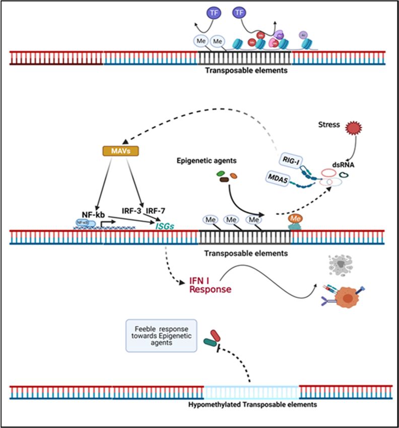
Biomedicines 2022, 10, 211                                                                                          2 of 18

                                   Aberrantly methylated genes can serve as biomarkers to predict therapeutic efficacy in
                             cancers. A well-known example is the MGMT (O6 -methylguanine–DNA methyltransferase)
                             promoter methylation. Hegi et al. discovered that a subset of glioblastoma tumors with
                             methylated MGMT responded better to the alkylating agent temozolomide (TMZ). The
                             rationale was that the TMZ-mediated DNA mismatch is not corrected due to the hyperme-
                             thylated and silenced MGMT gene [10]. The methylated DNA product of TMZ, termed
                             as O6 -methyguanine, can potentiate DNA single-strand break during mismatch repair,
                             which in turn favors the p53-mediated apoptosis in p53 wild-type tumors or facilitates the
                             mitochondrial pathway of apoptosis in p53 mutant tumors [11].
                                   Promoter methylation of the mismatch repair gene MLH1 correlates with microsatel-
                             lite repeat instability in sporadic endometrial cancers [12]. Bisulfite sequencing of acute
                             myeloid leukemia (AML) revealed that the extensive methylation of MLH1 promoter might
                             occur as a common molecular event in some AML patients [13]. Similarly, in microsatellite-
                             instable colorectal cancers, the epigenetic silencing of MLH1 together with concomitant
                             gene repression in the chromosome 3p22 region likely contributes to the development of
                             this tumor [14]. Acquired methylation of tumor suppressor genes may also be involved in
                             tumor progression. For example, CDKN2A promoter hypermethylation increases when ab-
                             normal lesions progress from basal cell hyperplasia to carcinoma in situ in lung cancer [15].
                             While promoter methylation is associated with gene silencing, gene body methylation may
                             facilitate gene transcription [16,17]. While intragenic and intergenic methylation may occur
                             in the CpGi and non-CpGi regions, the distribution of methylated CpGi is more enriched
                             in the gene body than in the 50 -promoters [18]. Methylation in the non-CpGi regions
                             is a classic case for gene silencing of transposable elements (TEs) [17]. Local epigenetic
                             alterations may be a specific feature of cancer. For example, promoter hypermethylation of
                             p15 was exclusively found in leukemia while that of p16 only in colon cancers; in addition,
                             aberrant methylation outside the promoters can distinguish the tumor-associated epige-
                             netic changes [19]. Furthermore, DNA methylation patterns in tumor-initiating cells and
                             senescent cells are distinct, suggesting that methylation changes inherent in cells which
                             escape oncogene-induced senescence may contribute to tumorigenesis [20].
                                   Indeed, both hypermethylation and hypomethylation could occur as a result of cancer-
                             specific epigenetic modifications. Aberrant hypermethylated DNA occurs in CpG island-
                             enriched promoters of tumor suppressor genes, whereas hypomethylation is usually global
                             and hypomethylated repeat elements correlate with tumor progression. Sporadic DNA
                             hypomethylation can be located in the gene body regions of oncogenes [21,22]. For example,
                             DNMT1-depleted mice develop intestinal adenomas representing the onset of malignancy.
                             The resulting DNA hypomethylation leads to genomic instability, which is considered
                             characteristic of colorectal cancer [23]. Similarly, by comparing methylation levels at
                             abundant CpG sites between hepatocellular carcinoma (HCC) and normal tissues, it was
                             shown that the predominant cancer-associated epigenetic alteration is hypomethylation,
                             which occurs mainly in intergenic regions, and contributes to genomic instability and
                             tumorigenesis of HCC, while hypermethylation constitutes the rest of the cancer epigenome
                             and is more likely to be found at promoters [24]. Interestingly, in the mouse model of
                             hepatocarcinogenesis, significant hypermethylation was observed at CpG islands within
                             the gene body and associated with overexpression of specific oncogenes such as SCN8A,
                             NFKB2, NEURL1B, and CDKN2B. Notably, overexpression of these oncogenes also occurs in
                             HCC patients, accompanied by hypermethylation of CpG islands within the gene body [25].
                             Hypermethylation at promoters of tumor suppressor genes such as RASSF1A and APC
                             correlates with tumor grade of bladder cancer. The extent of hypermethylation in these
                             regions may reflect the grade and invasiveness of bladder cancer [26]. In glioblastoma,
                             recurrent hypomethylation within the gene body promoter was identified, including TERT,
                             GLI3, and TP73, which may lead to oncogenic epigenetic and transcriptomic changes [27].
                                   Epigenome-modifying genes are also altered in a number of tumors [28]. For example,
                             recurrent isocitrate dehydrogenase (IDH) mutations are found in a number of cancers
                             including myeloid malignancies and lower-grade gliomas (LGGs) [29,30] and establishes
Biomedicines 2022, 10, 211 3 of 18
the CpG island methylator phenotype in glioma [31]. In prostate cancer, DNMT1 acts as
tumor suppressor in the early stages of tumorigenesis, while contributing to metastasis as
an oncogenic factor in the later stage [32]. In prostate cancer, a number of epigenetically
silenced tumor suppressors have also been shown to be predictive of clinical features such
as Gleason score and tumor stage [33].
DNA binding protein, the CCCTC-binding factor (CTCF), is a protein family that
promotes or pauses DNA transcription and, together with other transcription factors, con-
stitutes a complex network that determines specific transcriptional activity [34,35]. As
a transcription regulator, CTCF represses MYC, by linking the promoter and enhancer
domains [16]. Intergenic CTCF contributes to transcriptional repression by protecting local
DNA hypermethylation. Interestingly, CTCF binding at exon 5 of CD45 was inhibited by
DNA methylation, and this was inversely correlated with local 5-methylcytosine (5-mc)
levels [36]. CTCF binding patterns in cancers may differ from those in normal tissues. Re-
duced CTCF binding is usually located within gene promoters while enhanced binding can
be induced by oncogenic transcription factors and is related to enhancer regions [37]. Fur-
thermore, CTCF deletion facilitates regional DNA hypermethylation in prostate and breast
cancers and correlates with decreased expression of TNFAIP3, FGF5, and EPHA3. Inhibition
of DNA methylation can facilitate re-expression of genes harboring CTCF binding sites [38].
The MBD (methyl CpG binding domain) proteins recognize DNA methylation sites
and participate in transcriptional repression and heterochromatin formation [39]. They
include MeCp2, MBD1, MBD2, MBD3, and MBD4. The distribution of MBD proteins and
their affinity for methylated DNA depend on the gene promoter and cell type. For example,
MeCp2 and MBD2 preferentially bind to promoters; however, no strong association of a par-
ticular MBD with specific promoters is indicated [40]. Following catalysis by relevant CpG
methylation recognition enzymes, 5-mc can give rise to diverse modifications, including
5-hydroxymethylcytosine, 5-formylcytosine, and 5-carboxylcytosine, all of which together
comprise 15 possible combinations. MBD1 and MeCp2 preferentially bind to 5mc/5mc or
other 5mc-containing cytosine combinations [41].
2.2. Histone Modifications
Epigenetic deregulation of histones is also observed in cancer. In this review, we
focus on the epigenetic regulation of histone methylation and histone acetylation. Histone
acetylation mostly implies an active chromatin state (H3K9Ac, H3K14Ac, and H4K16Ac)
and histone methylation marks can either be active and lead to gene activation (e.g.,
H3K4me2/3, H3K36me3, and H3K79me3) or repressive and lead to gene repression
(H3K9me2/3, H3K27me3, and H4K20me3) [42].
Histone acetylation occurs by the addition of an acetyl group to H3 or H4, which
interferes with the interaction between the core histones and DNA and facilitates tran-
scription [11]. Histone methylation states have different functionalities. For example,
H4K20me and H4K20me2 play a role in DNA replication and DNA damage repair, while
H4K20me3 characterizes silenced heterochromatin [43]. H3K27me3 is mainly a mark for
transcriptional repression, while H3K27me2 is involved in the control of enhancer activ-
ity. Co-expression of H3K27me1 and H3K36me3 is associated with open chromatin that
promotes transcription [44].
Interestingly, G9a, a lysine methyltransferase that dimethylates H3K9, cooperates and
colocalizes with DNMT1 during DNA replication. DNMT1 knockdown negatively affects
G9a loading and H3 methylation [45]. Meanwhile, lysine methyltransferase SETDB1 is
unable to convert its substrate to H3K9me3 in the presence of H3K4me3, and H3K9me3
catalyzing enzymes such as G9a, GLP, and SUV39H1 are unable to bind to DNA regions
containing H3K4me3 [46]. Protein lysine methyltransferases and protein arginine methyl-
transferases are the two major classes of enzymes that catalyze histone methylation. In
contrast, lysine demethylases remove the methyl-group from modified histones, which
include KDM1A, KDM1B, and Jumonji C domain-containing demethylases [44]. The En-
hancer of Zester homolog2 (EZH2), which contributes to the di- and trimethylation ofBiomedicines 2022, 10, 211 4 of 18
H3K27 in mammals, is overexpressed in several tumors, including breast cancer, bladder
cancer, and malignant melanoma. Moreover, EZH2 overexpression is associated with poor
survival in melanoma patients and its deletion results in slower tumor progression [47,48].
Similarly, low EZH2 and H3K27me3 levels are predictors of better chemotherapy outcomes
in ovarian cancer [49]. In addition, H3K27me3 has been shown to be an independent his-
tone methylation marker for poor prognosis in bladder cancer [50] and has been associated
with carcinogenesis and progression of prostate cancer [51]. H3K27me3 can function as
a surrogate modification of hypomethylated TEs in taxane-resistant breast cancer cells,
repressing their transcription and the resulting antiviral response. Breast cancer cells can
be resensitized to taxane upon EZH2 inhibition [52]. On the other hand, loss of H3K27me3
in AML patients suggests shorter overall survival. Multivariate analysis revealed that
reduced H3K27me3 in AML patients could serve as an independent unfavorable prognostic
factor associated with an enhanced anti-apoptotic phenotype [53]. Loss of H3K27me3 has
also been found to be an indicator of recurrence in meningiomas and a poor prognostic
marker [54]. Concurrent high levels of H3K27ac and H3K27me3 were associated with
aberrant p53 and tumor aggressiveness in a subset of HCC [55]. The promoters of PD-1,
CTLA-4, and Tim3 were significantly hypomethylated in breast cancer compared to normal
tissues, and increased expression of these genes and epi-modifications are likely to lead
to tumor immune evasion. Additionally, the repressive histone markers H3K9me3 and
H3K27me3 exhibited attenuated binding in these promoter regions [56]. H3K27 mutations
lead to a global depletion of H3K27me3 and acquisition of H3K27ac, which is usually
enriched in repeat elements, and the acquired H3K27ac increases the susceptibility of cells
to epigenetic agents [57].
Furthermore, histone modifications, as essential components of the nucleosome, can
affect DNA methylation and influence the accessibility of dinucleotides to DNA methyl-
transferases [17]. DNA methylation attracts MBD proteins, which in turn recruit the
histone deacetylases (HDACs) and remove acetylation from histones, thereby repressing
gene transcription [44].
3. The Cancer Epigenome Contributes to Antitumor Immunity
Epigenetic changes in immune cells may influence antitumor immunity. Inherent
epigenetic events contribute to the regulation of several immune-related events, such
as Ig expression, Th1 and Th2 differentiation, B cell maturation, cytokine expression,
MHC I and II expression, and VDJ recombination [58]. Epigenetic changes in immune
cells coordinate with pathogenic stimuli to alter immune cell plasticity. Epigenetic pat-
terns distinguish innate and adaptive immune cells. Interferon-γ (IFN-γ) is essential for
Th1 cells and these cells usually have a demethylated IFN-γ promoter; whereas memory
CD8+ cells are characterized by having high H3K4me3 and low H3K27me3 at specific
loci (PRDM1, IFNG, and GZMB) compared to naive T cells [59]. Naive CD4+ T cells often
exhibit epigenetic suppression of both IFN-γ and IL4. The IFN-γ and IL4 loci undergo
DNA demethylation and histone acetylation when cells differentiate into Th1 and Th2,
respectively. Notably, STAT4 and T-bet, STAT6 and GATA3 may serve as downstream
signaling transducers in this process [60]. In another study, demethylation of H3K27me3 by
demethylase JMJD3 was found to be a representative feature during CD4+ T cell activation
through the JAK/STAT pathway [61].
Epigenetic agents have been shown to modify the anticancer immunity by enhancing
the tumor-associated antigen presentation and recognition, as well as the effective function
of cytotoxic T cells [62]. Compared to normal lymphocytes, increased DNA methylation
has been found in chronic lymphocytic leukemia in association with higher DNMT1 levels.
As a result, certain genes such as E-Cadherin, p15, and p16 are silenced, whereas in normal
cells these genes are demethylated and their expression is tightly regulated [63]. Aberrant
methylation of the CXCL14 promoter and subsequent gene repression have been observed
in certain tumor types, including prostate cancer, gastric cancer, and colorectal cancer [64].
Meanwhile, Peng et al. found that the level of DNMT1 and EZH2 negatively affectedBiomedicines 2022, 10, 211 5 of 18
the number of tumor infiltrating CD8+ lymphocytes and prognosis in an ovarian cancer
model, and that epigenetic silencing of the chemokines CXCL9 and CXCL10 resulted in
immunosuppression [65]. Epigenetic deregulation resulting from mutations of chromatin-
modifying enzymes such as MLL2, EZH2, and EP300 is observed in follicular lymphoma
and diffuse large B-cell lymphoma [66]. Overexpression of MeCP2 negatively affects Th1
cell differentiation in mice and leads to dysfunction of the cellular IFN-γ response by
dampening the accessibility of the IFN-γ gene for transcription factor binding [67].
Bunsen et al. showed that autocrine 2-hydroxyglutarate (2-HG) produced by IDH-
mutant gliomas inhibited the T cell activation and immunity [68]. Interestingly, de novo
DNA methylation acquisition occurs during progressive T cell exhaustion and impedes T
cell expansion and rejuvenation upon immune-checkpoint blockade (ICB). Furthermore, the
exhaustion-related DNA modification was characteristic of tumor-infiltrating PD-1hi CD8+ T
cells. DNA demethylating agents could reverse the exhaustion-associated changes, thereby
bypassing effector T cell resistance to ICB, leading to improved tumor control [69]. Similarly,
the upregulation of several immune checkpoints such as Tim3, TIGIT, and PD-L1 was found
in the peripheral blood of breast cancer and colorectal cancer patients in association with
the corresponding promoter hypomethylation, while the DNA demethylating enzymes
TET2 and TET3 also showed upregulation [70]. In summary, the epigenome is likely to play
an integral part in shaping the immune cell landscape, immune evasion, and establishing
tumor immunogenic signatures (“hot or cold” tumors) [71].
4. Targeting Cancer Epigenetics
4.1. Epigenetics: A Versatile Therapeutic Target
Several malignancies harbor mutations that lead to epigenetic alterations, such as the
IDH mutation in gliomas [29,30]. IDH mutation leads to extensive DNA hypermethylation
and blocks cellular differentiation [72]. The metabolite product of mutated IDH, 2-HG, is a
competitive inhibitor of enzymes that utilize α-ketoglutarate, such as the Jumanji C domain
containing histone demethylases JHDM1, lysine demethylase 4, and DNA demethylase
TET2. DNMT inhibitors (DNMTi) alter the epigenetic landscape, which promotes cellular
differentiation and suppresses cell growth [73]. Surprisingly, the attenuated DNMT1
achieved by DNMTi may not be sufficient to lead to global hypomethylation and re-express
silenced genes. Therefore, epigenetic agents could be combined with alternate therapies to
achieve the desired antitumor effect [74]. Due to the immunogenic nature of IDH1 (R132H),
vaccine-based therapy targeting the mutation may be a potent therapeutic regimen [75].
In addition, DNMT1i azacytidine represses the growth of IDH1 mutant gliomas in vivo,
accompanied by hypomethylation and marked cellular differentiation, with no tumor
recurrence observed up to 7 weeks after drug withdrawal [76].
Inhibition of the histone methyltransferase G9a has been shown to be a potential target
for the treatment of several malignancies [77–79]. Knockdown of G9a inhibited tumor
growth and progression in mouse models of pancreatic carcinoma [80]. G9a expression
was enriched in tumor-initiating cells (TICs) of non-small cell lung cancer (NSCLC), and
loss of G9a led to reduced proliferation and sphere-forming capacity of TICs. In addition,
the hypomethylation of select genes in response to G9a deprivation associates with favor-
able clinical prognosis in NSCLC patients [77]. Oncogenic driver MYC cooperates with
G9a to epigenetically silence gene expression. Depletion of G9a abrogates the binding
of MYC to chromatin, reverses the repression of MYC-suppressed genes, and hinders
tumor growth [81]. Some preliminary studies have shown promising results suggesting
dual inhibition of DNMT and G9a may be a therapeutic strategy in certain cancer models.
Co-inhibition of DNMT and G9a showed high potential to inhibit cellular proliferation,
to promote INF-stimulating genes and prolong the survival of tumor models in hema-
tological lesions [82]. The expression of DNMT1 and G9a was shown to correlate with
poor prognosis in HCC and dual inhibition of these two targets impaired cell growth
in vitro and in vivo [83]. Similarly, G9a was associated with poor prognosis in bladder
cancer and exhibited resistance to anti-PD1 therapy. The combination of dual inhibition ofBiomedicines 2022, 10, 211 6 of 18
G9a and DNMT1 together with PD-L1 showed promising results, especially in inducing
immunogenic cell death and adjusting the endogenous antitumor immune response [84].
Epigenetic drugs (Figure 1) are expected to have versatile potential, especially in pre-
Biomedicines 2022, 10, x FOR PEER REVIEW 7 of 19
venting cancer recurrence and resistance, as well as in sensitizing to therapeutics during
long-term treatments [5].
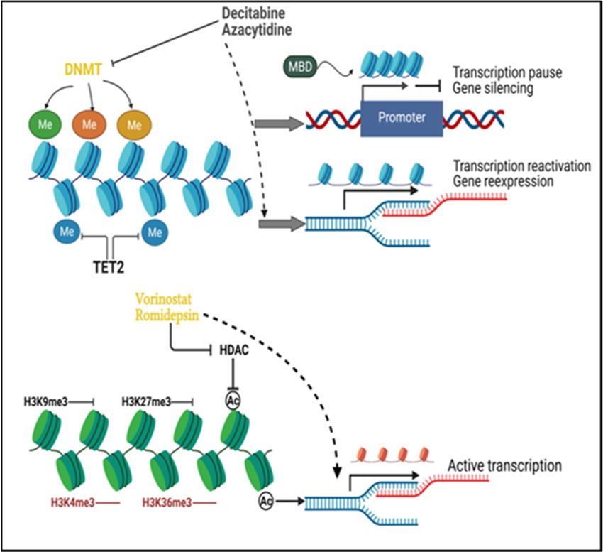
Figure1. 1.
Figure Principles
Principles of cancer
of cancer epigenetic
epigenetic modifications
modifications and theiranddrug their drug
targets. DNAtargets. DNA
methyltrans-
methyltransferases (DNMTs) add methyl groups to DNA and maintain methylated DNA, while Tet
ferases (DNMTs) add methyl groups to DNA and maintain methylated DNA, while Tet methylcy-
methylcytosine dioxygenase 2 (TET2) removes the methyl groups from DNA. DNA methylation at
tosine dioxygenase 2 (TET2) removes the methyl groups from DNA. DNA methylation at the gene
the gene promoter impairs local binding of transcription factors and blocks transcription.
promoter
Recruitmentimpairs local CpG
of methyl binding of transcription
binding domain (MBD) factors and by
protein blocks transcription.
the methylated DNA Recruitment of
facilitates the
methyl CpG binding
heterochromatin domain (MBD)
formation proteininbytranscription
and results the methylated DNA facilitates
repression. DNMTthe heterochromatin
inhibitors such as
formation
decitabineandandresults in transcription
azacytidine repression.
will incorporate into DNMT inhibitors
the genome such as decitabine
and degrade the activityand
of azacyti-
DNMT,
reverse
dine will the aberrantinto
incorporate DNA hypermethylation,
the genome and degrade and
the enable
activitythe re-expression
of DNMT, reverse of
thesilenced
aberrant genes.
DNA
H3K9me3 and H3K27me3
hypermethylation, and enable serve as repressive of
the re-expression histone marks,
silenced genes.while H3K4me3
H3K9me3 and H3K36me3
and H3K27me3 serve are
as
active marks.
repressive Histone
histone marks,deacetylation
while H3K4me3 is and
among the major
H3K36me3 repressive
are active marks.mechanisms of histone
Histone deacetylation
ismodification. Histone
among the major deacetylases
repressive (HDAC)
mechanisms inhibitors
of histone (e.g., vorinostat
modification. Histoneand romidepsin)
deacetylases inhibit
(HDAC)
histone deacetylation caused by HDAC to maintain active chromatin status for transcription. Me:
inhibitors (e.g., vorinostat and romidepsin) inhibit histone deacetylation caused by HDAC to maintain
DNA methylation; Ac: histone acetylation.
active chromatin status for transcription. Me: DNA methylation; Ac: histone acetylation.
Histone Agents
4.2. Epigenetic acetylation and deacetylation are catalyzed by histone acetyltransferases
(HATs) and histone deacetyltransferases (HDACs). In addition to histones, HDACs can
5-Aza-2-deoxycytidine (decitabine) and 5-azacytosine (azacytidine) are two classical
also bind to and catalyze non-histone proteins; the binding partners include p53 and
DNMTi, both of which can reverse DNA hypermethylation by covalently trapping the
transcription
DNMTs to DNA factors such astoSTAT,
and leading GATA1-3 [88].
their degradation [3].HDAC inhibitors
In the cells, these (HDACi)
agents arerepresent
converteda
group of epi-drugs that are extensively studied. They are categorized
to the triphosphate form and become physiologically active. Decitabine is incorporatedinto three classes:
hydroxamates (vorinostat, belinostat, panobinostat), benzamides (entinostat,
into DNA while azacytidine binds mostly to RNA, but a small percentage of the converted chidamide),
cyclic peptides
product (romidepsin),
is incorporated into DNA andasaliphatic acids.
well [59]. Some
Of note, of these possesses
decitabine compounds are being
a half-life of
tested in clinical trials either alone or in combination with other anticancer
only 12–25 min in patients, due to degradation by cytidine deaminase in the liver after drugsthe in
various malignancies, from multiple myeloma and myelodysplastic
drug enters the bloodstream [85]. Decitabine has shown clinical benefit in hematological syndrome to
glioblastoma,[86]
malignancies ovarian cancer, potential
and showed and sometoother epithelial/solid
sensitize tumors
to therapeutic [89]. in
response Several HDACi
solid tumors,
have
for been approved
example, by the
by improving Food and DruginAdministration
chemosensitivity refractory ovarian(FDA) for patients
cancer the treatment
[87]. of
cutaneous T-cell lymphoma, including vorinostat, romidepsin, and belinostat [90].
Although vorinostat has not been shown to be as effective as single agent therapy in
solid tumors in clinical trials, it has been proposed that this epi-agent be combined with
other chemotherapy drugs to optimize therapeutic benefit. Most importantly,
downregulation of oncogenes and upregulation of tumor suppressors is considered to beBiomedicines 2022, 10, 211 7 of 18
Histone acetylation and deacetylation are catalyzed by histone acetyltransferases
(HATs) and histone deacetyltransferases (HDACs). In addition to histones, HDACs can
also bind to and catalyze non-histone proteins; the binding partners include p53 and
transcription factors such as STAT, GATA1-3 [88]. HDAC inhibitors (HDACi) represent a
group of epi-drugs that are extensively studied. They are categorized into three classes:
hydroxamates (vorinostat, belinostat, panobinostat), benzamides (entinostat, chidamide),
cyclic peptides (romidepsin), and aliphatic acids. Some of these compounds are being
tested in clinical trials either alone or in combination with other anticancer drugs in various
malignancies, from multiple myeloma and myelodysplastic syndrome to glioblastoma,
ovarian cancer, and some other epithelial/solid tumors [89]. Several HDACi have been
approved by the Food and Drug Administration (FDA) for the treatment of cutaneous
T-cell lymphoma, including vorinostat, romidepsin, and belinostat [90].
Although vorinostat has not been shown to be as effective as single agent therapy in
solid tumors in clinical trials, it has been proposed that this epi-agent be combined with
other chemotherapy drugs to optimize therapeutic benefit. Most importantly, downregu-
lation of oncogenes and upregulation of tumor suppressors is considered to be the main
mechanism of action of vorinostat [90,91]. Interestingly, depsipeptide (romidepsin) not only
caused histone deacetylation but also strongly demethylated the promoter of some genes,
including p16, SALL3, and GATA4. Moreover, attenuated binding of DNMT1 together with
decreased expression of H3K9 methyltransferases G9a and SUV39H1 was suggested to
underlie the indirect demethylating activity of depsipeptide [92].
5. Transposable Elements
Endogenous retroviruses (ERVs), as a subset of TEs, may account for up to 8% of
the human genome. TEs were once interpreted as “genetic parasites” because of their
non-coding roles. However, it was later found that these elements can be actively tran-
scribed into nucleic acids or proteins that resemble pathogen-associated molecular patterns
(PAMPs) and are recognized by pathogen recognition receptor, resulting in an immune
response that resembles an antiviral response [7,93]. The TE can be divided into two classes:
1. Class I, also known as retrotransposons, contains long terminal repeats (LTR)/ERV, long
and short interspersed nuclear elements (LINEs and SINEs); 2. Class II, the main compo-
nent is DNA transposons [7]. Retrotransposons are classified according to an alternative
classification into either autonomous or non-autonomous elements. The former contains
long terminal repeats (LTR) and non-LTR retrotransposons—also referred to as LINEs and
the latter contains SINEs [94]. The ability of ERV to elicit an antiviral immune response
(Figure 2) can be explained by the fact that nucleic acids produced by viral infections or
endogenous retroelements are normally distinct from host cellular RNA and are therefore
recognized as PAMP. Retinoic acid-inducible gene I (RIG-I) and Toll-like receptors (TLR) are
two important RNA sensors. RIG-I recognizes cytosolic viral RNA, while TLR recognizes
extracellular viral RNA endocytosed in endolysosomes [95]. Indeed, dsRNA is recognized
by TLR-3, ssRNA is recognized by TLR-7 and TLR-8, and foreign DNA is recognized by
TLR-9. Melanoma differentiation-associated gene 5 (MDA5) also serves as a sensor for
intracellular dsRNA [96]. DNA demethylating agents can restore the expression of ERVs
in tumor cells, placing the cells in a mock virus-infected state that then impairs the cell
growth and proliferation [97,98]. Viral or endogenous RNA sensing leads to downstream
activation of NF-kB and interferon-regulated factors, coupled with an IFN type I response
and activation of a number of interferon stimulated genes (ISGs) [99,100]. Type I and III
IFN responses activate transcription of ISGs through JAK/STAT pathways, and type II
IFN (IFN-γ) response transduces signaling through STAT1 phosphorylation and nuclear
translocation and subsequent binding to the promoters of IFN-γ induced genes [101].Biomedicines 2022, 10, 211 8 of 18
Biomedicines 2022, 10, x FOR PEER REVIEW 9 of 19
Figure2.2.Transposable
Figure Transposableelements
elementsdetermine
determinethe theinherent
inherentimmunogenicity
immunogenicityand andresponse
responseofoftumor
tumor
cells to epigenetic agents. Transposable elements (TE) in the genome are typically not actively
cells to epigenetic agents. Transposable elements (TE) in the genome are typically not actively
transcribed but can be stimulated by stress and epigenetic agents. Endogenous retroviruses (ERVs)
transcribed but can be stimulated by stress and epigenetic agents. Endogenous retroviruses (ERVs)
compose a major part of TE. Regionally hypermethylated ERVs are transcriptionally inactive, and
compose
repressivea major
histone part of TE. Regionally
modifications at ERVs hypermethylated ERVs are
loci disturb the access transcriptionally
of genome inactive,
for transcription and
factors
repressive histoneagents
(TF). Epigenetic modifications at ERVs
potentiate loci disturb of
the transcription theERVs
accessinto
of genome for transcription
nucleic acids that mimic factors
a virus
(TF). Epigenetic
infection. agents potentiate
The transcription producttheoftranscription
ERVs, dsRNA, of are
ERVs into by
sensed nucleic acids
cytosolic that mimic
sensors: a virus
retinoic acid-
inducible gene
infection. I (RIG-I) or melanoma
The transcription product differentiation-associated
of ERVs, dsRNA, are sensed geneby5 (MDA5).
cytosolicThe resulting
sensors: signal
retinoic
is transducedgene
acid-inducible by mitochondrial antiviraldifferentiation-associated
I (RIG-I) or melanoma proteins (MAVs) and leads gene to NF-kb and
5 (MDA5). The interferon
resulting
regulated factors (IRF) translocation into the nucleus, inducing the expression
signal is transduced by mitochondrial antiviral proteins (MAVs) and leads to NF-kb and interferon of interferon-
stimulated
regulated genes
factors (ISGs)
(IRF) and type Iinto
translocation IFNtheresponse
nucleus,and results
inducing theinexpression
tumor cellofapoptosis or enhanced
interferon-stimulated
expression of tumor associated antigens. Hypomethylated ERVs may be a characteristic epigenetic
genes (ISGs) and type I IFN response and results in tumor cell apoptosis or enhanced expression of
feature in tumor cells and may perturb cellular responses to epigenetic agents. Inherent ERV
tumor associated antigens. Hypomethylated ERVs may be a characteristic epigenetic feature in tumor
patterns and regional epigenetic modifications may provide predictive value for epigenetic therapy
cells and may perturb cellular responses to epigenetic agents. Inherent ERV patterns and regional
[52,100,101].
epigenetic modifications may provide predictive value for epigenetic therapy [52,100,101].
Endogenous dsRNA, which is triggered and reactivated by stimuli, represents an
A recent analysis from The Cancer Genome Atlas (TCGA) demonstrated the predictive
important element of the antiviral immune response. Apart from viral infections, dsRNA
value of human ERVs (hERVs) present in clear cell renal carcinoma cells in response to
may arise from tissue stress, damage, and necrosis [93,104]. It has been suggested that
anti-PD1 therapy and showed that variable signatures of hERVs correlated with differential
dsRNA shares the same signaling pathway as dsDNA, which can be activated by viral
survival: the RIG-I like up (up 50th percentiles) implied longer overall survival compared
DNA infection [99]. Notably, recognition of viral RNA leads not only to apoptosis but also
to the RIG-I like down (down 50th percentiles) group [102]. Interestingly, only a basal level
to pyroptosis, a state of inflammasome-mediated cell death accompanied by disrupted
of genes within the antiviral pathway was shown in the DNMT1 hypomorph cells and the
cell membrane integrity and release of cytoplasmic content from cells [105]. A recent
upregulation of antiviral related genes can be achieved by DNMT1 depletion [74]. High
finding reported
expression of ERVs inthat the positively
tumors protein correlated
expressionwithof an
AGO1x
efficientinterfered with dsRNA
antiviral response [103].
accumulation in breast cancer cells and hampered the dsRNA-induced
Recently, an interesting discovery was made regarding how epigenetic modifications interferon
and
response,
agents leading
can alter the to refractory cell
transcription growth.
of TEs. This Loss
studyofshowed
AGO1xthatexpression restored
resistance the dsRNA
of triple-negative
and interferon
breast response
cancer cells and eventually
to taxanes led to more
can be attributed apoptosis
to loci enriched [106].
in hypomethylated TEs
Epi-agents induce retroelements sensed by RIG-I
but abundant in H3K27me3, which negatively affected TE transcriptionand MDA5and which
viralaffect the
mimicry,
intracellular glucose hydrolysis, resulting in energy depletion and tumor cell death. InBiomedicines 2022, 10, 211 9 of 18
thereby attenuating intracellular antiviral immunity and enhancing the sustainability of
taxane-resistant cells [52].
Endogenous dsRNA, which is triggered and reactivated by stimuli, represents an
important element of the antiviral immune response. Apart from viral infections, dsRNA
may arise from tissue stress, damage, and necrosis [93,104]. It has been suggested that
dsRNA shares the same signaling pathway as dsDNA, which can be activated by viral
DNA infection [99]. Notably, recognition of viral RNA leads not only to apoptosis but also
to pyroptosis, a state of inflammasome-mediated cell death accompanied by disrupted cell
membrane integrity and release of cytoplasmic content from cells [105]. A recent finding
reported that the protein expression of AGO1x interfered with dsRNA accumulation in
breast cancer cells and hampered the dsRNA-induced interferon response, leading to
refractory cell growth. Loss of AGO1x expression restored the dsRNA and interferon
response and eventually led to more apoptosis [106].
Epi-agents induce retroelements sensed by RIG-I and MDA5 which affect the intracel-
lular glucose hydrolysis, resulting in energy depletion and tumor cell death. In addition,
this effect is coupled with altered mitochondrial metabolism to compensate for ATP, and
tumor cell death (necroptosis) is independent of caspase-mediated apoptosis but closely
associated with BCL2 [107].
6. Epigenetic Targeting Meets Immune Check Point Inhibition: Does the
Union Empower?
6.1. Tumor-Infiltrating Immune Cells in Gliomas
The tumor microenvironment of gliomas is unique in part due to the blood–brain
barrier (BBB). Of note, glioblastoma is referred to as an immunogenic “cold tumor” because
of the lack of tumor antigen expression, the absence of antigen presentation to T cells, and
the high level of immune checkpoints on infiltrating lymphocytes [108]. Indeed, lympho-
cytes require adhesion signals on endothelial cells to migrate into the brain, additionally,
naive T cells are not normally present in the central nervous system while T cells that
penetrate the BBB are patrolling T cells and regulatory T cells that prevent inappropriate
inflammatory responses [109].
A relevant analysis of TCGA data revealed that monocytes, activated NK cells,
macrophages, and eosinophils among other infiltrative immune cells, correlated with
survival of glioblastoma patients, with the abundance of macrophages indicating poorer
survival, while the others were associated with better survival [110].
A retrospective study of immunohistochemical analysis of tissue samples from 43 glioblas-
toma patients concluded that among the infiltrating immune cells, lymphocytes were sparsely
distributed compared to macrophages, but a lower amount of CD4/CD8 infiltrating lympho-
cytes (TILs) was associated with better survival [111].
Genetic alterations in tumors also correlate with TILs in the tumor microenviron-
ment. For instance, TILs are enriched in NF1 and RB1 mutated gliomas but depleted in
EGFR-amplified and PTEN-deleted gliomas. Interestingly, IDH-wildtype glioma is usually
associated with more lymphocyte infiltration and PD-L1 expression while IDH-mutant
gliomas have less IFN-γ and lower infiltration of CD8+ and CD4+ T cells [112]. Methy-
lation chip-based analysis of gliomas found that there was no dramatic difference in the
extent of immune cell infiltration between long-term and short-term survivors [113]. A
study of 519 glioblastoma patients indicated that long-term survivors were more likely
to have extensive T cell infiltration than short-term survivors, with high CD8+ infiltrat-
ing T cells indicating long-term survival [114]. A recent study uncovered the correlation
between infiltrated T cells and overall survival in glioma patients. Patients with T cell-
deficient gliomas presented a longer survival than the T cell-enriched group; nevertheless,
CD8+ T cell-dominant group predicted a better survival as compared with the CD4+ T
cell-dominant group. Notably, fewer infiltrated macrophages were found in the IDH-
mutated gliomas [115].Biomedicines 2022, 10, 211 10 of 18
IDH-mutated tumors were found to express less IFN-γ inducible chemokines such
as CXCL10, which was further confirmed by the introduction of IDH1 mutation which
decreased CXCL10 expression and reduced the number of T cells in a glioma mouse model.
Furthermore, mutant IDH1 inhibitor led to increased survival in preclinical glioma models
and led to increased CXCL10 expression and TILs [116]. Similarly, Weenink et al. quantified
the TILs in both lower and high-grade glioma (LGG and HGG) samples and discovered that
LGG contained fewer CD8+ T cells, which was related to the lower expression of CXCL9,
CXCL10, and ICAM1, the relative absence of TILs in LGG was thought to potentially affect
the therapeutic efficacy of immune checkpoint inhibitors in this context [117].
6.2. Combination of Epigenetic Drugs with Immune Checkpoint Inhibitors
The epigenetic modifications inherent to the tumor may reflect its immunogenic prop-
erties in the antitumor microenvironment. Epigenetic agents have been shown to restore
the vulnerability of tumor tissues to therapeutic modalities. For example, treatment of
colon and ovarian cancer cell lines with DNMTi enhanced antigen presentation and cancer
testis antigens at the transcriptional and translational levels [118]. In non-small cell lung
cancer, analysis of CpG-methylation assays and bisulfite sequencing revealed that CTLA-4
and PD1 methylation levels were reduced compared to normal tissues and epigenetic
changes were inversely correlated with gene expression [119]. Encouraging results showed
that epigenetic agents enhance antitumor immunity, especially when combined with cer-
tain compounds, as demonstrated in a number of reports. In a study with decitabine on
glioblastoma cells and patient samples, it was found that tumor cells showed increased
expression of MHC I and tumor-associated antigens after decitabine treatment, and T cells
presented an upregulated Fas ligand (CD95) in association with increased levels of INF-γ,
TNF-α, IL-5, and CD107A (functional parameters of degranulation of cytotoxic T cells via
the Fas pathway) of NY-ESO-1 specific T cells and concluded that the epigenetic agent
sensitizes glioblastoma to the functionality of specific T cells [120].
Treatment of lung cancer cells with azacytidine was shown to alter a variety of immune-
related gene expression, including upregulation of HLA and IFN-γ and its downstream
signaling factors. Azacytidine led to increased PD-L1 and CD80/CD86 (CTLA-ligands),
providing a rationale for combining of azacytidine with immune checkpoint blockade to
overcome immune evasion of tumor cells [121]. Ishibashi et al. showed the inverse correla-
tion of HLA-G expression with prognosis in breast cancer patients. Decitabine treatment
increased HLA-G expression in tumor cells and enhanced recognition of these cells by
specific CD4+ helper T lymphocytes, suggesting a combination of decitabine with HLA-G
targeting to improve T cell-based immunotherapy [122]. Similar efficacy was observed in
mouse GL261 glioma cells. Decitabine potentiated the immunogenic signature in glioma-
initiating GL261 cells by increasing the expression of FasL and MHCI which enhanced
tumor recognition and killing by CTLs [123]. The new generation DNMTi guadecitabine
has shown the potential to alter the antitumor microenvironment by increasing MHCI
expression and enhancing IFN-γ response in breast cancer cells. Moreover, tumor growth
was significantly slowed when guadecitabine was used together with anti-PD-L1 ther-
apy in a mouse model [124]. Similarly, azacytidine enriched effective immune cells via
type I IFN signaling. Moreover, the triple combination of azacytidine, HDACi, and PD-1
antibody showed the greatest antitumor potency in a mouse ovarian tumor model [125].
Combinatorial use of HDACi (SAHA and CI994) with the PD-1 inhibitor showed promising
efficacy in the mouse model of urothelial bladder cancer. HDACi was shown to facilitate
delayed immune recognition by upregulating the expression of associated genes such as
NGK2D and HSP70. Meanwhile, it has been suggested that fully activated T cells are not
sufficient for intact antitumor immunity, but that pre-exposure of tumor cells to agents
such as HDACs will optimize the antitumor immunity [126]. As such, HDACi CG-745
modulated the immune microenvironment by increasing the proportion of cytotoxic T
cells and NK cells and decreasing the suppressive immune components such as regula-Biomedicines 2022, 10, 211 11 of 18
tory T cells and myeloid-derived suppressor cells and favored the anti-PD1 therapy in a
synergistic fashion [127].
The elementary factors that determine the response of cancer cells to immune check-
point blockade include the tumor mutational burden, immune phenotype of tumor mi-
croenvironment, and immune escape of tumors [71]. The inherent and acquired epigenetic
modifications within the loci of immune checkpoint genes may contribute to resistance to
immune checkpoint inhibitors as only a subset of patients respond to immunotherapy. The
relevant epigenetic modifications may be potent predictive biomarkers for immune check-
point therapy and can be targets in a combination strategy to increase therapeutic benefit.
Increased expression of PD-L1 upon azacytidine was shown to elevate the response to anti-
PD1 therapy [128]; similarly, high levels of PD-L1 and TIL were associated with positive
response to anti-PD1/PD-L1 therapy [129]. Similarly, melanoma patients with low PD-L1
expression and low TIL count did not respond to anti-PD1 therapy. Notably, abundant
miRNA negatively regulated PD-L1 expression across multiple cancers and contributed to
resistance to immune checkpoint inhibitors [130]. Lower CTLA-4 methylation in melanoma
samples indicated a better response to anti-PD1 or anti-CTLA-4 therapy [131] and increased
level of PD-1, CTLA-4, or PD-L1 was found to correlate with DNA hypomethylation across
many types of tumors such as non-small cell lung cancer, lower grade gliomas (LGG), and
head and neck squamous cell carcinoma [132].
Several clinical trials on the combinatorial approach of epigenetic agents and immune
checkpoint inhibitors for various tumors are still ongoing, and their therapeutic effects and
potential side effects are being monitored (Table 1). As a novel concept that showed exciting
results in several preclinical and clinical studies, the combination of immune-checkpoint
inhibitors with epigenetic agents may provide increased therapeutic benefit. Hopefully,
these studies will add to our current knowledge of the clinical utility and limitations of
epigenetic agents and combinatorial strategies for the benefit of patients.
Table 1. Clinical trials of epigenetic agents combined with immune checkpoint inhibitors for cancer
therapy.
Therapeutics
Identifier Malignant Conditions Start Date Results
(Single or Combined)
1. MBG453 (Tim3 antibody)
NCT02608268 Advanced solid tumors 2. PDR001 (PD-1 antibody) November 2015 Recruiting
3. Decitabine
1. Decitabine/Azacytidine
Acute myeloid leukemia or high
NCT03066648 2. PDR001 July 2017 Recruiting
risk myelodysplastic syndrome
3. MBG453
1. ASTX 727 (oral decitabine)
NCT03019003 Head and neck cancer March 2017 Recruiting
2. Durvalumab (PD-L1 antibody)
1. Durvalumab (PD-L1 inhibitor)
Relapsed or refractory peripheral 2. Romidepsin
NCT03161223 May 2018 Recruiting
T-cell lymphomas (PTCL) 3. 5-azacytidine
4. Pralatrexate
1. Azacytidine
Non-small cell lung
NCT01928576 2. Entinostat August 2013 Recruiting
cancer (NSCLC)
3. Nivolumab
PD-1 monoclonal 1. Decitabine
NCT04611711 antibody-resistant digestive 2. TQB2450 (PD-1 inhibitor) November 2020 Recruiting
system tumors 3. Anlotinib (VEGFR inhibitor)
Relapsed or refractory
1. Decitabine
NCT02890329 myelodysplastic syndrome or September 2016 Recruiting
2. Ipilimumab (CTLA-4 antibody)
acute myeloid leukemiaBiomedicines 2022, 10, 211 12 of 18
Table 1. Cont.
Therapeutics
Identifier Malignant Conditions Start Date Results
(Single or Combined)
1. Decitabine
Newly diagnosed TP53 mutated
NCT04277442 2. Nivolumab (PD-1 inhibitor) February 2020 Recruiting
acute myeloid leukemia
3. Venetoclax (Bcl-2 inhibitor)
1. Azacytidine
Refractory/relapsed or newly
NCT02397720 2. Ipilimumab April 2015 Recruiting
diagnosed acute myeloid leukemia
3. Nivolumab
1. Azacytidine
NCT02816021 Metastatic melanoma February 2017 Recruiting
2. Pembrolizumab (PD-1 inhibitor)
Inoperable locally advanced or 1. Oral decitabine
metastatic NSCLC, and 2. Tetrahydrouridine (inhibitor of
NCT03233724 April 2018 Recruiting
esophageal carcinomas, or pleural cytidine deaminase)
mesotheliomas 3. Pembrolizumab (PD-1 inhibitor)
1. Azacytidine
Advanced solid tumors and 2. Pembrolizumab
previously treated stage IIIB or 3. Epacadostat
NCT02959437 stage IV non-small cell lung cancer (indoleamine2,3-dioxygenase February 2017 Recruiting
and stage IV microsatellite-stable inhibitor)
colorectal cancer 4. INCB057643 (BET inhibitor)
5. INCB059872 (LSD1 inhibitor)
Locally advanced or metastatic 1. CC-486 (oral azacytidine)
NCT02546986 October 2015 Recruiting
non-small cell lung cancer 2. Pembrolizumab
1. Ipilimumab + Nivolumab +
Melanoma and NSCLC resistant to
NCT04250246 Guadecitabine March 2020 Recruiting
anti-PD1/PDL1
2. Ipilimumab + Nivolumab
1. Entinostat
NCT03765229 Melanoma March 2019 Recruiting
2. Pembrolizumab
NSCLC, melanoma and
1. Entinostat
NCT02437136 mismatch repair-proficient July 2015 Recruiting
2. Pembrolizumab
colorectal cancer
1. Atezolizumab (PD-L1 inhibitor)
NCT03024437 Advanced renal cell carcinoma 2. Bevacizumab (VEGF inhibitor) May 2017 Recruiting
3. Entinostat
1. Bintrafusp Alfa (bifunctional
fusion protein composed of the
Solid tumors, metastatic
extracellular domain of the TGF-β
checkpoint refractory
NCT04708470 receptor II fused to an IgG1 August 2021 Recruiting
HPV-associated tumors,
antibody blocking PD-L1)
microsatellite stable colon cancer
2. NHS-IL12
3. Entinostat
Advanced epithelial 1. Entinostat
NCT02915523 January 2017 Recruiting
ovarian cancer 2. Avelumab (PD-L1 inhibitor)
Previously treated
unresectable/metastatic 1. Entinostat
NCT03250273 November 2017 Recruiting
cholangiocarcinoma and 2. Nivolumab
pancreatic cancer
Locally advanced and metastatic 1. Pembrolizumab
NCT03854474 May 2019 Recruiting
urothelial carcinoma 2. Tazemetostat (EZH2 inhibitor)
Unresectable or locally advanced 1. Entinostat
NCT02453620 or metastatic Her2-negative breast 2. Ipilimumab November 2015 Recruiting
cancer 3. NivolumabBiomedicines 2022, 10, 211 13 of 18
Table 1. Cont.
Therapeutics
Identifier Malignant Conditions Start Date Results
(Single or Combined)
1. Vorinostat
Hormone receptor expressing
NCT02395627 2. Tamoxifen May 2015 Recruiting
advanced breast cancer
3. Pembrolizumab
7. Conclusions
Cancer-associated epigenetic modifications play a central role in suggesting an appro-
priate therapeutic strategy. Previous studies have shown that epigenetic agents, in addition
to being an “epigenetic editor”, also can activate silenced tumor suppressor genes and cellu-
lar antiviral signaling pathways, and tumor-associated antigens and immune-checkpoints.
The epigenetic landscape of tumors and its influence on tumor phenotype, microenvi-
ronment, and the interaction between epigenetics and immune plasticity with respect to
tumorigenesis and progression are of great scientific interest. Given the complexity and
diversity of epigenetic modifications in different tissues, tumor grades, and therapy-related
potential alterations, more comprehensive knowledge is needed to appropriately design
preclinical studies and clinical trials accompanied by interdisciplinary expertise.
Author Contributions: Y.L. and S.T. contributed to the discussion of content and wrote, reviewed, and
edited the manuscript. All authors have read and agreed to the published version of the manuscript.
Funding: Y.Y.L. is supported by a grant from the Else Kröner-Fresenius Foundation.
Institutional Review Board Statement: Not applicable.
Informed Consent Statement: Not applicable.
Data Availability Statement: Not applicable.
Acknowledgments: The figures were created with BioRender.com.
Conflicts of Interest: The authors declare no conflict of interest.
References
1. Kanwal, R.; Gupta, S. Epigenetic modifications in cancer. Clin. Genet. 2011, 81, 303–311. [CrossRef]
2. Issa, J.-P. Introduction: Cancer as an epigenetic disease. Cancer J. 2017, 23, 255–256. [CrossRef] [PubMed]
3. Chan, T.; Ho, A.S.; Turcan, S. Epigenetic therapy: Use of agents targeting deacetylation and methylation in cancer management.
Oncol. Targets Ther. 2013, 6, 223–232. [CrossRef] [PubMed]
4. Baylin, S.B. The cancer epigenome: Its origins, contributions to tumorigenesis, and translational implications. Proc. Am. Thorac.
Soc. 2012, 9, 64–65. [CrossRef]
5. Easwaran, H.; Tsai, H.-C.; Baylin, S.B. Cancer epigenetics: Tumor heterogeneity, plasticity of stem-like states, and drug resistance.
Mol. Cell 2014, 54, 716–727. [CrossRef] [PubMed]
6. Baylin, S.B. DNA Methylation and Gene Silencing in Cancer. Chem. Inform. 2006, 2, S4–S11. [CrossRef]
7. Rebollo, R.; Romanish, M.T.; Mager, D.L. Transposable Elements: An Abundant and Natural Source of Regulatory Sequences for
Host Genes. Annu. Rev. Genet. 2012, 46, 21–42. [CrossRef] [PubMed]
8. Ehrlich, M. DNA hypomethylation in cancer cells. Epigenomics 2009, 1, 239–259. [CrossRef]
9. Ehrlich, M. DNA methylation in cancer: Too much, but also too little. Oncogene 2002, 21, 5400–5413. [CrossRef]
10. Hegi, M.E.; Diserens, A.-C.; Stupp, R. MGMT gene silencing and benefit from temozolomide in glioblastoma. N. Engl. J. Med.
2005, 352, 997–1003. [CrossRef]
11. Romani, M.; Pistillo, M.P.; Banelli, B. Epigenetic Targeting of Glioblastoma. Front. Oncol. 2018, 8, 448. [CrossRef]
12. Simpkins, S.B.; Bocker, T.; Swisher, E.M.; Mutch, D.G.; Gersell, D.J.; Kovatich, A.J.; Palazzo, J.P.; Fishel, R.; Goodfellow, P.J. MLH1
Promoter Methylation and Gene Silencing is the Primary Cause of Microsatellite Instability in Sporadic Endometrial Cancers.
Hum. Mol. Genet. 1999, 8, 661–666. [CrossRef]
13. Seedhouse, C.H.; Das-Gupta, E.; Russell, N. Methylation of the hMLH1 promoter and its association with microsatellite instability
in acute myeloid leukemia. Leukemia 2003, 17, 83–88. [CrossRef]
14. Hitchins, M.P.; Ap Lin, V.; Buckle, A.; Cheong, K.; Halani, N.; Ku, S.; Kwok, C.-T.; Packham, D.; Suter, C.M.; Meagher, A.; et al.
Epigenetic Inactivation of a Cluster of Genes Flanking MLH1 in Microsatellite-Unstable Colorectal Cancer. Cancer Res. 2007, 67,
9107–9116. [CrossRef]Biomedicines 2022, 10, 211 14 of 18
15. Belinsky, S.A.; Nikula, K.J.; Palmisano, W.A.; Michels, R.; Saccomanno, G.; Gabrielson, E.; Baylin, S.B.; Herman, J.G. Aberrant
methylation of p16INK4a is an early event in lung cancer and a potential biomarker for early diagnosis. Proc. Natl. Acad. Sci. USA
1998, 95, 11891–11896. [CrossRef]
16. Lay, F.D.; Liang, G. Rethinking demethylating agents in epigenetic cancer therapy. J. Mol. Pharm. Org. Process Res. 2016, 4, 133.
17. Jones, P.A. Functions of DNA methylation:islands, start sites, gene bodies and beyond. Nature 2012, 13, 484–492. [CrossRef]
18. Maunakea, A.K.; Nagarajan, R.P.; Bilenky, M.; Ballinger, T.J.; D’Souza, C.; Fouse, S.D.; Johnson, B.E.; Hong, C.; Nielsen, C.; Zhao,
Y.; et al. Conserved role of intragenic DNA methylation in regulating alternative promoters. Nature 2010, 466, 253–257. [CrossRef]
19. Nguyen, C.; Liang, G.; Jones, P.A. Susceptibility of nonpromoter CpG islands to De Novo methylation in normal and neoplastic
cells. J. Natl. Cancer Inst. 2001, 93, 1465–1472. [CrossRef] [PubMed]
20. Xie, W.; Kagiampakis, I.; Pan, L.; Zhang, Y.W.; Murphy, L.; Tao, Y.; Kong, X.; Kang, B.; Xia, L.; Carvalho, F.L.; et al. DNA
Methylation Patterns Separate Senescence from Transformation Potential and Indicate Cancer Risk. Cancer Cell 2018, 33,
309–321.e5. [CrossRef]
21. Smet, D.; Loriot, A. DNA hypomethylation in cancer: Epigenetic scars of a neoplastic journey. Epigenetics 2010, 5, 206–213.
[CrossRef]
22. Esteller, M. Epigenetic gene silencing in cancer: The DNA hypermethylome. Hum. Mol. Genet. 2007, 16, R50–R59. [CrossRef]
23. Sheaffer, K.L.; Elliott, E.N.; Kaestner, K.H. DNA Hypomethylation Contributes to Genomic Instability and Intestinal Cancer
Initiation. Cancer Prev. Res. 2016, 9, 534–546. [CrossRef] [PubMed]
24. Nishida, N.; Nishimura, T.; Nakai, T.; Chishina, H.; Arizumi, T.; Takita, M.; Kitai, S.; Yada, N.; Hagiwara, S.; Inoue, T.; et al.
Genome-Wide Profiling of DNA Methylation and Tumor Progression in Human Hepatocellular Carcinoma. Dig. Dis. 2014, 32,
658–663. [CrossRef] [PubMed]
25. Arechederra, M.; Daian, F.; Yim, A.; Bazai, S.K.; Richelme, S.; Dono, R.; Saurin, A.J.; Habermann, B.H.; Maina, F. Hypermethylation
of gene body CpG islands predicts high dosage of functional oncogenes in liver cancer. Nat. Commun. 2018, 9, 3164. [CrossRef]
26. Bilgrami, S.M.; A Qureshi, S.; Pervez, S.; Abbas, F. Promoter hypermethylation of tumor suppressor genes correlates with tumor
grade and invasiveness in patients with urothelial bladder cancer. Springerplus 2014, 3, 178. [CrossRef] [PubMed]
27. Nagarajan, R.P.; Zhang, B.; Bell, R.J.; Johnson, B.E.; Olshen, A.B.; Sundaram, V.; Li, D.; Graham, A.E.; Diaz, A.; Fouse, S.D.;
et al. Recurrent epimutations activate gene body promoters in primary glioblastoma. Genome Res. 2014, 24, 761–774. [CrossRef]
[PubMed]
28. Baylin, S.B.; Jones, P.A. Epigenetic Determinants of Cancer. Cold Spring Harb. Perspect. Biol. 2016, 8, a019505. [CrossRef]
29. Yan, H.; Parsons, D.W.; Jin, G.; McLendon, R.; Rasheed, B.A.; Yuan, W.; Kos, I.; Batinic-Haberle, I.; Jones, S.; Riggins, G.J.; et al.
IDH1 and IDH2 Mutations in Gliomas. N. Engl. J. Med. 2009, 360, 765–773. [CrossRef]
30. Dang, L.; Yen, K.; Attar, E.C. IDH mutations in cancer and progress toward development of targeted therapeutics. Ann. Oncol.
2016, 27, 599–608. [CrossRef]
31. Turcan, S.; Rohle, D.; Goenka, A.; Walsh, L.; Fang, F.; Yilmaz, E.; Campos, C.; Fabius, A.W.M.; Lu, C.; Ward, P.; et al. IDH1
mutation is sufficient to establish the glioma hypermethylator phenotype. Nature 2012, 483, 479–483. [CrossRef] [PubMed]
32. Ge, R.; Wang, Z.; Montironi, R.; Jiang, Z.; Cheng, M.; Santoni, M.; Huang, K.; Massari, F.; Lu, X.; Cimadamore, A.; et al. Epigenetic
modulations and lineage plasticity in advanced prostate cancer. Ann. Oncol. 2020, 31, 470–479. [CrossRef]
33. Zhang, W.; Flemington, E.K.; Deng, H.-W.; Zhang, K. Epigenetically Silenced Candidate Tumor Suppressor Genes in Prostate
Cancer: Identified by Modeling Methylation Stratification and Applied to Progression Prediction. Cancer Epidemiol. Biomark. Prev.
2018, 28, 198–207. [CrossRef]
34. Ohlsson, R.; Renkawitz, R.; Lobanenkov, V. CTCF is a uniquely versatile transcription regulator linked to epigenetics and disease.
Trends Genet. 2001, 17, 520–527. [CrossRef]
35. Holwerda, S.J.B.; de Laat, W. CTCF: The protein, the binding partners, the binding sites and their chromation loops. Philos. Trans.
R. Soc. B 2013, 368, 20120369. [CrossRef]
36. Shukla, S.; Kavak, E.; Gregory, M.; Imashimizu, M.; Shutinoski, B.; Kashlev, M.; Oberdoerffer, P.; Sandberg, R.; Oberdoerffer, S.
CTCF-promoted RNA polymerase II pausing links DNA methylation to splicing. Nature 2011, 479, 74–79. [CrossRef]
37. Fang, C.; Wang, Z.; Zang, C. Cancer-specific CTCF binding facilitates oncogenic transcriptional dysregulation. Genome Biol. 2020,
21, 247. [CrossRef] [PubMed]
38. Damaschke, N.A.; Gawdzik, J.; Avilla, M.; Yang, B.; Svaren, J.; Roopra, A.; Luo, J.-H.; Yu, Y.P.; Keles, S.; Jarrard, D.F. CTCF loss
mediates unique DNA hypermethylation landscapes in human cancers. Clin. Epigenetics 2020, 12, 1–13. [CrossRef]
39. Fatemi, M.; Wade, P. MBD family proteins: Reading the epigenetic code. J. Cell Sci. 2006, 119, 3033–3037. [CrossRef]
40. Lopez-Serra, L.; Ballestar, E.; Fraga, M.F.; Alaminos, M.; Setién, F.; Esteller, M. A Profile of Methyl-CpG Binding Domain Protein
Occupancy of Hypermethylated Promoter CpG Islands of Tumor Suppressor Genes in Human Cancer. Cancer Res. 2006, 66,
8342–8346. [CrossRef]
41. Buchmuller, B.C.; Kosel, B.; Summerer, D. Complete Profiling of Methyl-CpG-Binding Domains for Combinations of Cytosine
Modifications at CpG Dinucleotides Reveals Differential Read-out in Normal and Rett-Associated States. Sci. Rep. 2020, 10, 4053.
[CrossRef] [PubMed]
42. Orouji, E.; Utikal, J. Tackling malignant melanoma epigenetically: Histone lysine methylation. Clin. Epigenetics 2018, 10, 145.
[CrossRef]You can also read