Lipid Self-Assemblies under the Atomic Force Microscope
←
→
Page content transcription
If your browser does not render page correctly, please read the page content below
International Journal of
               Molecular Sciences

Review
Lipid Self-Assemblies under the Atomic Force Microscope †
Aritz B. García-Arribas                   , Félix M. Goñi and Alicia Alonso *

                                             Instituto Biofisika (CSIC, UPV/EHU), Universidad del País Vasco, 48940 Leioa, Spain;
                                             aritzgarciaar@hotmail.com (A.B.G.-A.); felix.goni@ehu.es (F.M.G.)
                                             * Correspondence: alicia.alonso@ehu.eus
                                             † In memoriam. Professor J.L.R. Arrondo (1953–2021).

                                             Abstract: Lipid model membranes are important tools in the study of biophysical processes such
                                             as lipid self-assembly and lipid–lipid interactions in cell membranes. The use of model systems to
                                             adequate and modulate complexity helps in the understanding of many events that occur in cellular
                                             membranes, that exhibit a wide variety of components, including lipids of different subfamilies (e.g.,
                                             phospholipids, sphingolipids, sterols . . . ), in addition to proteins and sugars. The capacity of lipids
                                             to segregate by themselves into different phases at the nanoscale (nanodomains) is an intriguing
                                             feature that is yet to be fully characterized in vivo due to the proposed transient nature of these
                                             domains in living systems. Model lipid membranes, instead, have the advantage of (usually) greater
                                             phase stability, together with the possibility of fully controlling the system lipid composition. Atomic
                                             force microscopy (AFM) is a powerful tool to detect the presence of meso- and nanodomains in a
                                             lipid membrane. It also allows the direct quantification of nanomechanical resistance in each phase
                                             present. In this review, we explore the main kinds of lipid assemblies used as model membranes and
                                             describe AFM experiments on model membranes. In addition, we discuss how these assemblies have

                                      extended our knowledge of membrane biophysics over the last two decades, particularly in issues
Citation: García-Arribas, A.B.; Goñi,
                                             related to the variability of different model membranes and the impact of supports/cytoskeleton on
F.M.; Alonso, A. Lipid Self-                 lipid behavior, such as segregated domain size or bilayer leaflet uncoupling.
Assemblies under the Atomic Force
Microscope. Int. J. Mol. Sci. 2021, 22,      Keywords: lipid assemblies; cell membranes; model membranes; nanodomains; atomic force mi-
10085. https://doi.org/10.3390/              croscopy; supported planar bilayers; phospholipids; sphingolipids
ijms221810085

Academic Editors: Ian A. Nicholls
and Vladimir N. Uversky                      1. Introduction: Membranes
                                                   The cell membrane concept has evolved along the decades, from the first ideas on
Received: 10 July 2021
                                             how cells need to isolate themselves from the surrounding medium [1], in the late 19th
Accepted: 27 August 2021
                                             century, to the groundbreaking proposal of the fluid-mosaic model [2] in 1972. Further-
Published: 18 September 2021
                                             more, over the last 50 years, new findings have modified this model towards a greater
                                             degree of complexity [3–6], in which aspects such as protein–protein interactions and lipid
Publisher’s Note: MDPI stays neutral
                                             leaflet asymmetry have gained importance [7]. Among these relevant novelties, bilayer
with regard to jurisdictional claims in
published maps and institutional affil-
                                             heterogeneity is perhaps one of the most intriguing, as lipids may be present in different
iations.
                                             phases and govern the properties of the membrane at different local points [8]. The lipid
                                             raft hypothesis has been one of the most impactful contributions to this area [9], leading to
                                             the current concept of nanodomains. The somehow related concept of lipid phase refers to
                                             the physical state of lipid assemblies in aqueous media, in the same way as H2 O may be
                                             present in either solid, liquid or gas phases, or sometimes in a combination of them (e.g.,
Copyright: © 2021 by the authors.
                                             an ice cube floating in water). However, lipids are often found in phases whose properties
Licensee MDPI, Basel, Switzerland.
                                             are intermediate between solid and liquid, the so-called mesophases.
This article is an open access article
distributed under the terms and
                                                   The most common lipid phases in biology are the lamellar ones (bilayers), particularly
conditions of the Creative Commons
                                             the liquid-disordered (‘fluid’, or ‘liquid-crystalline’, the most prevalent one) and the liquid-
Attribution (CC BY) license (https://        ordered ones, the latter usually related to the presence of cholesterol [10]. Bilayers often
creativecommons.org/licenses/by/             exhibit a melting temperature (Tm ), below which an additional, solid-ordered lamellar
4.0/).                                       phase (commonly known as ‘gel phase’) exists [11]. Tm depends on the lipids present

Int. J. Mol. Sci. 2021, 22, 10085. https://doi.org/10.3390/ijms221810085                                           https://www.mdpi.com/journal/ijms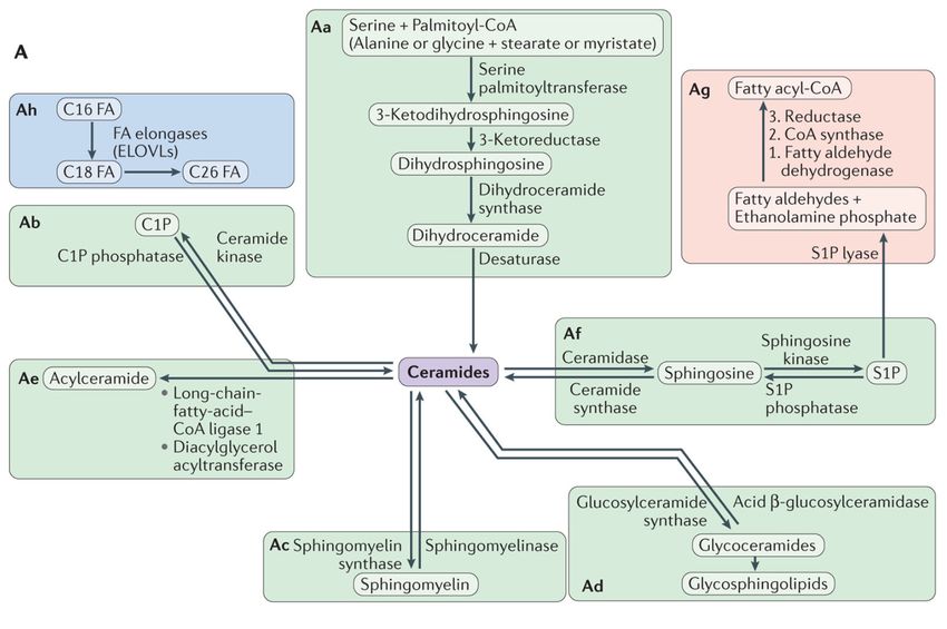
Int. J. Mol. Sci. 2021, 22, x FOR PEER REVIEW 2 of 16
Int. J. Mol. Sci. 2021, 22, 10085 2 of 17
liquid-ordered ones, the latter usually related to the presence of cholesterol [10]. Bilayers
often exhibit a melting temperature (Tm), below which an additional, solid-ordered lamel-
lar phase (commonly known as ‘gel phase’) exists [11]. Tm depends on the lipids present
ininthe
thesystem.
system. This
Thisisis relevant
relevant as
as lipid distribution is extremely
extremely variable
variable ininmembranes,
membranes,
evenmore
even moreififwewe compare
compare not not only different cell lines
lines but
but also
alsodifferent
differentorganelles
organelleswithin
within
aasingle
singlecell
cell (Figure
(Figure 1),
1), or distinct lipid leaflets
leaflets of
of the
the very
verysame
samemembrane
membrane(membrane
(membrane
lipidasymmetry).
lipid asymmetry). In Inaddition
addition other,
other, non-lamellar,
non-lamellar, lipid
lipid morphologies—e.g.,
morphologies—e.g.,hexagonal,
hexagonal,
or cubic [3]—may be present transiently (e.g., in membrane fusion and fissionprocesses,
or cubic [3]—may be present transiently (e.g., in membrane fusion and fission processes,
ororto
tofacilitate
facilitateprotein
protein insertion
insertion in
in membranes
membranes [12,13]),
[12,13]), which
which greatly
greatlyincrease
increasemembrane
membrane
complexityand
complexity and variability.
variability.
Figure
Figure1.1.Glycerophospholipid
Glycerophospholipidcomposition
compositionofoforganelles.
organelles.The
Thebar
bargraph
graphhighlights
highlightsthe
thesubcellular
subcellulardistribution
distributionofofglycer-
glyc-
ophospholipids between the different organelles in baby hamster kidney cells. The pie charts display
erophospholipids between the different organelles in baby hamster kidney cells. The pie charts display the the relative
relativeabun-
abun-
dance
danceofofeach
eachlipid
lipidclass
classin
inorganelles,
organelles,based
based on
on composite
composite data
data from rat hepatocytes
from rat hepatocytes and
and (for
(forlipid
lipiddroplets)
droplets)from
frommurine
murine
hepatocytes. The figure shows the variability of lipids, with phosphatidylcholines (PCs) and phosphatidylethanolamines
hepatocytes. The figure shows the variability of lipids, with phosphatidylcholines (PCs) and phosphatidylethanolamines
(PEs) as the most predominant species. Adapted from Yang, et al. [14].
(PEs) as the most predominant species. Adapted from Yang, et al. [14].
2.2.Lipids:
Lipids:Building
BuildingBlocks
Blocksand and More
More
Lipids,
Lipids, particularly phospholipidsand
particularly phospholipids andsterols,
sterols,play
play a fundamental
a fundamental rolerole as the
as the build-
building
ing blocks
blocks of the
of the cellcell membrane
membrane lipid lipid matrix.
matrix. ThisThis occurs
occurs because
because of amphiphilic
of the the amphiphilic naturena-
ture of these molecules, that favors their self-assembly in aqueous
of these molecules, that favors their self-assembly in aqueous media [2,3]. Lipid self-media [2,3]. Lipid self-
assembly,
assembly, mainly
mainly driven
driven byby entropy,
entropy, is is the
the basis
basis for
for the
the spontaneous
spontaneousformation
formationofofnot notonly
only
cell
cellmembranes
membranesbut but also
also of
of the lipid bilayers
the lipid bilayers used
used asas ‘model
‘modelmembranes’
membranes’ininbiophysical
biophysical
studies
studies(see(seebelow).
below).Moreover,
Moreover, a widespread
a widespread misconception
misconception of lipids in membranes
of lipids in membranes is that
is
they
that have
they only
have aonly
strictly structural
a strictly function.
structural As stated
function. As previously, the structural
stated previously, concept
the structural
ofconcept
cell membranes has evolved
of cell membranes hasover the last
evolved decades
over the lasttowards
decades a more complex
towards a more perspective.
complex
perspective. Accordingly, the role of lipids has also been expanded with the discovery of
their bioactive effects, particularly for sphingolipids, a subfamily of lipids structurally based
on a sphingosine backbone that undergo many metabolic modifications (Figure 2). While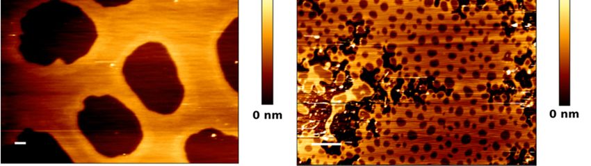
Int. J. Mol. Sci. 2021, 22, x FOR PEER REVIEW 3 of 16
Accordingly, the role of lipids has also been expanded with the discovery of their bioac-
Int. J. Mol. Sci. 2021, 22, 10085 tive effects, particularly for sphingolipids, a subfamily of lipids structurally based 3 ofon
17 a
sphingosine backbone that undergo many metabolic modifications (Figure 2). While
sphingolipids had been discovered over a century ago [15], they were considered as
simply structural lipids, thus attracting scarce interest for 100 years. However, their bio-
sphingolipids had been discovered over a century ago [15], they were considered as simply
active role was described in the 80s of the past century [16,17] suddenly bringing them to
structural lipids, thus attracting scarce interest for 100 years. However, their bioactive role
the spotlight as bioactive protagonists. Sphingolipids such as ceramides [18] have a pro-
was described in the 80s of the past century [16,17] suddenly bringing them to the spotlight
apoptotic role [19,20] and are used as chemotherapeutic agents for cancer treatments
as bioactive protagonists. Sphingolipids such as ceramides [18] have a pro-apoptotic
[21,22]. Other
role [19,20] andsphingolipids, like ceramide-1-phosphate,
are used as chemotherapeutic have treatments
agents for cancer the opposite tendency,
[21,22]. Otheras
they constitute like
sphingolipids, pro-survival signals [23] have
ceramide-1-phosphate, and may be of interest
the opposite as as
tendency, pro-inflammatory
they constitute
agents [24]. Moreover, lipids have more recently attracted a renewed interest
pro-survival signals [23] and may be of interest as pro-inflammatory agents [24]. Moreover, as the met-
abolic impact of lipid droplets (lipid reservoirs within cells) has been unveiled
lipids have more recently attracted a renewed interest as the metabolic impact of lipid [25]. Lipids
could also(lipid
droplets play areservoirs
role in thewithin
immune response
cells) has been(e.g., for virus-infected
unveiled [25]. Lipidscells
could[26])
alsoand
playsomea
reports point
role in the to a functional
immune response relationship between lipid
(e.g., for virus-infected cellsdroplets
[26]) andand
somesphingolipid
reports point metab-
to a
olism [27]. relationship between lipid droplets and sphingolipid metabolism [27].
functional
Overviewofofsphingolipid
Figure2.2.Overview
Figure sphingolipid metabolism.
metabolism. Being
Being the
theprecursors
precursorsofofallallcomplex
complex sphingolipids,
sphingolipids,ceramides
ceramidesconstitute a
constitute
a family
family of
ofclosely
closelyrelated
relatedmolecules
molecules that contain
that a sphingoid
contain basebase
a sphingoid and and
an amino-linked
an amino-linkedfatty acid.
fatty Ceramide can be can
acid. Ceramide generated
be gen-
through
erated different
through pathways
different but most
pathways but of them
most can also
of them canoperate in reverse
also operate and use
in reverse andceramide to produce
use ceramide other other
to produce metabolic
meta-
bolic products
products (A).
(A). In In addition,
addition, different
different enzymesenzymes may introduce
may introduce variations
variations to the basicto the basic structure,
structure, increasing increasing variability
variability (B). From
(B). From Hannun
Hannun and Obeid and Obeid [28].
[28].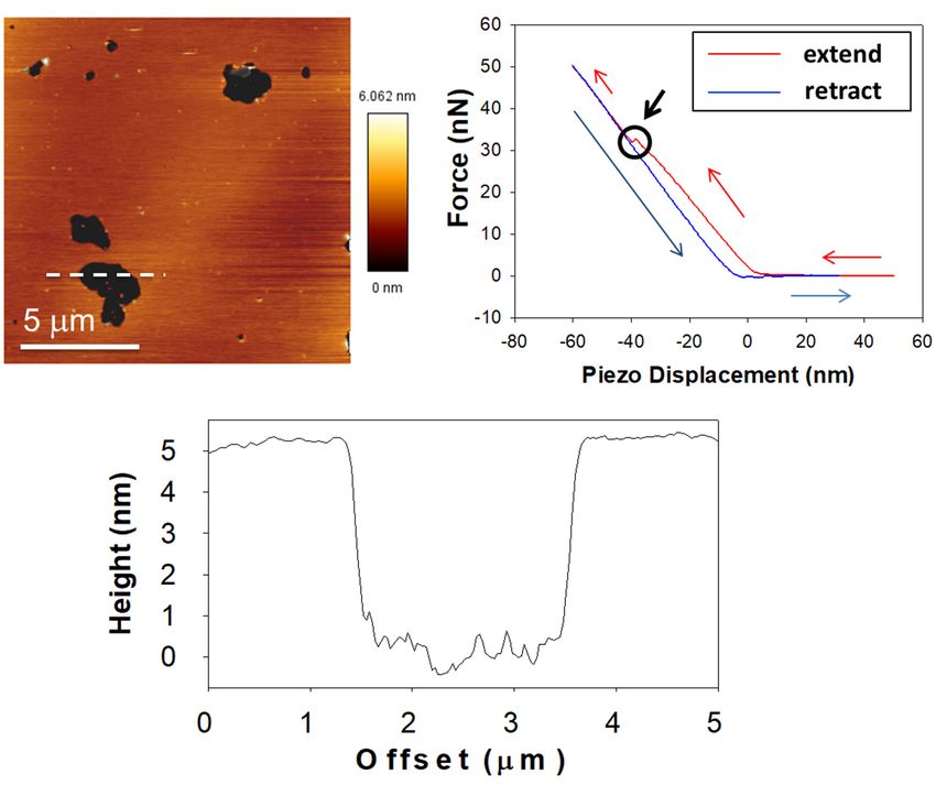
Int. J. Mol. Sci. 2021, 22, 10085                                                                                           4 of 17

                                          Although the biological impact of lipid signaling is currently beyond discussion, how
                                    does this relate to cell membrane structure? That is perhaps the most important question
                                    regarding this topic, not yet fully understood. What we actually do know is that sphin-
                                    golipids have (usually) a higher Tm than their glycerolipid counterparts, and accordingly
                                    the former have a tendency to form membrane domains. This makes them extremely
                                    impactful for the biophysical properties of membranes [18]. Ceramides, due to their high
                                    hydrophobicity as well as their high Tm values, tend to form gel phase platforms [29–32]
                                    depending on their carbon chain length [33–35], while also inducing membrane permeabil-
                                    ity [36–40] and lipid flip-flop (loss of bilayer asymmetry) [41]. Sphingomyelins (which are
                                    non-bioactive per se) are considered to be an integral part of the liquid-ordered domains
                                    as they have a strong affinity for cholesterol [42], but they are also reservoirs for quick
                                    ceramide generation via sphingomyelinases [43]. This means that ceramide formation
                                    may not only cause ceramide-enriched ‘gel’ domains but also contribute to ‘liquid-ordered’
                                    nanodomains to coalesce due to sphingomyelin cleavage and recruitment. In fact, a com-
                                    petition between cholesterol and ceramide for sphingomyelin binding was proposed as
                                    they shared the tendency to occupy the same hydrophobic pockets for sphingomyelin
                                    interaction [44–46], but later reports indicated that this displacement might not be occur-
                                    ring in many relevant cases where both molecules can be accommodated and interact in a
                                    stabilizing manner [30,47–49], as discussed in a previous report [50].
                                          To further assess the relationship between membrane structure and the bioactive
                                    (apoptotic) effects of its constituting lipids, the current consensus hypothesis is that sph-
                                    ingolipids play a key role in the formation of the transient MOMP (Mitochondrial Outer
                                    Membrane Pore), essential for cytochrome c translocation from mitochondria to cytosol,
                                    thereby triggering the intrinsic apoptotic cascade [51]. For this purpose, ceramides have
                                    been proposed to interact with Bcl-2 family apoptotic proteins such as Bak [52] and Bax [53].
                                    How ceramides are transported to the mitochondrial outer membrane from the endo-
                                    plasmic reticulum is an important target of current research [54]. In addition, ceramide
                                    platforms have also been proposed to interact with Fas [55] for receptor-mediated extrinsic
                                    apoptosis, which is non-cytochrome c dependent.

                                    3. Membrane Biophysics: Model Membranes
                                          The study of membrane structural properties and characterization is usually named
                                    ‘membrane biophysics’. The complexity of membranes in living cells requires the use of
                                    model membranes to understand the basic principles of molecular interactions in those
                                    structures. ‘Model membranes’ is a wide concept that may include a vast array of sim-
                                    plified systems, from those composed of a single lipid, to lipid membrane extracts that
                                    may also include proteins. One common feature of model membranes is that they are
                                    dependent on the self-assembly properties of membrane lipids, mainly phospholipids and
                                    cholesterol. The usual approaches in membrane biophysics through model membranes are:
                                    (i) to gradually increase complexity of the systems when the most basic ones have been
                                    understood (a Cartesian approach) and/or (ii) to compare different sets of systems where
                                    only one component (or parameter) has been changed (a comparative approach).
                                          Lipid polymorphism is a factor that increases even more the possibilities for model
                                    membranes. As previously stated, lipids have their own phase behavior, and lipid geometry
                                    is very important in this respect, as it governs membrane curvature, thus the mode(s) of
                                    lipid self-assembly [56]. Depending on the intrinsic curvature of lipids present in a sample,
                                    different non-lamellar structures can be induced, such as micelles, tubules (tethers), or
                                    hexagonal (normal or inverted) phases [57,58].
                                          The most commonly used model system in membrane biophysics is the liposome, as
                                    it shares the basic structural principles of a biological membrane: a lamellar lipid structure
                                    which isolates an internal aqueous medium from an external one. While the liposome
                                    does not present the internal architecture of a cell (it lacks cytoskeleton, for instance)
                                    and usually forms spherical structures, it allows a controlled lipid environment as we
                                    define the lipid composition of the system. Liposomes are also used as drug carriers in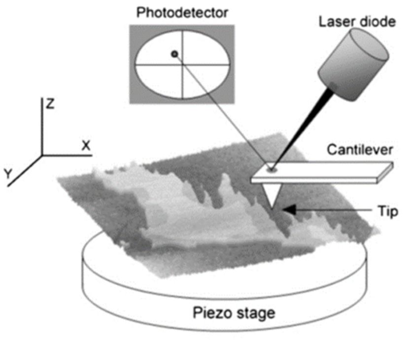
Int. J. Mol. Sci. 2021, 22, 10085                                                                                            5 of 17

                                    pharmaceutical industry [59,60]—e.g., in cancer treatments [61] or COVID-19 vaccines,
                                    from Pfizer (BNT162b1) [62] or Moderna [63].
                                         Liposomes can be classified in two groups: (i) multilamellar vesicles (MLV), which
                                    are spontaneously formed when a lamellar-inducing lipid mixture is dispersed in an
                                    aqueous medium; or (ii) unilamellar vesicles, which in most cases come from MLV but
                                    require further treatment, and are classified by size into small, large, or giant unilamellar
                                    vesicles. For instance, giant unilamellar vesicles (GUV, >1 micron diameter) are often used
                                    under fluorescence confocal microscopy, combined with fluorescent probes [64,65]. Large
                                    unilamellar vesicles (LUV, from 1 micron to 100 nm diameter) are useful for membrane
                                    permeation (leakage) assays [66]. Small unilamellar vesicles (SUV,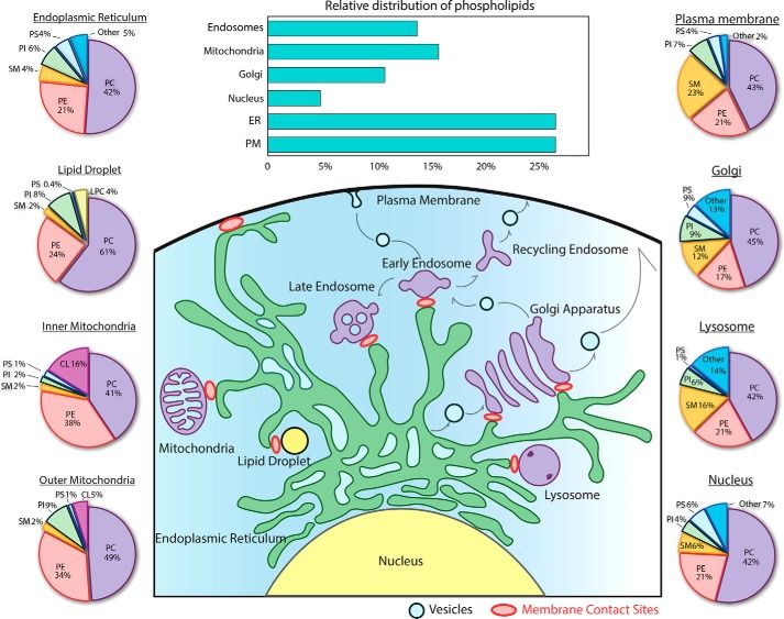
The basic parts of an AFM setup are shown in Figure 3. In summary, the sample is
scanned by a tip attached to a cantilever, which is focused by a laser beam. When a change
in the height profile of the surface (Z axis) is encountered, the cantilever bends accordingly
and this causes a deflection of the laser. This deflection is quantified by a photodetector6 of 17
Int. J. Mol. Sci. 2021, 22, 10085
and interpreted, with the supporting software included in the AFM. Depending on the
mode of use, the AFM interface will generate by itself the result—e.g., an image or a force
curve. The basic parts of an AFM setup are shown in Figure 3. In summary, the sample is
Another interesting
scanned feature of AFM
by a tip attached to ausage is the
cantilever, whichavailability
is focused by of adifferent
laser beam.modes
When afor change
data retrieval, particularly
in the height during
profileimage acquisition,
of the surface (Z axis) depending
is encountered, onthe
the nature bends
cantilever of theaccordingly
sam-
ple under study, and and onthis tip–surface
causes a deflection of the laser.The
interactions. This ‘contact
deflection mode’
is quantified by a photodetector
is commonly used, and
interpreted, with the supporting software included in the AFM. Depending on the mode of
in it the tip is constantly in contact with the sample. However, in biological samples, this
use, the AFM interface will generate by itself the result—e.g., an image or a force curve.
mode may be intrusiveAnother as, depending
interesting on the of
feature applied
AFM usage force and
is the the nature
availability of ourmodes
of different sample, for data
the tip can damageretrieval,
the sample and dramatically
particularly affect its depending
during image acquisition, integrity: on objects mayofbe
the nature thedis-
sample
placed from their original position or partially disassembled. For this purpose, a more in it
under study, and on tip–surface interactions. The ‘contact mode’ is commonly used,
the tip is constantly in contact with the sample. However, in biological samples, this mode
convenient mode would be the use of intermittent contact, such as tapping or other more
may be intrusive as, depending on the applied force and the nature of our sample, the tip
advanced methodscan that combine
damage imaging
the sample and force spectroscopy,
and dramatically affect its integrity:which
objectswill
may be later de-from
be displaced
scribed. their original position or partially disassembled. For this purpose, a more convenient mode
Due to the scanning
would beprobe the usenature of thecontact,
of intermittent AFM, such it also exhibits
as tapping or some
other moremethodological
advanced methods
that combine imaging and force spectroscopy, which
problems or liabilities. AFM tips can spontaneously ‘pick up’ debris or aggregates affect- will be later described.
Due to the scanning probe nature of the AFM, it also exhibits some methodologi-
ing the original properties of the tip or the cantilever, e.g., the spring constant of the can-
cal problems or liabilities. AFM tips can spontaneously ‘pick up’ debris or aggregates
tilever may be altered (although
affecting in many
the original propertiescasesof a
there-calibration of thee.g.,
tip or the cantilever, system is enough
the spring constantto of the
circumvent the issue) or themay
cantilever tip may lose(although
be altered its intended
in many shape.
casesThus, tip or cantilever
a re-calibration of the systemfouling
is enough
is a recurring problem to circumvent
for AFMthe usersissue)
as itoroften
the tipgenerates
may lose its intended
artifacts andshape. Thus,immediate
requires tip or cantilever
fouling is a recurring problem for AFM users as it often generates artifacts and requires
washing of the tip/cantilever or, usually, a replacement with a clean new one. Tip func-
immediate washing of the tip/cantilever or, usually, a replacement with a clean new one.
tionalization (i.e., the
Tip chemical modification
functionalization of the AFM
(i.e., the chemical tip with
modification a specific
of the AFM tipagent) is also agent)
with a specific a
common way to limit undesired
is also a commoninteractions [87]. interactions [87].
way to limit undesired
Figure 3. Overview of the basic parts of an AFM. A laser is focused on the tip that reflects the beam
Figure 3. Overview of the basic parts of an AFM. A laser is focused on the tip that reflects the beam
towards a photodetector. Tip–surface interactions bend the cantilever, which in turn generates a laser
towards a photodetector. Tip–surface interactions bend the cantilever, which in turn generates a
beam displacement. Then, the photodetector and the AFM software transform laser movements into
laser beam displacement. Then,
the desired the photodetector
information. and theand
From Garcia-Manyes AFMSanzsoftware
[88]. transform laser move-
ments into the desired information. From Garcia-Manyes and Sanz [88].
5. Membranes under AFM: The Basics
5. Membranes under AFM:
The useThe Basics
of AFM on membrane models has acquired a particular prominence in the
The use of AFMlast on
twomembrane
decades [70,88]. The basic
models has principle
acquired was to form SUVprominence
a particular of a defined composition
in the
and then extend them to form SPBs (vesicle adsorption method), as previously mentioned.
last two decades [70,88]. The basic principle was to form SUV of a defined composition
However, different methodologies were developed later, such as the direct spin-coating of
and then extend them toonto
lipids formtheSPBs (vesicle
support adsorption
surface [89]. This ismethod),
of interestas
as previously mentioned.
the vesicle adsorption method
However, different methodologies were developed later, such as the direct spin-coating
of lipids onto the support surface [89]. This is of interest as the vesicle adsorption method
requires heating of the SUV over the Tm, which in some samples may be difficult to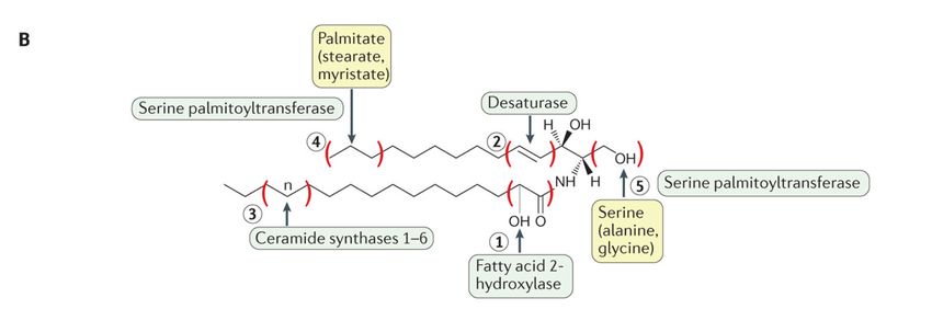
Int. J. Mol. Sci. 2021, 22, 10085                                                                                              7 of 17

                                     requires heating of the SUV over the Tm , which in some samples may be difficult to achieve,
                                    particularly if the mixture presents a high concentration of saturated long chain ceramides,
                                     because of their high Tm values [90]. Another advantage of the alternative spin-coating
                                     method is the absence of divalent cations, which are required in the vesicle adsorption
                                     method and, if not washed properly afterwards, may affect measurements [91,92]. Sample
                                    preparation methods are in constant improvement as in most cases protocol optimization
                                     is the easiest way to increase the quality of an experiment. For instance, SPB preparation
                                     may lead (depending on the chosen method, the details of the protocol, and the nature of
                                     the lipids involved) to inefficient support coverage, as bilayers appear as distinct ‘patches’,
                                     sometimes with insufficient area to perform reproducible measurements and generating
                                     artifacts due to inefficient lipid mixing if two or more lipids are present. These small
                                    patches often exhibit, for instance, abnormal thicknesses [93], pointing to the presence of an
                                     artifact. A common way to improve bilayer extensions for the vesicle adsorption method
                                     is the use of divalent cations, typically calcium (II) [94–96], although in many cases these
                                     cations require to be washed away after SPB formation, as previously mentioned. SPB are
                                     also required to exhibit the lowest possible roughness in order to decrease any background
                                    ‘noise’, this being particularly important in high-resolution AFM imaging. Thus, the nature
                                     of the support is also a key factor, and SPB experiments in most cases use atomically flat
                                     mica sheets, while supports such as silicon, HOPG (highly oriented pyrolytic graphite) [97]
                                     or silicon oxide are used for AFM characterization of other types of biological samples.
                                           What is the precise information retrieved when AFM is applied to membranes? What
                                     is the particular purpose of this technique? AFM, as a scanning probe technique, can
                                    provide information on two general aspects: topography and nanomechanics. These
                                     two are respectively linked to image acquisition and force spectroscopy. On one hand,
                                    AFM provides direct images of any surface under scan, through different modes (contact,
                                     non-contact, intermittent contact . . . ). On the other hand, AFM can ‘touch’ the surface
                                     to test its nanomechanical properties (nanomechanical resistance, elasticity . . . ). These
                                     modes are not mutually exclusive, as image acquisition may be followed by localized force
                                     spectroscopy on a specific point of the previous image, but a stable sample is required.
                                    Thus, AFM provides information about membrane thickness (including the presence
                                     of segregated domain phases and the detection of proteins, characterizing subunits in
                                     some cases [98]) and about membrane stiffness (localized, for example, for every phase
                                    present [99]). Figure 4 shows an example of this combined approach. AFM can also be
                                     associated with fluorescence-related techniques, with an appropriate setup [100].
                                           Over the last decade, improvements in AFM technology point in the same direction,
                                     namely reducing the time required for a full characterization of any sample, not only
                                     because of the fast nature of biological processes, but also to reduce the possibility of an
                                     impact of the AFM scan on the state of the sample over time, such as: (i) sample alteration
                                     by tip contact, (ii) local heating produced by laser focalization, or (iii) (if the experiment is
                                     combined with fluorescence monitoring) fluorescent probe photobleaching by the laser.
                                           In addition to the aforementioned problems, the time scale of conventional time-
                                     resolved AFM experiments may also present issues not necessarily related to sample
                                     alteration but associated to the AFM itself. Particularly for experiments in aqueous media,
                                    which is the case for biological experiments, laser alignment has a tendency to drift over
                                     time. Although this drift may seem a small problem at first glance, if a conventional time-
                                     resolved AFM image takes, for instance, around 10 min to complete, this means a significant
                                     difference between starting vs. final laser alignment. Why is this relevant? Because laser
                                     alignment defines the force that the AFM system applies on the sample: the drift causes
                                     the starting force to change during the experiment and it has to be constantly readjusted to
                                     keep it at the desired value (commonly the lowest possible one to avoid sample damaging,
                                     as drift usually causes higher forces to be applied) [101]. This is particularly important
                                     because once the AFM scan starts, force readjustment is also subject to drifting, which
                                     means that the adjustment has to be performed as an empirical approximation at the risk
                                     of losing contact with the sample if the force is excessively decreased.
J. Mol. Sci. 2021, 22, x FOR PEER REVIEW 8 of
Int. J. Mol. Sci. 2021, 22, 10085 8 of 17
Thus, AFM technology has evolved into more complex (faster) modes that can co
bine both image acquisition and force spectroscopy at the same time [102]. Another use
improvement Thus, AFM technology
for the has evolved
technique is the into more complex
development of(faster) modes that
high-speed can combine
AFM, allowing the
both image acquisition and force spectroscopy at the same time [102]. Another useful
trieval of multiple successive images per second (while the conventional AFM usua
improvement for the technique is the development of high-speed AFM, allowing the
takesretrieval
some min to perform
of multiple a single
successive imagesimage) in such
per second a way
(while that biological
the conventional AFMprocesses
usually may
monitored andmin
takes some recorded onavideos
to perform [103].inThese
single image) such a advances successfully
way that biological overcome
processes may be most
monitored and recorded
the aforementioned issueson videosto
related [103]. Thesealteration
sample advances successfully overcomealthough
or AFM drifting, most of syste
the aforementioned issues related to sample alteration or AFM drifting, although
are still subject to methodological AFM imitations (undesired tip-sample interactions, d systems
are still subject to methodological AFM imitations (undesired tip-sample interactions,
bris attachment to the tip), so there is probably still room for further improvements in t
debris attachment to the tip), so there is probably still room for further improvements in
regard.
this regard.
Classic experimental
Figure 4.experimental
Figure 4. Classic approach
approach forfor
SPBSPBcharacterization
characterization under the AFM.
under Upper-left
the AFM. panel shows
Upper-left an AFM
panel shows image of
an AFM image
a lipid bilayer in a gel phase (pSM:Chol:pCer 54:23:23, as published in García-Arribas, et al. [30]). The height
of a lipid bilayer in a gel phase (pSM:Chol:pCer 54:23:23, as published in García-Arribas, et al. [30]). The height profile of profile of the
the dasheddashed
line isline is depicted in the bottom panel; the black area is a bilayer defect that allows direct quantification of bilayer
depicted in the bottom panel; the black area is a bilayer defect that allows direct quantification of bilayer
thickness (≈5 nm). The upper-right panel is a force-distance curve where the extension is colored in red and the retraction
thickness (≈5 nm). The upper-right panel is a force-distance curve where the extension is colored in red and the retraction
in blue. The circle indicated by the black arrow highlights the breakthrough event, the latter marking the force required to
in blue. The circle indicated by the black arrow highlights the breakthrough event, the latter marking the force required
pierce through the bilayer (which is related to bilayer stiffness).
to pierce through the bilayer (which is related to bilayer stiffness).
6. Membranes under the AFM: Findings
It would be difficult to review the whole relevant research currently performed w
AFM on membranes, within reasonable length limits, thus our contribution will focus
some specific points of interest that the AFM has revealed or confirmed, beyond what h
been discovered or observed using other techniques.Int. J. Mol. Sci. 2021, 22, 10085 9 of 17
6. Membranes under the AFM: Findings
It would be difficult to review the whole relevant research currently performed with
AFM on membranes, within reasonable length limits, thus our contribution will focus on
some specific points of interest that the AFM has revealed or confirmed, beyond what had
been discovered or observed using other techniques.
One interesting aspect is related to the aforementioned variability among different
membrane models. As previously explained, liposomes and liposome-derived models
are most commonly used in membrane biophysics, but they are not the only ones and,
even when using liposomes, there are different preparation methods, each affecting the
final outcome. A recent report by Monasterio et al. [104] makes an interesting compari-
son of three well-established models for cell membrane analysis: vesicles obtained from
lipid extracts, inside-out membrane patches and membrane blebbing. At first glance, it
would be easily assumable that the most ‘different’ of the three would be the pure lipid
samples. Moreover, considering that both patches and lipid-extract-derived SPBs are,
indeed, supported samples, while the blebs are vesicles ‘attached’ to the surface but not
fully supported, the values of bleb stiffness would be expected to be much lower than
in the other two preparations. However, the study proves that bleb vesicles have the
highest stiffness measured by AFM force spectroscopy, and, indeed, the biggest difference
in bilayer stiffness is detected between blebs and lipid extracts. This study demonstrates,
with the use of AFM, that different membrane models present clearly distinct properties.
AFM characterization of SPB has also revealed unexpected behavior even in the
simplest systems. The biophysical properties of sphingolipids—e.g., sphingomyelin, or
ceramide—are markedly influenced by the length of their N-acyl chain [105]. A report by
Jiménez-Rojo et al. [90] showed that pure C18:0 SM SPB had the capacity to achieve a stable
phase separation due to an unusual complex transition (two peaks detected in DSC thermo-
grams). The appearance of domains when monitoring a gel-to-fluid transition with AFM
had been documented earlier [106], but they were typically unstable as they represented
the transition itself (one of the phases ended being predominant and the other disappeared).
The capacity of a single lipid to maintain a stable phase separation at a specific temperature
opens new possibilities for an increased membrane heterogeneity in living cells. Another
unexpected finding, this time using force spectroscopy, appeared in a report by Balleza
et al. [107], revealing that bilayers made of ‘branched’ lipids (the ether-based diphytanyl
and the ester-based diphytanoyl PC), both exhibited breakthroughs at two different forces
despite being apparently homogenous under the AFM. The same phenomenon had also
been reported by Redondo-Morata et al. [108], albeit in a binary system: DPPC/Chol
at an equimolar ratio presented two different nanomechanical resistance values, despite
appearing homogenous under AFM imaging. These findings point to two possibilities:
(i) two different but equally favored ways (from the point of view of thermodynamics)
to achieve an AFM tip breakthrough or, perhaps more probable; (ii) nanoscopic domains
below the resolution capacity of AFM tips (nominal radius typically around 20 nm). We
should also consider that nanoscopic domains could be more likely to appear when two or
more lipid species are present, just as in the report by Redondo-Morata et al. [108], rather
than in bilayers made of a single lipid such as those studied by Balleza et al. [107].
Force spectroscopy of lipid bilayers under the AFM has also revealed a particularly
interesting finding related to the capacity of both lipid leaflets to interact between them
in a single bilayer. Briefly, lipid membranes are often considered a single entity, and,
while leaflet asymmetry has been thoroughly documented over the last decades, the
general consensus was that the whole membrane was a single physico-chemical structure,
regardless of the lipid asymmetry present. Then, the idea of each lipid leaflet of a bilayer
presenting different biophysical properties was suggested, and finally demonstrated in a
report by Alessandrini et al. [109] in 2012. This study described that AFM force spectroscopy
of lipid bilayers made of a single lipid could exhibit a two-step breakthrough. This was
interpreted as the tip first piercing the proximal leaflet (the one farther away from the
support, and closer to the AFM tip) and then, at a higher force, the distal leaflet (inInt. J. Mol. Sci. 2021, 22, 10085 10 of 17
direct contact with the support, farther from the AFM tip). The same report indicated
that a SPB could exhibit either a one-step or a two-step piercing process, depending on
the temperature used for the vesicle adsorption method. This phenomenon was called
‘membrane uncoupling’ and seemed to occur when using lower temperatures during
bilayer formation. Interestingly, the T threshold for a lipid to form coupled or uncoupled
membranes was related to Tm in some cases; for instance, POPE bilayers were uncoupled
when formed at T = 15 ◦ C but coupled at T = 30 ◦ C (POPE Tm = 25 ◦ C), pointing to the
importance of achieving a fluid phase for a coupled SPB preparation. However, other
lipids, such as POPG, exhibited uncoupled bilayers well above the Tm in the same report,
which indicates that not every fluid lipid gives rise to coupled mechanics. The same study
also pointed to AFM tip velocity as a factor to detect uncoupled bilayers, as higher tip
speeds showed but a single event, which the authors explained by an increased friction
between leaflets.
The relevance of the unexpected membrane uncoupling was clear, as it demonstrated
that different leaflets of a single bilayer could have different properties, and the support
could exert a stiffening effect on the distal leaflet. It also meant that the membrane was able
to achieve a metastable state when the tip was piercing the first leaflet only, demonstrating
that the distal leaflet was not immediately collapsing. Membrane uncoupling was later
detected in samples formed by the spin-coating procedure, and, in some cases, it was
even detected for both the spin-coating and the vesicle adsorption procedures of the same
lipid sample, which essentially discarded any contribution caused by the divalent cations
used in the vesicle adsorption method [30]. In addition, lipid samples that could exhibit
membrane uncoupling included pure gel-phase samples and also liquid-ordered ones,
demonstrating that the Tm dependency was not as clear as previously presumed [30]. All
these results point to a complex nanomechanical behavior of SPB, resulting from at least (i)
the preparation procedures; (ii) the nature of the lipids; (iii) the properties of the support;
and (iv) an important contribution from the instrumental setup and settings. This issue
was thoroughly analyzed in a report by Relat-Goberna et al. [110]. A more recent report
by Vázquez et al. [111] showed that lipid leaflets could become locally uncoupled after
preparation, thus inducing bilayer asymmetry. This report points to lipid phase mismatch
between leaflets as an additional factor to bilayer uncoupling, which greatly increases the
complexity of these events as lipid phases may be locally segregated not only laterally as
we would initially think, but also in an asymmetrical fashion along each leaflet. This ‘local
uncoupling’ presents itself as a novel, interesting topic in membrane heterogeneity studies.
Hopefully, further research will focus on the impact of this phenomenon on lipid–protein
interactions within the membrane, particularly for transmembrane proteins.
Regarding lipid–protein interactions, a recent report from Banerjee and Lyubchenko [93]
showed the importance of the presence of a SPB during the amyloid fibril (Aβ42) for-
mation process. Aβ42 reportedly adsorbs to POPC-containing bilayers, without being
inserted [112]. Using an optimized protocol for multicomponent SPB preparation in order to
have smooth and large lipid extensions on the mica support, Banerjee and Lyubchenko [93]
monitored on-membrane aggregation of protofibrils under the AFM. Interestingly, the
aggregation process of oligomers generated the formation of protofibrils, and through
quantitative analysis of AFM images, the authors demonstrated that these protofibrils
exhibited a shorter length when compared to the mature solution-generated fibrils. In
addition, on-membrane generated protofibrils appeared straight, non-twisted and non-
branched. This is relevant because it supports the hypothesis of the governing role of
membrane and protein-lipid interaction for Aβ42 fibril formation.
7. Impact on Membrane Biophysics ‘Hot Topics’
The influence of the support in SPB has been a hot topic of research, not only because
of the nature of SPB as model systems, but also due to the great underlying question of
‘how does cytoskeleton affect living membranes?’ This is not an easy question to answer
for membrane biophysicists, as most membrane models are unsupported or, if supported,Int. J. Mol. Sci. 2021, 22, 10085 11 of 17
are not cytoskeleton-bound. In this particular instance, the use of supported models
(such as SPB) may be more interesting than unsupported ones, as they could to some
extent reproduce some of the effects that cytoskeleton could exert [113]. Not unexpectedly,
different supports show different properties. Reports indicate that rougher supports tend
to decrease the size of segregated phases in a lipid SPB [114,115]. This is a relevant finding
because it could explain why the size of lipid domains exhibits such a great variability
between different models: liposomes (such as GUV) present domains of up to several
microns [31], while mica-supported bilayers visualized by either AFM or fluorescence often
exhibit domains up to 10 times larger [30,116].
Domain size is also a recurrent topic of discussion, as one of the main criticisms
received by the ‘lipid raft’ hypothesis is the current near-impossibility of directly visualizing
lipid domains on living cell membranes, while in model systems they can be clearly
assessed. This has been traditionally explained by the transient nature of the domains and
the nanoscopic size, below the resolution limit of many techniques. Regarding the latter,
there is a further hypothesis that states the possibility that the cytoskeleton is the cause
of the small size of the domains, as the cytoskeleton would define ‘boundaries’ within
the membrane (‘picket fence model’ [117]) and the domains would appear at the contact
points between lipids and the cytoskeleton meshwork [114]. Recent reports on membrane
diffusion dynamics further support this hypothesis [72].
It should be noted that some of these phenomena have already appeared in non-
cytoskeleton supported bilayers as well. As already stated, previous reports by Balleza
et al. [107] and Redondo-Morata et al. [108] indicate that nanoscopic domains are a pos-
sibility. It should also be taken into consideration that some of the previously described
findings, such as multi-step breakthrough events due to bilayer uncoupling [30,109–111] or
two-modal breakthrough events in apparently homogenous samples [107,108], are almost
certainly related to the fact that SPB are, indeed, supported. Would these events also hap-
pen in cytoskeleton-supported bilayers? Further experiments will probably try to answer
this question in the future, but it seems a reasonable assumption that if nanodomains are
present in both SPB and cytoskeleton-based bilayers, then cytoskeleton may also have a
role in local membrane uncoupling [111] as the leaflet in contact with the cytoskeleton will
likely have some different biophysical properties than the opposite one.
Regarding non-cytoskeleton supported SPB, a recent publication from our lab [34]
also demonstrated, using AFM, that domains might be unstable in SPB for some lipid
mixtures (related to the combined presence of C24:0 and C24:1 sphingolipids), to the
point of completely disappearing over time, while non-supported liposomes of the same
composition were perfectly stable. This would point to a support-driven effect that reduces
domain size over time until they become undetectable by AFM imaging (the possibility of
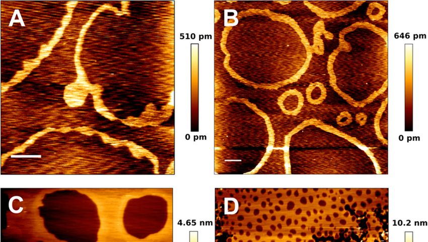
nanodomains under the AFM resolution limit cannot be discarded). Furthermore, another
AFM study revealed recently that some lipid compositions of sphingolipids and cholesterol
may form ring-like phases (Figure 5), even in the absence of any cytoskeleton molecule [99].
These ring-like gel phases greatly affected the nanomechanical properties of the lipid phase
‘inside’ the boundary defined by the ring, increasing the resistance to a point that the AFM
tip was unable to pierce through it, indicating an extremely packed and stiff phase [118,119].
All these results considered, the influence of supports is undeniable and the complexity of
the supported lipids even in the absence of proteins cannot be underestimated.Int. J. Mol. Sci. 2021, 22, 10085 12 of 17
Int. J. Mol. Sci. 2021, 22, x FOR PEER REVIEW 12 of 16
Figure 5. AFM
Figure images
5. AFM of pSM:pCer:Chol-based
images of pSM:pCer:Chol-basedSPB SPBatat different molratios.
different mol ratios.pSM:pCer:Chol
pSM:pCer:Chol (54:23:23)
(54:23:23) (A), (A), (60:20:20)
(60:20:20) (B), (B),
(66:17:17) (C), and (70:15:15) (D). Scale bars: 1 μm. Panels A and B show the spontaneous formation of ‘ring-like’
(66:17:17) (C), and (70:15:15) (D). Scale bars: 1 µm. Panels A and B show the spontaneous formation of ‘ring-like’ phases in phases
in absence of proteins. Adapted from González-Ramírez et
absence of proteins. Adapted from González-Ramírez et al. [99]. al. [99].
8. Conclusion and
8. Conclusions Perspectives
and Perspectives
The
The useofofAFM
use AFMin in membrane
membrane biophysics,
biophysics,and and particularly in the
particularly study
in the of lipid
study self- self-
of lipid
assemblies, has helped in gaining a more thorough understanding on
assemblies, has helped in gaining a more thorough understanding on how membranes how membranes are
are built. However, more importantly perhaps, it has also pointed to new relevanttodetails
built. However, more importantly perhaps, it has also pointed to new relevant details and
a higher degree of complexity in totally unexpected issues that went previously undetected
and to a higher degree of complexity in totally unexpected issues that went previously
due to the limitations of other techniques. Moreover, as AFM technology evolves towards
undetected
faster and due
bettertoresolved
the limitations of other techniques.
systems, supported by advancedMoreover, as AFMsuch
imaging techniques technology
as
evolves
STED,towards
we should faster and
expect better
that theseresolved
issues aresystems,
solved andsupported by advanced
suitable answers imaging tech-
are conveniently
niques
found such as STED,
for our we should
questions. expect
The increase that these issues
in complexity are solved
of the samples andstudy,
under suitable answers
as well
areasconveniently
the improvementsfoundinfor
theour questions.
scanning The increase
techniques, in complexity
will probably of theunexpected
generate new, samples under
questions,
study, as wellas further
as the research targets will
improvements surely
in the appear. techniques, will probably generate
scanning
new, unexpected questions, as further research targets will surely appear.
Funding: This research received no external funding.
Funding: This research
Institutional received
Review Board no external
Statement: funding.
Not applicable.
Informed Consent
Institutional ReviewStatement: Not applicable.
Board Statement: Not applicable
Acknowledgments:
Informed A.B.G.A. was
Consent Statement: Nota post-doctoral
applicable scientist supported by the University of the Basque
Country and the Basque Government. This work was supported in part by grants from the Spanish
Acknowledgments: A.B.G.A. was a post-doctoral scientist supported by the University of the
Basque Country and the Basque Government. This work was supported in part by grants from the
Spanish Ministry of Economy (grant FEDER MINECO PGC2018-099857-B-I00) and the Basque Gov-
ernment (grants no. IT1264-19 and IT1270-19), as well as Fundación Biofísica Bizkaia and the BasqueInt. J. Mol. Sci. 2021, 22, 10085 13 of 17
Ministry of Economy (grant FEDER MINECO PGC2018-099857-B-I00) and the Basque Government
(grants no. IT1264-19 and IT1270-19), as well as Fundación Biofísica Bizkaia and the Basque Excellence
Research Centre (BERC) program of the Basque Government.
Conflicts of Interest: The authors have stated explicitly that there are no conflict of interest in
connection with this article.
References
1. Overton, C.E. Über die osmotischen Eigenschaften der lebenden Pflanzen-und Tierzelle; Fäsi & Beer: Zurich, Switzerland, 1895.
2. Singer, S.J.; Nicolson, G.L. The fluid mosaic model of the structure of cell membranes. Science 1972, 175, 720–731. [CrossRef]
3. Goñi, F.M. The basic structure and dynamics of cell membranes: An update of the Singer—Nicolson model. Biochim. Et Biophys.
Acta (BBA)-Biomembr. 2014, 1838, 1467–1476. [CrossRef] [PubMed]
4. Chapman, D.; Gomez-Fernandez, J.C.; Goni, F.M. Intrinsic protein—Lipid interactions: Physical and biochemical evidence. FEBS
Lett. 1979, 98, 211–223. [CrossRef]
5. Engelman, D.M. Membranes are more mosaic than fluid. Nature 2005, 438, 578–580. [CrossRef]
6. Bagatolli, L.A.; Mouritsen, O.G. Is the fluid mosaic (and the accompanying raft hypothesis) a suitable model to describe
fundamental features of biological membranes? What may be missing? Front. Plant Sci. 2013, 4, 457. [CrossRef] [PubMed]
7. Van Meer, G. Dynamic transbilayer lipid asymmetry. Cold Spring Harb. Perspect. Biol. 2011, 3, a004671. [CrossRef]
8. Goñi, F.M.; Alonso, A.; Contreras, F.X. Membrane Nanodomains. In eLS; John Wiley & Sons, Ltd.: Chichester, UK, 2020.
9. Simons, K.; Ikonen, E. Functional rafts in cell membranes. Nature 1997, 387, 569–572. [CrossRef]
10. Shimshick, E.J.; McConnell, H.M. Lateral phase separations in binary mixtures of cholesterol and phospholipids. Biochem. Biophys.
Res. Commun. 1973, 53, 446–451. [CrossRef]
11. Chapman, D.; Collin, D.T. Differential thermal analysis of phospholipids. Nature 1965, 206, 189. [CrossRef]
12. Epand, R.M. Lipid polymorphism and protein-lipid interactions. Biochim. Et Biophys. Acta (BBA)-Rev. Biomembr. 1998, 1376,
353–368. [CrossRef]
13. Chernomordik, L.V.; Zimmerberg, J.; Kozlov, M.M. Membranes of the world unite! J. Cell Biol. 2006, 175, 201–207. [CrossRef]
14. Yang, Y.; Lee, M.; Fairn, G.D. Phospholipid subcellular localization and dynamics. J. Biol. Chem. 2018, 293, 6230–6240. [CrossRef]
15. Thudichum, J.L.W. A Treatise on the Chemical Constitution of the Brain: Based Throughout Upon Original Researches; Baillière, Tindall,
and Cox: London, UK, 1884.
16. Merrill, A.; Sereni, A.; Stevens, V.; Hannun, Y.; Bell, R.; Kinkade, J. Inhibition of phorbol ester-dependent differentiation of human
promyelocytic leukemic (HL-60) cells by sphinganine and other long-chain bases. J. Biol. Chem. 1986, 261, 12610–12615. [CrossRef]
17. Hannun, Y.A.; Loomis, C.R.; Merrill, A.H., Jr.; Bell, R.M. Sphingosine inhibition of protein kinase C activity and of phorbol
dibutyrate binding in vitro and in human platelets. J. Biol. Chem. 1986, 261, 12604–12609. [CrossRef]
18. Castro, B.M.; Prieto, M.; Silva, L.C. Ceramide: A simple sphingolipid with unique biophysical properties. Prog. Lipid Res. 2014,
54, 53–67. [CrossRef]
19. Obeid, L.M.; Linardic, C.M.; Karolak, L.A.; Hannun, Y.A. Programmed cell death induced by ceramide. Science 1993, 259,
1769–1771. [CrossRef] [PubMed]
20. Summers, S.A.; Chaurasia, B.; Holland, W.L. Metabolic messengers: Ceramides. Nat. Metab. 2019, 1, 1051–1058. [CrossRef]
[PubMed]
21. Barth, B.M.; Cabot, M.C.; Kester, M. Ceramide-based therapeutics for the treatment of cancer. Anti-Cancer Agents Med. Chem.
2011, 11, 911–919. [CrossRef] [PubMed]
22. Shaw, J.; Costa-Pinheiro, P.; Patterson, L.; Drews, K.; Spiegel, S.; Kester, M. Novel sphingolipid-based cancer therapeutics in the
personalized medicine era. In Advances in Cancer Research; Elsevier: Amsterdam, The Netherlands, 2018; Volume 140, pp. 327–366.
23. Gómez-Muñoz, A. Ceramide-1-phosphate: A novel regulator of cell activation. FEBS Lett. 2004, 562, 5–10. [CrossRef]
24. Gómez-Muñoz, A.; Gangoiti, P.; Arana, L.; Ouro, A.; Rivera, I.G.; Ordoñez, M.; Trueba, M. New insights on the role of ceramide
1-phosphate in inflammation. Biochim. Et Biophys. Acta (BBA)-Mol. Cell Biol. Lipids 2013, 1831, 1060–1066. [CrossRef] [PubMed]
25. Zhang, C.; Boppart, S.A. Dynamic Signatures of Lipid Droplets as New Markers to Quantify Cellular Metabolic Changes. Anal.
Chem. 2020, 92, 15943–15952. [CrossRef]
26. Cloherty, A.P.; Olmstead, A.D.; Ribeiro, C.; Jean, F. Hijacking of Lipid Droplets by Hepatitis C, Dengue and Zika Viruses—From
Viral Protein Moonlighting to Extracellular Release. Int. J. Mol. Sci. 2020, 21, 7901. [CrossRef] [PubMed]
27. Chaurasia, B.; Talbot, C.L.; Summers, S.A. Adipocyte Ceramides—The Nexus of Inflammation and Metabolic Disease. Front.
Immunol. 2020, 11, 2282. [CrossRef] [PubMed]
28. Hannun, Y.A.; Obeid, L.M. Sphingolipids and their metabolism in physiology and disease. Nat. Rev. Mol. Cell Biol. 2018, 19, 175.
[CrossRef]
29. Fidorra, M.; Duelund, L.; Leidy, C.; Simonsen, A.C.; Bagatolli, L.A. Absence of fluid-ordered/fluid-disordered phase coexistence
in ceramide/POPC mixtures containing cholesterol. Biophys. J. 2006, 90, 4437–4451. [CrossRef] [PubMed]
30. García-Arribas, A.B.; Busto, J.V.; Alonso, A.; Goñi, F.M. Atomic force microscopy characterization of palmitoylceramide and
cholesterol effects on phospholipid bilayers: A topographic and nanomechanical study. Langmuir 2015, 31, 3135–3145. [CrossRef]
31. Sot, J.; Bagatolli, L.A.; Goñi, F.M.; Alonso, A. Detergent-resistant, ceramide-enriched domains in sphingomyelin/ceramide
bilayers. Biophys. J. 2006, 90, 903–914. [CrossRef] [PubMed]Int. J. Mol. Sci. 2021, 22, 10085 14 of 17
32. Fanani, M.L.; Maggio, B. The many faces (and phases) of ceramide and sphingomyelin II—Binary mixtures. Biophys. Rev. 2017, 9,
601–616. [CrossRef]
33. Al Sazzad, M.A.; Yasuda, T.; Murata, M.; Slotte, J.P. The long-chain sphingoid base of ceramides determines their propensity for
lateral segregation. Biophys. J. 2017, 112, 976–983. [CrossRef]
34. González-Ramírez, E.J.; García-Arribas, A.B.; Sot, J.; Goñi, F.M.; Alonso, A. C24: 0 and C24: 1 sphingolipids in cholesterol-
containing, five-and six-component lipid membranes. Sci. Rep. 2020, 10, 14085. [CrossRef]
35. García-Arribas, A.B.; González-Ramírez, E.J.; Sot, J.; Areso, I.; Alonso, A.; Goñi, F.M. Complex Effects of 24:1 Sphingolipids in
Membranes Containing Dioleoylphosphatidylcholine and Cholesterol. Langmuir 2017, 33, 5545–5554. [CrossRef]
36. Veiga, M.P.; Arrondo, J.L.R.; Goñi, F.M.; Alonso, A. Ceramides in phospholipid membranes: Effects on bilayer stability and
transition to nonlamellar phases. Biophys. J. 1999, 76, 342–350. [CrossRef]
37. Siskind, L.J.; Kolesnick, R.N.; Colombini, M. Ceramide channels increase the permeability of the mitochondrial outer membrane
to small proteins. J. Biol. Chem. 2002, 277, 26796–26803. [CrossRef] [PubMed]
38. Montes, L.R.; Ruiz-Arguello, M.B.; Goñi, F.M.; Alonso, A. Membrane restructuring via ceramide results in enhanced solute efflux.
J. Biol. Chem. 2002, 277, 11788–11794. [CrossRef]
39. Colombini, M. Ceramide channels and mitochondrial outer membrane permeability. J. Bioenerg. Biomembr. 2017, 49, 57–64.
[CrossRef] [PubMed]
40. Artetxe, I.; Ugarte-Uribe, B.; Gil, D.; Valle, M.; Alonso, A.; García-Sáez, A.J.; Goñi, F.M. Does ceramide form channels? The
ceramide-induced membrane permeabilization mechanism. Biophys. J. 2017, 113, 860–868. [CrossRef]
41. Contreras, F.X.; Villar, A.V.; Alonso, A.; Kolesnick, R.N.; Goñi, F.M. Sphingomyelinase activity causes transbilayer lipid transloca-
tion in model and cell membranes. J. Biol. Chem. 2003, 278, 37169–37174. [CrossRef]
42. Ohvo-Rekilä, H.; Ramstedt, B.; Leppimäki, P.; Peter Slotte, J. Cholesterol interactions with phospholipids in membranes. Prog.
Lipid Res. 2002, 41, 66–97. [CrossRef]
43. Bienias, K.; Fiedorowicz, A.; Sadowska, A.; Prokopiuk, S.; Car, H. Regulation of sphingomyelin metabolism. Pharmacol. Rep. 2016,
68, 570–581. [CrossRef] [PubMed]
44. Megha; London, E. Ceramide selectively displaces cholesterol from ordered lipid domains (rafts): Implications for lipid raft
structure and function. J. Biol. Chem. 2004, 279, 9997–10004. [CrossRef] [PubMed]
45. Silva, L.C.; de Almeida, R.F.; Castro, B.M.; Fedorov, A.; Prieto, M. Ceramide-domain formation and collapse in lipid rafts:
Membrane reorganization by an apoptotic lipid. Biophys. J. 2007, 92, 502–516. [CrossRef]
46. Sot, J.; Ibarguren, M.; Busto, J.V.; Montes, L.; Goñi, F.M.; Alonso, A. Cholesterol displacement by ceramide in sphingomyelin-
containing liquid-ordered domains, and generation of gel regions in giant lipidic vesicles. FEBS Lett. 2008, 582, 3230–3236.
[CrossRef] [PubMed]
47. García-Arribas, A.B.; Axpe, E.; Mujika, J.I.; Mérida, D.; Busto, J.V.; Sot, J.; Alonso, A.; Lopez, X.; García, J.Á.; Ugalde, J.M.; et al.
Cholesterol–Ceramide Interactions in Phospholipid and Sphingolipid Bilayers As Observed by Positron Annihilation Lifetime
Spectroscopy and Molecular Dynamics Simulations. Langmuir 2016, 32, 5434–5444. [CrossRef] [PubMed]
48. Busto, J.V.; Garcia-Arribas, A.B.; Sot, J.; Torrecillas, A.; Gomez-Fernandez, J.C.; Goñi, F.M.; Alonso, A. Lamellar gel (Lb ) phases of
ternary lipid composition containing ceramide and cholesterol. Biophys. J. 2014, 106, 621–630. [CrossRef]
49. Goldschmidt-Arzi, M.; Shimoni, E.; Sabanay, H.; Futerman, A.H.; Addadi, L. Intracellular localization of organized lipid domains
of C16-ceramide/cholesterol. J. Struct. Biol. 2011, 175, 21–30. [CrossRef]
50. García-Arribas, A.B.; Alonso, A.; Goñi, F.M. Cholesterol interactions with ceramide and sphingomyelin. Chem. Phys. Lipids 2016,
199, 26–34. [CrossRef] [PubMed]
51. Chipuk, J.E.; McStay, G.P.; Bharti, A.; Kuwana, T.; Clarke, C.J.; Siskind, L.J.; Obeid, L.M.; Green, D.R. Sphingolipid metabolism
cooperates with BAK and BAX to promote the mitochondrial pathway of apoptosis. Cell 2012, 148, 988–1000. [CrossRef]
52. Lee, H.; Rotolo, J.A.; Mesicek, J.; Penate-Medina, T.; Rimner, A.; Liao, W.-C.; Yin, X.; Ragupathi, G.; Ehleiter, D.; Gulbins, E.
Mitochondrial ceramide-rich macrodomains functionalize Bax upon irradiation. PLoS ONE 2011, 6, e19783. [CrossRef]
53. Jain, A.; Beutel, O.; Ebell, K.; Korneev, S.; Holthuis, J.C. Diverting CERT-mediated ceramide transport to mitochondria triggers
Bax-dependent apoptosis. J. Cell Sci. 2017, 130, 360–371. [CrossRef]
54. Jain, A.; Dadsena, S.; Holthuis, J.C.M. A switchable ceramide transfer protein for dissecting the mechanism of ceramide-induced
mitochondrial apoptosis. FEBS Lett. 2020, 594, 3739–3750. [CrossRef]
55. Cremesti, A.; Paris, F.; Grassme, H.; Holler, N.; Tschopp, J.; Fuks, Z.; Gulbins, E.; Kolesnick, R. Ceramide enables fas to cap and
kill. J. Biol. Chem. 2001, 276, 23954–23961. [CrossRef] [PubMed]
56. Israelachvili, J.N.; Marcelja, S.; Horn, R.G. Physical principles of membrane organization. Q. Rev. Biophys. 1980, 13, 121–200.
[CrossRef]
57. Luzzati, V. Biological significance of lipid polymorphism: The cubic phases. Curr. Opin. Struct. Biol. 1997, 7, 661–668. [CrossRef]
58. Luzzati, V. X-ray diffraction studies of lipid-water systems. Biol. Membr. 1968, 1, 71–123.
59. Tan, M.L.; Choong, P.F.; Dass, C.R. Recent developments in liposomes, microparticles and nanoparticles for protein and peptide
drug delivery. Peptides 2010, 31, 184–193. [CrossRef]
60. Pardi, N.; Tuyishime, S.; Muramatsu, H.; Kariko, K.; Mui, B.L.; Tam, Y.K.; Madden, T.D.; Hope, M.J.; Weissman, D. Expression
kinetics of nucleoside-modified mRNA delivered in lipid nanoparticles to mice by various routes. J. Control. Release 2015, 217,
345–351. [CrossRef] [PubMed]You can also read