Multiple Myeloma Associated Bone Disease - MDPI
←
→
Page content transcription
If your browser does not render page correctly, please read the page content below
cancers
Review
Multiple Myeloma Associated Bone Disease
Stine Rasch 1,2 , Thomas Lund 1,3 , Jon Thor Asmussen 4 , Anne Lerberg Nielsen 5 ,
Rikke Faebo Larsen 1 , Mikkel Østerheden Andersen 6 and Niels Abildgaard 1,3, *
 1    Department of Haematology, Odense University Hospital, Kloevervaenget 10, 12th Floor,
      DK-5000 Odense, Denmark; Stine.Rasch@rsyd.dk (S.R.); Thomas.Lund2@rsyd.dk (T.L.);
      Rikke.Faebo.Larsen@rsyd.dk (R.F.L.)
 2    Department of Internal Medicine, Division of Haematology, Sydvestjysk Sygehus, Finsensgade 35,
      DK-6700 Esbjerg, Denmark
 3    Haematology Research Unit, Department of Clinical Research, University of Southern Denmark,
      Kloevervaenget 10, 12th Floor, DK-5000 Odense, Denmark
 4    Department of Clinical Radiology, Odense University Hospital, Sdr. Boulevard 29,
      DK-5000 Odense, Denmark; jon.asmussen@rsyd.dk
 5    Department of Nuclear Medicine, Odense University Hospital, Sdr. Boulevard 29,
      DK-5000 Odense, Denmark; anne.l.nielsen@rsyd.dk
 6    Center for Spine Surgery & Research, Lillebaelt Hospital, Østre Hougvel 55, DK-5500 Middelfart, Denmark;
      Mikkel.Andersen2@rsyd.dk
 *    Correspondence: niels.abildgaard@rsyd.dk

 Received: 2 July 2020; Accepted: 27 July 2020; Published: 30 July 2020

 Abstract: The lytic bone disease is a hallmark of multiple myeloma, being present in about 80% of
 patients with newly diagnosed MM, and in more during the disease course. The myeloma associated
 bone disease (MBD) severely affects the morbidity and quality of life of the patients. MBD defines
 treatment demanding MM. In recent years, knowledge of the underlying pathophysiology has
 increased, and novel imaging technologies, medical and non-pharmaceutical treatments have
 improved. In this review, we highlight the major achievements in understanding, diagnosing and
 treating MBD. For diagnosing MBD, low-dose whole-body CT is now recommended over conventional
 skeletal survey, but also more advanced functional imaging modalities, such as diffusion-weighted MRI
 and PET/CT are increasingly important in the assessment and monitoring of MBD. Bisphosphonates
 have, for many years, played a key role in management of MBD, but denosumab is now an alternative to
 bisphosphonates, especially in patients with renal impairment. Radiotherapy is used for uncontrolled
 pain, for impeding fractures and in treatment of impeding or symptomatic spinal cord compression.
 Cement augmentation has been shown to reduce pain from vertebral compression fractures. Cautious
 exercise programs are safe and feasible and may have the potential to improve the status of patients
 with MM.

 Keywords: multiple myeloma; myeloma bone disease; pathophysiology; osteolysis; imaging;
 zoledronic acid; denosumab; vertebral augmentation; rehabilitation; exercise

1. Introduction
     Multiple myeloma is an incurable B-cell malignancy characterized by proliferation and expansion
of clonal plasma cells in the bone marrow [1]. The presence of osteolytic lesions is a hallmark of
multiple myeloma and occurs in up to 80% of patients at diagnosis [2]. The axial skeleton, particularly
the spine, and the proximal long bones, are most often affected, but any bone can be involved [3].
Myeloma bone disease also includes hypercalcemia, pathological fractures, bone pain and risk of
spinal cord compression, all of which are associated with reduced quality of life [4,5]. Furthermore,

Cancers 2020, 12, 2113; doi:10.3390/cancers12082113                              www.mdpi.com/journal/cancers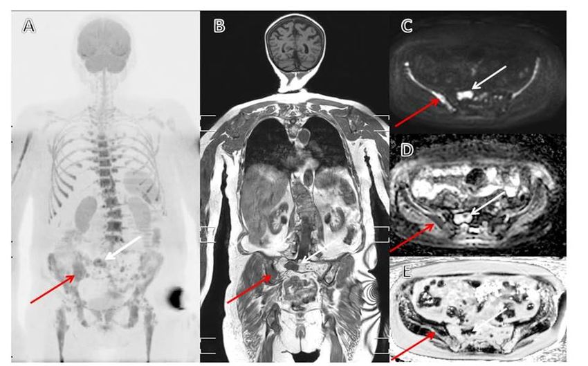
Cancers 2020, 12, 2113 2 of 19
skeletal-related events may have a negative impact on survival [6,7]. Despite the new, more targeted
anti-myeloma treatments, which have significantly improved the overall survival for patients with
multiple myeloma [8,9], MBD remains a major problem [10].
2. Pathophysiology
Bone remodeling is a continuous, lifelong process where old bone is resorbed by osteoclasts
and replaced by new bone created by the osteoblasts. The process is well balanced and mediated by
crosstalk between osteoblasts, osteoclasts, osteocytes, immune cells and bone matrix bound factors,
and is partly mediated by certain cytokines and hormones [11]. In patients with MBD, the harmonious
coupling of osteoclast and osteoblast activity is lost. Increased osteoclast activity and suppressed
osteoblast activity lead to increased bone resorption that is not compensated for by bone formation [12].
A crucial regulatory system of bone remodeling is the receptor activator of nuclear factor kappa B
(RANK)/RANK ligand (RANKL) signaling pathway. Through the RANK receptor on the precursor
osteoclasts, RANKL stimulates recruitment, differentiation and activity of the osteoclasts. The bone
marrow stromal cells (BMSC) and osteoblasts secrete osteoprotegerin (OPG), a decoy receptor for
RANKL, which inactivates RANKL, thereby reducing osteoclast activation [13,14]. Myeloma cells
interact with the bone marrow microenvironment, activating molecular cascades that lead to increased
RANKL and decreased OPG expression [15,16]. Consequently, RANKL/OPG ratio is increased as the
key element in the increased osteoclast hyper-activation.
Secondly, osteoblast inhibition, and thereby reduced bone formation, plays an important role
in the severity of MBD. Several factors are involved in downregulation of osteoblastic activity by
interfering with the Wingless (Wnt)/(DKK1) signaling pathway, which is a key pathway for osteoblast
recruitment and activation [17]. Dickkopf-1 (DKK1), expressed by the myeloma cells and BMSC,
antagonizes the WNT-pathway, blocks the differentiation of osteoblasts, and high DKK1 expression in
the bone marrow is associated with more severe MBD [18–20].
Besides the signaling abnormalities involved in the control of osteoclast and osteoblast activity,
it has been suggested that direct myeloma cell invasion into the bone remodeling compartment is
involved in the pathophysiology [21]. The remodeling compartment is a closed microenvironment,
which is shielded against the bone marrow space by a thin canopy. It has been shown that these
canopies may be infiltrated and disrupted by myeloma cells, thereby causing uncoupling of the normal
remodeling process [21].
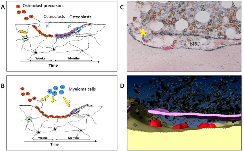
Figure 1 summarizes the key pathophysiological abnormalities in MBD. Beside the abovementioned
pathways, many other molecular pathways and signaling molecules are hypothesized to be involved
in the pathophysiology of MBD, and some data even indicate that the involved mechanisms may differ
between patients as summarized in a thorough, recent review [22]. Understanding these mechanisms
is crucial to improve the management of MBD.Cancers 2020, 12, 2113 3 of 19
Cancers 2020, 12, x FOR PEER REVIEW 3 of 18
Figure1.1. (A)
Figure (A) Cartoon
Cartoon illustrating
illustrating the
the normal
normal bonebone remodeling
remodeling taking
taking place
place in
in bone
bone remodeling
remodeling
compartments(BRC)
compartments (BRC)that
that are
are separated
separated from
from the
the bone
bonemarrow
marrowenvironment
environmentby byaathin
thinroofing
roofingcanopy.
canopy.
(B) summarizes the major pathophysiological events in myeloma bone
(B) summarizes the major pathophysiological events in myeloma bone disease: (1) The multipledisease: 1) The multiple
myeloma(MM)
myeloma (MM) cells
cells increase
increase recruitment
recruitment ofof osteoclast
osteoclast precursors,
precursors, 2)
(2)MMMMcells
cellsinfiltrate
infiltratethe
theBRC,
BRC,
disrupt the canopy and stimulate osteoclast activity, and (3) MM cells inhibit the osteoblasts,cause
disrupt the canopy and stimulate osteoclast activity, and 3) MM cells inhibit the osteoblasts, cause
osteoblastopenia,and
osteoblastopenia, andMM MM cell
cell invasion
invasion into
into the
the BRC
BRC contributes
contributes to
to the
the uncoupling
uncouplingof ofosteoclast
osteoclastand
and
osteoblastactivity,
osteoblast activity,(C)
(C)shows
shows thethe microscopic
microscopic findings
findings where
where MM
MM cells
cells (brown)
(brown)disrupt
disruptthethecanopy
canopy
(yellowasterisk)
(yellow asterisk)and
andinvade
invade into
into the
the BRC.
BRC. (D)
(D) A A computerized
computerized reconstruction
reconstruction of ofcanopy
canopydisruption
disruption
and invasion of MM cells into the BRC. (C,D) are reproduced by permission fromthe
and invasion of MM cells into the BRC. (C,D) are reproduced by permission from theoriginal
originalwork
work
publishedininBritish
published BritishJournal
Journalof ofHaematology
Haematology fromfrom 2010
2010 [21].
[21].
3.3.Imaging
Imaging
Imaging
Imagingplays
playsaacrucial
crucial role
role when
when diagnosing multiple myeloma
myeloma (MM).
(MM).First
Firstof
ofall,
all,identification
identification
ofoflytic
lyticlesions
lesionsisisone
oneof
ofthe
the CRAB-criteria
CRAB-criteria (Calcium, Renal, Anemia,
Anemia, Bone)
Bone)that
thatdefine
defineorgan
organdamage
damage
andthe
and theneed
needfor
forstarting
startinganti-myeloma
anti-myeloma therapy
therapy [23]. Imaging
Imaging is
is also
also essential
essentialto
todistinguish
distinguishsolitary
solitary
plasmacytomafrom
plasmacytoma from multiple
multiple myeloma,
myeloma, and for identifying
identifying extramedullary
extramedullarydisease
disease[24,25].
[24,25].Finally,
Finally,
imagingisisincreasingly
imaging increasinglyimportant
important in
in post-treatment
post-treatment response
response evaluation [26].
3.1.
3.1.Definition
DefinitionofofMyeloma
Myeloma Associated
Associated Bone
Bone Disease
In
In2014,
2014,thethe
International
InternationalMyeloma
Myeloma Working
WorkingGroup (IMWG)
Group updated
(IMWG) the criteria
updated the for the diagnosis
criteria for the
ofdiagnosis
multiple myeloma
of multiple andmyeloma
stated thatand
onestated
or more thattypical
one punched-out
or more typicallytic bone destructions
punched-out lytic(≥5 mm
bone
indestructions
size) on CT/low-dose CT or
(≥ 5 mm in size) onPET/CT wouldCT
CT/low-dose meet the CRAB-criteria
or PET/CT would meetregardless of its visualization
the CRAB-criteria regardless
onof skeletal
its visualization
radiographyon skeletal radiography
[27]. Increased focal[27].
FDG Increased
uptake on focal FDG uptake
PET-CT alone ison PET-CT
not alone
sufficient to is not
define
sufficient
bone to define
disease; boneofdisease;
evidence evidence
lytic bone of lyticmust
destruction bone bedestruction
present on mustthebe present The
CT-part. on the CT-part.of
presence
The presenceorofvertebral
osteoporosis osteoporosis or vertebral
compression compression
fractures fractures
in the absence of in thelesions
lytic absence is of
notlytic lesionsofisMBD.
evidence not
evidence of MBD. Additionally, more than one focal lesion on magnetic resonance
Additionally, more than one focal lesion on magnetic resonance imaging (MRI), reflecting “tumoral” imaging (MRI),
reflecting
changes “tumoral”
in the bone marrow,changes in the
fulfils the imaging
bone marrow,
criteria for fulfils the imaging criteria
treatment-demanding MMfor [27].treatment-
Both MRI
demanding
and PET/CT MM [27].toBoth
are able MRI
detect andisPET/CT
what referred aretoable to detect
as focal what
lesions, is referred
however onlytolytic
as focal
bonelesions,
lesions
however only lytic bone lesions detected
detected by CT are truly evidence of MBD [28]. by CT are truly evidence of MBD [28].Cancers 2020, 12, 2113 4 of 19
Cancers 2020, 12, x FOR PEER REVIEW 4 of 18
3.2.
3.2.From
FromConventional
ConventionalSkeletal
SkeletalSurvey
SurveytotoWhole-Body
Whole-BodyCT
CT
Conventional
Conventionalskeletal
skeletalsurvey
survey(CSS)
(CSS)hashasbeen
beenthe thestandard
standardimaging
imagingtechnique
techniquein inthe
theradiological
radiological
diagnosis
diagnosis of multiple myeloma for many years [29]. A definite advantage of CSS has beenits
of multiple myeloma for many years [29]. A definite advantage of CSS has been itsgeneral
general
availability
availability and low cost. However, CSS has limitations, especially in relation to sensitivity. Anolder
and low cost. However, CSS has limitations, especially in relation to sensitivity. An older
study
study from
from 1967
1967 [30]
[30] showed
showed that that lytic
lytic bone
bone disease
diseaseonly
onlybecomes
becomesdetectible
detectibleby byCSSCSSwhen
whenover over 30%
30% of
of the trabecular bone
the trabecular bone is lost. is lost.
Particularly
Particularlyin inthe
thespine
spineand and pelvis,
pelvis, whole-body
whole-bodylow lowdose
doseCT CT (WBLDCT)
(WBLDCT)has hasbeen
been shown
shown to to
have
have superior sensitivity in detecting osteolytic lesions. For instance, superimposed air in thebowel
superior sensitivity in detecting osteolytic lesions. For instance, superimposed air in the bowel
can
canchallenge
challengethe theinterpretation
interpretationof ofthe
thepelvis
pelvis(Figure
(Figure2). 2). In
Inaastudy
studyof of32
32patients
patientswith
withMM, MM,ititwaswas
shown that osteolytic lesions in the pelvis or spine were found in 50% of the
shown that osteolytic lesions in the pelvis or spine were found in 50% of the patients examined with patients examined with
radiographs,
radiographs, and andinin 74% 74% of patients
of patients examinedexamined with WBLDCT
with WBLDCT [31].retrospective,
[31]. A large, A large, retrospective,
international,
international, multicenter study performed a blinded comparison
multicenter study performed a blinded comparison of CSS and WBLDCT in patients with of CSS and WBLDCT in patients
newly
with newly MM
diagnosed diagnosed
[32]. InMM [32]. In
general, general, was
WBLDCT WBLDCT wastosuperior
superior to CSS in identifying
CSS in identifying lyticHowever,
lytic lesions. lesions.
However, the difference
the difference in the sensitivity
in the sensitivity depended depended on the
on the location of location
the lytic of the lytic
lesions. lesions. was
WBLDCT WBLDCTsuperiorwasin
superior in detecting lesions in the spine and pelvis, whereas no significant difference
detecting lesions in the spine and pelvis, whereas no significant difference in sensitivity was observed in sensitivity
was observed
in long bones.inInlong bones.
a large In a largeofsub-cohort
sub-cohort patients withof patients
apparent with apparent MM
smoldering smoldering
(SMM),MM lytic(SMM),
lesions
lytic lesions were identified by WBLDCT, but not by CSS, in 22.2% of the
were identified by WBLDCT, but not by CSS, in 22.2% of the patients. These patients had a higher patients. These patients had
aprobability
higher probability
of progressionof progression
to symptomatic to symptomatic
myeloma comparedmyeloma compared
to those without to bone
thosedestructions
without bone [32].
destructions
These and similar, small cohort study observations caused a change in diagnostic practiceinindiagnostic
[32]. These and similar, small cohort study observations caused a change many MM
practice
centers. in many MM
WBLDCT was centers. WBLDCT
implemented wasstandard
as the implemented as the standard
for diagnostic screening for for
diagnostic
MBD. Also, screening
in the
for MBD. IMWG
updated Also, in2014
the updated
guideline, IMWG
WBLDCT 2014 was
guideline, WBLDCT
recommended wasCSS
over recommended
[27]. over CSS [27].
Figure 2. (A) A radiograph of the pelvis is assessed by the radiologist as normal. (B) CT of the pelvis in
Figure 2. (A) A radiograph of the pelvis is assessed by the radiologist as normal. (B) CT of the pelvis
the same patient identifies a large lytic lesion with soft tumor in right crista region. Super-imposed air
in the same patient identifies a large lytic lesion with soft tumor in right crista region. Super-imposed
in the bowel hides the destruction on the conventional radiograph.
air in the bowel hides the destruction on the conventional radiograph.
The appendicular bone marrow consists partly of adipose tissue, but in multiple myeloma patients,
The appendicular bone marrow consists partly of adipose tissue, but in multiple myeloma
the bone marrow is diffusely or focally infiltrated by neoplastic plasma cells to varying degrees. Bone
patients, the bone marrow is diffusely or focally infiltrated by neoplastic plasma cells to varying
marrow changes are traditionally mostly investigated and reported by magnetic resonance imaging
degrees. Bone marrow changes are traditionally mostly investigated and reported by magnetic
techniques (see below), but nodular or diffuse infiltration of long bones can also be detected by
resonance imaging techniques (see below), but nodular or diffuse infiltration of long bones can also
WBLDCT and has been reported to have prognostic significance. Identified focal and diffuse pattern in
be detected by WBLDCT and has been reported to have prognostic significance. Identified focal and
the appendicular bone marrow by WCLDCT is associated with a shorter PFS and OS [33].
diffuse pattern in the appendicular bone marrow by WCLDCT is associated with a shorter PFS and
Today, WBLDCT is considered standard of care in diagnostic screening for MBD [28]. If WBLDCT
OS [33].
is not available, CSS can still be used [28].
Today, WBLDCT is considered standard of care in diagnostic screening for MBD [28]. If
WBLDCT is not available, CSS can still be used [28].Cancers 2020, 12, 2113 5 of 19
3.3. MRI as a Diagnostic and Prognostic Tool in Patients with Multiple Myeloma
Magnetic resonance imaging (MRI) has the ability to detect early focal and diffuse infiltration
patterns of the bone marrow [34]. Studies have shown that MRI, either axial or whole body, has a
higher sensitivity in detecting bone marrow involvement in multiple myeloma compared to CSS and
WBLDCT [35–37]. Thus, a study of 611 patients concluded that MRI was able to detect more focal
lesions than CSS, and the presence of more than seven focal lesions on MRI was an independent
adverse feature for survival [36]. However, it should be noticed that a focal lesion in the bone marrow
on MRI is not evidence of an established lytic destruction; it reflects a dense cellular infiltration that
may or may not have a connected lytic lesion, or may (or may not) precede development of a lytic
lesion. Lytic destruction is identified by loss of bone on CT or radiographs. Thus, it is important to
realize that MRI and CT offer complementary information in many patients [38].
In line with this, MRI may identify focal lesions in patients with presumed SMM and normal
WBLDCT. Two independent studies found that the finding of more than one focal lesion on axial or
whole-body MRI was associated with a 70–80% risk of progression to symptomatic disease within
2 years [39,40]. Based on this observation, the IMWG included the criteria “more than one focal lesion
on MRI” in the updated 2014 criteria for treatment demanding disease [27]. Therefore, whole-body
MRI should be the next diagnostic procedure in a patient with normal findings on WBLDCT and no
other CRAB-criteria. This patient would traditionally have been diagnosed as a SMM patient; however,
whole body MRI may up-classify the patient to have treatment-demanding disease. However, it should
be realized that “more than one focal lesion” on MRI is not an unequivocal finding; MRI findings are
not specific, and there will be a role for interpretation. Dubious findings may require confirmation
by biopsy, or a wait-and-watch strategy with repeated MRI after 3-6 months. Progression of focal
lesions or appearance of new focal lesions identify a subgroup of patients with true active disease,
whereas unchanged findings indicate low risk and SMM phenotype [41]. In contrary to focal lesions,
diffuse infiltration of the bone marrow on MRI is not considered a myeloma-defining event, but should
lead to follow-up imaging in 3–6 month [27].
Figure 3 illustrates typical findings on whole-body MRI (WBMRI). WBMRI is recommended over
combined spinal and pelvic MRI as lesions in rib cage, shoulder girdles and long bones could otherwise
be missed.
The NICE-guidelines suggest considering whole-body MRI as first-line imaging when multiple
myeloma is suspected [42]. At least in particular clinical settings MRI will be the preferred
methodology. Whole-body MRI is recommended as the first choice in patients with suspected solitary
bone plasmacytoma (whereas FDG-PET/CT is recommended in suspected solitary extramedullary
plasmacytoma) [28] and MRI is recommended as the first-line investigation if spinal cord compression
is suspected and is the chosen imaging technique to characterize whether vertebral compression
fractures are caused by osteopenia only or are myeloma infiltrated [43,44].
3.4. The Evolving Role of FDG-PET/CT in Multiple Myeloma
Positron Emission Tomography (PET)/CT using 18 Fluoro-deoxy-glucose (FDG) as the radioactively
labelled tracer (FDG-PET/CT) permits whole-body assessment and is able to visualize both
extramedullary and skeletal disease. FDG-PET offers dynamic information on metabolic active
sites of disease, and CT contributes with precise anatomic information, thereby making the combined
investigation able to identify and differentiate between active and inactive sites and provide information
about extramedullary involvement [45]. Due to the CT part, PET/CT is superior to CSS in diagnosing
lytic bone lesions [46]. Compared to MRI, PET/CT has a lower sensitivity for detection of bone marrow
involvement [46]. A recent systematic review compared whole-body MRI and FDG PET/CT in their
ability to detect myeloma skeletal lesions and suggested that MRI is more sensitive but less specific
than FDG PET/CT. Yet, it also concluded that most of the included studies were heterogeneous and
lacking an independent reference standard [47].patients with true active disease, whereas unchanged findings indicate low risk and SMM phenotype
[41]. In contrary to focal lesions, diffuse infiltration of the bone marrow on MRI is not considered a
myeloma-defining event, but should lead to follow-up imaging in 3–6 month [27].
Figure 3 illustrates typical findings on whole-body MRI (WBMRI). WBMRI is recommended
over combined spinal and pelvic MRI as lesions in rib cage, shoulder girdles and long bones could
Cancers 2020, 12, 2113 6 of 19
otherwise be missed.
Figure3.3.Whole
Figure Wholebody
bodyMRIMRIofofrelapsing
relapsingMyeloma,
Myeloma,multiple
multiplenew
newlesions
lesionsprimarily
primarilyin
inright
rightarm,
arm,spine,
spine,
ribs
ribsand
andright
rightside
sideofofpelvis.
pelvis.Many
Manypreviously
previouslytreated
treatedlesions
lesionsininspine,
spine,pelvis
pelvisand
andlegs.
legs.Red
Redarrow:
arrow:
typical
typicalnew
newlesion
lesionwith
withmyeloma
myelomacells
cells(low,
(low,homogenous
homogenousADC),
ADC),White
Whitearrow:
arrow:typical
typicalold
oldlesion
lesionwith
with
cell free water content (high ADC) and possible focal recurrence. (A) MIP of DWI-sequence with high
b-value, (B) T1-DIXON in-phase sequence, (C) DWI-sequence high b-value, (D) ADC (parametric map
calculated from DWI), and (E) Fat fraction (parametric map calculated from T1-DIXON).
However, several studies have shown that PET-positive lesions offer prognostic information, both at
diagnosis, during and after treatment. The number of lesions, the intensity of tracer uptake, and the
presence of extra-medullary disease has been shown to be associated with inferior survival [48–51].
In the response criteria of minimal residual disease negativity, FDG PET/CT is included and requires
disappearance of abnormal tracer uptake found on baseline scan or decrease to less than mediastinal
blood pool or surrounding normal tissue [26].
The IMWG recommends that PET/CT can be used in place of WBCT, but also in place of WBMRI
if imaging with MRI is not possible [28].
Sodium 18 F-Fluoride (NaF) is a bone-seeking agent introduced in 1962 [52]. The uptake of
18 F-fluoride reflects blood flow and osteoblastic activity and thereby bone remodeling [53,54]. NaF-PET
is used in the assessment of malignant and benign skeletal disease and has been suggested as a
potentially valuable tool in the assessment of MM as well [53,55]. Hypothetically, post-treatment
NaF-PET could identify bone healing activity in lytic lesions [25]. However, so far, studies have not
been able to demonstrate that NaF-PET provides additional clinical information when assessing MBD
or evaluating treatment response compared to FDG-PET [56–58]. Figure 4 shows typical findings on
FDG-PET/CT and NaF-PET/CT in the same patient and illustrates how the findings differentiate.
Other PET tracers, such as choline-based tracers, have been proposed for PET/CT imaging in
patients with MM. 11 C-Choline and 18 F-Fluorocholine PET/CT were initially developed for prostate
cancer imaging [59]. Choline is actively incorporated into the new cell membranes [60]. Results from
two smaller studies suggest that Choline PET/CT detects up to 75% more focal lesions than FDG
PET/CT in patients with MM suspected of progression or relapse [61,62]. Thus, potentially there is a
value in using other tracers than FDG in MM; however, this needs to be explored further and validated
in clinical trials.is used in the assessment of malignant and benign skeletal disease and has been suggested as a
potentially valuable tool in the assessment of MM as well [53,55]. Hypothetically, post-treatment
NaF-PET could identify bone healing activity in lytic lesions.[25]. However, so far, studies have not
been able to demonstrate that NaF-PET provides additional clinical information when assessing MBD
or evaluating
Cancers treatment response compared to FDG-PET [56–58]. Figure 4 shows typical findings
2020, 12, 2113 7 ofon
19
FDG-PET/CT and NaF-PET/CT in the same patient and illustrates how the findings differentiate.
Figure 4. (A)
(A) CT
CT of the pelvis showed a lytic
lytic lesion
lesion of
of the
the left
left pubic
pubicarea. (B) 18
area. (B) 18F-FDG PET/CT
demonstrated
demonstrated increased metabolism
metabolism localized
localized within the osteolytic lesion consistent with myeloma
18
cells. (C) PET/CT with F-Sodium
18
F-Sodium Fluoride
Fluoride (NaF)
(NaF) revealed
revealed increased
increased patchy
patchy uptake
uptake in the periphery
of the lytic lesion indicating areas bone remodeling
remodeling activity.
activity.
3.5. Follow-Up, Response Assessment, and Relapse
At the moment, there are no clear recommendations regarding routine follow-up, but in general,
CSS should not be used for disease monitoring [42]. It is recommended to repeat relevant imaging
of the same modality, PET/CT or WBMRI, as part of response evaluation in patients where active
disease sites or extramedullary disease were identified prior to start of therapy. In patients with known
extramedullary manifestations, imaging must be repeated for response assessment. Oppositely, for now,
there is no consensus that whole body imaging should be performed as part of response evaluation in
all patients. However, in patients with achieved complete remission after high-dose chemotherapy
and autologous stem-cell transplantation (ASCT), PET-positivity may persist and predict early relapse
and inferior outcome [63]. Moreover, IMWG has included FDG-PET/CT into response assessment
when evaluating MRD status [26] and recommends PET/CT assessment at baseline and for response
assessment in all patients included in clinical trials [28]. If PET/CT is not available, diffusion-weighted
WBMRI can be used and has shown some promising ability to assess response to therapy [64].
For response assessment with FDG PET/CT as well as with WBMRI it applies that there is
a continued need for standardization of the techniques, clear definition of response criteria and
prospective evaluation hereof.
4. Medical Treatment
4.1. Bisphosphonates
Since Berenson’s pamidronate trial in 1996, bisphosphonates have played a key role and been
standard of care in management of MBD [65]. Bisphosphonates are pyrophosphate analogues that
bind to bone and are ingested by the osteoclasts, leading to inhibition of osteoclastic activity. There
are different types of bisphosphonates: Pamidronate, alendronate, ibandronate and zoledronate
are all examples of nitrogen-containing bisphosphonates and inhibit the mevalonate pathway.
Non-nitrogen-containing bisphosphonate, like clodronate, results in accumulation of hydrolytical
stable analogues of adenosine triphosphate. Zoledronate, pamidronate and clodronate have been most
intensively studied in MM. Both types of bisphosphonates cause inhibition and apoptosis of osteoclasts.
Furthermore, data indicate that bisphosphonates, in addition to their bone-protective effects, may have
antitumor activity due to an uncoupling of the hypothesized vicious circle between bone resorption
and tumor growth in MM [66].
Few prospective, randomized trials comparing the different bisphosphonates head to head have
been conducted. The Rosen study from 2003 [67] compared zoledronic acid to pamidronate, in patients
with either MM or breast cancer. Zoledronic acid was superior to pamidronate in reducing the risk of
skeletal related events (SRE), but the subgroup analysis only found a significant difference in the breast
cancer population. No data on overall survival (OS) were provided. The UK MRC Myeloma XI study
from 2011 compared zoledronic acid with clodronate [68,69]. Zoledronic acid was found to be superior
to clodronate both in regard to SRE and overall survival. The lower risk of SRE was also observed in
patients without bone lesions at baseline [69].Cancers 2020, 12, 2113 8 of 19
A meta-analysis by the Cochrane database from 2017, including 24 randomized controlled trials
with a total of 7293 patients, investigated the beneficial and adverse effects associated with the use of
different types of bisphosphonates in patients with MM [70]. They concluded that bisphosphonates
reduce overall fractures and pain, and that zoledronic acid improves overall survival compared
to no bisphosphonate treatment. The meta-analysis showed no significant difference between the
different types of bisphosphonate [70]. In contrast, a retrospective cohort study, of over 1000 patients
who had been treated with either zoledronic acid or pamidronate, reported that zoledronic acid
compared to pamidronate reduced the risk of SRE by 25% and was associated with an increased overall
survival [71]. The current recommendation by IMWG is to initiate treatment with either zoledronic
acid or pamidronate in all patients with symptomatic MM, regardless of detectible osteolytic lesions
on baseline imaging [72].
In patients with smoldering myeloma, it has not been shown that bisphosphonates prolong the
time to progression to symptomatic disease [73,74] and it is therefore not recommended.
The optimal duration of bisphosphonate treatment is still controversial. In most randomized,
controlled trials, bisphosphonates were administered up to 2 years. In the Myeloma IX trial however,
bisphosphonates were given until progression. A sub-analysis conducted in patients receiving
treatment from year 2 and onward demonstrated persistent superiority of the more potent zoledronic
acid, both in regard to SRE and OS [75]. Interestingly, the cumulative incidence of renal complication
and osteonecrosis of the jaw (ONJ) seemed to reach a plateau between year 2 to 3 [76]. Another group
investigated if 4 years treatment with zoledronic acid was superior to treatment for only 2 years.
Prolonged treatment reduced SRE but no difference in OS was observed [77]. Some experts argue for
less bisphosphonate treatment in cases where the myeloma is well treated [78]. Indeed, data from the
Myeloma IX trial showed that a reduction in SRE was not observed in patients achieving at least CR
after ASCT, and that no survival benefit was seen in patients achieving VGPR or better after ASCT [79].
All the referred studies used the standard dosing of pamidronate and zoledronic acid every
3–4 weeks. However, an open-label study by Himelstein et al. comprised 1154 patients with bone
metastases, including 278 patients with MM, and compared zoledronic acid administrated every
4 weeks to every 12 weeks for up to 2 years [80]. No differences in SRE or side effects were observed.
Unfortunately, the study had a relatively high drop-out rate of 31%, and because only about 25% of the
included patients had MM, it is difficult to draw firm conclusions about the possible adjustment of
zoledronic acid scheduling in MM. Other groups have proposed that the interval between zoledronic
acid infusions could be guided by the levels of the bone resorption marker Ntx-1 (N-terminal telopeptide
of type 1 collagen) in the urine. [81]. Though this strategy is appealing and could reduce the risk of
developing ONJ, the evidence for doing this is still insufficient.
IMWG recommends that in patients who do not achieve very good partial response or better,
zoledronic acid should be administrated monthly until disease progression [72]. Otherwise, it is
suggested that bisphosphonates should be administered for up to 2 years and should be reinitiated at
relapse, if discontinued earlier [72]. Rationally, and because bisphosphonate treatment is prophylactic,
re-initiation of zoledronic acid should be at biochemical relapse and not postponed until clinical
relapse. This is supported by a Spanish study that randomized patients to zoledronic acid versus no
bisphosphonate at first sign of biochemical relapse. Although no effects were demonstrated on time to
need of treatment or survival, the patients who were re-initiated early with zoledronic acid had less
SREs at the time of treatment demanding relapse [82].
As mentioned, a serious but rare adverse event of bisphosphonate use is ONJ. Recent, randomized,
controlled trials showed an incidence of 3–4% in myeloma patients receiving zoledronic acid [68,83].
The median time from start of treatment to ONJ was found to be 13.6 months [83]. Invasive dental
procedures, dental prostheses and intravenous bisphosphonate administration and long-term treatment
as well as the myeloma itself are all risk factors associated with ONJ [84]. A case-control study showed
that patients, who were assessed by their dentist and had all necessary dental procedures done
before initiating treatment with zoledronic acid, had a three-fold decrease in the risk of developingCancers 2020, 12, 2113 9 of 19 ONJ [85]. If invasive dental procedures are required during bisphosphonate treatment a “drug holiday” before and after invasive dental procedures is generally recommended [72], despite the fact that bisphosphonates remain in the skeleton for many years [86,87]. A retrospective study indicated that prophylactic antibiotics during invasive dental procedures may reduce the risk of developing ONJ [88]. Bisphosphonate-induced nephrotoxicity is another major concern when treating patients with MM. Zoledronic acid should be dose reduced already with a mild to moderate renal impairment (CrCl 30–60 mL/min) and is not recommended in patients with severe renal impairment (
Cancers 2020, 12, 2113 10 of 19
Some concerns regarding depletion of the bone marrow reserve after concurrent chemotherapy
and radiotherapy exist. A smaller study, including 39 patients with myeloma receiving radiotherapy
alone or radiotherapy with concurrent novel agents-based chemotherapy, concluded that concurrent
treatment
Cancers 2020,with radiotherapy
12, x FOR PEER REVIEW and systemic treatment was safe regarding hematologic toxicity and 10 was
of 18
well tolerated in the majority of patients (87.5%) [101].
5.2. Vertebral Augmentation
5.2. Vertebral Augmentation
Vertebroplasty and kyphoplasty are both minimal invasive fluoroscopic guided percutaneous
surgical procedures and
Vertebroplasty usedkyphoplasty
to reduce pain arecaused
both minimal invasive
by vertebral fluoroscopic
compression guided
fractures in percutaneous
patients with
surgical
myeloma.procedures used to reduce
Cement augmentation of thepain
spinecaused by vertebral
is possible compression
at all spinal levels. In thefractures
cervicalin patients
region, the
with myeloma. Cement augmentation of the spine is possible at all spinal
vertebral bodies can be accessed through an anterior approach. Thoracic and lumbar vertebrae are levels. In the cervical
region,
reachedthethrough
vertebral abodies can be accessed
transpedicular throughwith
approach an anterior approach.
a Jamshidi Thoracic
needle. and lumbar
Bone cement
vertebrae are reached through
(polymethylmethacrylate) a transpedicular
is injected approach
into the vertebral bodywith
under a imaging
Jamshidiguidance.
needle. Bone cement
Kyphoplasty
(polymethylmethacrylate)
differs from vertebroplasty is as
injected into the
the height of vertebral body vertebra
the fractured under imaging guidance.
is restored with Kyphoplasty
an inflatable
differs from vertebroplasty as the height of the fractured vertebra
balloon catheter prior to injection of bone cement. The void created by the balloonis restored with an inflatable
catheterballoon
while
catheter
restoringprior to injection
the vertebral of bone
height cement.
allows The controlled
for more void created by the of
delivery balloon
cement,catheter
reducingwhile
therestoring the
risk of bone
vertebral height
cement leakage. allows for more controlled delivery of cement, reducing the risk of bone cement leakage.
Both
Both procedures
procedures can can bebe performed
performed under
under local
local anesthesia
anesthesia in in anan outpatient
outpatient setting.
setting. However,
However,
kyphoplasty is often performed under general anesthesia as some patients
kyphoplasty is often performed under general anesthesia as some patients experience pain while experience pain while
the
the vertebral height is restored. For patient safety reasons, the procedure is performed
vertebral height is restored. For patient safety reasons, the procedure is performed under local anesthesia, under local
anesthesia,
allowing theallowing
patient tothe patient to communicate
communicate radiating pain,radiating
which couldpain, which that
indicate couldtheindicate
needles that theofneedles
are out target,
are out of target, thereby minimizing the risk of neurological injury. Figure 5 illustrates
thereby minimizing the risk of neurological injury. Figure 5 illustrates typical lumbar spine MRI findings typical lumbar
spine MRI
prior to findings prior
vertebroplasty, and tothe
vertebroplasty, and appearance
final radiological the final radiological appearance after the procedure.
after the procedure.
5. (A)
Figure 5. (A) MRI
MRIscanscanofofmultiple myeloma
multiple myeloma patient with
patient several
with lesions
several of lumbar
lesions vertebrae
of lumbar and Th12.
vertebrae and
(B) Post-operative
Th12. X-ray of
(B) Post-operative the same
X-ray of thepatient after vertebroplasty
same patient in Th12in
after vertebroplasty to Th12
L5 vertebrae. Bone cement
to L5 vertebrae. Bone
leakage is visibleisrelated
cement leakage visible to L1. to L1.
related
A randomized, controlled trial, including 134 cancer patients with vertebral fractures, of whom
49 had multiple myeloma, found that kyphoplasty resulted in significant pain relief, improved back-
specific functional status, quality of life (QoL) and self-reported physical activity, compared to non-
surgical management. These improvements persisted throughout the entire study period until the
end of the study at 12 months [102]. Similar results with reduced pain and improved QoL afterCancers 2020, 12, 2113 11 of 19
A randomized, controlled trial, including 134 cancer patients with vertebral fractures, of whom
49 had multiple myeloma, found that kyphoplasty resulted in significant pain relief, improved
back-specific functional status, quality of life (QoL) and self-reported physical activity, compared to
non-surgical management. These improvements persisted throughout the entire study period until the
end of the study at 12 months [102]. Similar results with reduced pain and improved QoL after cement
augmentation by vertebroplasty or kyphoplasty have been reported in MM cohort studies [103–107].
A meta-analysis from 2014 found vertebroplasty and kyphoplasty to be equally effective in
reducing pain in myeloma patients [104]. In favor of kyphoplasty is a potentially better correction of
the patients’ sagittal balance. Bone cement leakage is commonly reported (4–26% per treated vertebra),
but is mostly asymptomatic [108].
A retrospective analysis, with 18 myeloma patients who underwent vertebroplasty prior
to autologous stem cell transplant showed that vertebroplasty could be done without affecting
peripheral blood stem cell collection and transplant [109]. The current recommendation in myeloma
associated vertebral collapse is to consider vertebral augmentation if it causes moderate or severe
pain, and particularly if it affects mobilization [110] This is supported by a recently published national
guideline based on the GRADE-approach [111] recommending vertebral augmentation as treatment in
patients with painful vertebral lesions and malignant hematologic disease [112].
5.3. Rehabilitation and Exercise
Exercise has been demonstrated to have a significant beneficial effect on QoL and physical
function in patients with cancer [113,114], but only few studies have been conducted on patients with
MM [115,116]. This is probably explained by the MBD and suspected increased risk of pathological
fractures. However, two literature reviews found that exercise appeared safe and acceptable for patients
with MM, but also concluded that data are limited and that no conclusion regarding the effectiveness
of exercise could be drawn [115,116]. All studies in patients with MM have been conducted in patients
before, during or after ASCT.
Baseline data from a randomized controlled trial indicate that patients with newly diagnosed
multiple myeloma generally had lower physical function compared to the normal population, and this
goes particularly for patients with bone disease and fractures [117]. A feasibility study, evaluated
30 patients with newly diagnosed myeloma who were randomized 1:1 to usual care or usual care and
individualized, supervised exercise combined with home-based exercise for 10 weeks. Sixty-seven
percent of the patients had bone involvement. The study showed that even in older patients and
in patients with MBD, individualized physical exercise is feasible and safe around the time of
diagnosis [118]. The following expanded effect trial included 100 patients with newly diagnosed MM
in a randomized setting did not show effect on physical function, physical activity, QoL, or pain [119].
However, the results of physical function indicated a trend for less loss of muscle strengths in the
intervention group, but there is a need to pay attention to pain, since this might be worsened by the
intervention [119].
6. Conclusions
Despite improved anti-myeloma treatments, MBD remains a significant problem.
The understanding of the pathophysiology has improved and may lead the way for development
of new bone directed treatments. Until then, anti-resorptive treatment with bisphosphonates or
denosumab is standard of care. Modern imaging with CT, PET/CT, and MRI play an essential role in
diagnosing and monitoring MBD and help to guide supplementary treatment with irradiation and
vertebral augmentation. Exercise in patients with MM is safe and feasible when relevant restrictions
are taken into account; however, so far, no studies have demonstrated definite benefit of training.
Author Contributions: Wrote the first draft: S.R., T.L. and N.A.; PET imaging: A.L.N.; Radiology and MR imaging:
J.T.A.; Vertebral augmentation: M.Ø.A.; Rehabilitation and exercise: R.F.L.; All authors have read and agreed to
the published version of the manuscript.Cancers 2020, 12, 2113 12 of 19
Funding: This research received no external funding.
Acknowledgments: Secretary Vicky Svane Kristensen, Haematology Research Unit, is highly acknowledged for
critical English reading.
Conflicts of Interest: The authors declare no conflicts of interest.
References
1. Raab, M.S.; Podar, K.; Breitkreutz, I.; Richardson, P.G.; Anderson, K.C. Multiple myeloma. Lancet 2009, 374,
324–339. [CrossRef]
2. Kyle, R.A.; Gertz, M.A.; Witzig, T.E.; Lust, J.A.; Lacy, M.Q.; Dispenzieri, A.; Fonseca, R.; Rajkumar, S.V.;
Offord, J.R.; Larson, D.R.; et al. Review of 1027 patients with newly diagnosed multiple myeloma.
Mayo Clin. Proc. 2003, 78, 21–33. [CrossRef]
3. Shortt, C.P.; Carty, F.; Murray, J.G. The Role of Whole-Body Imaging in the Diagnosis, Staging, and Follow-Up
of Multiple Myeloma. Semin. Musculoskelet. Radiol. 2010, 14, 37–46. [CrossRef] [PubMed]
4. Cocks, K.; Cohen, D.; Wisløff, F.; Sezer, O.; Lee, S.; Hippe, E.; Gimsing, P.; Turesson, I.; Hajek, R.; Smith, A.; et al.
An international field study of the reliability and validity of a disease-specific questionnaire module (the
QLQ-MY20) in assessing the quality of life of patients with multiple myeloma. Eur. J. Cancer 2007, 43,
1670–1678. [CrossRef] [PubMed]
5. Jordan, K.; Proskorovsky, I.; Lewis, P.; Ishak, J.; Payne, K.; Lordan, N.; Kyriakou, C.; Williams, C.D.; Peters, S.;
Davies, F.E. Effect of general symptom level, specific adverse events, treatment patterns, and patient
characteristics on health-related quality of life in patients with multiple myeloma: Results of a European,
multicenter cohort study. Support. Care Cancer 2014, 22, 417–426. [CrossRef] [PubMed]
6. Terpos, E.; Berenson, J.; Cook, R.J.; Lipton, A.; Coleman, R.E. Prognostic variables for survival and skeletal
complications in patients with multiple myeloma osteolytic bone disease. Leukemia 2010, 24, 1043–1049.
[CrossRef] [PubMed]
7. Augustson, B.M.; Begum, G.; Dunn, J.A.; Barth, N.J.; Davies, F.; Morgan, G.; Behrens, J.; Smith, A.; Child, J.A.;
Drayson, M.T. Early mortality after diagnosis of multiple myeloma: Analysis of patients entered onto the
United Kingdom Medical Research Council trials between 1980 and 2002—Medical Research Council Adult
Leukaemia Working Party. J. Clin. Oncol. 2005, 23, 9219–9226. [CrossRef] [PubMed]
8. Naymagon, L.; Abdul-Hay, M. Novel agents in the treatment of multiple myeloma: A review about the
future. J. Hematol. Oncol. 2016, 9, 52. [CrossRef]
9. Landgren, O.; Iskander, K. Modern multiple myeloma therapy: Deep, sustained treatment response and
good clinical outcomes. J. Intern. Med. 2017, 281, 365–382. [CrossRef]
10. Vallet, S.; Filzmoser, J.-M.; Pecherstorfer, M.; Podar, K. Myeloma Bone Disease: Update on Pathogenesis and
Novel Treatment Strategies. Pharmaceutics 2018, 10, 202. [CrossRef]
11. Xiao, W.; Wang, Y.; Pacios, S.; Li, S.; Graves, D.T. Cellular and Molecular Aspects of Bone Remodeling.
Front. Oral. Biol. 2016, 18, 9–16. [PubMed]
12. Giuliani, N.; Rizzoli, V.; Roodman, G.D. Multiple myeloma bone disease: Pathophysiology of osteoblast
inhibition. Blood 2006, 108, 3992–3996. [CrossRef] [PubMed]
13. Boyle, W.J.; Simonet, W.S.; Lacey, D.L. Osteoclast differentiation and activation. Nature 2003, 423, 337–342.
[CrossRef] [PubMed]
14. Han, Y.; You, X.; Xing, W.; Zhang, Z.; Zou, W. Paracrine and endocrine actions of bone-the functions of
secretory proteins from osteoblasts, osteocytes, and osteoclasts. Bone Res. 2018, 6, 16. [CrossRef]
15. Giuliani, N.; Bataille, R.; Mancini, C.; Lazzaretti, M.; Barillé, S. Myeloma cells induce imbalance in the
osteoprotegerin/osteoprotegerin ligand system in the human bone marrow environment. Blood 2001, 98,
3527–3533. [CrossRef]
16. Terpos, E.; Szydlo, R.; Apperley, J.F.; Hatjiharissi, E.; Politou, M.; Meletis, J.; Viniou, N.; Yataganas, X.;
Goldman, J.M.; Rahemtulla, A. Soluble receptor activator of nuclear factor kappaB ligand-osteoprotegerin
ratio predicts survival in multiple myeloma: Proposal for a novel prognostic index. Blood 2003, 102, 1064–1069.
[CrossRef]
17. Kim, J.H.; Liu, X.; Wang, J.; Chen, X.; Zhang, H.; Kim, S.H.; Cui, J.; Li, R.; Zhang, W.; Kong, Y.; et al. Wnt
signaling in bone formation and its therapeutic potential for bone diseases. Ther. Adv. Musculoskelet. Dis.
2013, 5, 13–31. [CrossRef]Cancers 2020, 12, 2113 13 of 19
18. Qiang, Y.-W.; Chen, Y.; Stephens, O.; Brown, N.; Chen, B.; Epstein, J.; Barlogie, B.; Shaughnessy, J.D.
Myeloma-derived Dickkopf-1 disrupts Wnt-regulated osteoprotegerin and RANKL production by osteoblasts:
A potential mechanism underlying osteolytic bone lesions in multiple myeloma. Blood 2008, 112, 196–207.
[CrossRef]
19. Kristensen, I.B.; Christensen, J.H.; Lyng, M.B.; Møller, M.B.; Pedersen, L.M.; Rasmussen, L.M.; Ditzel, H.J.;
Abildgaard, N. Expression of osteoblast and osteoclast regulatory genes in the bone marrow microenvironment
in multiple myeloma: Only up-regulation of Wnt inhibitors SFRP3 and DKK1 is associated with lytic bone
disease. Leuk. Lymphoma 2014, 55, 911–919. [CrossRef]
20. Palma, B.D.; Guasco, D.; Pedrazzoni, M.; Bolzoni, M.; Accardi, F.; Costa, F.; Sammarelli, G.; Craviotto, L.;
Filippo, M.D.; Ruffini, L.; et al. Osteolytic lesions, cytogenetic features and bone marrow levels of cytokines
and chemokines in multiple myeloma patients: Role of chemokine (C-C motif) ligand 20. Leukemia 2016, 30,
409–416. [CrossRef]
21. Andersen, T.L.; Søe, K.; Sondergaard, T.E.; Plesner, T.; Delaisse, J.-M. Myeloma cell-induced disruption of
bone remodelling compartments leads to osteolytic lesions and generation of osteoclast-myeloma hybrid
cells. Br. J. Haematol. 2010, 148, 551–561. [CrossRef] [PubMed]
22. Børset, M.; Sundan, A.; Waage, A.; Standal, T. Why do myeloma patients have bone disease? A historical
perspective. Blood Rev. 2020, 41, 100646. [CrossRef]
23. Criteria for the classification of monoclonal gammopathies, multiple myeloma and related disorders: A report
of the International Myeloma Working Group. Br. J. Haematol. 2003, 121, 749–757. [CrossRef]
24. Caers, J.; Paiva, B.; Zamagni, E.; Leleu, X.; Bladé, J.; Kristinsson, S.Y.; Touzeau, C.; Abildgaard, N.; Terpos, E.;
Heusschen, R.; et al. Diagnosis, treatment, and response assessment in solitary plasmacytoma: Updated
recommendations from a European Expert Panel. J. Hematol. Oncol. 2018, 11, 140. [CrossRef] [PubMed]
25. Sevcikova, S.; Minarik, J.; Stork, M.; Jelinek, T.; Pour, L.; Hajek, R. Extramedullary disease in multiple
myeloma-controversies and future directions. Blood Rev. 2019, 36, 32–39. [CrossRef]
26. Kumar, S.; Paiva, B.; Anderson, K.C.; Durie, B.; Landgren, O.; Moreau, P.; Munshi, N.; Lonial, S.; Bladé, J.;
Mateos, M.-V.; et al. International Myeloma Working Group consensus criteria for response and minimal
residual disease assessment in multiple myeloma. Lancet Oncol. 2016, 17, e328–e346. [CrossRef]
27. Rajkumar, S.V.; Dimopoulos, M.A.; Palumbo, A.; Blade, J.; Merlini, G.; Mateos, M.-V.; Kumar, S.; Hillengass, J.;
Kastritis, E.; Richardson, P.; et al. International Myeloma Working Group updated criteria for the diagnosis
of multiple myeloma. Lancet Oncol. 2014, 15, e538–e548. [CrossRef]
28. Hillengass, J.; Usmani, S.; Rajkumar, S.V.; Durie, B.G.M.; Mateos, M.-V.; Lonial, S.; Joao, C.; Anderson, K.C.;
García-Sanz, R.; Riva, E.; et al. International myeloma working group consensus recommendations on
imaging in monoclonal plasma cell disorders. Lancet Oncol. 2019, 20, e302–e312. [CrossRef]
29. Dimopoulos, M.; Terpos, E.; Comenzo, R.L.; Tosi, P.; Beksac, M.; Sezer, O.; Siegel, D.; Lokhorst, H.; Kumar, S.;
Rajkumar, S.V.; et al. International myeloma working group consensus statement and guidelines regarding
the current role of imaging techniques in the diagnosis and monitoring of multiple Myeloma. Leukemia 2009,
23, 1545–1556. [CrossRef]
30. Edelstyn, G.A.; Gillespie, P.J.; Grebbell, F.S. The radiological demonstration of osseous metastases.
Experimental observations. Clin. Radiol. 1967, 18, 158–162. [CrossRef]
31. Hinge, M.; Andersen, K.T.; Lund, T.; Jørgensen, H.B.; Holdgaard, P.C.; Ormstrup, T.E.; Østergaard, L.L.;
Plesner, T. Baseline bone involvement in multiple myeloma—A prospectiv prospective comparison
of conventional X-ray, low-dose computed tomography, and 18flourodeoxyglucose positron emission
tomography in previously untreated patients. Haematologica 2016, 101, e415–e418. [CrossRef] [PubMed]
32. Hillengass, J.; Moulopoulos, L.A.; Delorme, S.; Koutoulidis, V.; Mosebach, J.; Hielscher, T.; Drake, M.;
Rajkumar, S.V.; Oestergaard, B.; Abildgaard, N.; et al. Whole-body computed tomography versus conventional
skeletal survey in patients with multiple myeloma: A study of the International Myeloma Working Group.
Blood Cancer J. 2017, 7, e599. [CrossRef] [PubMed]
33. Matsue, K.; Kobayashi, H.; Matsue, Y.; Abe, Y.; Narita, K.; Kitadate, A.; Takeuchi, M. Prognostic significance
of bone marrow abnormalities in the appendicular skeleton of patients with multiple myeloma. Blood Adv.
2018, 2, 1032–1039. [CrossRef] [PubMed]
34. Moulopoulos, L.A.; Dimopoulos, M.A. Magnetic Resonance Imaging of the Bone Marrow in Hematologic
Malignancies. Blood 1997, 90, 2127–2147. [CrossRef]Cancers 2020, 12, 2113 14 of 19
35. Wolf, M.B.; Murray, F.; Kilk, K.; Hillengass, J.; Delorme, S.; Heiss, C.; Neben, K.; Goldschmidt, H.; Kauczor, H.-U.;
Weber, M.-A. Sensitivity of whole-body CT and MRI versus projection radiography in the detection of osteolyses
in patients with monoclonal plasma cell disease. Eur. J. Radiol. 2014, 83, 1222–1230. [CrossRef]
36. Walker, R.; Barlogie, B.; Haessler, J.; Tricot, G.; Anaissie, E.; Shaughnessy, J.D.; Epstein, J.; van Hemert, R.;
Erdem, E.; Hoering, A.; et al. Magnetic Resonance Imaging in Multiple Myeloma: Diagnostic and Clinical
Implications. J. Clin. Oncol. 2007, 25, 1121–1128. [CrossRef]
37. Baur-Melnyk, A.; Buhmann, S.; Becker, C.; Schoenberg, S.O.; Lang, N.; Bartl, R.; Reiser, M.F. Whole-Body MRI
Versus Whole-Body MDCT for Staging of Multiple Myeloma. Am. J. Roentgenol. 2008, 190, 1097–1104. [CrossRef]
38. Lecouvet, F.E.; Vande Berg, B.C.; Malghem, J.; Maldague, B.E. Magnetic resonance and computed tomography
imaging in multiple myeloma. Semin. Musculoskelet. Radiol. 2001, 5, 43–55. [CrossRef]
39. Kastritis, E.; Moulopoulos, L.A.; Terpos, E.; Koutoulidis, V.; Dimopoulos, M.A. The prognostic importance of
the presence of more than one focal lesion in spine MRI of patients with asymptomatic (smoldering) multiple
myeloma. Leukemia 2014, 28, 2402–2403. [CrossRef]
40. Hillengass, J.; Fechtner, K.; Weber, M.-A.; Bäuerle, T.; Ayyaz, S.; Heiss, C.; Hielscher, T.; Moehler, T.M.;
Egerer, G.; Neben, K.; et al. Prognostic significance of focal lesions in whole-body magnetic resonance
imaging in patients with asymptomatic multiple myeloma. J. Clin. Oncol. 2010, 28, 1606–1610. [CrossRef]
41. Merz, M.; Hielscher, T.; Wagner, B.; Sauer, S.; Shah, S.; Raab, M.S.; Jauch, A.; Neben, K.; Hose, D.;
Egerer, G.; et al. Predictive value of longitudinal whole-body magnetic resonance imaging in patients with
smoldering multiple myeloma. Leukemia 2014, 28, 1902–1908. [CrossRef]
42. Pratt, G.; Morris, T.C. Review of the NICE guidelines for multiple myeloma. Int. J. Lab. Hematol. 2017, 39,
3–13. [CrossRef] [PubMed]
43. 1 Guidance|Metastatic Spinal Cord Compression in Adults: Risk Assessment, Diagnosis and
Management|Guidance|NICE. Available online: https://www.nice.org.uk/guidance/CG75/chapter/1-
Guidance#imaging (accessed on 4 March 2020).
44. Mauch, J.T.; Carr, C.M.; Cloft, H.; Diehn, F.E. Review of the Imaging Features of Benign Osteoporotic and
Malignant Vertebral Compression Fractures. Am. J. Neuroradiol. 2018, 39, 1584–1592. [CrossRef]
45. Role of 18 F-FDG PET/CT in the Diagnosis and Management of Multiple Myeloma and Other
Plasma Cell Disorders: A Consensus Statement by the International Myeloma Working Group-Clinical
Key. Available online: https://www-clinicalkey-com.proxy2-bib.sdu.dk/#!/content/journal/1-s2.0-
S1470204517301894 (accessed on 5 March 2020).
46. Zamagni, E.; Nanni, C.; Patriarca, F.; Englaro, E.; Castellucci, P.; Geatti, O.; Tosi, P.; Tacchetti, P.;
Cangini, D.; Perrone, G.; et al. A prospective comparison of 18F-fluorodeoxyglucose positron emission
tomography-computed tomography, magnetic resonance imaging and whole-body planar radiographs in the
assessment of bone disease in newly diagnosed multiple myeloma. Haematologica 2007, 92, 50–55. [CrossRef]
[PubMed]
47. Gariani, J.; Westerland, O.; Natas, S.; Verma, H.; Cook, G.; Goh, V. Comparison of whole body magnetic
resonance imaging (WBMRI) to whole body computed tomography (WBCT) or 18F-fluorodeoxyglucose
positron emission tomography/CT (18F-FDG PET/CT) in patients with myeloma: Systematic review of
diagnostic performance. Crit. Rev. Oncol./Hematol. 2018, 124, 66–72. [CrossRef] [PubMed]
48. Bartel, T.B.; Haessler, J.; Brown, T.L.Y.; Shaughnessy, J.D.; van Rhee, F.; Anaissie, E.; Alpe, T.; Angtuaco, E.;
Walker, R.; Epstein, J.; et al. F18-fluorodeoxyglucose positron emission tomography in the context of other
imaging techniques and prognostic factors in multiple myeloma. Blood 2009, 114, 2068–2076. [CrossRef]
49. Zamagni, E.; Patriarca, F.; Nanni, C.; Zannetti, B.; Englaro, E.; Pezzi, A.; Tacchetti, P.; Buttignol, S.; Perrone, G.;
Brioli, A.; et al. Prognostic relevance of 18-F FDG PET/CT in newly diagnosed multiple myeloma patients
treated with up-front autologous transplantation. Blood 2011, 118, 5989–5995. [CrossRef]
50. Usmani, S.Z.; Mitchell, A.; Waheed, S.; Crowley, J.; Hoering, A.; Petty, N.; Brown, T.; Bartel, T.; Anaissie, E.;
van Rhee, F.; et al. Prognostic implications of serial 18-fluoro-deoxyglucose emission tomography in multiple
myeloma treated with total therapy 3. Blood 2013, 121, 1819–1823. [CrossRef]
51. Moon, S.H.; Choi, W.H.; Yoo, I.R.; Lee, S.J.; Paeng, J.C.; Jeong, S.Y.; Lee, S.-W.; Kim, K.; Choi, J.Y. Prognostic
Value of Baseline 18F-Fluorodeoxyglucose PET/CT in Patients with Multiple Myeloma: A Multicenter Cohort
Study. Korean J. Radiol. 2018, 19, 481–488. [CrossRef]
52. Blau, M.; Nagler, W.; Bender, M.A. Fluorine-18: A new isotope for bone scanning. J. Nucl. Med. 1962, 3,
332–334. [PubMed]You can also read