History of Feline infectious Peritonitis 1963-2022 - First description to Successful Treatment
←
→
Page content transcription
If your browser does not render page correctly, please read the page content below
1

History of Feline infectious Peritonitis 1963-2022 – First description to
Successful Treatment
Niels C. Pedersen

Center for Companion Animal Health, School of Veterinary Medicine, University of
California, 944 Garrod Drive, Davis, CA, 95616, USA
April 17, 2022

Abstract

This article highlights knowledge of feline infectious peritonitis (FIP) as it evolved,
starting at its recognition in 1963 to present time, and was prepared with veterinarians,
cat rescuers and guardians, shelter staff, and cat lovers in mind. A brief mention is
made of the causative feline coronavirus and its relationship to a ubiquitous and
minimally pathogenic enteric coronavirus of felids, epizootiology, pathogenesis,
pathology, clinical features, and diagnostics. Major emphasis is on risk factors affecting
FIP prevalence, and the role of modern antiviral drugs in successful treatment.

Introduction

Feline infectious peritonitis (FIP) was described as a specific disease entity in 1963 by
veterinarians at the Angell Memorial Animal Hospital in Boston (Holzworth 1963) (Fig.
1). Pathology records from this institution and The Ohio State University failed to identify
earlier cases (Wolfe and Griesemer 1966), although identical cases were soon
recognized worldwide. The initial pathologic descriptions were of a diffuse inflammation
of the tissues lining the peritoneal cavity and abdominal organs with extensive
inflammatory fluid effusion, from which the disease was ultimately named (Wolfe and
Griesemer 1966, 1971) (Figs. 2,3). A second, and less common clinical form of FIP,
manifested by less diffuse and more widespread granulomatous lesions involving organ
parenchyma was first described in 1972 (Montali and Strandberg 1972) (Figs.3,4). The
presence of inflammatory effusions in body cavity in the common form, and lack of
effusions in the less common form, led to the names wet (effusive, non-
parenchymatous) and dry (non-effusive, parenchymatous) FIP.
2

Figure 1. Photograph of the author and Dr. Jean Holzworth (1915-2007) taken in 1991. Dr. Holzworth was the
finest feline veterinarian that the author has known and was responsible for the first report of FIP as a
specific disease entity. She spent her entire career at the Angell Memorial Animal Hospital in Boston.

Figure 2. Gross necropsy appearance of the abdominal cavity of a cat with acute onset wet FIP. The
abdomen is filled with several hundred ml of a yellow, viscous fluid, the omentum is reddened, edematous
and contracted, and fibrin plaques are evident on the surfaces of the spleen and edges of liver (arrows). A
strand of fibrin is seen on the spleen

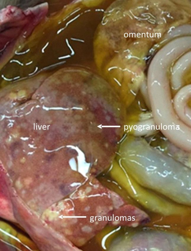
3

Figure 3. Appearance of the open abdomen at necropsy of a cat dying of a more chronic form of effusive FIP.
The abdomen is filled with a viscous, yellow-tinged exudate and the omentum is thickened and contracted.
The major lesions are in the liver, with numerous plaque-like structures (pyogranulomas) on the capsule.
More defined lesions (granulomas), also serosal surface orientated, appear fleshier and are raised above the
surface. These lesions also extend into the underlying liver parenchyma and are more typical of dry
FIP. This is an example of a case of FIP that is transitioning between wet and dry (arrow).
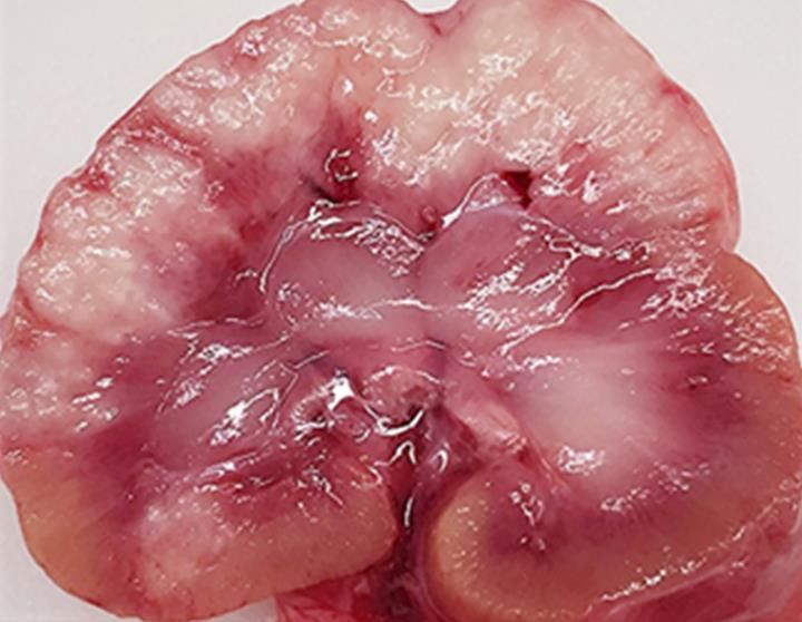
4

A.                                            B.
Figure 4, A, B - Gross cross-section of a kidneys from two cats with the dry form of FIP. Lesions are surface
orientated and extend down into the underlying parenchyma. B- The lesions of dry FIP in organs such as
kidney, cecum, colon, and intestinal lymph nodes (Fig. 5) have been confused grossly with renal lymphoma.

Figure 5. Gross enlargement of ileo-cecal-colic lymph nodes in a cat with dry FIP.

The prevalence of FIP appeared to increase during the feline leukemia virus (FeLV)
panzootic of the 1960s-80s, when many FIP cases were found to be FeLV associated
(Cotter et al., 1973; Pedersen 1976a). The subsequent control of FeLV infection in
owned cats with rapid testing and vaccination saw a plateau in FIP cases. However,
5

recent interest in foster/rescue, coupled with an effective treatment, has led to an
increased awareness of the disease and its diagnosis.

Etiologic agent

Early attempts failed to identify a causative agent for FIP but confirmed its infectious
nature (Wolfe and Griesemer 1966). A viral etiology was established in 1968 using
ultrafiltrates of infectious material (Zook et al., 1968). The causative virus was
subsequently identified as a coronavirus (Ward 1970) closely related to enteric
coronaviruses of dogs and swine (Pedersen et al., 1978).

Confusion arose when a feline enteric coronavirus (FECV) was isolated from the feces
of healthy cats and found indistinguishable to the feline infectious peritonitis virus (FIPV)
(Pedersen et al., 1981). Unlike FIPVs, which readily recreated FIP in laboratory cats,
experimental FECV infections were largely asymptomatic. The relationship of the two
viruses was resolved when FIPVs were found to be mutants of FECVs that arise within
the body of each cat with FIP (Vennema et al., 1995; Poland et al., 1996).

FECV is ubiquitous in cat populations around the world and is first shed in the feces
starting at around 9-10 weeks of age coincident with the loss of maternal immunity
(Pedersen et al., 2008;). Infection is by the fecal-oral route and targets the intestinal
epithelium, and primary signs of enteritis are mild or inapparent (Pedersen et al., 2008;
Vogel et al., 2010). Subsequent fecal shedding is from the colon, and usually ceases
after several weeks or months (Herrewegh et al.,1997; Pedersen et al., 2008; Vogel et
al., 2010). Immunity is short-lived, and recurrent infections are common (Pedersen et
al., 2008; Pearson et al., 2016). Stronger immunity eventually develops over time and
cats older than 3 years are less likely to be fecal shedders (Addie et al., 2003). FECV is
continuously undergoing genetic drift into locally and regionally identifiable clades
(Herrewegh et al.,1997; Pedersen et al., 2009).

FECV and FIPV are classified as biotypes of the subspecies feline coronavirus (FCoV).
The genomes of FECV and FIPV biotypes are >98% related, yet with unique host cell
tropisms and pathogenicity (Chang et al., 2012; Pedersen et al., 2009). FECVs infect
mature intestinal epithelium, while FIPVs lose intestinal tropism and gain the ability to
replicate in monocyte/macrophages. The published names FECV or FIPV will be used
herein when referring to aspects of disease specific to each biotype, while the term
FCoV will be used when referring to features common to both.

Three types of mutations have been implicated in the FECV-to-FIPV biotype change.
The first type, which is unique to each cat with FIP (Poland et al., 1996), consists of an
accumulation of missense and nonsense mutations in the c-terminus of the 3c
accessory gene that frequently lead to a truncated 3c gene products (Pedersen et al.,
2012; Vennema et al., 1995). The second type of mutations consist of two specific
single nucleotide polymorphisms in the fusion peptide of the S gene, with one or the
other forms shared by >95% of FIPVs and absent from FECVs (Chang et al., 2012).
The third type of mutations, unique to each FIPV isolate and not found in FECVs, occur
6

in and around the furin cleavage motif between the receptor-binding (S1) and fusion
(S2) domains of the spike (S) gene (Licitra et al., 2013). These mutations have variable
effects on furin cleavage activity. Collectively, and in a yet to be determined manner,
they are responsible for a shift in host cell tropism from enterocyte to macrophage, and
a profound change in disease form.

FCoVs, and therefore FECVs and FIPVs, exist in two serotypes identified by antibodies
to the virus neutralizing epitope on the S gene (Herrewegh et al., 1998; Terada et al.,
2014). Serotype I FCoVs are recognized by cat sera and predominate in most countries.
Serotype II FCoVs result from recombination with a portion of the S gene of canine
coronavirus (Herrewegh et al., 1998; Terada et al., 2014), and identified by antibodies to
canine coronavirus. Serotype II FIPVs are readily cultivated in tissue culture, while
serotype I FIPVs are difficult to adapt to in vitro growth. Serotype I and II FECVs have
not been grown in conventional cell cultures (Tekes et al., 2020).

FIPVs are found solely in activated monocytes and macrophages within diseased
tissues and effusions and not shed externally. Therefore, cat-to-cat (horizontal)
transmission of FIPVs is not the major mode of spread. Rather, FIP follows the pattern
of the underlying enzootic FECV infection, with sporadic cases and occasional small
clusters of disease (Foley et al., 1997). These clusters of cases can be mistaken for
epizootics. A single report of epizootic FIP has been linked to a single serotype II virus
that appeared to evolve within a shelter housing both dogs and cats (Wang et al., 2013).
Horizontal transmission in this instance followed an epizootic rather than enzootic
disease pattern, with infections spreading rapidly to cats of all ages and in close contact
with an index case (Wang et al., 2013).

The low prevalence of FIP cases in a population suggests that FIPV mutants are
generated infrequently. However, studies involving FECV infection in
immunocompromised FIV and FeLV infected cats indicate that FIP mutants may be
common but cause disease only under certain circumstances. Nineteen cats infected
with feline immunodeficiency virus (FIV) for 6 years, and a control group of 20 FIV-naive
siblings, were orally infected with FECV (Poland et al., 1996). Cats in both groups
remained asymptomatic for two months, when two cats in the FIV-infected group
developed FIP. In a second study, 26 young cats with enzootic FECV infection, and
from a breeding colony with no history of FIP, were contact exposed to FeLV carriers
(Pedersen et al., 1977). Two kittens in the group subsequently developed FIP 2-10
weeks after becoming FeLV viremic. A remaining question is how long FIPVs can
survive in the body before being eliminated? One theory is that they persist in the body
for some time and only become pathologic if immunity to them is impaired (Healey et
al., 2022). This theory is supported by how immunity to FeLV develops. Most cats past
kittenhood will resist FeLV viremia and develop a solid and lasting immunity, but this
occurs over a period of weeks during which time the virus persists in a subclinical or
latent state (Pedersen et al., 1982; Rojko et al., 1982). Methylprednisolone administered
during this period, but not after, will abolish developing immunity and lead to a state of
persistent viremia.

7

Epizootiology

Epizootiology is the study of incidence, distribution, and possible control of diseases of
animals and the influence of environmental, host and agent factors. FIP is touted as one
of the most important infectious causes of death in cats, although there are no accurate
figures on prevalence. It has been estimated that 0.3-1.4% of deaths in cats presented
to veterinary institutions are associated with FIP (Rohrbach et al., 2001; Pesteanu-
Somogyi et al., 2006; Riemer et al., 2016), and as high as 3.6-7.8% in some shelters
and catteries (Cave et al., 2002). FIP is also described as a disease of denser multi-cat
environments. Three-fourths of FIP cases in a current ongoing treatment study came
from the field via cat foster/rescues and shelters, 14% from catteries, and only 11%
from homes.1

Studies based on cases seen at academic institutions have shown the effect of age and
sex on FIP prevalence (Rohrbach et al., 2001; Pesteanu-Somogyi et al., 2006;
Pedersen 1976a; Worthing et al., 2012; Riemer et al., 2016). Three-fourths of cases in
these cohorts occurred in cats under 3 years of age and few after 7 years. This was
confirmed by a current and ongoing field study from the Czech Republic and Slovakia,
which found that over 80% of FIP cases occurred in cats 3 years of age or younger and
only 5% in cats older than 7 years (Fig. 6).1 Earlier institutional studies varied as to the
effect of sex, but indications were that males were somewhat more prone to FIP than
females. This was confirmed by current data from the field showing a male to female
ratio of 1.3:1.1. It is unclear whether neutering affects FIP prevalence, with some reports
suggesting that it may increase susceptibility (Riemer et al., 2016), while others do not
show such a clear effect.1

Figure 6. Ages of over 607 cats from the Czech Republic and Slovakia at the time of diagnosis and treatment
for FIP.1 Thirty percent of infections were seen in cats six-months of age or younger, 50% one-year and 85%
three years of age or younger.
8

Additional environmental and virus-related risk factors have been implicated in
increased prevalence of FIP, but their importance requires knowing disease incidence in
their absence. A possible baseline may have been provided by a study of an enzootic
FECV infection that was unknowingly present for many years in a well-managed specific
pathogen free breeding colony (Hickman et al., 1995). This colony was maintained
under strict quarantine free of other infections and the level of nutrition and husbandry
were high. This colony produced hundreds of kittens each year before the first case of
FIP was diagnosed. Such observations suggest that FIP may be a rare occurrence in
the absence of risk factors.

The importance of re-homing as a risk factor for FIP is only now being appreciated.
Pedigreed cat breeders, many of whom have suffered no cases of FIP within their
catteries, have as their greatest fear notification that one of their kittens developed FIP
shortly after going to a new home. A recent study found that over one-half of cats with
FIP had experienced a change in environment, a stay in a shelter, or capture within the
weeks preceding their illness.1 Cats are known for hiding outward signs of stress, even
when suffering serious internal effects. Procedures as simple as changing cages will
suppress immunity and reactivate latent herpes virus shedding and disease signs in
cats (Gaskell and Povey, 1977). Stressful situations, even those that seem minor, can
cause decreased levels of lymphocytes and ‘sickness behaviors’ (Stella et al., 2013).

Variations in the genetic makeup of enzootic strains of FCoVs may also be involved in
FIP prevalence in a population. Serotype II FIPVs are thought to be more virulent than
serotype I and more likely to be transmitted cat-to-cat (Lin et al., 2009; Wang et al.,
2013). It is also possible that certain clades of FECV are more apt to mutate to FIPVs,
and this should be studied. The author has also observed an inordinately high
proportion of cats with neurological FIP in some regions such of the world, suggesting
genetic determinants in certain strains of FCoVs may be more neurotropic.

Retrovirus-associated immunodeficiencies have been linked to FIP susceptibility. Up to
one-half of FIP cases during height of the FeLV panzootic were persistently FeLV
infected (Cotter et al., 1973; Pedersen 1976a; Hardy 1981). FeLV infection causes
suppression of T-cell immunity, which can inhibit a protective immune response to FIP.
The significance of FeLV infection for FIP incidence was greatly diminished starting in
1980s, when the removal of carriers and vaccination forced FeLV back into nature
where exposures are less severe and immunity the usual outcome. Chronic feline
immunodeficiency virus (FIV) infection has also been shown to be a risk factor for FIP in
FECV infected cats under experimental conditions (Poland et al., 1996). FeLV infection
was recognized in 2% and FIV in 1% of cats being treated for FIP in one recent field
study.1

The incidence of FIP purebred cats is reportedly higher than for random bred cats, with
some breeds appearing more susceptible than others (Pesteanu-Somogyi et al., 2006;
Worthing et al., 2012), suggesting a genetic component to susceptibility. Genetic

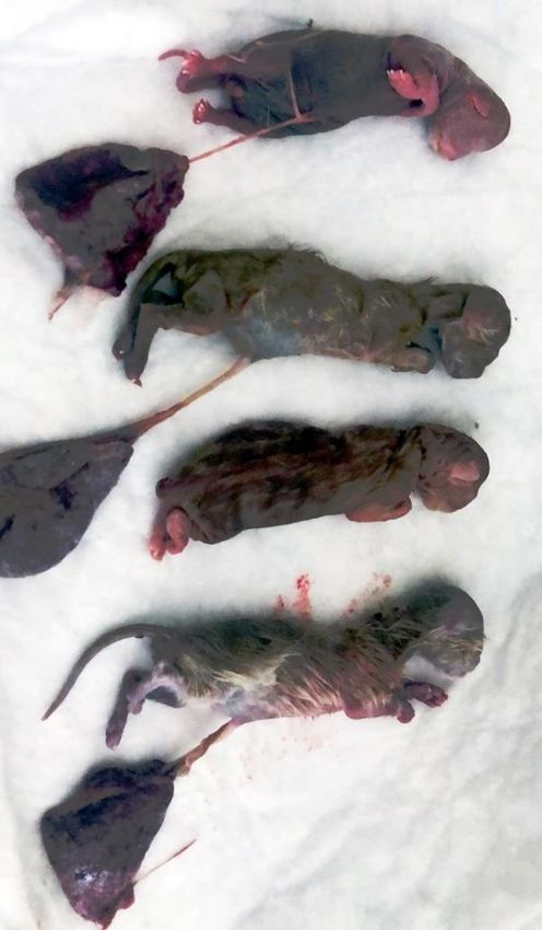
9

predisposition to FIP has been studied in several Persian catteries and was estimated
to account for one-half of the disease risk (Foley et al., 1997). Some breeds, such as the
Birman, are more apt to develop dry than wet FIP (Golovko et al., 2013). Attempts to
identify specific genes associated with FIP susceptibility in Birman cats implicated
several genes involved with immunity, but none achieved required significance (Golovko
et al., 2013). The largest study of genetic susceptibility to FIP showed it to be extremely
polymorphic and implicated inbreeding as the major risk factor (Pedersen et al., 2016).
Specific polymorphisms in several genes have also been linked to high level FECV
shedding among several pedigreed breeds of cats (Bubenikova et al., 2020).

Queens can develop FIP, usually the wet form, during pregnancy or in the perinatal
period. This phenomenon resembles the suppression of immunity in pregnant women
and predisposition to certain infections (Mor and Cardenas 2010). It is unclear whether
subclinical FIP is activated by pregnancy or to increased susceptibility to a new
infection. Infection of the queen early in pregnancy has led to fetal death and resorption,
while later infections often result in abortion (Fig. 7). Kittens may also be born healthy
but develop disease and die in the perinatal period. Some litters are born uninfected,
due to the efficiency of the placental maternal/fetal blood barrier or the aid of antiviral
treatment (Fig. 8).

Figure 7. Aborted kittens from a queen that developed wet FIP in the later stage of pregnancy. The abortion
was the first indication of FIP, with classical signs of abdominal wet FIP rapidly following. The queen was
successfully cured of FIP with the antiviral GS-441524.

10

Figure 8. This queen developed signs of wet abdominal FIP 3 weeks into pregnancy and was successfully
cured with GS-441524. She subsequently delivered a litter of four kittens by C-section, one of which was
dead and three that survived and grew up healthy. Treatment was given for the remaining 6 weeks of
pregnancy and continued for 6 weeks during which kittens were successfully nursed. GS-441524 had no
apparent side-effects to queen or kittens.

A possible surge in FIP cases has been observed in cats over 10 years of age in
studies reported 50 years ago (Pedersen 1976a). Somewhat over 3% of FIP cases in a
recent study occurred in cats 10 years of age and older and 1.5% in cats 12 years or
older (Fig. 6).1 The occurrence of FIP in aged often involves two different scenarios. The
first scenario also involves exposure to FECV fecal shedders, but in a unique manner. It
is common for aged cats to be paired as kittens and live together in relative isolation not
exposed to FECV for many years. One cat of the pair dies, leaving it alone, and a much
younger companion obtained from a rescue organization, shelter, or cattery and with a
high likelihood that it is shedding FECV is brought into the home. Aged cats are also
susceptible to the same FIP risk factors of younger cats, but also to additional factors
associated with old age. The first of these is the effect of aging on the immune system,
the most consistent being an impairment of cell-mediated immune function (Day 2010).
Additional risk factors associated with aged cats are include the debilitating and
potentially immunosuppressive effects of diseases such as cancer, and chronic
diseases of kidney, liver, oral cavity, and bowel. Some diseases of aged cats can be
confused for FIP or complicate the treatment of FIP if present at the same time.

Additional risk factors needing further study include loss of maternal systemic immunity
by separation at birth, early weaning and loss of lactogenic immunity, malnutrition,
common infectious diseases of kittenhood, early neutering, vaccinations, congenital
heart disease, and even a shelter fire (Drechsler et al., 2011; Healey et al., 2022;
Pedersen 2009, Pedersen et al. 2019).1 However, the most important positive risk factor
remains the presence of FECV within a population (Addie et al., 1995). The prevalence
of FIP in several Persian catteries was also related in one study to the proportion of cats
that shed FECV at a given time and the fraction of those cats that are chronic shedders
11

(Foley et al., 1997). The importance of FECV exposure supports the need to find ways
to either prevent infection or decrease its severity. One of the first steps is to better
understand FECV immunity (Pearson et al., 2019).

Pathogenesis

The first interface between FECV and the immune system is the lymphoid tissues of the
intestine (Malbon et al., 2019, 2020). Although subsequent events leading to FIP are not
entirely understood, it is possible to speculate from what is already known about FECV
and FIPV infections, other macrophage-tropic infections, and viral immunity in general.
FECV particles and proteins will be brought to regional lymphoid tissues during
intestinal infection and processed by phagocytic cells, first to peptides, and ultimately to
amino acids. Certain of these peptides will be recognized as foreign when arrayed on
the cell surface, triggering innate (natural or non-specific) and adaptive (acquired or
specific) immune responses (Pearson et al., 2016). FECVs are also undergoing
mutation to FIPVs at the same time and the same type cells. Some of these mutations
will allow the virus to replicate within these or closely related cells of a specific
monocyte/macrophage lineage.

The host cell for FIPVs appears to be a specific class of activated monocytes found
around venules in surfaces of intestinal and thoracic organs, mesenteries, omentum,
uveal tract, meninges, choroid and ependyma of brain and spinal cord, and free in
effusions. These cells are of the activated (M1) class (Watanabe et al., 2018) and
resemble a subpopulation of small peritoneal macrophages described in mice (Cassado
et al., 2015). This cell type is generated from circulating bone-marrow-derived
monocytes that are rapidly mobilized from blood in response to infectious or
inflammatory stimuli. An identical appearing population of activated monocytes has
been described around blood vessels in FIP diseased retinas (Ziolkowska et al., 2017).
These cells stained for Calprotectin, indicating their blood origin. Although FIPV
infection occurs initially in smaller activated monocytes, virus replication is most intense
in large, vacuolated, terminally differentiated macrophages (Watanabe et al., 2018).
Virus released from these cells will rapidly infect activated monocytes produced in bone
marrow and drawn to the site from the bloodstream.

The cellular receptor used by FECVs to infect intestinal epithelial cells has yet to be
determined. The cellular receptor used by FIPVs to infect activated monocytes is
likewise unknown. RNAs for conventional coronavirus receptors such as
aminopeptidase N (APN), angiotensin converting enzyme 2 (ACE2), and CD209L (L-
SIGN) were not upregulated in infected peritoneal cells from cats with experimental FIP,
and CD209 (DC-SIGN) was significantly under-expressed (Watanabe et al., 2018). An
alternative route of activated monocyte infection may involve immune complexing of the
virus and cell entry by phagocytosis (Dewerchin et al., 2008, 2014; Van Hamme et al.,
2008). Activated monocytes in lesions stain strongly positive for FIPV antigen, IgG, and
complement (Pedersen 2009) and the mRNA for FcγRIIIA (CD16A/ADCC receptor) is
significantly upregulated in infected cells (Watanabe et al., 2018), supporting infection
by immune complexing and alternative receptors related to phagocytosis.
12

Macrophage pathogens are intracellular, and elimination of infected cells is by
lymphocyte-mediated killing. The first line of defense is non-specific natural killer
lymphocytes, and if this fails, an adaptive immune response by FIPV specific T-killer
lymphocytes will ensue. Failure to contain and eliminate infected activated monocytes
and macrophages will allow them to spread locally in the abdomen, presumably from
lymph nodes regional to the lower intestine and site of FECV replication. Dissemination,
locally and to distant sites through the bloodstream, is by infected monocytic cells (Kipar
et al., 2005).

FIP occurs in two basic forms, wet (effusive, non-parenchymatous) (Figs. 2, 3) or dry
(non-effusive, parenchymatous) (Figs. 4. 5), with wet FIP wet FIP accounting for 80% of
cases.1 The term ‘wet’ applies to the characteristic fluid effusion in the abdomen or
chest (Wolfe and Griesemer 1966, 1971). Lesions of wet FIP are dominated by
inflammation resembling immediate- or Arthus-type hypersensitivity (Pedersen and
Boyle, 1980), while lesions of dry FIP resemble delayed-type hypersensitivity reactions
(Montali and Strandberg 1972; Pedersen 2009). Therefore, wet, and dry forms of FIP
reflect the competing influences of antibody-mediated and cell-mediated immunity and
associated cytokine pathways (Malbon et al., 2020, Pedersen 2009). Immunity to FIPV
infected cells, which is the norm, is presumed to involve strong cell mediated responses
(Kamal et al. 2019). Dry FIP is presumed to occur when cell mediated immunity is
partially effective in containing the infection, and wet FIP when cellular immunity is
ineffective and humoral immune responses dominate.

FIP is viewed as unique among macrophage infections for being viral, but the dry form
shares many clinical and pathogenic features with diseases of cats caused by systemic
mycobacterial (Gunn-Moore et al., 2012) and fungal infections (Lloret et al., 2013).
Similarities in pathogenesis also exist between wet FIP and antibody-enhanced viral
infections such as Dengue fever and Dengue hemorrhagic shock syndrome (Pedersen
and Boyle 1980; Rothman et al., 1999, Weiss and Scott 1981).

It is assumed that host responses solely determine the outcome of FIPV infection and
resultant disease forms. However, macrophage-tropic pathogens have evolved their
own unique defense mechanisms against the host (Leseigneur et al., 2020). One
mechanism is to delay programmed cell death (apoptosis). Delayed apoptosis allows for
sustained microbial replication and eventual release of larger numbers of infectious
agents, as also described in FIPV infected macrophages (Watanabe et al., 2018). FIPV
may also control the recognition and destruction of infected activated monocytes by
specific or non-specific T-cells. The cell surface target for T-cell killing of infected cells is
presumably FIPV proteins (antigens) expressed on major histocompatibility-class I
(MHC-I) receptors. However, no surface expression of viral antigens by MHC-I
receptors was detected on FIPV-positive cells harvested from FIP tissues or effusions
(Cornelissen et al., 2007). DC-Sign has been suggested as a receptor for FIPV (Regan
and Whitaker, 2008), but RNA for DC-Sign is significantly under-expressed by infected
peritoneal cells, while Fc (MHC-II) receptor RNAs are significantly over-expressed and
MHC-I RNA down-regulated (Watanabe et al., 2018). This suggests that the normal
13

route of infection of host cells may be altered by FIPVs to favor infection by
phagocytosis rather than by binding to specific viral receptors on the cell surface, cell
membrane fusion, and internalization.

Pathology

Detailed descriptions of gross and microscopic lesions in the wet form of FIP were first
reported by Wolfe and Griesemer (1966, 1971). Disease is characterized by vasculitis
involving venules in tissues lining the abdominal or thoracic cavity, surfaces of organs,
and supporting tissues such as mesenteries, omentum, and mediastinum. The
inflammatory process leads to abdominal or thoracic effusions up to a liter or more
(Figs. 2, 3). The basic lesion is the pyogranuloma, consisting of a focal accumulation of
activated monocytic cells in various stages of differentiation, interspersed with non-
degenerate neutrophils and sparse numbers of lymphocytes. Pyogranulomas are
surface orientated and appear grossly and microscopically as individual and coalescing
plaques (Fig. 2).

FIPV antigen is seen by immunohistochemistry (IHC) only in activated monocytes within
lesions and in effusions (Litster et al., 2013). Large vacuolated terminally differentiated
macrophages are particularly rich in virus (Watanabe et al., 2018), a feature reminiscent
of the lepromatous form of leprosy (deSousa et al., 2017). Lymph nodes regional to
sites of inflammation are hyperplastic and enlarged.

The relationship of dry to wet FIP was first described in 1972 in a report of cases of
unknown etiology with similar pathology (Montali and Strandberg 1972). As reported by
the authors, ‘This pathologic syndrome was characterized by granulomatous
inflammation in a variety of organs, but principally affected the kidneys, visceral lymph
nodes, lungs, liver, eyes, and leptomeninges.’ Tissue extracts of these lesions produced
wet FIP in laboratory cats, confirming that wet and dry FIP were caused by the same
agent.

The gross and microscopic pathology of dry FIP resembles that of other macrophage-
tropic infections such as feline systemic blastomycosis, histoplasmosis,
coccidioidomycosis (Lloret et al., 2013), tuberculosis and leprosy (Gunn-Moore et al.,
2012). Lesions of dry FIP involve mainly abdominal organs (Figs. 5, 6) and are
uncommon in the thoracic cavity (Montali and Strandberg 1972; Pedersen 2009).
Lesions are less widespread and focal than in wet FIP, with a tendency to extend from
serosal surfaces into underlying organ parenchyma (Figs. 5, 6). The targets of the host
immune response are small aggregates of infected monocytic cells associated with
venules, like pyogranulomas in wet FIP, but surrounded by dense accumulations of
lymphocytes and plasma cells and variable fibrosis. The florid hyperemia, edema and
microhemorrhage associated with wet FIP are largely absent, hence the lack of
significant body cavity effusions. The host response to foci of infection gives lesions a
gross tumor-like appearance (Figs. 5, 6). Infected activated monocytes within the
central focus of infection are less dense and contain lower levels of virus than in the wet
form (Pedersen 2009;), a feature of the tuberculoid form of leprosy (de Sousa et al.,
14

2017). Lesions in some sites, such as the wall of the colon may elicit a dense
surrounding zone of fibrosis, reminiscent of the classic granulomas of tuberculosis.
Transitional forms also exist between wet to dry forms in a small proportion of cases
and most recognizable at necropsy (Fig. 3).

Ocular and neurological FIP are categorized as forms of dry FIP (Montali and
Strandberg 1972). However, pathology in the uveal tract and retina of the eye, and
ependyma and meninges of the brain and spinal cord, is intermediate in appearance
between wet and dry FIP (Fankhauser and Fatzer 1977; Peiffer and Wilcock 1991). This
can be explained by the effectiveness of blood-eye and blood-brain barriers in shielding
these areas from systemic immune responses.

Clinical features of FIP

The five most common presenting signs in cats with FIP, regardless of clinical form and
in order of frequency, are lethargy, inappetence, enlarged abdominal lymph nodes,
weight loss, fever, and deteriorating coat.1 These signs may occur rapidly, over a week
or so, or exist for many weeks and even months before a diagnosis is made. The
disease course tends to be more rapid in cats with wet FIP than dry FIP and retarded
growth is common in young cats, especially those with more chronic disease. Twenty
percent of cats with fever as a major presenting sign will ultimately be diagnosed with
FIP (Spencer et al., 2017).

Wet FIP is the presenting form in about 80% of cases, more common in younger cats,
and tends to be more severe and rapidly progressive than the dry form. Abdominal
effusion (ascites) is four times more common than pleural effusion, with abdominal
distension (Fig. 9) and dyspnea being frequent presenting signs for the respective
forms. Pyrexia and jaundice are more common presenting signs in cats with wet than
dry FIP (Tasker, 2018).
15

Figure 9. (left) An adult long-haired cat with chronic abdominal wet FIP. The cat was in reasonable health
other than mild weight loss, lethargy, decline in coat quality, and intermittent low-grade fever. Abdominal
distension was not appreciated for some time and the abdominal fluid had a relatively low protein and white
cell count. (right) A young cat that presented with a rapid onset of high fever, inappetence, distension of the
abdomen and abdominal fluid containing high levels of protein and white cells.

 Most cats with dry FIP have disease signs limited to the abdomen and/or chest at
presentation. The most common clinical features of dry FIP are palpable or ultrasound
identifiable masses in kidney (Fig. 4), cecum, colon, liver, and associated lymph nodes
(Fig. 5). Lesions of dry FIP tend to spare the thoracic cavity, and rarely seen in skin,
nasal passages, pericardium, and testicles as part of a wider systemic disease.

Neurological and ocular disease are sole or secondary features of 10% of total FIP
cases and 10 times more likely to be associated with dry than wet FIP (Pedersen 2009).
Neurological, and ocular forms of FIP have been classified as forms of dry FIP, but it
may be preferable to classify them as distinct forms of FIP resulting from the modifying
effects of the blood/eye and blood/brain barriers behind which they occur. These
barriers have a strong influence on the nature of disease in eyes and central nervous
system (CNS) and response to anti-viral drug therapies.

Clinical signs of neurological FIP relate to both brain and spinal cord and include
posterior weakness and ataxia, generalized incoordination, seizures, mental dullness,
anisocoria, and varying degrees of fecal and/or urinary incontinence (Foley et al., 1998;
Dickinson et al., 2020) (Fig. 10). Extreme intracranial pressure can lead to sudden
herniation of the cerebellum and brainstem into the spinal canal and spinal shock
syndrome. Prodromal signs include compulsive licking of walls or floors, eating litter,
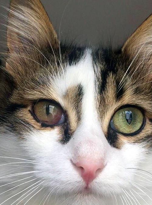
16

involuntary muscle twitching, and reluctance or inability to jump to high places. Ocular
involvement can precede or accompany neurological disease. Neurological FIP is a
common occurrence in antiviral drug therapy, either appearing during treatment of non-
CNS forms of FIP or as a manifestation of disease relapse after treatment has ended
(Pedersen et al., 2018, 2019; Dickinson et al., 2020).

Figure 10. A young cat with dry FIP and neurological involvement. The cat is lethargic, thin, and with a poor
coat. Hair in the perineal area is wet and stained from urinary incontinence.

Ocular involvement is usually grossly apparent and confirmed on ophthalmoscopic
examination of anterior and posterior chambers. Ocular FIP targets the iris, ciliary body,
retina, and optic disc to varying degrees (Peiffer and Wilcock 1991; Ziółkowska et al.,
2017; Andrew, 2000). The earliest sign is often unilateral discoloration of the iris (Fig.
11). The anterior chamber may appear cloudy and exhibit high protein levels and
aqueous flare with refracted light. There is an outpouring of inflammatory products into
the anterior chamber in the form of activated macrophages, red cells, fibrin tags, and
small blood clots. This material often adheres to the back side of the cornea as keratic
precipitates (Fig. 12). The disease may also involve the retina in tapetal and non-tapetal
areas and lead to retinal detachments. Intraocular pressures are usually low, except in
cases complicated by involvement of the ciliary body and glaucoma (Figs. 12, 13).
17

Figure 11. Discoloration of the iris in the right eye of this cat was the earliest sign of FIP-associated uveitis.
There is a slight haziness to the anterior chamber and deposits of red cell rich fibrin on the inside of the
cornea. The pupils are also uneven (anisocoria).

Figure 12. A young cat with ocular FIP manifested in the right eye as anterior uveitis with secondary
glaucoma causing enlargement of the globe. The iris has changed color due to inflammation, blood vessels
at the base of the iris are engorged, and there is a cloudiness to the aqueous humor and inflammatory
products on the back side of the cornea. Intraocular pressure is usually low in uncomplicated uveitis but
increased in cats with glaucoma.
18

Figure 13. This young cat presented with anterior uveitis but treatment for FIP with GS-441524 was delayed,
which allowed glaucoma to develop in both eyes. Treatment eliminated the underlying uveitis and greatly
improved outward health, but secondary glaucoma and blindness persisted.

Diagnosis of FIP

The signalment, environmental history, clinical signs, and findings on physical
examination are often indicative of FIP (Tasker, 2018). A thorough physical examination
should include body weight and temperature, status of coat and flesh, manual palpation
of the abdomen and abdominal organs, gross evaluation of cardiac and pulmonary
function, and cursory examination of the eyes and neurological system. A strong
suspicion of effusion in the abdomen or chest may warrant a confirmatory aspiration and
even in-house fluid analysis as part of the initial examination.

Abnormalities in a complete blood count (CBC) and a basic serum biochemistry panel
are important for FIP diagnosis (Tasker, 2018; Felten and Hartmann, 2019) and
monitoring antiviral drug therapy (Pedersen et al., 2018, 2019; Jones et al., 2021;
Krentz et al., 2021) (Fig. 14). The total leukocyte count is most likely to be high in cats
with wet FIP, but low counts can occur with severe inflammation. High leukocyte counts
are often associated with neutrophilia, lymphopenia and eosinopenia. A mild to
moderately severe non-regenerative anemia is also frequently observed in both wet and
dry FIP. Total proteins are usually elevated due to increased levels of globulin, while
albumin values tend to be low (Fig. 14). This results in an A:G ratio that is often below
0.5-0.6 and considered one of the most consistent indicators of FIP. However, a low
A:G ratio can occur in situations where both albumin and globulin are in their reference
interval or in other diseases. Therefore, the A:G ratio should not be the sole indicator of
FIP and should always be evaluated in context with other indicators of FIP (Tasker,
2018; Felten and Hartmann, 2019). Serum protein values obtained from most serum
chemistry panels are usually sufficient. Serum protein electrophoresis can provide
19

additional information, especially when protein values from serum chemistry are
questionable (Stranieri et al., 2017).

Figure 14. Complete blood count (CBC) (a) and serum chemistry (b) panels from a young cat with acute wet
abdominal FIP. Although the leukocyte count was not elevated, there was a relative but not absolute
neutrophilia, relative and absolute lymphopenia, relative and absolute eosinopenia and non-responsive
anemia as indicated by low red blood cells, hematocrit, and hemoglobin with normal reticulocyte count. The
relevant values in the serum chemistry panel were the elevated total protein, low albumin, high globulin, low
Albumin/Globulin (A:G) ratio, and an elevated total and direct bilirubin. Liver enzymes were normal except for
mild elevation of AST, and BUN and Creatinine are normal, indicating absence of significant liver or renal
disease. Globulin values are not always provided but a reasonable estimate can be calculated by subtracting
albumin levels from the total protein.
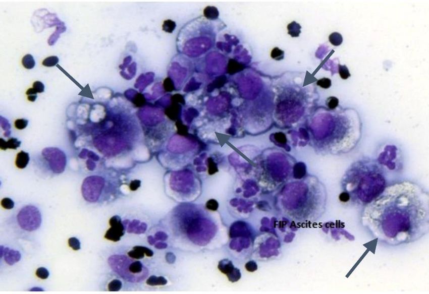
20

Over-reliance on abnormalities in CBC and serum biochemistry panels can lead to
diagnostic uncertainty when absent, even with the realization that no test value is
consistently abnormal in all FIP cases (Tasker, 2018).1 The greatest variation is
between the presenting clinical form, with leukocytosis and lymphopenia being more
common in cats with wet than dry FIP (Riemer et al., 2016). Hyperbilirubinemia is
common in cats with FIP, but mainly in those with wet FIP (Tasker, 2018). The author
has also found that many cats with primary neurological FIP have minor or no blood
abnormalities. Blood test values for FIP also vary between studies (Tasker, 2018).

Complete analysis of the effusion is important for diagnosing wet FIP and for ruling out
other potential causes of fluid accumulation (Dempsey and Ewing, 2011). This includes
color (clear or yellow), viscosity (thin or viscous), presence of precipitates, ability to form
partial clot on standing, protein content, leukocyte count and differential. The character
of the fluid can vary based on duration of the disease and its severity. Effusions in cats
with more severe disease signs usually have protein values near serum levels, are more
viscous, contain higher numbers of leukocytes, more yellow-tinged, and more apt to
form partial clots on standing. Chronic effusions tend to be less inflammatory in nature,
with lower levels of protein and leukocytes, less viscous, and clearer. These values can
be determined in-house in most clinics. Clotting property is determined by comparing
fluid collected in serum and anticoagulant tubes after standing. Color and viscosity can
be grossly determined, and protein levels estimated with a handheld total solids’
refractometer. Cells are pelleted from fluid and analyzed on a quick-stained slide by light
microscopy and leukocyte numbers and differential estimated. Cells include non-septic
appearing neutrophils, small and medium size mononuclear cells, and large vacuolated
macrophages (Fig. 15). It is important to note that effusions may occur with a wide
range of diseases, such as heart failure, cancer, hypoproteinemia, and bacterial
infections. Effusions from these other disorders usually have distinct identifying
characteristics.

Figure 15. Stained smear of peritoneal cells centrifuged from abdominal fluid of a cat with wet FIP and
examined on a quick stained slide by light microscopy. The predominant cells are large heavily vacuolated
macrophages, smaller differentiating activated monocytes, and neutrophils. The largest concentration of
viral particles is within intracytoplasmic vacuoles of macrophages (arrows).
21

A positive Rivalta test on abdominal or thoracic fluid is frequently used to diagnose FIP
as the cause of an effusion and a negative test tends to exclude it (Fischer et al., 2010)
(Fig.16). However, the test may be positive in inflammatory effusions of other cause and
negative in some cats with FIP. Therefore, the Rivalta test is most helpful when coupled
with other clinical findings of FIP and should not be a replacement for a thorough fluid
analysis (Felten and Hartmann, 2019).

Figure 16. A positive Rivalta test result. A small sample of abdominal or thoracic fluid is dropped gently into
a small glass filled with dilute acetic acid (8 ml distilled water and 1 drop of concentrated acetic acid).
Inflammatory proteins will almost immediately congeal and sink to the bottom (positive). Less inflammatory
fluids will form diffuse clots (questionable) or diffuse freely in the solution (negative).

Serum total and direct bilirubin levels are frequently elevated, especially in cats with wet
FIP (Fig. 14) and may be associated with jaundice and bilirubinuria. Hyperbilirubinemia
in FIP is not due to liver disease (Tasker, 2018), but more likely to vasculitis, micro-
hemorrhaging, hemolysis, and destruction of damaged red blood cells by macrophages
locally and in the liver. The released hemoglobin is ultimately metabolized to bilirubin,
which is then conjugated in hepatocytes and excreted in urine. Glucuronidation is
essential for bilirubin secretion and genetic disorders affecting glucuronidation in
humans will impede its excretion (Kalakonda et al., 2021). Cats, as a species, are
deficient in the enzymes required for glucuronidation, which makes it more difficult for
them to excrete substances such as bilirubin (Court and Greenblatt 2000).

Although FIP can involve kidneys and liver, it is not severe enough to cause significant
loss of renal or hepatic function. However, serum tests for blood urea nitrogen (BUN)
and creatinine as a measure of kidney disease and alanine aminotransferase (ALT),
alkaline phosphatase (ALP), and gamma glutamyl transferase (GGT) for liver disease
may be mildly elevated in cats with FIP, especially in cats with more acute and severe
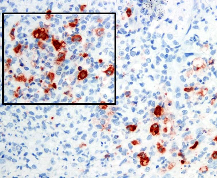
22

disease (Fig. 14). Therefore, mildly abnormal test values should not be over-interpreted
in the absence of other clinical features of liver or kidney disease, while significant
increases should point to the possibility of concurrent, and possibly predisposing,
diseases of those organs.

Serum can also be tested for additional markers of systemic inflammation, such as
elevated levels of alpha-1-acid glycoprotein (AGP) (Paltrinieri et al., 2007) and feline
serum amyloid A (fSAA) (Yuki et al., 2020). They may also prove useful in monitoring
response to antiviral drug treatment (Krentz et al., 2021).

Radiography can be helpful in identifying thoracic and abdominal effusions. Abdominal
ultrasound can detect smaller amounts of effusion be identify enlarged mesenteric and
ileo-cecal-colic lymph nodes, colonic wall thickening, and lesions in organs such as the
kidney, liver, and spleen (Lewis and O’Brien 2010). It can also be useful in scanning the
chest for lesions. and assist with needle aspiration or biopsy.

The value placed on FCoV antibody titers has diminished since first reported almost 50
years ago (Pedersen 1976b). The reference antibody test employs indirect fluorescent
antibody (IFA) staining IFA titers ≥1:3200 in cats with FIP are higher than for most
FECV exposed cats (1:25-1:400). Newer tests often use ELISA procedures for rapid in-
house or laboratory testing, but they are more qualitative than quantitative. IFA antibody
titers decrease during successful antiviral drug treatment in many cats but remain high
in others (Dickinson et al., 2020; Krentz et al., 2021). Sequential titers may show a
progressive rise in titers as FIP develops (Pedersen et al., 1977), but prior serum
samples are seldom available for comparison. Like most tests, FCoV antibody levels
should not be used as the sole criterion for diagnosing or excluding FIP (Felten and
Hartmann, 2019) or for evaluating treatment success (Krentz et al., 2021).

The reverse transcriptase polymerase chain reaction (RT-PCR) is the primary means to
identify FCoV RNA in inflammatory effusions, fluids, or diseased tissues (Felten and
Hartmann, 2019). RNA of the 7b accessory gene is present at the highest level in both
FECV or FIPV infected tissues, fluids, or excretions, which makes it the most sensitive
target for detecting low levels of virus (Gut et al., 1999). RT-PCR for the FIPV S gene
mutations is often used in samples testing positive for 7b RNA to make it specific for
FIPV (Felten et al., 2017). Other studies suggest that RT-PCR tests for FIPV specific S
gene mutations have similar specificity for FIP but at a significant loss of sensitivity
(Barker et al., 2017). A decrease in sensitivity relates to an increase in false negatives.
False negative RT-PCR tests also occur in samples that contain insufficient infected
macrophages or in cats with very low virus levels. False negatives are particularly
common when testing whole blood.

Immunohistochemistry (IHC) detects feline coronavirus nucleocapsid protein in formalin
fixed tissues with high sensitivity and specificity but has not been as popular as RT-PCR
(Litster et al., 2013; Ziółkowska et al., 2019). Samples for IHC must contain intact
infected macrophages (Fig.17), which requires careful separation of cells from effusions
and placement on slides, or diseased tissues fixed in formalin and embedded in paraffin
23

that show lesions compatible with FIP. Coronavirus antigen in a macrophage within a
typical FIP lesion or fluid, is observed only with FIP, giving IHC a high level of specificity.

Figure 17. Histologic section from the thickened colon of a cat with the colonic form of FIP. The thickened
wall contained foci of macrophages (square area) that stained positive (brownish red) by immunoperoxidase
for the nucleocapsid protein of FIPV.

A thorough ophthalmologic examination is essential for diagnosing characteristic lesions
of FIP (Pfeiffer and Wilcock 1991; Andrew, 2000). A sample of aqueous humor from the
anterior chamber of an inflamed eye can is also useful for cytology, PCR, and IHC.

Neurological FIP is frequently diagnosed by magnetic resonance imaging (MRI) with
contrast enhancement and is often coupled with cerebrospinal fluid (CSF) analysis
(Crawford et al., 2017; Tasker, 2018; Dickinson et al., 2020). However, these are
expensive procedures that are not always available and carry some risk to the cat. MRI
lesions include obstructive hydrocephalus, syringomyelia, and foramen magnum
herniation, with contrast enhancement of the meninges of the brain and spinal cord and
ependyma of the third ventricle, mesencephalic aqueduct, and brainstem. CSF shows
increased protein and cell count (neutrophils, lymphocytes, monocyte/macrophages),
and when these are present, it can be a reliable source for PCR or IHC testing.

The neurological and/or ocular forms of FIP is often confused with feline systemic
toxoplasmosis and many cats with FIP are treated empirically for toxoplasmosis before
the diagnosis of FIP is made. Fortunately, the availability of an effective treatment for
FIP has reduced this practice. Systemic toxoplasmosis is much less prevalent than FIP
and less than 1% of cats with FIP in one field study were serologically positive. 1
Therefore, testing or treatment for toxoplasmosis should be considered only after the
diagnosis of FIP has been adequately pursued.

Antiviral treatment as a diagnostic tool
24

Situations commonly arrive where clinical findings point to FIP, but doubt remains. A
choice at that point is to do more diagnostic tests that may not lead to a more definitive
diagnosis. An alternative diagnostic approach is to treat with an appropriate antiviral
drug for 1-2 weeks at a correct dosage for the suspected form of FIP.2 Treatment will
often cause clinical improvement in as little as 24-48h and this will rapidly progress over
the next 2 weeks and the full treatment given (Fig. 18). No response to test treatment
and/or a deterioration in health would indicate a need to further investigate the cause(s)
of ill-health.

Figure 18. Cat with FIP at the start of treatment with GS-441524 (a) and after 1 week (b). The response is rapid
with fever disappearing in 24-48h and significant improvement in outward health within 1-2 weeks. This type
of response is often used to confirm a diagnosis of FIP.

Treatment of FIP

There was no cure for FIP before 2017 and treatments were directed mainly at
lessening disease signs (Izes et al., 2020). Such supportive care was focused on
maintaining good nutrition, controlling inflammation (corticosteroids), altering immune
responses (interferons, cyclophosphamide, chlorambucil), and inhibiting key cytokine
responses (pentoxifylline and other TNF-alpha inhibitors). Dietary supplements claiming
to help specific organ functions were also commonly used, such as one claiming to
improve immunity and prolong survival in cats with dry, but not wet, FIP (Legendre et
al., 2017). The effect of good supportive care on survival was impossible to determine,
because most cats have been euthanized upon diagnosis or within several days or
weeks. The survival rate even with the mildest forms of dry FIP and most persistent
25

treatment in one study was only 13% at 200 days and 6% at 300 days (Legendre et al.,
2017).

Many commercially available drugs and compounds will inhibit FIPV infection or
replication in vitro, some being repurposed drugs known to inhibit specific proteins of
HIV or hepatitis C virus, while others work by inhibiting normal cellular processes
usurped by the virus for its own life cycle (Hsieh et al., 2010; Izes et al., 2020; Delaplace
et al., 2021). These various drugs and substances include cyclosporine and related
immunophilins, several nucleosides and protease inhibitors, vioporin inhibitors, pyridine
N-oxide derivatives, chloroquine, and related compounds, ivermectin, several plant
lectins, ubiquitin inhibitors, itraconazole, and several antibiotics. However, the
concentrations needed to inhibit virus replication in vitro are often near toxic levels to
cells. It has also been difficult to translate favorable in vitro findings to animals and
studies on diseased cats seldom follow. Ribavarin will inhibit FIPV replication in vitro but
was not effective as a treatment of experimental FIP (Weiss et al., 1993). Chloroquine
was tested for efficacy in FIPV infected laboratory cats, but clinical scores in treated
cats were only slightly better than untreated cats and there was evidence of
hepatotoxicity (Takano et al., 2013). A 3-month-old kitten with thoracic wet FIP was
treated with itraconazole and prednisolone developed neurological FIP and was
euthanized after 38 days of treatment (Kameshima et al., 2020). Mefloquine has also
inhibited FIPV replication at low concentrations in cultured feline cells without cytotoxic
effects and preliminary pharmacokinetic studies in cats appeared favorable (Yu et al.,
2020), but evidence of its safety and efficacy in clinical trials of cats with FIP has yet to
be published.

A breakthrough in the treatment of FIP came about in 2016-2019 with reports of antiviral
drugs that targeted specific FIPV proteins essential for replication. The first of these
drugs was GC376, an inhibitor of the main protease (Mpro ) of FIPV (Kim et al., 2016;
Pedersen et al., 2018). Protease inhibitors prevent the formation of individual viral
proteins by inhibiting their cleavage from polyprotein precursors. GC376 was able to
cure all experimentally infected cats and 7 of 21 cats with naturally occurring wet and
dry FIP but it was less efficacious for cats with ocular or neurological signs (Pedersen et
al., 2018). The second of these drugs was GS-441514, the active moiety of the prodrug
remdesivir (Gilead Sciences; Murphy et al., 2018; Pedersen et al., 2019). GS-441524 is
an adenine nucleotide analog that blocks replication of FIPV by inserting a nonsense
adenine in the developing viral RNA. GS-441524 was also able to cure all
experimentally infected cats (Murphy et al., 2018) and 25/31 cats with naturally
occurring wet and dry FIP (Pedersen et al., 2019). It also appeared to be effective in
several cats with ocular and neurological FIP at a higher dosage (Pedersen et al., 2019)
and is currently the drug of choice for cats with neurological FIP (Dickinson et al., 2020).
GS-441524 has treated thousands of cats with FIP from around the world over the last
three years with an overall cure rate just over 90% (Jones et al., 2021).1

Even though the ability of GC376 and GS-441524 to cure cats has been known for
several years, neither drug is legally available at this time in most countries. The rights
for GC376 were purchased by Anivive, but is yet to be marketed.3 Potential conflicts
26

with remdesivir’s development for treatment of COVID-19 led Gilead Sciences to
withhold animal rights for GS-441524, which prompted the creation of an unapproved
source for GS-441524 out of China (Jones et al., 2021).1,2,4 Remdesivir is rapidly
converted to GS-441524 in the body and has been allowed for FIP treatment in some
countries.2 GS-441524 can also be given orally at a higher dosage and now commonly
used in the field (Krentz et al., 2021).1

The efficacy of drugs like GC376 and GS-441524 for FIP of cats, whose use predated
the COVID-19 pandemic, has been recognized by researchers studying related
inhibitors of SARS-CoV 2 (Yan et al., 2020; Vuong et al., 2021). Remdesivir, an
injectable drug marketed under the name veklury (Gilead), has been used worldwide to
lower mortality to COVID-19 (Beigel et al., 2020). GC373, the active form of the prodrug
GC376, has undergone simple modifications to enhance efficacy and oral bioavailability
(Vuong et al., 2021). A GC373-related drug, nirmatrelvir, was successfully tested
against early COVID-19 infections and has been approved for early COVID-19 and
marketed under the name paxlovid (Pfizer). Paxlovid consists of two drugs, nirmatrelvir
and the HIV protease inhibitor ritonavir. Ritonovir is not a significant inhibitor of SARS-
CoV 2, but reportedly extends the half-life of Mpro inhibitors when used in combination
(Vuong et al., 2020). Nirmatrelvir and paxlovid have not been tested in cats with FIP at
this time, but based on experience with the closely related GC376, it may be an
important oral treatment for some forms of FIP in the future.

Two additional nucleoside analogs. EIDD-1931 and EIDD-2801, have been researched
for treatment of several RNA virus infections in people and animals (Painter et al.,
2021). EIDD-1931 is the experimental designation for beta-D-N4-hydroxycytidine, a
compound widely studied since the 1970s. Beta-D-N4-hydroxycytidine is metabolized into
a ribonucleoside analog, which is incorporated into RNA in place of cytidine and results in fatal
mutations in the viral RNA chain.The compound is inhibitory to a broad range of human
and animal RNA viruses including all known coronaviruses. EIDD-1931 was modified to
increase oral absorption and named EIDD-2801 (molnupiravir) (Painter et al., 2021).
Molnupiravir is de-esterified in the body to its active ingredient, beta-D-N4-
hyroxycytidine. Therefore, EIDD-1931 and molnupiravir are analogous to GS-441524
and remdesivir, respectively. Molnupiravir is marketed for in-home treatment of primary
COVID-19 under the names lagevrio (Merck, USA) or molnulup (Lupin, India).

Both EIDD-1931 and EIDD-2801 have been shown to be effective in inhibiting FIPV in
tissue culture (Cook et al., 2021) and EIDD-2801 is currently being used to treat some
FIP cases in the field.5,7 The effective concentration-50% (EC50) for EIDD-1931 against
FIPV is 0.09 µM, EIDD-2801 0.4 µM and GS-441524 0.66 µM (Cook et al., 2021). The
percent cytotoxicity at 100 µM is 2.8, 3.8 and 0.0 for these compounds, respectively.
Therefore, EIDD-1931 and -2801 are slightly more virus inhibitory but more cytotoxic
than GS-441524. Resistance to GS-441524 is seen in some cases of FIP (Pedersen et
al., 2019) and to remdesivir in COVID-19 patients (Painter et al., 2021), but these
isolates remain susceptible to molnupiravir (Sheahan et al., 2020). This may prove
helpful in the countering GS-441524 resistance in cats and people, and in the
development of multi-drug treatments to prevent resistance from occurring.
You can also read