High-Light-Induced Stress Activates Lipid Deacylation at the Sn-2 Position in the Cyanobacterium Synechocystis Sp. PCC 6803
←
→
Page content transcription
If your browser does not render page correctly, please read the page content below
High-Light-Induced Stress Activates Lipid Deacylation at the
Sn-2 Position in the Cyanobacterium Synechocystis Sp. PCC
6803
Kouji Kojima1,2,6 , Ui Matsumoto1,2 , Sumie Keta1,2 , Kenji Nakahigashi2,3,7 , Kazutaka Ikeda2,4,8 ,
Regular Paper
Nobuyuki Takatani2,5 , Tatsuo Omata2,5 and Makiko Aichi1,2,*
Downloaded from https://academic.oup.com/pcp/advance-article/doi/10.1093/pcp/pcab147/6384518 by guest on 19 November 2021
1 Department of Biological Chemistry, Chubu University, 1200 Matsumoto-cho, Kasugai, 487-8501 Japan
2 Japan Science and Technology Agency, CREST, 4-1-8 Honmachi, Kwaguchi, Saitama 332-0012, Japan
3 Institute for Advanced Biosciences, Keio University, 246-2 Mizukami, Yamagata, 997-0052 Japan
4 Laboratory for Metabolomics, RIKEN Center for Integrative Medical Sciences, 1-7-22 Suehiro-cho, Tsurumi-ku, Yokohama, 230-0045 Japan
5 Graduate School of Bioagricultural Sciences, Nagoya University, Furo-cho, Chikusa-ku, Nagoya, 464-8601 Japan
6 Present address: Department of Applied Microbial Technology, Faculty of Biotechnology and Life Science, Sojo University, 4-22-1 Ikeda, Nishi-ku,
Kumamoto, 860-0082 Japan
7 Present address: Spiber Inc., Yamagata, 997-0052 Japan
8 Present address: Laboratory of Biomolecule Analysis, Department of Applied Genomics, Kazusa DNA Research Institute, 2-6-7 Kazusa-Kamatari, Kisarazu
Chiba, 292-0818 Japan
*Corresponding author: E-mail, makiko@isc.chubu.ac.jp
(Received 22 March 2021; Accepted 8 October 2021)
Cyanobacterial mutants defective in acyl-acyl carrier protein Introduction
synthetase (Aas) produce free fatty acids (FFAs) because the
FFAs generated by deacylation of membrane lipids cannot be Free fatty acids (FFAs) are naturally produced by cyanobacteria
recycled. An engineered Aas-deficient mutant of Synechocys- via deacylation of membrane lipids, but the cells do not nor-
tis sp. PCC 6803 grew normally under low-light (LL) condi- mally accumulate FFAs or secrete them out of the cells since
tions (50 µmol photons m−2 s−1 ) but was unable to sustain the FFAs are readily esterified to acyl carrier protein (ACP) by
growth under high-light (HL) conditions (400 µmol photons acyl-ACP synthetase (Aas) and recycled to lipids (Kaczmarzyk
m−2 s−1 ), revealing a crucial role of Aas in survival under and Fulda 2010). Initial physiological characterization of the
the HL conditions. Several-times larger amounts of FFAs were Aas-null mutants of model cyanobacterial species Synechococ-
produced by HL-exposed cultures than LL-grown cultures. cus elongatus PCC 7942 and Synechocystis sp. PCC 6803 revealed
Palmitic acid accounted for ∼85% of total FFAs in HL-exposed secretion of FFAs out of the cells but there was no noticeable
cultures, while C18 fatty acids (FAs) constituted ∼80% of the growth phenotype under illumination at 30–60 µmol photons
FFAs in LL-grown cultures. Since C16 FAs are esterified to m−2 s−1 (Kaczmarzyk and Fulda 2010, Ruffing 2014). Takatani
the sn-2 position of lipids in the Synechocystis species, it was et al. (2015) later showed that an Aas-deficient S. elongatus PCC
deduced that HL irradiation activated deacylation of lipids at 7942 grows slightly slower than the wild-type (WT) under low-
the sn-2 position. Heterologous expression of FarB, the FFA light (LL) conditions (50 µmol photons m−2 s−1 ), and unlike
exporter protein of Neisseria lactamica, prevented intracellu- WT, it cannot increase the growth rate in response to a shift
lar FFA accumulation and rescued the growth defect of the to high-light (HL) conditions (400 µmol photons m−2 s−1 ). It
mutant under HL, indicating that intracellular FFA was the was shown that lipid deacylation is enhanced under the HL
cause of growth inhibition. FarB expression also decreased condition and that the resulting FFAs destabilize photosystem
the ‘per-cell’ yield of FFA under HL by 90% and decreased II (PSII), making the cells more susceptible to photodamage
the proportion of palmitic acid to ∼15% of total FFA. These (Takatani et al. 2015). These results showed the importance
results indicated that the HL-induced lipid deacylation is trig- of Aas in acclimation of S. elongatus to the HL conditions. In
gered not by strong light per se but by HL-induced damage to this study, we characterized an Aas-deficient mutant (dAS11)
the cells. It was deduced that there is a positive feedback loop constructed from Synechocystis sp. PCC 6803. HL-responsive
between HL-induced damage and lipid deacylation, which is lipid deacylation is shown to take place also in Synechocystis
lethal unless FFA accumulation is prevented by Aas. sp. PCC 6803, causing severe growth inhibition of the mutant.
Involvement of an sn-2 specific lipase is deduced from the
Keywords: Acyl-ACP synthetase • Lipid deacylation FFA composition of the HL-exposed cells. We show that het-
• Photoinhibition • Toxicity to FFAs erologous expression of a bacterial FFA exporter prevents the
Plant Cell Physiol. 00(0): 1–10 (2021) doi:https://doi.org/10.1093/pcp/pcab147, Advance Access publication on 8 October 2021, available online at
https://academic.oup.com/pcp
© The Author(s) 2021. Published by Oxford University Press on behalf of Japanese Society of Plant Physiologists.
This is an Open Access article distributed under the terms of the Creative Commons Attribution-NonCommercial License
(https://creativecommons.org/licenses/by-nc/4.0/), which permits non-commercial re-use, distribution, and reproduction in any medium, provided the original
work is properly cited. For commercial re-use, please contact journals.permissions@oup.com
K. Kojima et al. | Lipid deacylation induced by high-light stress
accumulation of intracellular FFAs, largely rescues the growth dAS11 mutant, we planned expression in dAS11 of the tripartite
phenotype of the mutant, and at the same time attenuates the FFA efflux pump conserved among the bacteria of the Neisseria
lipase activation under HL. Based on these findings, we propose genus (Lee and Shafer 1999, see also Supplementary Fig. S1),
that there is a positive feedback between photodamage to the which is composed of FarB (an MFS family transporter), MtrE
cells, which is enhanced by intracellular FFA, and upregulation (an outer membrane channel protein, OMP) and FarA (a mem-
of an sn-2 specific lipase. Possible roles of lipid deacylation and brane fusion protein, MFP). Since heterologous expression of
FFA recycling in HL acclimation of cyanobacteria are discussed. the Neisseria FFA efflux pump had not been reported, we first
tried it in Escherichia coli. Construction of a plasmid carrying the
Results transcriptional fusion of the farAB operon and the mtrE gene
Downloaded from https://academic.oup.com/pcp/advance-article/doi/10.1093/pcp/pcab147/6384518 by guest on 19 November 2021
under the control of the Ptrc promoter was unsuccessful in E. coli
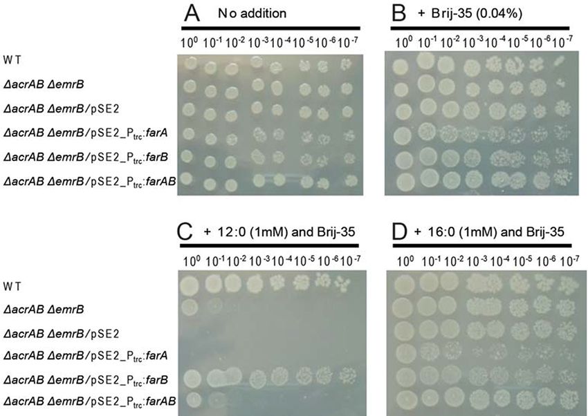
Sensitivity of an aas-deficient mutant of
even in the absence of isopropyl β-D-1-thiogalactopyranoside
Synechocystis sp. PCC 6803 to high-light conditions (IPTG) (data not shown), suggesting that expression of the
Fig. 1 represents the appearance of the cultures of the WT strain whole set of the proteins comprising the FFA pump might be
and an Aas-deficient mutant (dAS11) of Synechocystis sp. PCC toxic to the E. coli host. We hence examined the effects of
6803 during cultivation under LL (50 µmol photons m−2 s−1 ) plasmid-based expression of the Ptrc -farAB, Ptrc -farA or Ptrc -farB
and HL (400 µmol photons m−2 s−1 ) conditions. The dAS11 genes in an E. coli K12 ∆acrAB ∆emrB mutant, which lacks
culture became bluish after 3–4 days of cultivation under the HL two (AcrAB-TolC and EmrAB-TolC) of the three efflux pumps
conditions and completely lost pigmentation in 7 days, whereas known to mediate FFA efflux (Lennen et al. 2013). While the WT
no decrease in pigmentation was observed under the LL condi- E. coli cells were resistant to lauric acid (12:0) and palmitic acid
tions (Fig. 1). In a previous study in S. elongatus PCC 7942, the (16:0) added to the medium to a concentration of 1 mM, the
aas-deficient mutant dAS1 was shown to suffer from enhanced ∆acrAB ∆emrB mutant was sensitive to 12:0 (Fig. 2). Plasmid-
photodamage to PSII under the HL conditions, but the cells based expression of farA or farAB did not confer the cells the
could sustain growth under HL albeit at a lower rate as com- tolerance to exogenously added 12:0, but surprisingly, that of
pared to the WT strain (Takatani et al. 2015). These results farB rendered the cells tolerant to 12:0 (Fig. 2C). These results
indicated that Aas plays a more crucial role in Synechocystis sp. indicated that expression of FarB supported export of 12:0 out
PCC 6803 than in S. elongatus PCC 7942 in the acclimation of of the cell, but co-expression of FarA interfered with it.
the cells to the HL conditions.
Negative effect of FarA on the tolerance of
Functional expression of a Neisseria lactamica FFA Synechocystis cells to extrinsic linolenic acid
exporter in Escherichia coli
Fig. 3 shows the effects of exogenously added linolenic acid
Aas-deficient cyanobacterial mutants produce FFAs, which can (18:3) on growth of the WT Synechocystis sp. PCC 6803 strain,
be lethal if accumulated at high levels in the cell (Kato et al. the dAS11 mutant and the dAS11 derivatives expressing either
2015, 2016, 2017). To examine whether an increase in the FFA farA and farB or both farA and farB. Aas has been shown to
export activity could rescue the HL-sensitive phenotype of the facilitate passive entrance of extrinsic FFAs into the cell by con-
suming intracellular FFAs (Kaczmarzyk and Fulda 2010). Exoge-
nously added 18:3 is generally toxic to cyanobacteria (Sakamoto
et al. 1998, Maeda et al. 2005), but as reported previously (von
Berlepsch et al. 2012, Kojima et al. 2016), the Aas-deficient
mutant was tolerant to the fatty acid (FA) added to the medium
to 100 µM. Expression of farA in dAS11 completely abolished
the ability of the cells to grow in the presence of the FA, while
that of farB did not affect it. Interestingly, the cells co-expressing
farA and farB were tolerant to 18:3, showing that FarB could
partially suppress the negative effect of FarA.
Positive effect of FarB on the tolerance of
Synechocystis cells to extrinsic FAs
Fig. 4 shows the growth curves of WT, dAS11 and the
dAS11_farB strains in liquid media supplemented with vari-
ous FAs under the LL conditions. Growth of the WT strain
Fig. 1 Appearance of the WT and dAS11 cultures during growth under was severely inhibited by 100 µM of 12:0 and 18:3 (Fig. 4B, E,
the HL and LL conditions. Cells grown under LL conditions (50 µmol black circles), but the inhibitory effects of these FAs were
photons m−2 s−1 ) were inoculated into new medium to give an partially attenuated by the deficiency of Aas as previously
OD730 value of 0.01 and incubated under the LL or HL (400 µmol reported (Fig. 4B, E, white circles; von Berlepsch et al. 2012,
photons m−2 s−1 ) conditions for the indicated period. Kojima et al. 2016; see also Fig. 3). Expression of farB was found
2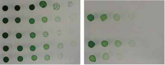
Plant Cell Physiol. 00(0): 1–10 (2021) doi:https://doi.org/10.1093/pcp/pcab147

                                                                                                                                                               Downloaded from https://academic.oup.com/pcp/advance-article/doi/10.1093/pcp/pcab147/6384518 by guest on 19 November 2021
Fig. 2 Effects of expression of either or both of the N. lactamica farA and farB genes on growth of the E. coli K12 ∆acrAB∆emrB mutant in the
presence of exogenously added FFAs. Three microliters of the E. coli cell suspensions (OD600 = 1.0) and their 10-fold serial dilutions were spotted
onto solid media and incubated for 1 day at 37◦ C. (A, B) Control experiments performed without (A) and with (B) 0.04% Brij-35. (C, D) Viability
test performed in the presence of 0.04% Brij-35 and 1 mM 12:0 (C) and 16:0 (D). Numbers on the top indicate the dilution factor. The results from
one of the three experiments, which yielded essentially the same results, are shown.

to further improve the growth of the aas-deficient strain in                  under the HL conditions increased the growth rates of both
the presence of these FAs (Fig. 4B, E, gray circles), suggesting             WT and dAS11 as compared to those under the LL conditions,
the involvement of FarB in the export of the toxic FAs. Unlike               but in the case of the mutant, cell growth ceased after 3 days
12:0 and 18:3, 16:0 is normally non-toxic to cyanobacteria (Ruff-            of exponential growth (Fig. 5B). The cells thereafter showed a
ing and Trahan 2014), but at a concentration of 500 µM, it
significantly inhibited the growth of dAS11 (Fig. 4C, white cir-
cles). Similar effects of exogenously added 16:0 were previously
reported for a FFA-producing mutant of S. elongatus PCC 7942
and ascribed to an interference of the secretion of the endoge-
nously produced FFA out of the cells (Kato et al. 2017). The
inhibitory effect of 16:0 on dAS11 was rescued by expression
of farB (Fig. 4C, gray circles), showing that FarB could mediate
export of 16:0 as well as of 12:0 and 18:3.

Effects of farB expression on growth and                                     Fig. 3 Effects of expression of either or both of the N. lactamica farA and
photosynthetic activity of the aas-deficient                                  farB genes on growth of the Aas-deficient Synechocystis sp. PCC 6803
Synechocystis mutant under high-light conditions                             mutant in the presence of 18:3. Three microliters of the Synechocys-
                                                                             tis cell suspensions (OD730 = 1.0) and their 5-fold serial dilutions were
Fig. 5 shows the growth curves of the WT, dAS11 and                          spotted onto solid media and incubated for 5 d at 30◦ C under illumi-
dAS11_farB strains under the LL and HL conditions. Under                     nation at 25 µmol photons m−2 s−1 . Growth of the cells in the absence
the LL conditions, the final optical density of the dAS11 cul-                (A) and the presence (B) of 0.1 mM 18:3 is compared. Numbers on the
ture was comparable to that of WT, although the growth of                    top indicate the dilution factor. A set of data from one of the three
the mutant was slower than that of WT (Fig. 5A). Cultivation                 experiments, which yielded essentially the same results, is shown.

                                                                                                                                                           3
K. Kojima et al. | Lipid deacylation induced by high-light stress
Downloaded from https://academic.oup.com/pcp/advance-article/doi/10.1093/pcp/pcab147/6384518 by guest on 19 November 2021
Fig. 4 Effects of Aas deficiency and farB expression on Synechocystis growth in the presence of various FAs. Growth curves of the WT (black circles),
dAS11 (white circles) and dAS11_farB (gray circles) strains were compared in the presence or absence of FAs (labeled on the top of the panel).
Data shown are mean ± SE from biological triplicates.
decrease in pigmentation as mentioned above (Fig. 1). Inter- which inhibits the repair of PSII (Fig. 6B). In the absence of
estingly, growth of the mutant resumed several days after the lincomycin (Fig. 6B), PSII activity of the LL-grown cells was
growth arrest, showing a recovery of pigmentation. Growth of decreased by about 40% after 20 min of exposure to illumina-
dAS11_farB was essentially the same as that of dAS11 under the tion of 1,500 µmol photons m−2 s−1 in all the strains. In WT and
LL conditions (Fig. 5A). Even under the HL conditions, the farB dAS11_farB, there was only a small decrease in the PSII activ-
expressing strain did not show the growth arrest or the bleach- ity thereafter, whereas dAS11 appeared to suffer from further
ing phenotype (Fig. 5B). Thus, expression of FarB largely rescued decrease in the PSII activity, which was decreased to ∼40% of
the growth defect caused by the deficiency of Aas, although the the initial level in 60 min of HL irradiation. However, the dif-
final cell density of the dAS11_farB strain was somewhat lower ference in PSII activity between dAS11 and the other strains
than that attained by WT (Fig. 5B). was not statistically significant. When lincomycin was added to
The PSII activity, as determined by measuring the Fv Fm−1 inhibit the repair of photodamaged PSII, on the other hand, the
ratio, showed a sharp decline between the third and fourth days HL-induced decrease of PSII activity was significantly faster in
of incubation of the dAS11 cells under HL (Fig. 5C, D), suggest- dAS11 than in WT or dAS11_farB (Fig. 6B), showing that PSII
ing that the decrease of PSII activity was the cause of the growth was more sensitive to photodamage in dAS11 than in the other
arrest. In contrast, no such decline was observed in the PSII yield two strains. Expression of farB thus stabilized the activity of PSII
of WT or the dAS11_farB strain. In the aas-deficient cells of S. in the aas-deficient mutant, suggesting that intracellular accu-
elongatus PCC 7942, intracellular FFA accumulation was shown mulation of FFA was responsible for the hypersensitivity of PSII
to render PSII unstable and to make the cells sensitive to HL to photodamage in dAS11.
(Takatani et al. 2015). The tolerance of PSII of the dAS11_farB
strain to the HL conditions therefore suggested that FarB pre-
vented FFA accumulation in the cells and thereby alleviated Effects of light intensity and farB expression on the
photoinhibition. intracellular and extracellular FFA levels
Fig. 7A compares the cellular FFA content of the WT and the
aas-deficient mutants after 7 days of cultivation. The intracellu-
Effects of farB expression on the PSII activity and lar FFAs were determined by LC-MS analysis of the extracts of
photodamage to PSII in the aas-deficient the cells collected by centrifugation. Under the HL conditions,
Synechocystis mutant dAS11 cells accumulated FFAs to a level as high as 250 µg per
Fig. 6A compares the PSII activity of LL-grown cells of WT, 1 × 10⁸ cells, which corresponded to 0.3 g of FFA ml−1 of cell
dAS11 and dAS11_farB. The PSII activity of dAS11 was lower volume. The cells of the farB-expressing aas-deficient mutant
than that of WT by about 40%, whereas that of the dAS11_farB also accumulated larger amounts of FFAs under the HL con-
strain was essentially the same as that of WT. Expression of farB ditions than under the LL conditions, but the FFA level in the
thus restored the activity of PSII of the aas-deficient strain to a HL-grown cells was only ∼20 µg per 1 × 10⁸ cells (Fig. 7A). The
level comparable to that observed in WT. amounts of total intracellular FFA were calculated for each of
To further investigate the effects of aas deficiency and the cyanobacterial cultures (Fig. 7C), using the data in Fig. 7A
farB expression on the process of photoinhibition of PSII, the and the cell density of each culture (Fig. 7B). In spite of the lower
time courses of photoinhibition of WT and the mutant strains cellular FFA content of dAS11_farB cells as compared to that of
were compared in the absence and presence of lincomycin, dAS11 (Fig. 7A), the total intracellular FFA in the dAS11_farB
4Plant Cell Physiol. 00(0): 1–10 (2021) doi:https://doi.org/10.1093/pcp/pcab147
Downloaded from https://academic.oup.com/pcp/advance-article/doi/10.1093/pcp/pcab147/6384518 by guest on 19 November 2021
Fig. 5 Rescue by farB expression of the HL-sensitive phenotype of the
Synechocystis aas mutant. Upper panels compare the growth curves
of the WT (black circles), dAS11 (white circles) and dAS11_farB (gray
circles) strains under the LL (A) and HL (B) conditions. Lower pan-
els show the photosynthetic yield of the three strains after 3 and 4 d Fig. 6 Effects of aas deficiency and farB expression on the PSII activity
of incubation under the LL (C) and HL (D) conditions. Data shown (A) and the sensitivity of PSII to photoinhibition (B) in Synechocystis
are mean ± SE from biological triplicates. The letters denote significant sp. PCC 6803. (A) PSII activity of the WT, dAS11 and dAS11_farB cells
differences (P < 0.05, Tukey’s test). grown under LL conditions (50 µmol photons m−2 s−1 ) for 24 h. The
letters denote significant differences (P < 0.01, Tukey’s test). (B) Suscep-
tibility of the WT (filled circles), dAS11 (open circles) and dAS11_farB
(open triangle) cells to photoinhibition. The LL-grown cells were incu-
culture was comparable to that in the dAS11 culture because bated under 1500 µmol photons m−2 s−1 light conditions for 60 min in
of the high cell density of the dAS11_farB culture (Fig. 7B, E). the absence or presence of 200 µg ml−1 lincomycin and the PSII activ-
The amounts of FFAs secreted from the cells, as determined ity was measured at the indicated time points. One hundred percent
enzymatically after removal of the cells from the cultures by cen- activity for each of the strains was in the range shown in (A). Data
trifugation, are shown in Fig. 7D. Low but significant amounts shown are mean ± SE (bars) from three independent experiments.
of extracellular FFAs (2–4 µg ml−1 of culture) were detected Asterisks indicate significant differences compared with WT (P < 0.01,
t-test).
in LL-grown cultures of all the strains including WT. When
grown under the HL conditions, the extracellular FFA level was
increased in all the strains, but the highest level was attained
by dAS11_farB; The HL-grown dAS11_farB cultures had 3 and was retained in the cells (Fig. 7C, D). The total amount of the
5 times more extracellular FFAs than the dAS11 and the WT FFA produced by the cells under HL, as calculated from the data
cultures, respectively. The higher extracellular FFA concentra- in Fig. 7C, D, was essentially the same in the two strains, being
tion and the lower cellular FFA content in HL-grown cultures about 190 mg l−1 (Fig. 7E), from which the average rate of FFA
of dAS11_farB than in those of dAS11 (Fig. 7A, D) verified the production was calculated to be ∼1.1 mg l−1 h−1 . Due to the
contribution of FarB to FFA secretion out of the cells. much higher cell density in the dAS11_farB cultures (Fig. 7B),
Comparison of the data in Fig. 7C, D indicated that in the dAS11_farB produced only 8.6% of FFA as compared to dAS11
HL-grown cultures of dAS11 and dAS11_farB, over 80% of FFA on a ‘per-cell’ basis (Fig. 7F).
5K. Kojima et al. | Lipid deacylation induced by high-light stress
Downloaded from https://academic.oup.com/pcp/advance-article/doi/10.1093/pcp/pcab147/6384518 by guest on 19 November 2021
Fig. 7 Effects of light intensity and farB expression on extracellular and intracellular FFA levels of the Synechocystis aas mutant cultures. Cells of
the WT, dAS11 and dAS11_farB strains were grown in the liquid medium under the LL or HL conditions as in Fig. 5. Intracellular FFA content (A),
the cell density of the culture (B) and the FFA concentration in the medium (D) were measured for each culture after 7 d of cultivation and used
to calculate total intracellular FFA l−1 of culture (C), total FFA in the culture (E) and FFA production per cell (F). Data shown are mean ± SE from
biological triplicates. Different letters denote significant differences (P < 0.05, Tukey’s multiple comparison test).
Effects of light intensity and farB expression on the it was deduced that HL irradiation activated deacylation of the
composition of the FFAs lipids at the sn-2 position. In the case of the dAS11_farB strain,
on the other hand, the major constituents of the FFAs were the
Table 1 shows the effects of light conditions on the composition
C18 FAs irrespective of the light conditions. The results indicated
of the FFA pool in the Aas-deficient mutants. In the LL-grown
that the strong light per se was not responsible for the activa-
dAS11 cells, the C18 FFAs including stearic acid (18:0), oleic acid
tion of lipid deacylation at the sn-2 position; it was deduced that
(18:1) and linoleic acid (18:2) and linolenic acid (18:3) consti-
HL-induced stresses triggered the lipid deacylation at sn-2.
tuted 86% of the FFAs. Since the C18 FAs are esterified to the
sn-1 position of the membrane lipids in Synechocystis sp. PCC
6803 (Wada and Murata 1989), the results indicated preferential
deacylation of the lipids at the sn-1 position under these condi- Discussion
tions. In the HL-grown cultures, by contrast, 16:0 was the major FFA production using cyanobacteria is considered to be
constituent of the FFAs accounting for 70% of the total. Given one of the promising approaches to sustainable produc-
that 16:0 is esterified to the sn-2 position of the membrane lipids tion of biofuels (Wang et al. 2020). Targeted inactivation
of Synechocystis sp. PCC 6803 except in SQDG where some 16:0 of the aas gene is an essential step in the engineering
is esterified also to the sn-1 position (Wada and Murata 1989), of FFA-producing cyanobacterial mutants. In addition, a
6Plant Cell Physiol. 00(0): 1–10 (2021) doi:https://doi.org/10.1093/pcp/pcab147
Table 1 Relative amounts of the intra- and extracellular FFAs in HL- and LL-grown Synechocystis mutants
FFA (mol%)
Strain Light conditions 14:0 16:0 16:1 18:0 18:1 18:2 18:3 Total C16 Total C18
Intracellular FFA
dAS11 LL 2 (1) 20 (12)K. Kojima et al. | Lipid deacylation induced by high-light stress
repair (Nishiyama and Murata 2014, Jimbo et al. 2019). By com- by the heterologous MFP on the function of the endogenous
paring the effects of exogenously added FFAs on PSII repair by efflux pumps.
Synechocystis sp. PCC 6803 cells, it was shown that saturated Increased FFA production under HL, which is presumably
FFAs, i.e. palmitic and stearic acids, enhance the repair of PSII by due to activation of lipase activity, has also been reported in an
accelerating de novo synthesis of the D1 protein, while linolenic aas-deficient mutant of S. elongatus PCC 7942 (Takatani et al.
acid is inhibitory to the repair process (Jimbo et al. 2020). In 2015), but as mentioned above, this mutant can sustain growth
Synechocystis sp. PCC 6803, C18 and C16 FAs are esterified to the under the HL conditions (Takatani et al. 2015) and hence is more
sn-1 and sn-2 positions of membrane lipids, respectively (Wada tolerant to high light conditions than the Synechocystis sp. PCC
and Murata 1989), and the fatty acyl moiety at the sn-1 position 6803 ∆aas mutant. The molecular basis of different light sen-
Downloaded from https://academic.oup.com/pcp/advance-article/doi/10.1093/pcp/pcab147/6384518 by guest on 19 November 2021
is unsaturated by the action of desaturases (Wada and Murata sitivities of the two mutants is currently unclear. In the case
1989, 1990). Since FFAs are not always available from the natu- of the S. elongatus PCC 7942 aas mutant, growth impairment
ral environment of cyanobacteria, the sn-2 specific HL-inducible due to accumulation of large amounts of intracellular FFA has
lipase may provide the saturated C16 FA to facilitate the repair been reported only when a foreign thioesterase is expressed to
of PSII. enhance FFA production (Kato et al. 2016, 2017). It is thus likely
FarB is an MFS-family transporter identified in Neisseria gon- that the activity of the endogenous lipase is lower in S. elongatus
orrhoeae as a component of a tripartite efflux pump FarAB- PCC 7942 than in Synechocystis sp. PCC 6803. Another possi-
MtrE, where MtrE is an OMP and FarA is a MFP (Lee and Shafer ble cause of the difference between the aas-deficient mutants
1999). It was therefore unexpected that introduction of FarB of the two cyanobacterial strains may be the difference in FA
alone could enhance FFA export of the E. coli K12 ∆acrAB∆emrB compositions of membrane lipids. One of the notable differ-
mutant (Fig. 2) and the Synechocystis sp. PCC 6803 dAS11 ences between the two strains is that S. elongatus PCC 7942
mutant (Fig. 4). It should be noted that gram-negative bacteria does not contain polyunsaturated FAs, whereas Synechocystis
have several efflux pumps associated with multidrug resistance, sp. PCC 6803 contain linoleic acid (18:2) and α-linolenic acid
among which some are capable of FFA export. Most of the (18:3) (Wada and Murata 1989, 1990). Although palmitic acid
multidrug resistance-associated pumps are RND-type pumps is by far the major FFA in the dAS11 cells exposed to HL, the
consisting of an Rnd family transporter, an inner membrane sum of 18:2 and 18:3 constitutes about 6% of total intracellu-
protein and an MFP. E. coli has several of this type of exporters, lar FFA (Table 1). Toxicity of extrinsically added 18:2 and 18:3
at least two of which (i.e. AcrAB-TolC and MdtEF-TolC) have on cyanobacteria is well documented (Sakamoto et al. 1998,
FFA export activity (Lennen et al. 2013). There also are tripartite Maeda et al. 2005, von Berlepsch et al. 2012, Kojima et al. 2016).
efflux pumps involving an MFS family protein as a transporter Given the high ‘per-cell’ FFA content of HL-grown dAS11 cells,
component, which are exemplified by FarAB-MtrE of Neisseria the endogenously produced polyunsaturated FAs may exert
lactamica. Since E. coli has a FarAB-MtrE homolog (i.e. EmrAB- photooxidative damage to the cells under HL conditions. Fur-
TolC), there are at least three efflux pumps for FFA. The E. coli ther studies are needed to elucidate the hypersensitivity of the
mutant used in this study is deficient in AcrAB-TolC and EmrAB- Synechocystis sp. PCC 6803 aas mutant to strong light.
TolC, but retains the MdtEF-TolC pump. The cyanobacterium S.
elongatus PCC 7942, on the other hand, has two sets of genes
encoding MFP and Rnd proteins, each forming an operon, and
Materials and Methods
one of these (i.e. rndA1B1) was shown to be involved in FFA Organisms and culture conditions
export (Kato et al. 2015). The expression level of rndA1B1 genes
Synechocystis sp. PCC 6803 cells were grown at 30◦ C under continuous illumi-
is low in the WT cells but inactivation of the operon results nation using nitrate as the nitrogen source. HL illumination of the cultures was
in loss of resistance to various exogenously added FFAs (Kato performed using white LED light at 400 µmol photons m−2 s−1 . For LL illumi-
et al. 2015). Synchocystis sp. PCC 6803 have several genes for nation of the cultures, the light intensity was 50 µmol photons m−2 s−1 unless
MFP and Rnd proteins, including the homologs of rndA1 and otherwise stated. The liquid medium used was a modification of BG-11 (Stanier
rndB1, and shows similar levels of FFA resistance as observed et al. 1971) described previously (Suzuki et al. 1995). The solid medium was
in the WT S. elongatus PCC 7942 cells (Kojima et al. 2016). It prepared by addition of 1.5% (w v−1 ) agar and 0.3% (w v−1 ) sodium thiosul-
fate to the liquid medium. Liquid cultures were bubbled with air supplemented
is therefore reasonable to assume that at least one RND-type
with 2% (v v−1 ) CO2 . Petri dishes containing the cells on the solid medium
FFA pump is functioning in Synechocystis sp. PCC 6803. It is, were incubated under the CO2 -enriched air. When appropriate, kanamycin and
however, unlikely that FarB formed a heterologous tripartite chloramphenicol were added to the medium at 15 µg ml−1 and 5 µg ml−1 ,
complex with the proteins of E. coli or Synechocystis sp. PCC respectively.
6803 to secrete FFA out of the cell. Given that RND-type pumps
secrete the substrates out of the cell not only from the cyto- Construction of the Neisseria farB-expressing
plasm but also from the periplasm (Alvarez-Ortega et al. 2013), dAS11 mutant
we presume that FarB enhanced FFA secretion by increasing
The engineered aas insertion mutant dAS11 (Kojima et al. 2016) was the
the rate of FFA transfer from the cytoplasm to the periplasm.
aas-deficient Synchocystis sp. PCC 6803 mutant used in this study. N. lactamica
In contrast to that of FarB, expression of FarA reduced resistance cells were obtained from ATCC and the genomic DNA was used for poly-
of E. coli and Synechocystis sp. PCC 6803 cells to exogenously merase chain reaction (PCR) amplification of the DNA fragments carrying the
added FFAs (Figs. 2, 3). This is likely to be due to interference farAB operon, the farA gene and the farB gene (Lee and Shafer 1999: GenBank
8Plant Cell Physiol. 00(0): 1–10 (2021) doi:https://doi.org/10.1093/pcp/pcab147
accession numbers AF132909 and AF132910), using the KOD plus DNA poly- Other methods
merase (Toyobo, Osaka, Japan) with the primer pairs a1/b3, a1/a2 and b1/b3,
The cell number and volume were determined by using a particle counter ana-
respectively (Supplementary Table S1). The amplified DNA fragments were
lyzer (CDA-1000, Sysmex). The photosynthetic yield of PSII was determined
cloned into the pGEM-T easy vector (Promega, Madison, WI, USA) and after
by measuring the Fv Fm −1 ratio using an AquaPen-C fluorometer (AP-C100,
verification of the nucleotide sequences excised from the plasmids by diges-
Photon Systems Instruments). The samples were dark-adapted for 5 min before
tion with EcoRI and ligated into the EcoRI site of the E. coli-S. elongatus PCC
measurement. For determination of the PSII activity of Synechocystis cells, the
7942 shuttle vector pSE2 (Maeda et al. 1998) so that the far genes are tran-
rate of oxygen evolution was measured by using an oxygen electrode (Oxygraph,
scriptionally fused to the Ptrc promoter on pSE2. The cat chloramphenicol
Hansatech Instruments Ltd) in the presence of 1 mM p-benzoquinone.
resistance gene was amplified by PCR from the plasmid pHSG399 (Takeshita
et al. 1987), using the primer pair d1/d2 (Supplementary Table S1) and ligated
Downloaded from https://academic.oup.com/pcp/advance-article/doi/10.1093/pcp/pcab147/6384518 by guest on 19 November 2021
into the XbaI site of the pSE2 derivatives carrying the Ptrc-far fusions. Using the Supplementary Data
resultant plasmids as the template, DNA fragments carrying the Ptrc-far tran-
scriptional fusions and the cat gene were amplified by PCR using the primer pair Supplementary data are available at PCP online.
e1/e2 (Supplementary Table S1) and ligated into the EcoRV site of the plasmid
pUM1, a cloning vector for gene introduction into the neutral site of the Syne- Data Availability
chocystis sp. PCC 6803 genome (Keta et al. unpublished results). The resultant
plasmid DNAs, which were referred to as pUM1_Ptrc-farAB_cat, pUM1_Ptrc- The data underlying this article are available in the article and
farA_cat and pUM1_Ptrc-farB_cat, respectively, were used to transform the in its online supplementary material.
dAS11 cells through homologous recombination, generating kanamycin- and
chloramphenicol-resistant transformants. After three rounds of streak purifica-
tion of single colonies, selected colonies were analyzed by PCR to confirm the
Funding
insertion of the Ptrc-far constructs into the neutral site of the chromosome with
Japan Science and Technology Agency (JST)-Core Research
complete genome segregation (Supplementary Fig. S1).
for Evolutionary Science and Technology (CREST) program
(JPMJCR11V5); JST-Mirai program (JPMJMI17EE) from JST.
FFA analysis
For the analysis of the intracellular and extracellular FFAs, 10-ml aliquots of the
cultures were centrifuged at 1,700× g for 15 min to separate the cells and the
Disclosures
medium. The supernatant was transferred to a 15-ml plastic tube and the cells The authors have no conflicts of interest to declare.
were resuspended in 1 ml of methanol. The samples were stored at −80◦ C until
use. For determination of the total concentration of FFAs in the medium, the
supernatant was analyzed using the Free Fatty Acid Quantification Kit (Biovi-
References
sion, California, USA) according to the manufacturer’s instructions. For analysis
Alvarez-Ortega, C., Olivares, J. and Martinez, J. (2013) RND multidrug efflux
of the extracellular and intracellular FFA profiles and the total FFA content in
pumps: what are they good for? Front Microbiol 5: 7.
the cells, samples were extracted with a modified Folch method (Folch et al.
Baba, T., Ara, T., Hasegawa, M., Takai, Y., Okumura, Y., Baba, M., et al. (2006)
1957, Ikeda 2015) and analyzed by liquid chromatography-mass spectrometry
Construction of Escherichia coli K-12 in-frame, single-gene knockout
(LC-MS) as described (Ikeda 2015, Takatani et al. 2015).
mutants: the Keio collection. Mol. Syst. Biol. 2: 2006.0008.
Folch, J., Lees, M. and Sloane Stanley, G.H. (1957) A simple method for the
Effects of FFAs on E. coli growth isolation and purification of total lipids from animal tissues. J. Biol. Chem.
Construction of the E. coli K-12 ∆acrAB∆emrB strain was performed accord- 226: 497–509.
ing to previously reported methods (Baba et al., Nakahigashi et al. 2009) using Ikeda, K. (2015) Mass-spectrometric analysis of phospholipids by target
BW25113 as a background strain. For expression of the Neisseria farAB, farA discovery approach. In Bioactive Lipid Mediators: Current Reviews and
or farB genes, pSE2_Ptrc-farAB_cat, pSE2_Ptrc-farA_cat or pSE2_Ptrc-farB_cat Protocols. Edited by Yokomizo, T. and Murakami, M. Part IV 25, pp.
was introduced into the ∆acrAB∆emrB strain, respectively. A control strain was 349–356. Springer, Tokyo.
obtained by transformation of the ∆acrAB∆emrB strain with the pSE2 plasmid. Jimbo, H., Izuhara, T., Hihara, Y., Hisabori, T. and Nishiyama, Y. (2019) Light-
For viability assays on the agar plates, cultures of E. coli in the late logarith- inducible expression of translation factor EF-Tu during acclimation to
mic phase of growth were incubated in the presence of 0.1 mM IPTG for 2 h strong light enhances the repair of photosystem II. Proc. Natl. Acad. Sci.
and diluted to give an optical density (OD) of 1.0 at 600 nm, followed by serial U. S. A. 116: 21268–21273.
dilution into fresh Luria–Bertani (LB) medium. About 3-µl aliquot from each Jimbo, H., Takagi, K., Hirashima, T., Nishiyama, Y. and Wada, H. (2020) Long-
dilution was spotted onto LB plates containing 1 mM lauric or palmitic acid and chain saturated fatty acids, palmitic and stearic acids, enhance the repair
0.04% (v v−1 ) polyoxyethyleneglycol dodecyl ether (Brij-35). The plates were of photosystem II. Int. J. Mol. Sci. 21: 7509.
incubated for 1 day at 37◦ C in the dark. Kaczmarzyk, D. and Fulda, M. (2010) Fatty acid activation in cyanobacte-
ria mediated by acyl-acyl carrier protein synthetase enables fatty acid
recycling. Plant Physiol. 152: 1598–1610.
Effects of FFAs on cyanobacteria growth Kato, A., Takatani, N., Ikeda, K., Maeda, S.I. and Omata, T. (2017) Removal of
For viability assays on the agar plates, liquid cultures in the late logarithmic the product from the culture medium strongly enhances free fatty acid
phase of growth were diluted to an optical density of 1.0 at 730 nm, followed production by genetically engineered Synechococcus elongatus. Biotech-
by serial dilution into fresh liquid medium. About 3-µl aliquot from each dilu- nol. Biofuels 10: 141.
tion was spotted onto solid media containing 0.1 mM 18:3. The plates were Kato, A., Takatani, N., Use, K., Uesaka, K., Ikeda, K., Chang, Y., et al. (2015)
incubated for 5 days at 30◦ C under illumination at a light intensity of 25 µmol Identification of a cyanobacterial RND-type efflux system involved in
photons m−2 s−1 . For growth assays in liquid media, cells were inoculated into export of free fatty acids. Plant Cell Physiol. 56: 2467–2477.
the media supplemented with 0.1 mM or 0.5 mM of various FAs to give an OD730 Kato, A., Use, K., Takatani, N., Ikeda, K., Matsuura, M., Kojima, K., et al.
value of 0.01 and incubated under illumination at 50 µmol photons m−2 s−1 . (2016) Modulation of the balance of fatty acid production and secretion
9K. Kojima et al. | Lipid deacylation induced by high-light stress
is crucial for enhancement of growth and productivity of the engineered Sakamoto, T., Delgaizo, V.B. and Bryant, D.A. (1998) Growth on
mutant of the cyanobacterium Synechococcus elongatus. Biotechnol. Bio- urea can trigger death and peroxidation of the cyanobacterium
fuels 9: 91. Synechococcus sp. strain PCC 7002. Appl. Environ. Microbiol. 64:
Kojima, K., Keta, S., Uesaka, K., Kato, A., Takatani, N., Ihara, K., et al. (2016) A 2361–2366.
simple method for isolation and construction of markerless cyanobac- Stanier, R.Y., Kunisawa, R., Mandel, M. and Cohen-Bazire, G. (1971) Purifica-
terial mutants defective in acyl-acyl carrier protein synthetase. Appl. tion and properties of unicellular blue-green algae (order Chroococcales).
Microbiol. Biotechnol. 100: 10107–10113. Bacteriol. Rev. 35: 171–205.
Lee, E.H. and Shafer, W.M. (1999) The farAB-encoded efflux pump medi- Suzuki, I., Horie, N., Sugiyama, T. and Omata, T. (1995) Identifica-
ates resistance of gonococci to long-chained antibacterial fatty acids. Mol. tion and characterization of two nitrogen-regulated genes of
Microbiol. 33: 839–845. the cyanobacterium Synechococcus sp. strain PCC7942 required
Downloaded from https://academic.oup.com/pcp/advance-article/doi/10.1093/pcp/pcab147/6384518 by guest on 19 November 2021
Lennen, R.M., Politz, M.G., Kruziki, M.A. and Pfleger, B.F. (2013) Identifica- for maximum efficiency of nitrogen assimilation. J. Bacteriol. 177:
tion of transport proteins involved in free fatty acid efflux in Escherichia 290–296.
coli. J. Bacteriol. 195: 135–144. Takatani, N., Use, K., Kato, A., Ikeda, K., Kojima, K., Aichi, M., et al. (2015)
Maeda, H., Sakuragi, Y., Bryant, D.A. and DellaPenna, D. (2005) Tocopherols Essential role of acyl-ACP synthetase in acclimation of the cyanobac-
protect Synechocystis sp. strain PCC 6803 from lipid peroxidation. Plant terium Synechococcus elongatus strain PCC 7942 to high-light condi-
Physiol. 138: 1422–1435. tions. Plant Cell Physiol. 56: 1608–1615.
Maeda, S., Kawaguchi, Y., Ohe, T. and Omata, T. (1998) cis-acting sequences Takeshita, S., Sato, M., Toba, M., Masahashi, W. and Hashimoto-Gotoh, T.
required for NtcB-dependent, nitrite-responsive positive regulation of (1987) High-copy-number and low-copy-number plasmid vectors for
the nitrate assimilation operon in the cyanobacterium Synechococcus sp. lacZ alpha-complementation and chloramphenicol- or kanamycin-
strain PCC 7942. J. Bacteriol. 180: 4080–4088. resistance selection. Gene 61: 63–74.
Nakahigashi, K., Toya, Y., Ishii, N., Soga, T., Hasegawa, M., Watanabe, H., von Berlepsch, S., Kunz, H.-H., Brodesser, S., Fink, P., Marin, K., Flügge,
et al. (2009) Systematic phenome analysis of Escherichia coli multiple- U.-I., et al. (2012) The acyl-acyl carrier protein synthetase from Syne-
knockout mutants reveals hidden reactions in central carbon chocystis sp. PCC 6803 mediates fatty acid import. Plant Physiol. 159:
metabolism. Mol. Syst. Biol. 5: 306. 606–617.
Nishiyama, Y. and Murata, N. (2014) Revised scheme for the mechanism Wada, H. and Murata, N. (1989) Synechocystis PCC6803 mutants defective
of photoinhibition and its application to enhance the abiotic stress tol- in desaturation of fatty acids. Plant Cell Physiol. 30: 971–978.
erance of the photosynthetic machinery. Appl. Microbiol. Biotechnol. 98: Wada, H. and Murata, N. (1990) Temperature-induced changes in the fatty
8777–8796. acid composition of the cyanobacterium, Synechocystis PCC6803. Plant
Ruffing, A.M. (2014) Improved free fatty acid production in cyanobacteria Physiol. 92: 1062–1069.
with Synechococcus sp. PCC 7002 as host. Front. Bioeng. Biotechnol. 2: 17. Wang, L., Chen, L., Yang, S. and Tan, X. (2020) Photosynthetic conversion of
Ruffing, A.M. and Trahan, C.A. (2014) Biofuel toxicity and mechanisms of carbon dioxide to oleochemicals by cyanobacteria: recent advances and
biofuel tolerance in three model cyanobacteria. Algal Res. 5: 121–132. future perspectives. Front. Microbiol. 11: 634.
10You can also read


