GDF-5 promotes epidermal stem cells proliferation via Foxg1-cyclin D1 signaling
←
→
Page content transcription
If your browser does not render page correctly, please read the page content below
Zhao et al. Stem Cell Research & Therapy   (2021) 12:42
https://doi.org/10.1186/s13287-020-02106-7

 RESEARCH                                                                                                                                           Open Access

GDF-5 promotes epidermal stem cells
proliferation via Foxg1-cyclin D1 signaling
Xiaohong Zhao1†, Ruyu Bian1†, Fan Wang2, Ying Wang1, Xue Li1, Yicheng Guo1, Xiaorong Zhang1,
Gaoxing Luo1* and Rixing Zhan1*

  Abstract
  Objective: Epidermal stem cells (EpSCs) can self-renew, which are responsible for the long-term maintenance of the
  skin, and it also plays a critical role in wound re-epithelization, but the mechanism underlying EpSCs proliferation is
  unclear. GDF-5, also known as BMP-14, is a member of the BMP family and can be used as a self-renewal supporter.
  Here, we studied the effects of GDF-5 on mouse EpSCs proliferation mechanism in wound healing.
  Methods: Firstly, the effects of GDF-5 on EpSCs proliferation was tested by using CCK8 reagent and PCNA expression was
  analyzed by Western blotting. Secondly, we screened genes that promote EpSCs proliferation in the FOX and cyclin family
  by qPCR, and then the protein expression level of the selected genes was further analyzed by Western blotting. Thirdly,
  siRNA plasmids and pAdEasy adenovirus were transfected or infected, respectively, into mouse EpSCs to detect the effect of
  target genes on GDF-5-induced cell proliferation. Furthermore, we injected GDF-5 to a deep partial thickness burn mouse
  model for finding out whether EpSCs proliferation can be detected by immunohistochemical. Finally, the relevant target
  genes were analyzed by qPCR, immunoblotting, and dual-luciferase reporter gene detection.
  Results: We discovered that 100 ng/ml recombinant mouse GDF-5 was the optimal concentration for promoting mouse
  EpSCs proliferation. Through preliminary screened by qPCR, we found that Foxg1 and cyclin D1 could be the downstream
  molecules of GDF-5, and the results were confirmed by Western blotting. And the effect of GDF-5 on mouse EpSCs
  proliferation was adjusted by Foxg1/cyclin D1 in vitro and in vivo. Besides, GDF-5-induced transcription of cyclin D1 was
  regulated by Foxg1-mediated cyclin D1 promoter activity.
  Conclusion: This paper showed that GDF-5 promotes mouse EpSCs proliferation via Foxg1-cyclin D1 signal pathway. It is
  suggested that GDF-5 may be a new approach to make EpSCs proliferation which can be used in wound healing.
  Keywords: GDF-5, Mouse epidermal stem cells, Foxg1, Cell proliferation, Cyclin D1

Introduction                                                                           which is responsible for immune regulation, pigmenta-
The epidermis is derived from ectoderm cells during                                    tion, and sensory function [2]. The self-renewal and
embryonic development, these cells go through a layer-                                 damage repairing of skin tissue mainly depend on the
ing process to form basal, spinous, and granular layers                                compensatory proliferation and differentiation of EpSCs
[1]. The skin epidermis contains different appendages in-                              [3]. After Billingham and Reynolds firstly reported skin
cluding sweat glands, hair follicles, and sebaceous glands,                            cell transplantation for wound healing in 1952 [4],
                                                                                       EpSCs have been used in clinical practice, repairing of
* Correspondence: logxw@yahoo.com; zhanrixing@sina.com                                 burns, acute trauma, and skin damage caused by certain
†
 Xiaohong Zhao and Ruyu Bian contributed equally to this work.                         diseases [5–7]. But the expansion of EpSCs always is a
1
 Institute of Burn Research; State Key Laboratory of Trauma, Burn and
Combined Injury; Southwest Hospital, The Third Military Medical University
                                                                                       choke point in its clinical application. The physiological
(Army Medical University), Chongqing 400038, China                                     state of EpSCs can be affected by different signaling
Full list of author information is available at the end of the article

                                        © The Author(s). 2021 Open Access This article is licensed under a Creative Commons Attribution 4.0 International License,
                                        which permits use, sharing, adaptation, distribution and reproduction in any medium or format, as long as you give
                                        appropriate credit to the original author(s) and the source, provide a link to the Creative Commons licence, and indicate if
                                        changes were made. The images or other third party material in this article are included in the article's Creative Commons
                                        licence, unless indicated otherwise in a credit line to the material. If material is not included in the article's Creative Commons
                                        licence and your intended use is not permitted by statutory regulation or exceeds the permitted use, you will need to obtain
                                        permission directly from the copyright holder. To view a copy of this licence, visit http://creativecommons.org/licenses/by/4.0/.
                                        The Creative Commons Public Domain Dedication waiver (http://creativecommons.org/publicdomain/zero/1.0/) applies to the
                                        data made available in this article, unless otherwise stated in a credit line to the data.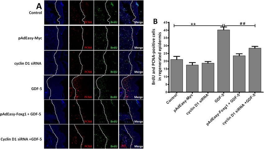
Zhao et al. Stem Cell Research & Therapy   (2021) 12:42                                                        Page 2 of 11

pathways, including MAPK (mitogen-activated protein             EpSCs proliferation mechanisms in wound healing. We
kinase) [8, 9], Wnt (wingless) [10, 11], and TGF-β (trans-      also hope to find new wound repair targets through our
forming growth factor-beta) [12] signaling. BMP (bone           research and provide new strategies for clinical research.
morphogenetic protein) belongs to the TGF-β family
and can stimulate cell proliferation [13].                      Methods and reagents
   Extracellular BMP binds with cell membrane receptors         Animals
to initiate downstream signaling pathways, and the signal       Both male and female C57BL/6 mice were used in this
molecule is translocated to the nucleus, where it is com-       study. They were obtained from the Experimental Ani-
bined with a nuclear transcription factor to regulate           mal Department of the Army Military Medical Univer-
gene expression [12]. According to the report, BMP-4            sity, China. All animal procedures were approved by the
supports self-renewal by inhibiting MAPK pathways in            Committee on the ethics of Animal Experiments of the
mouse embryonic stem cells [14]. Growth/differentiation         Third Military Medical University and were conducted
factor 5 (GDF-5) is a BMP family member [15], also              in accordance with the guidelines of the Experimental
known as CDMP-1 and BMP-14. Studies suggest that                Animal Department of the Army Military Medical Uni-
GDF-5 affects angiogenesis [16], migration [16], apop-          versity. The animals were individually housed in plastic
tosis [17], and differentiation [18] in vitro. Syed H. E and    cages under standard conditions (temperature, 25 °C;
his colleagues also discovered that GDF5-induced p38-           relative humidity, 50%; and circadian rhythm, 12 h). Ani-
MAPK signaling in fibroblasts regulates cardiac repairing       mals were provided cold boiled water and rodent food,
after myocardial infarction [15]. The predecessors con-         and allow them to acclimate to the facility for 1 week be-
ducted a preliminary study on the effect of GDF-5 in            fore the experiment. All surgeries were performed under
wound repairing [13], so we speculate that the increase         0.1% sodium pentobarbital anesthesia, and all efforts
of GDF-5 may promote the proliferation of EpSCs, but            were made to minimize suffering. The wound needs to
the specific proliferation mechanism of EpSCs promoted          be covered with sterile oil gauze to prevent infection.
by GDF-5 has not been reported. Recent studies have
found that FOX (Fork head box) and cyclin are involved          Preparation of mouse primary EpSCs
in the promotion of cell proliferation by BMP [19].             The preparation of primary EpSCs from newborn mice
   FOX is a kind of nuclear transcription factors family.       (0–2 days) was described in our previous studies [30].
The activity of FOX protein can be regulated by phos-           Firstly, the neonatal mice were euthanized by cervical
phorylation, acetylation, and protease hydrolysis [20]. It is   dislocation. Secondly, soaked for 1 min in 75% ethanol
known that PI3K-AKT/PKB (phosphoinositide-3-kinase–             and washed twice with sterile PBS. Thirdly, the back skin
protein kinase B/Akt), TGFβ-Smad and MAPK signaling             was separated with sterile surgical instruments and incu-
pathways can affect the level of FOX family proteins [19].      bated with 0.5% dispase II (Gibco, #17105041) overnight
Foxa1, Foxc1, Foxd3, Foxo3, Foxg1, Foxp1, and Foxm1             at 4 °C. Next, the skin was washed three times with ster-
are associated with cell proliferation [21]. The Foxg1 gene     ile PBS and separated carefully and the epidermis was
is a dose-sensitive gene, and it can antagonize the pro-        dissociated with 0.25% Trypsin (Gibco, #25200056) at
apoptotic effect of Foxo3 [22] and promote hepatocellular       37 °C for 10 min; the single-cell suspension was passed
carcinoma epithelial-mesenchymal transition [23]. As Sha-       through a 70-μm filter (BD Falcon #352350) into a ster-
sha Zhang’s reported, knocking out the Foxg1 gene will          ile 15-ml tube. Then, the cell suspension was centrifuged
increase differentiation of newborn mouse cells [24]. Stud-     at 1000 rpm for 5 min, removed supernatant, and resus-
ies have found that cyclin D plays an important role in cell    pended cells in K-SFM (Gibco, 10744019) supplemented
proliferation, which has three subfamilies: cyclin D1, cyc-     with 0.2 ng/ml recombinant mouse EGF (stem cell,
lin D2, and cyclin D3. It mainly initiates signal cascade       #78016), 100 ng/ml Cholera toxin, 30 mg/ml BPE (bo-
after binding and activating CDK (4 or 6) (cyclin-              vine pituitary extract), 0.05 mM calcium chloride, and
dependent kinase 4 or 6) which promoting cell prolifera-        100 U/ml of streptomycin and penicillin. Follow 2.5 ×
tion [25, 26]. Julie A. Siegenthaler reported that Foxg1 was    10^6 cells/T25 to quickly adhere to the bottom for 10
associated with cyclin in promoting intermediate progeni-       min, change the medium. Finally, the cells were cultured
tor cell proliferation [27]. Besides, the Wnt/cyclin D1         and the medium was changed every 2–3 days.
pathway has a dedifferentiating effect for differentiated
epidermal cells [28]. In our previous research, we found        Flow cytometry analysis
that cyclin D1 is an important downstream signaling mol-        When second-generation (P2) cells confluence become
ecule in the proliferation of EpSCs [29].                       ~ 70% after TrypLE™ select (Gibco, #12563029) passaged.
   In this paper, we conducted an integral study in vitro       Flow cytometry analysis of the purity of passaged EpSCs:
and in vivo conditions and carried out necessary tests.         Collect EpSCs at a density of 10^6 cell/ml, and then add
We sought to elucidate the effects of GDF-5 on mouse            antibodies, Santa SC23372-CD71-PE 5 μl/EP tube, Santa
Zhao et al. Stem Cell Research & Therapy   (2021) 12:42                                                        Page 3 of 11

SC19622-CD49f-FITC 5 μl/EPT tube, test after 60 min           (Cambridge, Massachusetts, USA). After incubating
incubation at 4 °C. Finally, resuspended in 0.5 ml of PBS,    the primary antibody, the membrane was washed
and then subjected to flow cytometry analysis.                three times and incubated with goat anti-rabbit IgG
                                                              (H + L) (1:10,000) (Abcam, ab6702) for 1 h. The bands
Cell proliferation assay                                      were visualized by using the BeyoECL Plus (Beyotime,
Possible proliferation was assessed by cell viability using   P0018M, China), and the bands were detected using
Cell Counting Kit-8 (CCK-8) (Beyotime, C0038, China)          Image Quant LAS 4000 s (GE, USA) [33].
according to the manufacturer’s instructions. When
first-generation (P1) cells confluence become ~ 70%, col-     Real-time quantitative PCR (qPCR)
lect EpSCs at a density of 2 × 10^5 cell/ml. Then, the        GDF-5 treatment EpSCs for 24 h, and then collect cells.
2000 EpSCs were seeded in 96-well plates (100 μl/well)        We used RNAiso Plus (Takara, # 9109) to extract RNA
and treated with 0, 1, 50, 100, 500, and 1000 ng/ml of        following the instructions, and measured the A260 and
GDF-5 (Beyotime, P6193, China) for 12, 24, 48, and 72         A280 values of the sample. Next, qPCR was performed.
h. After that, 10 μl CCK-8 solution was added to 96-well      The first step is to remove the genomic DNA (42 °C, 2
and incubated for 2–4 h at 37 °C. Absorbance was mea-         min; 4 °C hold), the second step is the reverse transcrip-
sured at 450 nm with a microplate reader (Spectra Max         tion reaction (37 °C, 15 min; 85 °C, 5 s; 4 °C hold), and fi-
190; Molecular Devices).                                      nally the real-time PCR reaction (95 °C, 30 s; go to 39
                                                              (40 cycles), 95 °C, 5 s, 60 °C, 30 s; melt curve). The prime-
Adenovirus infection and siRNA transient transfection         script RT reagent kit with gDNA Eraser (Takara, #
Adenovirus transfection and siRNA interference proto-         RR047A) and TB green Premix Ex Taq (Takara, #
col were as previously described [31, 32]. Adenovirus         RR820A) were used. Real-time PCR analysis of mouse
transfection was made when EpSCs reached 70% conflu-          cDNA was performed using the 7500 qPCR System
ence and then aspirated the medium and added fresh
medium. After that, added 10 μl Myc adenovirus and
                                                              Table 1 Primers for the RT-qPCR
10 μl Foxg1 adenovirus and 10 μl empty vector to each
                                                              Primer name    Sequence (5′ to 3′)         Length     Tm (°C)
group. After 24 h’ transfection, we observed the fluores-
cence intensity and expression ratio with a fluorescence      GAPDH-F        GGTTGTCTCCTGCGACTTCA        20         57.5
microscope and changed the medium. After 48 h’ trans-         GAPDH-R        TGGTCCAGGGTTTCTTACTCC       21         56.5
fection, cells were collected for subsequent experiments.     Foxa1-F        TTACAAGGATGCCTCTCCA         19         52.6
Specific cyclin D1 siRNA and control siRNA were pur-          Foxa1-R        TGGCTCTCTGAAAAGCAAG         19         52.4
chased from Thermo Fisher Scientific. EpSCs prepar-           Foxc1-F        GGATCGGCTTGAACAACT          18         52.4
ation method is as described above. Mouse EpSCs were
                                                              Foxc1-R        AGAGTGCCGGGAATAGG           17         54.0
transfected with siRNA according to the manufacturer’s
instructions. The efficiency of siRNA interference was        Foxd3-F        CGTAGAGAAGCGTCGAGGA         19         56.6
analyzed by the following Western blotting.                   Foxd3-R        GGCAAAGGAGGTGTGAGTG         19         56.5
                                                              Foxg1-F        AACGGGCTGAGTGTGGA           17         58.8
Western blotting (WB)                                         Foxg1-R        CAGGGGTTGAGGGAGTAGG         19         57.6
The levels of C Foxg1, cyclin D1, PCNA, and GAPD              Foxo3-F        GAGGATTCGGCCATGCT           17         55.4
H protein were detected by WB. GDF-5 was treat-
                                                              Foxo3-R        TTCCTTGGTTGCCCAGAG          18         55.1
ment EpSCs for 24 h, and then collect cells. EpSCs
protein samples were prepared using RIPA lysis buffer         Foxp1-F        TGCGCTGGACGATAGAA           17         53.4
(Beyotime, P0013B, China), which contained protease           Foxp1-R        ATGCAGGTGGGTCATCA           17         53.5
and phosphatase inhibitors. It was quantified using           Cyclin C-F     ATGCTTGGTAATTGATTTGCT       21         49.5
BCA protein evaluation kit (Beyotime, P0012S,                 Cyclin C-R     CAGGGGTTGAGGGAGTAGG         19         57.6
China). Next, 30 μg/each sample of protein was                Cyclin D1-F    ACCCTGACACCAATCTCCT         19         55.6
loaded onto 10% SDS-PAGE and transferred to PVDF
                                                              Cyclin D1-R    CTCCTTCTGCACGCACTT          18         55.6
membrane (Beyotime, FFP24, China). Then, the mem-
brane was blocked with a 5% milk solution (w/v) at            Cyclin D2-F    CCGTTCTTGGCTCTGGT           17         55.1
room temperature for 2 h. Then, the primary antibody          Cyclin D2-R    AGGCACCTGTTGAAACTGA         19         53.9
was incubated overnight at 4 °C. The primary anti-            Cyclin D3-F    AAACCACGCCCCTGACT           17         57.2
body was diluted according to the following ratio:            Cyclin D3-R    AGGTCCCACTTGAGCTTCC         19         57.4
PCNA (ab92552, 1:5000), Foxg1 (ab196868, 1:5000),             Cyclin E-F     CCCAAGTCCTGAGCCAT           17         54.7
cyclin D1 (ab16663, 1:5000), and GAPDH (ab181606,
                                                              Cyclin E-R     TCGGAGCCACCTTCTTC           17         54.5
1:10,000). All antibodies were purchased from Abcam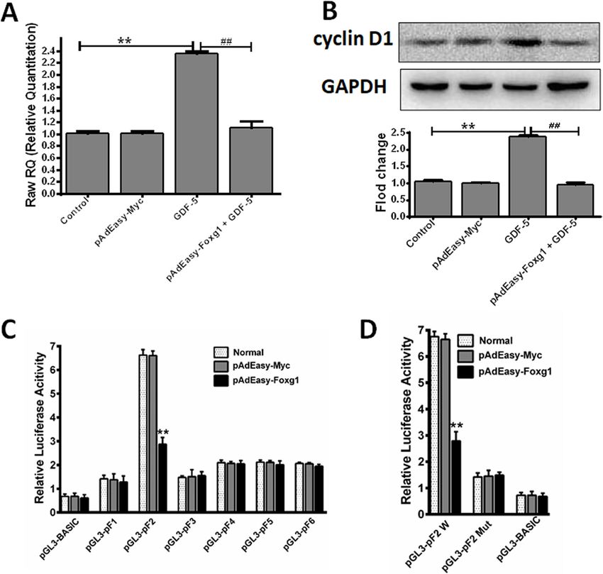
Zhao et al. Stem Cell Research & Therapy (2021) 12:42 Page 4 of 11
(Applied Biosystems). GAPDH serves as an internal ref- EpSCs proliferation assay in vivo
erence. Primers synthesized by Sangon Biotech (Shang- A deep partial-thickness burn mouse model was made
hai), and primer sequences are listed in Table 1. as follows description [34], and EpSCs were labeled
with BrdU in mouse skin. Neonatal C57BL/6 mice
were intraperitoneally injected with BrdU (50 mg/kg
Dual-luciferase assay body weight, Sigma) twice daily for 3 days, beginning
Follow our previous method [32]. Firstly, construction of on day 3 after birth. Skin cells retaining BrdU were
reporter gene plasmid. The mouse cyclin D1 promoter identified as EpSCs after 7 weeks. Next, an anesthetic
sequence was cloned into pGL3-basic plasmid vector to was injected intraperitoneally. Each gram of body
obtain pF1, pF2, pF3, pF4, pF5, and pF6 plasmid. Mu- weight was injected with 0.1% sodium pentobarbital
tated pF2 was generated by using the Mutant Best Kit at a dose of 10 μl; a metal plate (Shandong Academy
(Takara, China). Secondly, transfect cells. At 48 h after of Medical Science, China) with a diameter of 1.5 cm
pAdEasy-Foxg1 or pAdEasy-Myc transfection, the cells and weight of 0.5 kg was used to induce deep partial-
were transfected with the above luciferase reporter ex- thickness burns. The metal plate was heated to 70 °C
pression vectors using Lipofectamine 2000 for the pro- and was placed evenly on the shaved mouse dorsum
moter assay, respectively. And then, we used a multi- for 3 s to form a scald model. The skin wounds were
functional microplate reader to detect the expression covered with sterile oil gauze to prevent infection;
level of the reporter gene according to the manufac- mice were individually housed in plastic cages under
turer’s instructions (Promega, E2940). standard conditions.
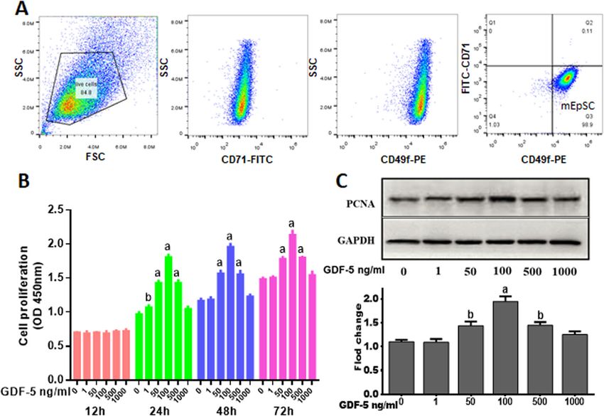
Fig. 1 Effect of GDF-5 on mouse EpSCs proliferation in vitro. a Flow cytometry was used to analyze the passaged EpSCs (P2). After the cells were passaged
twice, the cells were labeled with CD49f and CD71 antibodies. EpSCs expressed high levels of CD49f and low levels of CD71. b Mouse primary EpSCs were
treated with 0, 1, 50, 100, 500, and 1000 ng/ml of GDF-5 for 12, 24, 48, and 72 h. Cell proliferation was measured by CCK8. c EpSCs were treated by GDF-5 for
24 h, and the PCNA levels were analyzed by WB. The data were shown as the means ± SD of three independent experiments. aP < 0.01 vs. control (0 ng/ml
GDF-5 as control), bP < 0.05 vs. control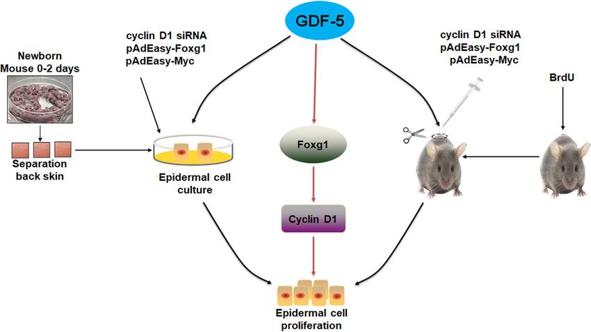
Zhao et al. Stem Cell Research & Therapy (2021) 12:42 Page 5 of 11
The 36 mice were divided randomly into 6 groups: 4% paraformaldehyde, and cut into 4 μm slices. Next,
control (normal saline), pAdEasy-Myc, cyclin D1 siRNA, antigen retrieval was performed using citrate buffer in a
GDF-5, pAdEasy-Foxg1 + GDF-5, and cyclin D1 siRNA pressure cooker at 95 °C for 30 min and each group was
+ GDF-5. After modeling, control group (normal saline blocked in 10% goat serum (16210064; Gibco) for 30
0.05 ml/g body weight, i.p.), pAdEasy-Myc and pAdEasy- min at 37 °C. Then, antibody incubation PCNA
Foxg1 and cyclin D1 siRNA (0.5 ml of PBS containing (ab92552,1:1000) at 4 °C overnight. And then drop the
2.5 × 10⁸ PFU virus, wound margin five points, s.c.), corresponding fluorescent secondary antibody (Goat
GDF-5 (0.05 ml/g body weight, i.p.), and GDF-5’s con- Anti-Rabbit IgG H&L (Cy5®) (ab6564)), DAPI counter-
centration is 10 μg/ml in normal saline. Mice were sacri- stain. Finally, dehydration and fix the mount, confocal
ficed after 24 h and using immunofluorescence assay laser observation.
BrdU and PCNA.
Statistical methods
Tissue immunofluorescence analysis All data were presented as the mean ± standard deviation
An anesthetic was injected intraperitoneally. Each gram (SD) with at least three independent experiments and
of body weight was injected with 0.1% sodium pentobar- analyzed using GraphPad Prism 7.0 software. Statistical
bital at a dose of 10 μl, and the mouse was euthanized significance was evaluated by one-way ANOVA or t test.
by cervical dislocation. The wound was biopsy, fixed in P < 0.05 was considered statistically significant.
Fig. 2 Changes of related factors after EpSCs were treated by GDF-5. Mouse primary EpSCs were treated with 0, 1, 50, 100, 500, and 1000 ng/ml
of GDF-5 for 24 h. a The FOX family and cyclins genes were analyzed by qPCR. b The Foxg1 and cyclin D1 levels were analyzed by WB. The data
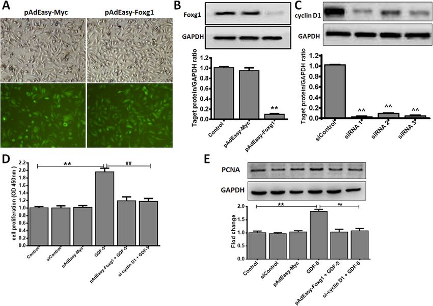
are shown as the means ± SD of three independent experiments. *P < 0.05 vs. control (0 ng/ml GDF-5 as control), **P < 0.01 vs. controlZhao et al. Stem Cell Research & Therapy (2021) 12:42 Page 6 of 11 Results The possible downstream molecules of GDF-5 Effect of GDF-5 on mouse EpSCs proliferation In order to detect the possible downstream molecules of Mouse EpSCs were defined as our previously de- GDF-5, we consulted references and found FOX/cyclin scribed [31, 35]. The purity of EpSCs was analyzed by may be the downstream molecules of GDF-5 [19]. flow cytometry, as shown in Fig. 1a, and the propor- Firstly, we screened downstream genes in the FOX and tion of EpSCs is about 99%. To illustrate the effect of cyclins family, the results discovered that Foxg1 and cyc- GDF-5 on mouse EpSCs proliferation, we used CCK-8 lin D1 were a dose-dependent relationship with GDF-5. assay the proliferation effect of GDF-5 at different In addition, Foxg1 expression increased to 4.79-fold and time points, and the results showed that 24 h has the cyclin D1 increased to 3.31-fold when the concentration best effect on promoting cell proliferation (0.84-fold) of GDF-5 was 100 ng/ml (Fig. 2a). Secondly, the protein when GDF-5 was 100 ng/ml (Fig. 1b), so 24 h was levels of Foxg1 and cyclin D1 in mouse EpSCs treated used in the following study. Because the PCNA is a by GDF-5 were detected by WB. The results showed marker that reflects the state of cell proliferation [36], that there was a dose-dependent relationship between our further analysis of GDF-5 promoting EpSCs pro- cells treated with GDF-5 and Foxg1/cyclin D1 protein liferation at 24 h found that the proliferation- expression. Moreover, Foxg1 increased to 1.79-fold and associated PCNA protein was significant at 100 ng/ml cyclin D1 increased to 1.68-fold when the optimal con- (P < 0.01) in response to GDF-5 (Fig. 1c). centration of GDF-5 was 100 ng/ml (P < 0.01) (Fig. 2b). Fig. 3 GDF-5 promotes EpSCs proliferation via Foxg1/cyclin D1 in vitro. Mouse EpSCs were infected with pAdEasy-Myc control adenovirus or pAdEasy-Foxg1. After the cells were transfected for 48 h with siRNA control plasmid or cyclin D1 siRNAs, mouse EpSCs were treated with 100 ng/ ml GDF-5 for 24 h. a Fluorescence effects of pAdEasy-Myc and pAdEasy-Foxg1 infected mouse EpSCs (× 100 magnification). b Foxg1 protein expression level was analyzed by WB. Control: non-infected normal mouse EpSCs. c Three siRNAs were synthesized to inhibit cyclin D1 expression and quantification of WB. d Cell proliferation was measured by CCK-8. e The PCNA levels were analyzed by WB. The data are shown as the means ± SD of three independent experiments. **P < 0.01 vs. Control, ^^P < 0.01 vs. siControl, ##P < 0.01 vs. the GDF-5 group
Zhao et al. Stem Cell Research & Therapy    (2021) 12:42                                                                        Page 7 of 11

GDF-5 promotes EpSCs proliferation via Foxg1/cyclin D1                  inhibited of pAdEasy-Foxg1 + GDF-5 group reduced 0.78
in vitro                                                                and cyclin D1 siRNA + GDF-5 group reduced 0.73 com-
The above part proved that Foxg1 and cyclin D1 are the                  pared with GDF-5 group (Fig. 3e).
downstream molecules of GDF-5. Here we discuss the
role of Foxg1 and cyclin D1 in GDF-5 promotes cell pro-                 The effect of GDF-5 on mouse EpSCs proliferation via
liferation. We used adenovirus and siRNA infection                      Foxg1/cyclin D1 in vivo
technology to verify the interrelationship of Foxg1/cyclin              To analyze the effect of GDF-5 on EpSCs proliferation
D1 during cell proliferation. The pAdEasy-Myc transfec-                 in vivo, BrdU-labeled EpSCs and mouse model of burn
tion mouse EpSCs as control virus, the results showed                   injury were established as our previously described [29].
that pAdEasy-Foxg1 and pAdEasy-Myc had a similar                        BrdU+ and PCNA+ EpSCs were presented by immuno-
transfection efficiency, reaching to 90% and 95% (Fig. 3a),             chemistry in the regenerated epidermis, and the double-
which indicated that each adenovirus successfully in-                   positive EpSCs were counted in the different re-
fected mouse EpSCs. The pAdEasy-Foxg1 group’s Foxg1                     epithelialization area. As can be seen from (Fig. 4a, b),
expression was reduced by 89% (P < 0.01) compared to                    the number of double-positive cells increased to 40.18-
the pAdEasy-Myc group, and there was no significance                    fold in the GDF-5 group compared with the control
between the pAdEasy-Myc and the control group (Fig.                     group; the results showed that GDF-5 can promote
3b). Three siRNAs were synthesized to inhibit cyclin D1                 EpSCs proliferation in vivo. However, the double-
gene expression. As shown in Fig. 3c, the siRNA1 (P <                   positive cells reduced 16.68 and 11.72 in the cyclin D1
0.01) with the best silencing efficiency was selected for               siRNA + GDF-5 group and the pAdEasy-Foxg1 + GDF-5
subsequent research. Finally, mouse EpSCs were treated                  group compared with the GDF-5 group, respectively.
with 100 ng/ml GDF-5 or not treated for 24 h, and cell                  The data showed that the pAdEasy-Foxg1 and cyclin D1
proliferation was evaluated by CCK-8 assay and PCNA                     siRNA abolished the effect of GDF-5 on the number of
protein analysis. Figure 3d and e showed that the GDF-5                 double-positive cells in the regenerated epidermis.
group had a significant proliferation compared with                     Moreover, the double-positive cells increased in the
other groups. In addition, the EpSCs proliferation was                  pAdEasy-Foxg1 + GDF-5 group and the cyclin D1 +

 Fig. 4 GDF-5 promotes EpSCs proliferation via Foxg1/cyclin D1 in vivo. The groups including control, pAdEasy-Myc, cyclin D1 siRNA, GDF-5,
 pAdEasy-Foxg1 + GDF-5, and cyclin D1 siRNA + GDF-5. a After mouse EpSCs were labeled by BrdU, BrdU+PCNA+-positive EpSCs were analyzed in
 wound. BrdU+ and PCNA+ cells in the regenerated epidermis are shown at the same magnification. Bar = 50 μm. b BrdU+ and PCNA+ cell count
 was performed using Image Pro Plus in the regenerated epidermis. The data are presented as the means ± SD of three independent
 experiments; **P < 0.01 vs. the control group, ##P < 0.01 vs. the GDF-5 group
Zhao et al. Stem Cell Research & Therapy    (2021) 12:42                                                                         Page 8 of 11

 Fig. 5 GDF-5 regulates cyclin D1 protein and mRNA expression through Foxg1. Mouse EpSCs were infected with pAdEasy-Myc control
 adenovirus or pAdEasy-Foxg1 and the cells were treated with 100 ng/ml GDF-5 for 24 h. a Cyclin D1 mRNA expression. b Cyclin D1 protein
 expression. c, d Mouse EpSCs were transfected with pAdEasy-Myc control adenovirus or pAdEasy-Foxg1 or the luciferase reporter expression
 vectors or mutated pGL3-pF2 vector using Lipofectamine 2000. The data are presented as the means ± SD of three independent experiments;
 **P < 0.01 vs. the control, ##P < 0.01 vs. the GDF-5 group

GDF-5 group compared with the control group, but the                    As can be seen from Fig. 5a, the expression of cyclinD1
difference is not obvious.                                              mRNA in the GDF-5 group increased to 2.34-fold com-
                                                                        pared with the control group (P < 0.01), but the cyclin
GDF-5 regulates cyclin D1 expression by Foxg1 and                       D1 mRNA expression of pAdEasy-Foxg1 + GDF-5 group
regulates transcriptional activity of the cyclin D1 gene                was reduced back to the control level. At the same time,
promoter                                                                the cyclin D1 protein expression level also had the same
In order to analyze the transcription relationship be-                  trend (Fig. 5b). This showed that pAdEasy-Foxg1
tween Foxg1 and cyclin D1, qPCR was used to detect                      blocked cyclin D1 expression. Next, we hypothesized
cyclin D1 mRNA, and WB was used to detect cyclin D1                     that pAdEasy-Foxg1 inhibits cyclin D1 by exerting in-
protein expression in the presence of pAdEasy-Foxg1.                    hibitory activities to the cyclin D1 promoter. We
Zhao et al. Stem Cell Research & Therapy (2021) 12:42 Page 9 of 11
constructed a pGL3-cyclin D1 (pF1, pF2, pF3, pF4, pF5, several subfamilies of the FOX family and cyclins related
pF6) luciferase reporter gene expression vector. The to cell proliferation by qPCR. Here, it was found that
dual-luciferase assay revealed that pAdEasy-Foxg1 sig- Foxg1 and cyclin D1 increased significantly (Fig. 2a).
nificantly inhibited the activity of the cyclin D1 pro- Wang Fan et al. found that cyclin D1 was significantly
moter (Fig. 5c). However, the mutant pGL3-pF2 did not expressed during the proliferation of human EpSCs [29].
respond to the pAdEasy-Foxg1 agonists (Fig. 5d). We previously reported that nitric oxide induces FoxG1
expression in human EpSCs [32]. In the analysis on
Discussion GDF-5 promoting EpSCs proliferation, we found that
At present, the effect of GDF-5 on wound healing has been Foxg1 and cyclin D1 could have prevented the prolifera-
reported [13, 15]. However, its specific mechanism for tion effect of GDF-5 (Figs. 3c, d and 4); further analysis
wound repairing is still unclear. In this paper, we discovered from the protein level found that Foxg1 and cyclin D1
that GDF-5 promoted mouse EpSCs proliferation via the were positively regulated by GDF-5 (Figs. 2b and 3e).
Foxg1/cyclin D1 signaling pathway in vivo and in vitro. Federica Verginelli et al. found that a transcriptional
Other studies had reported that GDF-5 promote cell program regulated by Foxg1 is significant for promoting
proliferation [13]; in this study, we found that GDF-5 glioblastoma growth [39]. Combined with the results of
can directly increase the number of EpSCs in vitro. We in vivo and in vitro studies, this indicated that after
detected the effect of GDF-5 on EpSCs when the con- GDF-5 stimulates EpSCs; the downstream molecules
centration of exogenous GDF-5 changed from 0 to 1000 Foxg1 and cyclin D1 were activated (Fig. 6).
ng/ml by CCK-8 assay at 12 h, 24 h, 48 h, and 72 h. The Besides, Foxg1 is involved in inhibiting the cell cycle exit
results showed that EpSCs had the best cell proliferation initiated by p21 [27]. Cyclin D1 is a key regulator of cell
effect after being treated with 100 ng/ml exogenous proliferation by promoting cell cycle transition, and its ex-
GDF-5 (Fig. 1b), the effective concentration of GDF-5 pression is regulated by transcription level [40, 41]. To clar-
on cells is similar to the previous reports [37]. In vivo, ify the upstream and downstream relationship between
through the study of a deep partial-thickness burn Foxg1 and cyclin D1, dual-luciferase reporter gene analysis
mouse model, we found that GDF-5 promoted the pro- was used, and we found that GDF-5 induced cyclin D1
liferation of EpSCs, which is consistent with the results transcription was regulated by Foxg1-mediated cyclin D1
of in vitro experiments (Fig. 4a, b). In addition, PCNA is promoter activity (Fig. 5c, d). There may be other signaling
a marker that reflects the state of cell proliferation [36]; pathways for GDF-5 to promote EpSCs proliferation. From
we tested the expression of PCNA protein after different Fig. 2a, discovered Foxo3/Foxp1 decreased significantly and
concentrations of GDF-5 treatment for 24 h (Fig. 1c). cyclin D2/cyclin D3 increased significantly at the transcrip-
Combining the results of cell count and PCNA protein tion level. Foxo3 and Foxp1 have been reported to play an
analysis, it was determined that GDF-5 promoted EpSCs inhibitory role in cell proliferation [19], and cyclin D2/cyc-
proliferation in vitro and in vivo. lin D3 helps isolate cell transplant factor p27 [42]. Whether
FOX and cyclin have important functions in the prolif- GDF-5 regulates these genes to promote the proliferation
eration of many cell types [21, 38]. Firstly, we screened of EpSCs will be analyzed in another project.
Fig. 6 GDF-5 promotes epidermal stem cell proliferation via Foxg1-cyclin D1 signalingZhao et al. Stem Cell Research & Therapy (2021) 12:42 Page 10 of 11
Conclusions 6. Hu P, Yang Q, Wang Q, Shi C, Wang D, Armato U, Prà ID, Chiarini A.
This study shows that GDF-5 plays an important role in Corrigendum to: ‘Mesenchymal stromal cells-exosomes: a promising cell-
free therapeutic tool for wound healing and cutaneous regeneration’. Burns
EpSCs proliferation in vitro and in vivo. The prolifera- Trauma. 2020;8:tkaa007.
tion is regulated by activating Foxg1-cyclin D1 signaling 7. Li Z, Maitz P. Cell therapy for severe burn wound healing. Burns Trauma.
pathway. The results can initially determine that GDF-5 2018;6(2):72–81.
8. Chen RE, Thorner J. Function and regulation in MAPK signaling
can be used as a new target for wound repairing. pathways: lessons learned from the yeast Saccharomyces cerevisiae.
Biochimica et Biophysica Acta (BBA) - Molecular Cell Research. 2007;
Abbreviations 1773(8):1311–40.
EpSCs: Epidermal stem cells; GDF-5: Growth/differentiation factor 5;
9. Zhu AJ, Haase I, Watt FM. Signaling via β 1 integrins and mitogen-activated
MAPK: Mitogen-activated protein kinase; PCNA: Proliferating Cell Nuclear
protein kinase determines human epidermal stem cell fate in vitro. Proc
Antigen; Wnt: Wingless/Integrated; BMP: Bone morphogenetic protein;
Natl Acad Sci U S A. 1999;96(12):6728–33.
FOX: Fork head box; TGF-β: Transforming growth factor-beta; CDK: (4 or 6)
10. Veltri A, Lang C, Lien WH. Concise review: Wnt signaling pathways in skin
Cyclin-dependent kinase 4 or 6; BPE: Bovine pituitary extract; CCK-8: Cell
development and epidermal stem cells. Stem Cells. 2018;36(1):22–35.
counting kit-8; qPCR: Real-time quantitative PCR; WB: Western blotting;
11. Thompson CC, Sisk JM, 3rd BG: Hairless and Wnt signaling: allies in epithelial
FC: Flow cytometry; PI3K-AKT/PKB: Phosphoinositide-3-kinase–protein kinase
stem cell differentiation. Cell Cycle 2006, 5(17):1913.
B/Akt
12. Wang RN, Green J, Wang Z, Deng Y, Qiao M, Peabody M, Zhang Q, Ye J,
Yan Z, Denduluri S et al. Bone Morphogenetic Protein (BMP) signaling in
Authors’ contributions
development and human diseases. Genes Dis. 2014;1(1):87–105.
Xiaohong Zhao and Ruyu Bian performed the experiments and analyzed the
13. Schiefer JL, Held M, Fuchs PC, Demir E, Plöger F, Schaller HE, Rahmanian-
data. Fan Wang and Ying Wang guided the WB and immunofluorescence
Schwarz A. Growth differentiation factor 5 accelerates wound closure and
assay. Xue Li, Yicheng Guo, and Xiaorong Zhang made the necessary
improves skin quality during repair of full-thickness skin defects. Adv Skin
corrections in the write-up. Gaoxing Luo and Rixing Zhan conceptualized
Wound Care. 2017;30(5):223–9.
and guided the research project. All authors proofread the manuscript. The
14. XX Q, Li TG, Hao J, Hu J, Wang J, Simmons H, Miura S, Mishina Y: BMP4
authors read and approved the final manuscript.
supports self-renewal of embryonic stem cells by inhibiting mitogen-
activated protein kinase pathways. Proc Natl Acad Sci U S A. 2004;101(16):
Funding
6027–32.
This work was supported by Chongqing Science and Health Joint Medical
Research Project in 2020 (2020MSXM001) and the National Natural Science 15. Zaidi SHE, Huang Q, Momen A, Riazi A, Husain M. Growth differentiation
Foundation of China (81920108022). factor 5 regulates cardiac repair after myocardial infarction. J Am Coll
Cardiol. 2010;55(2):135–43.
Availability of data and materials 16. Chen X, Zankl A, Niroomand F, Liu Z, Katus HA, Jahn L, Tiefenbacher C:
The data that support the findings of this study are available from the Upregulation of ID protein by growth and differentiation factor 5 (GDF5)
corresponding author upon reasonable request. through a smad-dependent and MAPK-independent pathway in HUVSMC. J
Molecular Cell Cardiol 2006, 41(1):0–33.
Ethics approval and consent to participate 17. Nakahara T, Tominaga K, Koseki T, Yamamoto M, Yamato K, Fukuda J,
All animal procedures were approved by the Committee on the ethics of Nishihara T. Growth/differentiation factor-5 induces growth arrest and
Animal Experiments of the Third Military Medical University and were apoptosis in mouse B lineage cells with modulation by Smad. Cell Signal.
conducted in accordance with the guidelines of the Experimental Animal 2003;15(2):181–7.
Department of the Army Military Medical University (Animal use protocol 18. Zeng Q, Li X, Beck G, Balian G, Shen FH: Growth and differentiation factor-5
approval number: amuwec20201412). (GDF-5) stimulates osteogenic differentiation and increases vascular
endothelial growth factor (VEGF) levels in fat-derived stromal cells in vitro.
Consent for publication Bone 2007, 40(2):0–381.
All patients signed a consent form for their data to be used for research or 19. Charvet C, Alberti I, Luciano F, Jacquel A, Bernard A, Auberger P, Deckert M.
publication. Proteolytic regulation of Forkhead transcription factor FOXO3a by caspase-
3-like proteases. Oncogene. 2003;22(29):4557–68.
Competing interests 20. Myatt SS, Lam EWF. The emerging roles of forkhead box (Fox) proteins in
The authors declare no competing financial interest. cancer. Nat Rev Cancer. 2007;7(11):847–59.
21. Katoh M, Katoh M. Human FOX gene family (review). Int J Oncol. 2004;25(5):
Author details 1495–500.
1
Institute of Burn Research; State Key Laboratory of Trauma, Burn and 22. Seoane J, Le H-V, Shen L, Anderson SA, Massagué J. Integration of Smad
Combined Injury; Southwest Hospital, The Third Military Medical University and Forkhead pathways in the control of neuroepithelial and glioblastoma
(Army Medical University), Chongqing 400038, China. 2Department of Plastic cell proliferation. Cell. 2004;117(2):211–23.
and Reconstructive Surgery, Southwest Hospital, The Third Military Medical 23. Zheng X, Lin J, Wu H, Mo Z, Lian Y, Wang P, Hu Z, Gao Z, Peng L, Xie C.
University (Army Medical University), Chongqing 400038, China. Forkhead box (FOX) G1 promotes hepatocellular carcinoma epithelial-
mesenchymal transition by activating Wnt signal through forming T-cell factor-
Received: 30 October 2020 Accepted: 15 December 2020 4/Beta-catenin/FOXG1 complex. J Experimental Clin Cancer Res. 2019;38(1):475.
24. Zhang S, Zhang Y, Dong Y, Guo L, Zhang Z, Shao B, Qi J, Zhou H, Zhu W,
Yan X, et al. Knockdown of Foxg1 in supporting cells increases the trans-
References differentiation of supporting cells into hair cells in the neonatal mouse
1. Beck B, Blanpain C. Mechanisms regulating epidermal stem cells. EMBO J. cochlea. Cell Molecular Life Sci. 2020;77(7):1401–19.
2012;31(9):2067–75. 25. Pestell RG. New roles of cyclin D1. Am J Pathol. 2013;183(1):3–9.
2. Horst BT, Chouhan G, Moiemen NS, Grover LM. Advances in keratinocyte 26. Witzel I-I, Koh LF, Perkins ND. Regulation of cyclin D1 gene expression.
delivery in burn wound care. Adv Drug Deliv Rev. 2017;123:18–32. Biochem Soc Trans. 2010;38(1):217.
3. Iwata Y, Akamatsu H, Hasebe Y, Hasegawa S, Sugiura K: [Skin-resident stem 27. Siegenthaler JA, Tremper-Wells BA, Miller MW. Foxg1 haploinsufficiency
cells and wound healing] Nihon Rinsho Men'eki Gakkai kaishi 2017, 40(1):1–11. reduces the population of cortical intermediate progenitor cells: effect of
4. Billingham RE, Reynolds J. Transplantation studies on sheets of pure increased p21 expression. Cereb Cortex. 2008;18(8):1865–75.
epidermal epithelium and on epidermal cell suspensions. Br J Plast Surg. 28. Zhao A, Yang L, Ma K, Sun M, Li L, Huang J, Li Y, Zhang C, Li H, Fu X.
1952;5(1):25–36. Overexpression of cyclin D1 induces the reprogramming of differentiated
5. Li Y, Zhang J, Yue J, Gou X, Wu X. Epidermal stem cells in skin wound epidermal cells into stem cell-like cells. Cell cycle (Georgetown, Tex). 2016;
healing. Advances Wound Care. 2017;6(9):297–307. 15(5):644–53.Zhao et al. Stem Cell Research & Therapy (2021) 12:42 Page 11 of 11
29. Wang F, Zhan R, Chen L, Dai X, Cao C. RhoA promotes epidermal stem cell
proliferation via PKN1-cyclin D1 signaling. PLoS One. 2017;12(2):e0172613.
30. Tudor D, Chaudry F, Harper L, Mackenzie IC. The in vitro behavior and
patterns of colony formation of murine epithelial stem cells. Cell Prolif. 2007;
40(5):706–20.
31. Yao Z, Li H, He W, Yang S, Zhang X, Zhan R, Xu R, Tan J, Zhou J, Wu J. P311
accelerates skin wound reepithelialization by promoting epidermal stem cell
migration through RhoA and Rac1 activation. Stem Cells Development.
2017;26(6):451–60.
32. Zhan, Rixing, Wang, Fan, Wu, Ying, Qian, Wei, Liu, Menglong: Nitric oxide
promotes epidermal stem cell proliferation via FOXG1-c-Myc signalling.
Nitric Oxide Biol Chemistry 2018.
33. Zhao X, Zou X, Li Q, Cai X, Li L, Wang J, Wang Y, Fang C, Xu F, Huang Y.
Total flavones of fermentation broth by co-culture of Coprinus comatus and
Morchella esculenta induces an anti-inflammatory effect on LPS-stimulated
RAW264.7 macrophages cells via the MAPK signaling pathway. Microb
Pathog. 2018;125:431–7.
34. Zhan R, He W, Wang F, Yao Z, Tan J, Xu R, Zhou J, Wang Y, Li H, Wu J. Nitric
oxide promotes epidermal stem cell migration via cGMP-rho GTPase
signalling. Sci Rep. 2016;6(1):30687.
35. Xu ZD, Li HS, Wang S, He WF, Wu J, Luo GX: [Effects of hypoxia inducible
factor-1α on P311 and its influence on the migration of murine epidermal
stem cells]. Zhonghua shao shang za zhi = Zhonghua shaoshang zazhi
2017, 33(5):287–294.
36. Lee KY, Myung K. PCNA modifications for regulation of post-replication
repair pathways. Molecules Cells. 2008;26(1):5–11.
37. Coleman CM, Vaughan EE, Browe DC, Mooney E, Howard L, Barry F. Growth
differentiation factor-5 enhances in vitro mesenchymal stromal cell
chondrogenesis and hypertrophy. Stem Cells Dev. 2013;22(13):1968–76.
38. Vezzali R, Weise SC, Hellbach N, Machado V, Heidrich S, Vogel T. The
FOXG1/FOXO/SMAD network balances proliferation and differentiation of
cortical progenitors and activatesKcnh3expression in mature neurons.
Oncotarget. 2016;7(25):37436–55.
39. Verginelli F, Perin A, Dali R, Fung KH, Stifani S. Transcription factors FOXG1
and Groucho/TLE promote glioblastoma growth. Nat Commun. 2013;4:2956.
40. Hernández-Hernández OT, Camacho-Arroyo I. Regulation of gene
expression by progesterone in cancer cells: effects on cyclin D1, EGFR and
VEGF. Mini Rev Med Chem. 2013;13(5):635–42.
41. Wee P, Wang Z. Epidermal growth factor receptor cell proliferation
signaling pathways. Cancers. 2017;9(5):52.
42. Chiles T. C: regulation and function of cyclin D2 in B lymphocyte subsets. J
Immunol. 2004;173(5):2901–7.
Publisher’s Note
Springer Nature remains neutral with regard to jurisdictional claims in
published maps and institutional affiliations.You can also read