Hair follicle germs containing vascular endothelial cells for hair regenerative medicine
←
→
Page content transcription
If your browser does not render page correctly, please read the page content below
www.nature.com/scientificreports
OPEN Hair follicle germs containing
vascular endothelial cells for hair
regenerative medicine
Tatsuto Kageyama1,2, Yang‑Sook Chun3 & Junji Fukuda1,2*
Hair regenerative medicine has emerged as a promising approach for the treatment of severe hair
loss. Recent advances in three-dimensional tissue engineering, such as formation of hair follicle
germs (HFGs), have considerably improved hair regeneration after transplantation in animal models.
Here, we proposed an approach for fabricating HFGs containing vascular endothelial cells. Epithelial,
dermal papilla, and vascular endothelial cells initially formed a single aggregate, which subsequently
became a dumbbell-shaped HFG, wherein the vascular endothelial cells localized in the region of
dermal papilla cells. The HFGs containing vascular endothelial cells exhibited higher expression
of hair morphogenesis-related genes in vitro, along with higher levels of hair shaft regeneration
upon transplantation to the dorsal side of nude mice, than those without vascular endothelial cells.
The generated hair follicles represented functional characteristics, such as piloerection, as well as
morphological characteristics comparable to those of natural hair shafts. This approach may provide
a promising strategy for fabricating tissue grafts with higher hair inductivity for hair regenerative
medicine.
Hair loss is a common concern worldwide. The International Society of Hair Restoration Surgery had estimated
approximately 600,000 patients to have been surgically treated for hair loss worldwide in 20161. Currently, severe
androgenetic alopecia is treated primarily by transplantation of the patient’s own hair follicles. However, the
outcomes are often unsatisfactory when there are not enough hair follicles available. Furthermore, since this
approach only translocates hair follicles from the back of head to the region with sparse hair, the total number of
hair follicles does not i ncrease2. Therefore, hair regenerative medicine, which in principle increases the number
of hair follicles, has emerged as a promising approach for treating alopecia, such as severe androgenetic alopecia,
female-pattern hair loss unresponsive to typical drugs, and scarring alopecia.
In vivo development of hair follicles is driven by the formation of hair follicle germs (HFGs) at a location
between the epidermal and mesenchymal l ayers3. Replication of such hair follicle development in vitro, includ-
ing epithelial-mesenchymal interactions, has been considered to engineer tissue grafts for hair regenerative
medicine4–6. One of the most advanced approaches in this regard is using a bioengineered HFG, prepared by
integrating aggregates of epithelial and mesenchymal cells7. The bioengineered HFGs efficiently regenerated
hair follicles after transplantation in nude mice, which reassembled connections to host arrector pili and nerve
fibers. Thus, this approach could partially replicate in vivo hair follicle development. However, since thousands
of HFGs are required for a single patient, this approach may be impractical, considering that HFG prepara-
tion includes laborious steps involving the handling of individual epithelial and mesenchymal cell aggregates
under a microscope. We have recently reported a more straightforward approach for large-scale preparation of
HFGs8,9. We observed that a mixture of epithelial and mesenchymal cells initially formed an aggregate, where
the two types of cells distributed randomly, however, spatially separated from each other following three days of
culture, and spontaneously formed HFGs. Due to the self-sorting formation of HFGs, this approach was scalable
to fabricate > 5,000 HFGs simultaneously by seeding a suspension of the two cell types in a lab-made microwell
array chip. The self-sorted HFGs regenerated hair follicles relatively efficiently, with normal hair cycles, when
transplanted into nude mice.
Murine embryonic cells generally exhibit a strong ability to regenerate hair follicles de novo. However, the
regenerative ability of adult human hair follicle stem cells often deteriorates even when the same approach is
used10. Thus, further studies using human cells would be necessary to better replicate in vivo development
1
Faculty of Engineering, Yokohama National University, 79‑5 Tokiwadai, Hodogaya‑ku, Yokohama,
Kanagawa 240‑8501, Japan. 2Kanagawa Institute of Industrial Science and Technology, 3‑2‑1 Sakado Takatsu‑ku,
Kawasaki, Kanagawa 213‑0012, Japan. 3Department of Physiology and Biomedical Sciences, Seoul National
University College of Medicine, 103 Daehak‑ro, Jongno‑gu, Seoul 110‑799, Korea. *email: fukuda@ynu.ac.jp
Scientific Reports | (2021) 11:624 | https://doi.org/10.1038/s41598-020-79722-z 1
Vol.:(0123456789)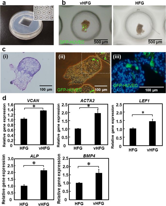
www.nature.com/scientificreports/
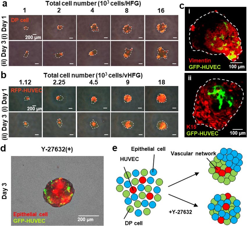
Figure 1. Preparation of vHFGs through self-organization. Human DP cells, murine fetal skin-derived
epithelial cells, and HUVECs were mixed and seeded in a non-cell-adhesive 96-well plate and lab-created HFG
chip. In each culture vessel, the cells formed single aggregates in the microwells, wherein the three types of cells
were randomly distributed after 1 day of culture and spontaneously formed vHFGs in the following 2 days of
culture. vHFGs were then transplanted into the dorsal skin of nude mice to evaluate hair regeneration potential.
and hair follicle microenvironment for clinical applications. Perivascular microenvironments are essential for
the development and maintenance of stem cells in many tissues. During development, newly forming ves-
sels approach the nascent follicles, and the vascular annulus is completely developed by b irth11. Reconstituted
hair follicles spontaneously form vascular loops around the upper bulge, recapitulating the pattern of normal
hair-bearing skin11. Furthermore, hair growth and hair follicle size significantly correlate with perifollicular
angiogenesis induced by vascular endothelial growth f actor12. Thus, involvement of vascular endothelial cells in
HFGs may be beneficial for replicating hair follicle development and improving de novo hair follicle generation.
This study aimed to determine whether vascular endothelial cells participate in the self-organization of HFGs
and improve hair regeneration. We added human vascular endothelial cells to a suspension of human dermal
papilla (DP) and mouse embryonic epithelial cells and allowed them to form an aggregate in a 96-well plate and
custom-designed microwell array plate (Fig. 1). Localization of vascular endothelial cells in an HFG and their
effects on follicular gene expression were assessed. Furthermore, we investigated the effects of vascular endothe-
lial cells on hair regeneration in animal models. Our findings provided insight into in vitro self-organization
and in vivo morphogenesis of hair follicles, thus providing a promising strategy for hair regenerative medicine.
Results and discussion
Self‑organization of the three cell types. Schematic representation of the experimental procedures is
shown in Fig. 1. Note that in vitro proliferation of human hair follicle epithelial stem cells with trichogenic abil-
ity has not been established yet, and thus we used mouse embryonic epithelial cells in the current study. Prior
to investigation with the three cell types, we examined whether the formation of HFGs through self-sorting
of epithelial and mesenchymal cells could occur even among cells from different species. Vybrant DiI-labeled
human DP and murine embryonic epithelial cells (1:1) were suspended and seeded in a non-cell-adhesive,
round-bottom, 96-well plate at different cell densities (1, 2, 4, 8, 16, 32, or 64 × 103 cells/0.1 mL). During 24 h
of culture, the two types of cells formed single aggregates in each well, wherein they were almost randomly
distributed and eventually formed small clusters (Fig. 2a (i)). Over the following two days of culture, the cells
spatially separated within the individual aggregates and formed HFG-like structures, although DP cells in larger
aggregates, composed of > 8 × 10³ cells/HFG, did not assemble into a single cluster and rather remained as a few
clusters (Fig. 2a (ii)). The dynamic and spatial changes in the chimeric human-mouse HFGs were consistent with
the results of our previous study involving HFGs composed of mouse embryonic epithelial and mesenchymal
cells, with the exception of larger a ggregates8. In our previous study, complete separation of the two cell types
and formation of dumbbell-like HFGs were observed even with 32 × 103 cells/HFG. Considering the cell sorting
mechanisms, these results suggested that human DP cells migrate less and/or show weak homotypic cell–cell
adhesion properties.
Human umbilical vein endothelial cells (HUVECs) were added to the suspension of DP and epithelial cells
(DP cells: epithelial cells: HUVECs = 4:4:1) to determine their effects on HFG formation in a 96-well plate (Fig. 1).
The total number of cells was varied (1.13, 2.25, 4.5, 9, 18, 36, or 72 × 103 cells/well) while maintaining the cell
ratio. The number of DP and epithelial cells used here were the same as in the experiments without HUVECs.
Scientific Reports | (2021) 11:624 | https://doi.org/10.1038/s41598-020-79722-z 2
Vol:.(1234567890)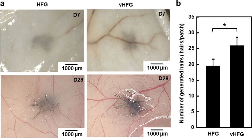
www.nature.com/scientificreports/
Figure 2. Self-organization of cells into vHFGs. (a) Changes in spatial distribution of DP and epithelial cells
(1:1 in cell number) in individual aggregates after 1 and 3 days of culture. The DP cells were labeled with
fluorescent Vybrant DiI dye before seeding. The fluorescence and phase-contrast images were overlaid. The
broken lines indicate the boundary of aggregates. (b) Changes in spatial distribution of DP cells, epithelial cells,
and HUVECs (4:4:1 in cell number) in individual aggregates at 1 and 3 days of culture. The RFP-HUVECs were
used to distinguish them from others. The fluorescence and phase-contrast images are overlaid. The broken
lines indicate the boundary of aggregates. (c) Confocal microscopic images of immunohistochemically stained
vHFGs (9 × 10³ cells/HFG). (i) Green, GFP-HUVEC; red, vimentin. (ii) Green, GFP-HUVEC; red, cytokeratin
15 (K15). (d) Effects of Y-27632 treatment on self-organization of the three cell types. Red, DiI-labeled mouse
epithelial cells; green, GFP-HUVECs. (e) Putative schematic of cell sorting behaviors in the presence/absence of
Y-27632.
The addition of HUVECs did not significantly affect cell aggregation or subsequent cell separation during three
days of culture (Fig. 2b). Interestingly, HUVECs localized predominantly in the region of DP cells after three
days of culture. Confocal laser microscopic analysis also revealed that HUVECs primarily resided in the DP cell
region (Fig. 2c). Vimentin is an extracellular matrix component secreted by DP cells while cytokeratin 15 (K15)
arker13,14. The cell aggregate containing HUVECs was designated as vHFG. Similar cell
is an epithelial stem-cell m
separation and HUVEC localization were observed in vHFGs when the number of HUVECs was doubled (DP
cells: epithelial cells: HUVECs = 2:2:1) with different densities (1.25, 2.5, 5, 10, 20, 40, or 80 × 103 cells/well, data
not shown). Same results were obtained when a series of different number of murine embryonic mesenchymal
cells were used rather than human DP cells (Fig. S1).
Spontaneous vHFG formation may be attributed to the differences in affinities between homotypic and het-
erotypic intercellular adhesion molecules, as observed in vivo in the mechanisms underlying the morphogenesis
of various tissues15. Cadherins are typical intercellular adhesion molecules16,17. We had previously reported that
N- and E-cadherins on mesenchymal and epithelial cells are responsible for HFG formation8. Vascular endothelial
cells express not only VE-cadherin, but also N-cadherin18,19. We confirmed this in HUVECs by immunohisto-
chemical staining of N-cadherin (Fig. S2). To further investigate the mechanism associated with vHFG formation,
it was examined in the presence of 30 µM Y-27632, which is a selective inhibitor of the Rho-associated coiled-coil-
containing protein kinase (ROCK) that downregulates N-cadherin expression in mesenchymal c ells20. Results
showed that separation of the three cell types was considerably disturbed by the inhibitor (Fig. 2d, e). Thus, the
mechanisms underlying vHFG formation, including the localization of HUVECs in DP and mesenchymal cell
regions, are likely governed by differences in the cadherin types rather than in the gradients of soluble factors
such as oxygen and growth factors.
Scientific Reports | (2021) 11:624 | https://doi.org/10.1038/s41598-020-79722-z 3
Vol.:(0123456789)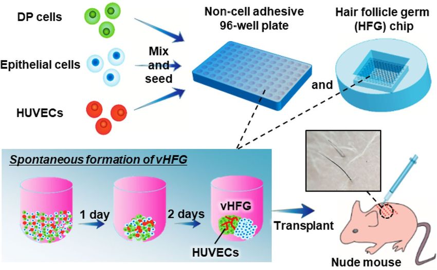
www.nature.com/scientificreports/
Figure 3. vHFG preparation in HFG chip. (a) Fabricated HFG chip. The inset shows a magnified view of
vHFGs in microwells. (b) Representative appearance of vHFGs and HFGs after 3 days of culture in the HFG
chip. The number of cells seeded in the HFG chip was 1.13 × 10⁶ total cells/chip for vHFG and 1 × 10⁶ total cells/
chip for HFG. GFP-HUVECs were visualized, and fluorescence and phase-contrast images were overlaid for
vHFG. (c) HE-staining (i) and fluorescence staining (ii, iii) of cross-sections of vHFGs after 3 days of culture in
the HFG chip. GFP-HUVECs (green) and nuclei (blue) were visualized. (d) Relative expression of trichogenic
genes. GAPDH was used as a reference gene to normalize expression. Error bars represent the standard error of
mean calculated from three independent experiments for each condition. Variables were statistically evaluated
using Student’s t-test. *p < 0.05 was considered significant.
Preparation of numerous vHFGs on HFG chip. We have previously reported a microwell array chip
(named as the HFG chip) for the large-scale preparation of HFGs (~ 5,000 HFGs/φ10-cm chip) via self-sorting
of epithelial and mesenchymal c ells8. Since the HFG chip is composed of polydimethylsiloxane (PDMS), an oxy-
gen-permeable silicone, oxygen is supplied not only through the surface of the culture medium, but also through
the bottom silicone layer. We predicted that the HFG chip could be used for large-scale preparation of vHFGs,
owing to the similar formation processes through self-organization, whereas vascular endothelial cells are gener-
ally activated under hypoxia rather than normoxia. Cell suspensions (2 mL) containing DP cells, epithelial cells,
and GFP-HUVECs (total density 1.13 × 10⁶ cells/mL, 4:4:1 ratio) were poured into the HFG chips and cultured
at 37 °C for three days. As shown in Fig. 3a,b, similar to the results obtained with 96-well plate, DP and epithelial
cells were spatially separated from each other, and the HUVECs localized in the DP cell region after three days
of culture. Notably, the DP and epithelial cell regions appeared dark and bright, respectively, under a phase-
Scientific Reports | (2021) 11:624 | https://doi.org/10.1038/s41598-020-79722-z 4
Vol:.(1234567890)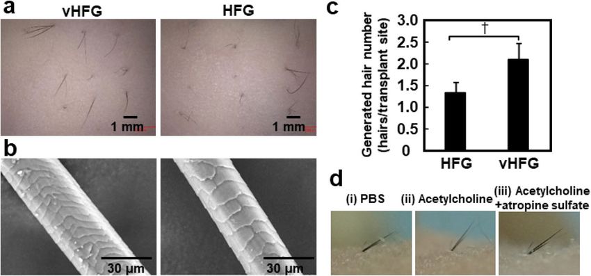
www.nature.com/scientificreports/
Figure 4. Hair-patch assay with vHFGs. (a) Hair shafts generated from 30 vHFGs and 30 HFGs in the lateral
dorsal skin. (b) Quantification of generated hair shafts. The values and error bars represent at least four
experiments for each condition. *p < 0.05 was considered significant. The values and error bars were calculated
based on at least five samples.
contrast microscope (IX-71, Olympus, Tokyo, Japan), whereas HUVECs were fluorescently labeled in these
images. Hematoxylin and eosin (HE) and fluorescence staining of the cross-sections of vHFGs revealed HUVEC
localization predominantly in the DP cell region (Fig. 3c). Considering that hair restoration surgery involves
the translocation of thousands of hair follicles per patient, a scalable and robust approach for the preparation of
thousands of tissue grafts would be essential. Since vHFGs were formed through self-organization, our approach
might provide a practical advantage in the large-scale preparation of tissue grafts for hair regenerative medicine.
Previous studies had shown that non-hair follicle cells improve the hair induction functions of hair follicle
cells. For example, co-culture of DP cells (as an inner DP core) with adipocyte progenitor cells (as an outer shell)
significantly promoted the hair induction ability of DP cells21. This was consistent with the fact that the telogen-
to-anagen transition is synchronized with differentiation of adipocyte progenitor cells to mature adipocytes
near the regenerating hair follicles22. Such cell–cell interactions are mediated via direct cell contact and soluble
signaling molecules, such as growth factors. Vascular endothelial cells secrete several growth factors, including
vascular endothelial growth factor, fibroblast growth factor, and transforming growth factor-β23,24. These growth
factors are widely used to treat hair loss12,25,26. We, therefore, hypothesized that vascular endothelial cells local-
ized in the DP cell region may upregulate follicular genes of DP cells in vHFG. We found that trichogenic DP
cell markers, versican (VCAN)27, alpha-smooth muscle actin 2 (ACTA2)28, alkaline phosphatase (ALP)29, bone
morphogenetic protein 4 (BMP4)29, and lymphoid enhancer factor (LEF1)30, were significantly upregulated in
vHFGs compared to HFGs without HUVECs (Fig. 3d). VCAN is a well-known hair-inductive marker of DP
cells, and HUVECs have also been reported to express VCAN31. Meanwhile, HUVECs did not significantly
affect WNT5A or noggin (NOG) expression (data not shown). A previous study had reported that DP signature
genes, including NOG, were upregulated not only by 3D aggregate culture, but also in a synergistic manner with
the canonical WNT signaling pathway activator CHIR9902132. Hence, a similar approach might be adaptable
to further improve trichogenic gene expression of DP cells in vHFGs. Although gene expression would be an
indicator of hair induction activity, these alone are not thoroughly reliable and animal experiments should be
conducted to estimate the activity of hair follicle generation in the skin.
Comparison between HFGs and vHFGs using the hair‑patch assay. The ability of vHFGs to gener-
ate hair follicles was determined using the hair-patch assay33. vHFGs (4.5 × 10³ cells/vHFG, 30 aggregates) or
HFGs (4 × 10³ cells/HFG, 30 aggregates) were transplanted into a wound pocket surgically generated on the
lateral dorsal skin of nude mice. Difference in the number of cells between vHFGs and HFGs arose solely from
the presence or absence of HUVECs. The vHFG and HFG transplanted sites were pigmented after seven days
of transplantation and black hair shafts were generated at all transplanted sites after four weeks of transplanta-
tion (Fig. 4a). The clusters of generated hair shafts were extracted after enzymatic dissociation of the tissues
and the hair shafts on each transplanted site were counted. The number of hairs generated from vHFGs was
significantly higher than that generated from HFGs without HUVECs (Fig. 4b). Recent studies have reported
pre-vascularization to significantly improve the engraftment of various three-dimensional tissues34,35. Vascular
structures in pre-vascularized tissues are typically immature, however, they readily connect to host vasculature
and attract blood flow from surrounding tissues to the entire tissue graft before severe depletion of o xygen34,35.
Scientific Reports | (2021) 11:624 | https://doi.org/10.1038/s41598-020-79722-z 5
Vol.:(0123456789)www.nature.com/scientificreports/
Figure 5. Intracutaneous transplantation of vHFGs and piloerection assay. (a) Hair shafts generated 3 weeks
after transplantation of vHFGs and HFGs into the dorsal murine skin. (b) Scanning electron microscopic
images of generated hair shafts. (c) Number of generated hair shafts. Values and error bars were calculated from
at least 15 HFG transplantations for each condition in three independent experiments. Numerical variables were
statistically evaluated using the Student’s t-test. p < 0.1 is represented by †. (d) Piloerection of generated hair
shafts after injection of (i) PBS, (ii) acetylcholine, and (iii) acetylcholine plus atropine sulfate.
A technical concern in this study was that, since the epithelial cells were isolated from mouse embryos, possible
contamination may have occurred from other cell types, including mesenchymal cells and vascular endothelial
cells. To estimate the adverse impact of contamination, spheroids composed of freshly isolated murine epithelial
cell suspension were transplanted into the back skin of mice in a similar manner as described for the vHFGs.
Three weeks after transplantation, no hair was observed at the transplanted sites (Fig. S4), suggesting that cell
contamination was negligible. Although further studies would be necessary to clarify the contributing mecha-
nisms of vascular endothelial cells, the success of engraftment may be attributed to both activation of vHFGs and
prompt anastomosis to host vasculature.
Intracutaneous transplantation of individual vHFGs. Further animal experiments were performed
to evaluate in situ hair follicle generation. We initially examined HFGs (without vascular endothelial cells) by
grafting them individually into the back skin of nude mice after three days of culture (Fig. S3). To investigate the
effect of cell number on hair regeneration, we transplanted HFGs with varying cell numbers into shallow stab
wounds generated on the back of mice using a 20-G ophthalmic V-Lance knife (Alcon, Geneva, Switzerland).
The transplanted sites were observed every 2–3 days and images of the generated hair were captured using a digi-
tal camera (Tough, Olympus). At three weeks after transplantation, HFGs with greater number of cells (> 4 × 10³
cells/HFG) generated black hair at the transplanted sites while those with lesser number of cells (< 2 × 10³ cells/
HFG) showed no hair growth. Hair cycle was observed at approximately 30-day interval (Fig. S3), and was equiv-
alent to that of typical mouse skin36. Based on these results, 8 × 10³ cells/HFG (DP cells: epithelial cells = 1:1) and
9 × 10³ cells/vHFG (DP cells: epithelial cells: HUVECs = 4:4:1) were used for subsequent experiments.
To compare the hair follicle generation capacity, individual vHFGs (9 × 10³ cells/vHFG) or HFGs (8 × 10³ cells/
HFG) were transplanted into shallow stab wounds generated in mouse dorsal skin. Black hair shafts appeared
from the skin surface three weeks after transplantation (Fig. 5a). The hair shafts in both approaches possessed
normal morphological characteristics, including the hair cuticle (Fig. 5b). There was no significant difference
between vHFGs and HFGs in hair generation efficiency, which was defined as the number of hair-generating
sites divided by the number of transplanted sites, whereas the number of generated hairs was higher with vHFGs
than with HFGs (Fig. 5c). Piloerection was monitored after intradermal administration of acetylcholine proximal
to the generated hair follicles (Fig. 5d). Hair shafts significantly moved vertically after acetylcholine injection,
though not after PBS injection (Fig. 5d (i) and (ii). Injection of an anti-cholinergic agent, atropine, suppressed
piloerection (Fig. 5d (iii)). Piloerection of regenerated hair shafts was equivalent to that of natural shafts in terms
of temporal and spatial c haracteristics37.
This study was inspired by elegant vascular networks surrounding hair follicles in vivo38 and by a report that
showed prevascularization of an engineered tissue graft containing hair follicle-like structures to significantly
improve human hair generation in mice39. Although our previous study had demonstrated that > 5000 HFGs
could be prepared in the lab-made c hip8, our approach lacked vascular cells. Our present study demonstrated
the significant potential of vascular endothelial cells in the formation of HFGs that would be beneficial for hair
regenerative medicine. Although we used accessible cell types as a proof of concept study, cell sources should
ideally be of human origin and most preferably autologous to cells in clinical setting. Hence, human DP c ells40
and dermal sheath cup c ells41 from patient’s own hair follicles would be the most promising candidates. Regard-
ing epithelial cell sources, hair follicle stem cells located in the bulge region reportedly possess de novo hair
generation ability42. Since isolation and proliferation of human hair follicle epithelial stem cells while maintaining
stemness is a critical issue43, iPS-derived epithelial cells could be another cell s ource44. The source of vascular
Scientific Reports | (2021) 11:624 | https://doi.org/10.1038/s41598-020-79722-z 6
Vol:.(1234567890)www.nature.com/scientificreports/
endothelial cells must also be considered. Microvascular endothelial cells surrounding hair follicles, iPS-derived
endothelial cells45, and endothelial progenitor cell (EPC)-derived endothelial cells46 could be used as autologous
or immunologically compatible cells.
In summary, we observed localization of HUVECs in the DP cell region in vHFGs. Due to the spontaneous
formation of vHFGs, a large number of vHFGs could be prepared by seeding a mixture of DP and epithelial cells,
with HUVECs, into a microwell-arrayed HFG chip. The presence of HUVECs upregulated follicular gene expres-
sion after three days of culture and significantly increased the number of hairs regenerated after three weeks of
transplantation into the dorsal skin of nude mice. The hair shafts possessed piloerection function and showed
typical morphological characteristics, such as hair cuticle and medulla. vHFGs may, therefore, be beneficial for
developing improved hair regenerative strategies.
Methods
Animals. Pregnant C57BL/6 mice were purchased from CLEA, Japan. Five-week-old ICR nu/nu mice were
purchased from Charles River, Japan. The animal study was approved by the committee on animal care and use,
Yokohama National University (Permit numbers: 2019-04 and 2019-06). Care and handling of mice conformed
to the requirements of the above-mentioned committee.
Preparation of murine epithelial cells. Mouse embryos (E18) were extracted from a pregnant C57BL/6
mouse, and small pieces of their dorsal skin were harvested under a dissecting microscope. Skin tissue was asep-
tically treated with 4.8 U/mL Dispase II (Sigma Aldrich, St. Louis, MO, USA) for 60 min, and the epithelial and
mesenchymal layers were separated using a pair of tweezers47. The epithelial layer was then treated with 100 U/
mL collagenase type I (Wako, Osaka, Japan) for 80 min and 0.25% trypsin (Thermo Fisher Scientific, Waltham,
MA, USA) for 10 min at 37 °C. Debris and tissue aggregates were removed using a 40-μm cell strainer (Corn-
ing, Corning, NY, USA). After centrifugation at 200 × g for 3 min, the epithelial cells were suspended in KG2
(Kurabo, Osaka, Japan) and counted.
Preparation of human DP cells and HUVECs. Human DP cells (PromoCell, Heidelberg, Germany)
were maintained in follicle DP cell growth medium (DPCGM) (PromoCell), which was changed every 2–3 days.
Cells from the fourth passage were used in the experiments. HUVECs (Angio-proteomie, Boston, MA, USA)
and GFP/RFP-induced HUVECs (GFP/RFP-HUVEC, Angio-proteomie) were maintained in endothelial cell
growth medium (EGM-2; Lonza, Basel, Switzerland), which was refreshed every 2–3 days. Cells from passages
5–7 were used in the experiments. Mixed culture medium, composed of DPCGM, KG2, and EGM-2 (1:1:1
ratio), was used for HFG culture in the HFG chips.
Fabrication of HFG chips. HFG chips were fabricated via micro-milling and molding, as described
reviously8. Briefly, configuration of the microwell arrays (diameter, 1 mm; pitch, 1.3 mm; depth, 500 µm) in
p
a 20 × 20-mm region (250 microwells) was designed using CAD/CAM software (VCarve pro, Vectric, Alcester,
UK). The design data were subsequently sent to a computer-aided micro-milling machine (MDX-540, Roland,
Irvine, CA, USA) to fabricate a negative mold in polyolefin plate (ZEONOR, Zeon, Tokyo, Japan). Epoxy resin
(Nissin Resin, Tokyo, Japan) was then poured on the negative mold and cured at room temperature for 24 h
to produce a positive mold. The PDMS solution (Shin-Etsu Silicones, Tokyo, Japan) (prepolymer solution and
curing agent were mixed in 10:1 ratio) was poured onto the positive mold and cured in an oven at 80 °C for 3 h.
Thickness of the PDMS substrate at the bottom of the microwells, which was responsible for supplying oxygen to
the culture, was set to 1.5 mm by adjusting the PDMS volume. The PDMS substrate was immersed in pure water
and autoclaved for sterilization. Surface of the PDMS substrate and microwells were treated with 4% pluronic
F-127 (Wako) solution for 12 h to prevent cell adhesion onto the surface. After substantial washing with PBS to
remove excess pluronic F-127, the PDMS substrate was used for cell culture.
Human‑mouse chimeric HFGs containing HUVECs (vHFG). To prepare HFGs, DP and epithelial
cells were suspended in 0.1 mL culture medium (1, 2, 4, 8, 16, 32, or 64 × 103 cells/0.1 mL; DP cells: epithelial
cells = 1:1). To prepare vHFGs, RFP/GFP-HUVECs, DP cells, and epithelial cells were suspended in 0.1 mL EGM2/
DMEM/KG2 (1.13, 2.25, 4.5, 9, 18, 36, or 72 × 103 cells/0.1 mL, DP cells: epithelial cells: RFP-HUVEC = 4:4:1;
1.25, 2.5, 5, 10, 20, 40, or 80 × 103 cells/0.1 mL, DP cells: epithelial cells: RFP-HUVEC = 2:2:1). Suspensions con-
taining the three cell types were mixed and seeded in wells of a non-cell-adhesive, round-bottom, 96-well plate
(prime surface 96U; Sumitomo Bakelite, Tokyo, Japan). After 3 days of culture, spatial distribution of the cells
was examined under a confocal microscope (LSM 700, Carl Zeiss, Oberkochen, Germany) and a fluorescence
microscope (DP-71, Olympus).
Large‑scale preparation of vHFGs. Cell suspensions (2 mL) containing DP cells, epithelial cells, and
GFP-HUVECs (total density 1.13 × 10⁶ cells/mL, 4:4:1 ratio) were poured into the HFG chips and cultured at
37 °C in mixed culture medium composed of DPCGM, KG2, and EGM-2 (1:1:1 ratio) for three days. Spatial
distribution of the cells was examined after three days using a phase-contrast microscope (IX-71, Olympus) and
fluorescence microscope (DP-71, Olympus), and the relative expression levels of genes associated with hair fol-
licle induction were assessed by real-time reverse-transcription polymerase chain reaction (RT-PCR).
Gene expression analysis using RT‑PCR. Total RNA was extracted from samples using an RNeasy mini-
kit (Qiagen, Hilden, Germany), and cDNA was synthesized by reverse-transcription using a ReverTraAce RT-
Scientific Reports | (2021) 11:624 | https://doi.org/10.1038/s41598-020-79722-z 7
Vol.:(0123456789)www.nature.com/scientificreports/
qPCR kit (Toyobo, Osaka, Japan), according to the manufacturer’s instructions. qPCR was performed using the
StepOne Plus RT-PCR system (Applied Biosystems, Foster City, CA, USA) and SYBR Premix Ex Taq II (Takara-
bio, Shiga, Japan). Gene expression was assessed using primers for the following genes: VCAN (versican), 5ʹ-CCA
GCAAGCACAAAATTTCA-3ʹ and 5ʹ-TGCACTGGATCTGTTTCTTCA-3ʹ; ACTA2 (α-smooth muscle actin,
α-SMA), 5ʹ-GCTTCCCTGAACACCACCCAGT-3ʹ and 5ʹ-GCCTTACAGAGCCCAGAGCCAT-3ʹ; LEF1 (lym-
phoid enhancer-binding factor 1), 5ʹ-CCCGATGACGGAAAGCAT-3ʹ and 5ʹ-TCGAGTAGGAGGGTCCCT
TGT-3ʹ; ALP (alkaline phosphatase), 5ʹ-ATTGACCACGGGCACCAT-3ʹ and 5ʹ-CTCCACCGCCTCATGCA-3ʹ;
BMP4 (bone morphogenetic protein 4), 5ʹ-GCCCGCAGCCTAGCAA-3ʹ and 5ʹ-CGGTAAAGATCCCGCATG
TAG-3ʹ; and GAPDH (glyceraldehyde-3-phosphate dehydrogenase), 5ʹ-GCACCGTCAAGGCTGAGAAC-3ʹ
and 5ʹ-TGGTGAAGACGCCAGTGGA-3ʹ. Expression levels of all the genes were normalized to that of GAPDH.
Relative gene expression was determined using the 2 −⊿⊿Ct method and presented as the mean ± standard error of
four independent experiments. Statistical analysis of variables was conducted using Student’s t-tests, and differ-
ences with p < 0.05 were considered statistically significant.
Hair‑patch assay. The hair induction ability of vHFGs was quantified using the hair-patch a ssay48. Briefly,
under isoflurane anesthesia, a wound (4–6 mm in diameter) was surgically generated on the lateral dorsal skin
of mice. The vHFGs (4.5 × 10³ cells/vHFG, 30 aggregates) and HFGs (4 × 10³ cells/HFG, 30 aggregates) were
transplanted separately into the wound. At four weeks after transplantation, all transplanted sites were examined
using a digital microscope (VHX-1000, Keyence, Osaka, Japan) and digital camera (Tough, Olympus). To disso-
ciate the generated hair shafts from the skin, a piece of skin (~ 1 × ~ 1 cm) was treated with 100 U/mL collagenase
at 37 °C for 2 h. The number of hair shafts per transplanted site was counted after hair dissociation.
Intracutaneous transplantation. The mice used in these experiments were raised under specific patho-
gen-free conditions and had access to chow and water ad libitum. Under isoflurane anesthesia, HFGs and vHFGs
were transplanted into the shallow stab wounds generated on the back of ICR nu/nu mice using a 20-G oph-
thalmic V-Lance knife (Alcon). The transplantation sites were then rubbed with ointment and observed every
2–3 days, and images of the generated hairs were captured using a digital camera (Tough, Olympus). Cuticles of
generated hairs were examined using a scanning electron microscope at 5–10 kV without any treatment.
Piloerection of regenerated hairs. Piloerection was assessed after injecting the mice with neurotrans-
mitters, as described previously37. PBS (1 µL) was injected to prevent possible cutaneous reflex movements. Sub-
sequently, 1 µL of 10 mg/mL acetylcholine solution (Sigma Aldrich) was injected intradermally near the regener-
ated hair follicles. To determine the specificity of acetylcholine for initiating piloerection, 1 µL of a 100 mg/mL
solution of atropine sulfate (acetylcholine inhibitor; Sigma Aldrich) was injected before acetylcholine injection.
The angles of piloerection were recorded using a digital camera (Tough, Olympus).
Histological and immunohistochemical staining. Samples were washed with PBS and fixed with 4%
formaldehyde (Sigma Aldrich) for 1 h at room temperature. After embedding in a paraffin block, 8-μm-thick
sections were cut and stained with Meyer’s hematoxylin and eosin Y solutions (Muto Pure Chemicals, Tokyo,
Japan). For immunohistochemical staining, the sections were fixed in 10% formaldehyde (pH 7) overnight at
room temperature. The samples were first blocked in PBS containing 1% bovine serum albumin (Thermo Fisher
Scientific) and 0.01% Triton X-100 (Thermo Fisher Scientific) for 1 h at room temperature and subsequently
incubated overnight with anti-vimentin (rabbit, primary antibody, Merck Millipore, Billerica, MA, USA) and
anti-cytokeratin 15 (rabbit, primary antibody, Abcam, Cambridge, UK) at 4 °C. The sections were incubated
with the corresponding secondary antibodies (highly cross-absorbed goat anti-IgG (H + L) Alexa Fluor 488 or
goat anti-IgG (H + L) Alexa Fluor 555, Thermo Fisher Scientific) in blocking solution for 3 h at room tempera-
ture, and finally stained with DAPI (Sigma Aldrich) in PBS for 8 min. A confocal microscope (LSM 700; Carl
Zeiss) was used for fluorescence imaging.
Received: 8 May 2020; Accepted: 3 December 2020
References
1. Relevant research, Inc. International society of hair restoration surgery: 2017 practice census results International society of hair
restoration surgery 2017, 1–31 (2017).
2. Orentreich, N. Autografts in alopecias and other selected dermatological conditions. Ann. N. Y. Acad. Sci. 83, 463–479 (1959).
3. Millar, S. E. Molecular mechanisms regulating hair follicle development. J. Invest. Dermatol. 118, 216–225 (2002).
4. Fan, S. M. Y. et al. Inducing hair follicle neogenesis with secreted proteins enriched in embryonic skin. Biomaterials 167, 121–131
(2018).
5. Lim, T. C. et al. Follicular dermal papilla structures by organization of epithelial and mesenchymal cells in interfacial polyelectrolyte
complex fibers. Biomaterials 34, 7064–7072 (2013).
6. Yen, C. M., Chan, C. C. & Lin, S. J. High-throughput reconstitution of epithelial-mesenchymal interaction in folliculoid microtis-
sues by biomaterial-facilitated self-assembly of dissociated heterotypic adult cells. Biomaterials 31, 4341–4352 (2010).
7. Toyoshima, K.-e et al. Fully functional hair follicle regeneration through the rearrangement of stem cells and their niches. Nat.
Commun. 3, 784. https://doi.org/10.1038/ncomms1784 (2012).
8. Kageyama, T. et al. Spontaneous hair follicle germ (HFG) formation in vitro, enabling the large-scale production of HFGs for
regenerative medicine. Biomaterials 154, 291–300 (2018).
Scientific Reports | (2021) 11:624 | https://doi.org/10.1038/s41598-020-79722-z 8
Vol:.(1234567890)www.nature.com/scientificreports/
9. Kageyama, T., Yan, L., Shimizu, A., Maruo, S. & Fukuda, J. Preparation of hair beads and hair follicle germs for regenerative
medicine. Biomaterials 212, 55–63 (2019).
10. Wu, X. W., Scott, L., Washenik, K. & Stenn, K. Full-thickness skin with mature hair follicles generated from tissue culture expanded
human cells. Tissue Eng. A 20, 3314–3321 (2014).
11. Xiao, Y. et al. Perivascular hair follicle stem cells associate with a venule annulus. J. Invest. Dermatol. 133, 2324–2331 (2013).
12. Yano, K., Brown, L. F. & Detmar, M. Control of hair growth and follicle size by VEGF-mediated angiogenesis. J. Clin. Invest. 107,
409–417 (2001).
13. Vahav, I. et al. Reconstructed human skin shows epidermal invagination towards integrated neopapillae indicating early hair fol-
licle formation in vitro. J. Tissue Eng. Regen. Med. 14, 761–773 (2020).
14. Bose, A., Teh, M. T., Mackenzie, I. C. & Waseem, A. Keratin K15 as a biomarker of epidermal stem cells. Int. J. Mol. Sci. 14,
19385–19398 (2013).
15. Halbleib, J. M. & Nelson, W. J. Cadherins in development: cell adhesion, sorting, and tissue morphogenesis. Genes Dev. 20,
3199–3214 (2006).
16. Steinberg, M. S. & Takeichi, M. Experimental specification of cell sorting, tissue spreading, and specific spatial patterning by
quantitative differences in cadherin expression. Proc. Natl. Acad. Sci. USA 91, 206–209 (1994).
17. Foty, R. A. & Steinberg, M. S. The differential adhesion hypothesis: a direct evaluation. Dev. Biol. 278, 255–263 (2005).
18. Corada, M. et al. Monoclonal antibodies directed to different regions of vascular endothelial cadherin extracellular domain affect
adhesion and clustering of the protein and modulate endothelial permeability. Blood 97, 1679–1684 (2001).
19. Ferreri, D. M., Minnear, F. L., Yin, T. F., Kowalczyk, A. P. & Vincent, P. A. N-Cadherin levels in endothelial cells are regulated by
monolayer maturity and p120 availability. Cell Commun. Adhes. 15, 333–349 (2008).
20. Hoffecker, I. T. & Iwata, H. Manipulation of cell sorting within mesenchymal stromal cell-islet cell multicellular spheroids. Tissue
Eng. A 20, 1643–1653 (2014).
21. Huang, C. F. et al. Assembling composite dermal papilla spheres with adipose-derived stem cells to enhance hair follicle induction.
Sci. Rep. 6, 26436. https://doi.org/10.1038/srep26436 (2016).
22. Festa, E. et al. Adipocyte lineage cells contribute to the skin stem cell niche to drive hair cycling. Cell 146, 761–771 (2011).
23. Du, H., Shi, H., Chen, D. L., Zhou, Y. B. & Che, G. W. Cross-talk between endothelial and tumor cells via basic fibroblast growth
factor and vascular endothelial growth factor signaling promotes lung cancer growth and angiogenesis. Oncol. Lett. 9, 1089–1094
(2015).
24. Lang, H. L. et al. Glioma cells enhance angiogenesis and inhibit endothelial cell apoptosis through the release of exosomes that
contain long non-coding RNA CCAT2. Oncol. Rep. 38, 785–798 (2017).
25. Lin, W. H. et al. Fibroblast growth factors stimulate hair growth through beta-catenin and Shh expression in C57BL/6 mice. Biomed.
Res. Int. 2015, 730139. https://doi.org/10.1155/2015/730139 (2015).
26. Oshimori, N. & Fuchs, E. Paracrine TGF-beta signaling counterbalances BMP-mediated repression in hair follicle stem cell activa-
tion. Cell Stem Cell 10, 63–75 (2012).
27. Kishimoto, J. et al. Selective activation of the versican promoter by epithelial-mesenchymal interactions during hair follicle devel-
opment. Proc. Natl. Acad. Sci. USA 96, 7336–7341 (1999).
28. Jahoda, C. A. B., Reynolds, A. J., Chaponnier, C., Forester, J. C. & Gabbiani, G. Smooth-muscle alpha-actin is a marker for hair
follicle dermis in vivo and in vitro. J. Cell Sci. 99, 627–636 (1991).
29. Veraitch, O. et al. Induction of hair follicle dermal papilla cell properties in human induced pluripotent stem cell-derived multi-
potent LNGFR(+) THY-1(+) mesenchymal cells. Sci. Rep. 7, 42777. https://doi.org/10.1038/srep42777 (2017).
30. Zhou, P. B., Byrne, C., Jacobs, J. & Fuchs, E. Llymphoid enhancer factor-1 directs hair follicle patterning and epithelial-cell fate.
Genes Dev. 9, 700–713 (1995).
31. Li, S. et al. Exosomes from hyperglycemia-stimulated vascular endothelial cells contain versican that regulate calcification/senes-
cence in vascular smooth muscle cells. Cell Biosci. https://doi.org/10.1186/s13578-018-0263-x (2019).
32. Yoshida, Y., Soma, T., Matsuzaki, T. & Kishimoto, J. Wnt activator CHIR99021-stimulated human dermal papilla spheroids con-
tribute to hair follicle formation and production of reconstituted follicle-enriched human skin. Biochem. Biophys. Res. Commun.
516, 599–605 (2019).
33. Ohyama, M., Zheng, Y., Paus, R. & Stenn, K. S. The mesenchymal component of hair follicle neogenesis: background, methods
and molecular characterization. Exp. Dermatol. 19, 89–99 (2010).
34. Laschke, M. W. & Menger, M. D. Prevascularization in tissue engineering: current concepts and future directions. Biotechnol. Adv.
34, 112–121 (2016).
35. Rouwkema, J., Rivron, N. C. & van Blitterswijk, C. A. Vascularization in tissue engineering. Trends Biotechnol. 26, 434–441 (2008).
36. Foitzik, K. et al. Control of murine hair follicle regression (catagen) by TGF-beta 1 in vivo. FASEB J. 14, 752–760 (2000).
37. Sato, A. et al. Single follicular unit transplantation reconstructs arrector pili muscle and nerve connections and restores functional
hair follicle piloerection. J. Dermatol. 39, 682–687 (2012).
38. Yoshida, Y., Soma, T. & Kishimoto, J. Characterization of human dermal sheath cells reveals CD36-expressing perivascular cells
associated with capillary blood vessel formation in hair follicles. Biochem. Biophys. Res. Commun. 516, 945–950 (2019).
39. Abaci, H. E. et al. Tissue engineering of human hair follicles using a biomimetic developmental approach. Nat. Commun. 9, 5301.
https://doi.org/10.1038/s41467-018-07579-y (2018).
40. Tsuboi, R. et al. Autologous cell-based therapy for male and female pattern hair loss using dermal sheath cup cells: a randomized
placebo-controlled double-blinded dose-finding clinical study. J. Am. Acad. Dermatol. 83, 109–116 (2020).
41. Castro, A. R. & Logarinho, E. Tissue engineering strategies for human hair follicle regeneration: how far from a hairy goal? Concise
review. Stem Cells Transl. Med. 9, 342–350 (2020).
42. An, L. Y. et al. ROCK inhibitor Y-27632 maintains the propagation and characteristics of hair follicle stem cells. Am. J. Transl. Res.
10, 3689–3700 (2018).
43. Sanchez-Danes, A. & Blanpain, C. Maintaining hair follicle stem cell identity in a dish. EMBO J. 36, 132–134 (2017).
44. Veraitch, O. et al. Human induced pluripotent stem cell-derived ectodermal precursor cells contribute to hair follicle morphogenesis
in vivo. J. Invest. Dermatol. 133, 1479–1488 (2013).
45. Rosa, S. et al. Functional characterization of iPSC-derived arterial- and venous-like endothelial cells. Sci. Rep. 9, 3826. https://doi.
org/10.1038/s41598-019-40417-9 (2019).
46. Williamson, K., Stringe, S. E. & Alexander, M. Y. Endothelial progenitor cells enter the aging arena. Front. Physiol. 3, 30. https://
doi.org/10.3389/fphys.2012.00030 (2012).
47. Lichti, U., Anders, J. & Yuspa, S. H. Isolation and short-term culture of primary keratinocytes, hair follicle populations and dermal
cells from newborn mice and keratinocytes from adult mice for in vitro analysis and for grafting to immunodeficient mice. Nat.
Protoc. 3, 799–810 (2008).
48. Inoue, K. et al. Evaluation of animal models for the hair-inducing capacity of cultured human dermal papilla cells. Cells Tissues
Organs. 190, 102–110 (2009).
Scientific Reports | (2021) 11:624 | https://doi.org/10.1038/s41598-020-79722-z 9
Vol.:(0123456789)www.nature.com/scientificreports/
Acknowledgements
This work was supported in part by the Ministry of Education, Culture, Sports, Science, and Technology of
Japan, Grants-in-Aid for Scientific Research (Kakenhi) (18K18971, 19K21107), Kanagawa Institute of Industrial
Science and Technology (KISTEC), and Japan Science and Technology Agency (JST)-PRESTO (JPMJPR19H2).
Author contributions
T.K., YS.C., and J.F. designed the experiments. T.K. conducted most of the experiments. T.K. and J.F. wrote the
manuscript.
Competing interests
The authors declare no competing interests.
Additional information
Supplementary Information The online version contains supplementary material available at https://doi.
org/10.1038/s41598-020-79722-z.
Correspondence and requests for materials should be addressed to J.F.
Reprints and permissions information is available at www.nature.com/reprints.
Publisher’s note Springer Nature remains neutral with regard to jurisdictional claims in published maps and
institutional affiliations.
Open Access This article is licensed under a Creative Commons Attribution 4.0 International
License, which permits use, sharing, adaptation, distribution and reproduction in any medium or
format, as long as you give appropriate credit to the original author(s) and the source, provide a link to the
Creative Commons licence, and indicate if changes were made. The images or other third party material in this
article are included in the article’s Creative Commons licence, unless indicated otherwise in a credit line to the
material. If material is not included in the article’s Creative Commons licence and your intended use is not
permitted by statutory regulation or exceeds the permitted use, you will need to obtain permission directly from
the copyright holder. To view a copy of this licence, visit http://creativecommons.org/licenses/by/4.0/.
© The Author(s) 2021
Scientific Reports | (2021) 11:624 | https://doi.org/10.1038/s41598-020-79722-z 10
Vol:.(1234567890)You can also read