CD33 Delineates Two Functionally Distinct NK Cell Populations Divergent in Cytokine Production and Antibody-Mediated Cellular Cytotoxicity - Frontiers
←
→
Page content transcription
If your browser does not render page correctly, please read the page content below
ORIGINAL RESEARCH
                                                                                                                                            published: 04 January 2022
                                                                                                                                      doi: 10.3389/fimmu.2021.798087

                                          CD33 Delineates Two Functionally
                                          Distinct NK Cell Populations
                                          Divergent in Cytokine Production
                                          and Antibody-Mediated Cellular
                                          Cytotoxicity
                           Edited by:     Maryam Hejazi 1, Congcong Zhang 2, Sabrina B. Bennstein 1, Vera Balz 1,
                         Marina Cella,
                                          Sarah B. Reusing 1,3, Melissa Quadflieg 2, Keven Hoerster 4, Stefan Heinrichs 4,
    Washington University School of
                                          Helmut Hanenberg 5, Sebastian Oberbeck 6, Marcus Nitsche 2, Sophie Cramer 2,
  Medicine in St. Louis, United States
                                          Rita Pfeifer 2, Pranav Oberoi 7, Heiko Rühl 8, Johannes Oldenburg 8,
                       Reviewed by:       Peter Brossart 6, Peter A. Horn 4, Florian Babor 3, Winfried S. Wels 7,
                Maya Caroline Andre,
                                          Johannes C. Fischer 1, Nina Möker 2 and Markus Uhrberg 1*
        University Children’s Hospital
                  Tübingen, Germany       1 Institute for Transplantation Diagnostics and Cell Therapeutics, Heinrich-Heine University, Düsseldorf, Germany, 2 Miltenyi
                   Frank M. Cichocki,     Biotec B.V. & Co. KG, Bergisch Gladbach, Germany, 3 Department of Pediatric Oncology, Hematology and Clinical
  University of Minnesota Twin Cities,    Immunology, Center for Child and Adolescent Health, Medical Faculty, Heinrich-Heine University, Düsseldorf, Germany,
                        United States     4 Institute for Transfusion Medicine, University Hospital Essen, University of Duisburg-Essen, Essen, Germany, 5 Department

                *Correspondence:          of Pediatrics III, University Children’s Hospital, University of Duisburg-Essen, Essen, Germany, 6 Department of Oncology,
                    Markus Uhrberg        Hematology, Immuno-Oncology and Rheumatology, University Hospital of Bonn, Bonn, Germany, 7 Georg-Speyer-Haus,
           markus.uhrberg@med.uni-        Institute for Tumor Biology and Experimental Therapy, Frankfurt am Main, Germany, 8 Institute of Experimental Hematology
                     duesseldorf.de       and Transfusion Medicine, University Hospital of Bonn, Bonn, Germany

                  Specialty section:
                                          The generation and expansion of functionally competent NK cells in vitro is of great interest
        This article was submitted to
NK and Innate Lymphoid Cell Biology,      for their application in immunotherapy of cancer. Since CD33 constitutes a promising
              a section of the journal    target for immunotherapy of myeloid malignancies, NK cells expressing a CD33-specific
             Frontiers in Immunology
                                          chimeric antigen receptor (CAR) were generated. Unexpectedly, we noted that CD33-
        Received: 19 October 2021
      Accepted: 29 November 2021
                                          CAR NK cells could not be efficiently expanded in vitro due to a fratricide-like process in
       Published: 04 January 2022         which CD33-CAR NK cells killed other CD33-CAR NK cells that had upregulated CD33 in
                              Citation:   culture. This upregulation was dependent on the stimulation protocol and encompassed
   Hejazi M, Zhang C, Bennstein SB,
                                          up to 50% of NK cells including CD56dim NK cells that do generally not express CD33 in
    Balz V, Reusing SB, Quadflieg M,
Hoerster K, Heinrichs S, Hanenberg H,     vivo. RNAseq analysis revealed that upregulation of CD33+ NK cells was accompanied by
  Oberbeck S, Nitsche M, Cramer S,        a unique transcriptional signature combining features of canonical CD56bright (CD117high,
           Pfeifer R, Oberoi P, Rühl H,
  Oldenburg J, Brossart P, Horn PA,
                                          CD16low) and CD56dim NK cells (high expression of granzyme B and perforin). CD33+ NK
      Babor F, Wels WS, Fischer JC,       cells exhibited significantly higher mobilization of cytotoxic granula and comparable levels
Möker N and Uhrberg M (2022) CD33
                                          of cytotoxicity against different leukemic target cells compared to the CD33− subset.
 Delineates Two Functionally Distinct
     NK Cell Populations Divergent in     Moreover, CD33+ NK cells showed superior production of IFNg and TNFa, whereas
  Cytokine Production and Antibody-       CD33− NK cells exerted increased antibody-dependent cellular cytotoxicity (ADCC). In
       Mediated Cellular Cytotoxicity.
          Front. Immunol. 12:798087.
                                          summary, the study delineates a novel functional divergence between NK cell subsets
   doi: 10.3389/fimmu.2021.798087         upon in vitro stimulation that is marked by CD33 expression. By choosing suitable

Frontiers in Immunology | www.frontiersin.org                                     1                                        January 2022 | Volume 12 | Article 798087
Hejazi et al.                                                                                                          Characterization of CD33+ NK Cells

                                          stimulation protocols, it is possible to preferentially generate CD33+ NK cells combining
                                          efficient target cell killing and cytokine production, or alternatively CD33− NK cells, which
                                          produce less cytokines but are more efficient in antibody-dependent applications.
                                          Keywords: NK cell, CD33-CAR, cytokine production and cytotoxicity, RNAseq analysis, NK cell expansion

INTRODUCTION                                                                        Here, we studied the relevance of CD33 expression on in vitro
                                                                                stimulated NK cells, triggered by the initial observation of
Natural Killer (NK) cells are getting increasingly into the focus of            fratricide in cultures of CD33-CAR NK cells. We demonstrate
allogeneic cell-based therapy due to their powerful effector                    that CD33 can be exploited to define two functionally distinct
mechanisms enabling eradication of tumor and virus-infected                     NK cell subsets representing CD33+ polyfunctional NK cells
cells without eliciting graft-versus-host disease (1). Classically,             capable of strong cytokine production and target-based
circulating NK cells are divided into CD56bright NK cells that are              cytotoxicity and a CD33− subset exhibiting efficient antibody-
non-cytotoxic and primarily respond with IFNg and TNFa                          dependent cellular cytotoxicity (ADCC) due to strong expression
production in response to exogenous cytokines and CD56dim                       of CD16. Notably, the CD33+ subset becomes highly abundant
NK cells that are uniquely able to exert cytotoxic effector                     when using a commercially available medium-based protocol,
functions via CD16-mediated antibody-dependent cellular                         whereas it remains only a minor subset when employing
cytotoxicity (ADCC) as well as direct targeting of aberrant cells               protocols using established stimulator cell lines. Thus, the
on the basis of missing self-recognition and expression of stress-              frequency of the CD33 subset in the expanded NK cell product
induced ligands (2). Beyond these two major subsets, a multitude                can be controlled a priori by choosing a suitable protocol for NK
of less well-defined NK cell subpopulations with more subtle                     cell stimulation.
differences exist along a continuum of different functional states
(3). Unfortunately, in NK cells stimulated and expanded in vitro,
functional distinction of NK cell subsets on the basis of CD56                  MATERIALS AND METHODS
expression is not useful due to unspecific upregulation on
virtually all NK cells. Similarly, other function-associated                    Human Samples
surface molecules are highly sensitive to shedding, such as                     Buffy coats of healthy donors were kindly provided by the
CD16, or are quickly downregulated in culture, such as CD62L                    Blutspendezentrale at the University Hospital Düsseldorf. The
(4, 5). Thus, although it is apparent that the available protocols to           protocol was accepted by the institutional review board at
expand NK cells do not lead to a homogenous pool of NK cells                    the University of Düsseldorf (study number 2019-383) and is
but rather result in a heterogeneous mixture of NK cell subsets                 in accordance with the Declaration of Helsinki. Peripheral blood
with divergent functional capabilities, the tools to differentiate              mononuclear cells (PBMC) were isolated using density gradient
between functional subsets on the basis of surface molecules                    centrifugation with Lymphocyte Separation Medium
are scarce.                                                                     (PromoCell, Heidelberg, Germany).
    CD33 (siglec-3) is the smallest member of the sialic acid-
binding immunoglobulin-like lectin (Siglec) family with only two                Flow Cytometry
Ig superfamily (IgSF)-like domains, a distal V domain mediating                 The following fluorescence-labeled monoclonal antibodies
sialic acid binding and a membrane-proximal C2 domain (6).                      (mAb) were used: CD3-FITC or PE/Cy5 (clone UCHT1),
Besides the Siglec family-defining binding of CD33 to sialylated                 CD11c-FITC (3.9), CD16-APC/Cy7 (3G8), CD30-PE/Cy7
ligands, such as glycoproteins, glycolipids, and gangliosides,                  (BY88), CD33-PE, BV605 or PE/Cy5 (P67.6), CD56-PE/
more specific protein ligands are currently unknown, with the                    Dazzle™594 (N901), CD57-FITC (HCD57), CD62L-PE/Cy7
exception of the recently defined complement component 1q                        (DREG56), CD107-BV510 (H4A3), CD117-BV421 (104D2),
(C1q) (7). The presence of an immunoreceptor tyrosine-based                     KLRG1-APC/Fire 750 (SA231A2), PD1-APC/Cy7 (EH12-2H7),
inhibitory motif (ITIM) and a second ITIM-like motif in the                     NKG2D-PE (1D11), NKp46-BV510 (9E2), NKp44-APC (P44-
cytoplasmic domain suggests that CD33 is an inhibitory                          8), NKp30-BV785 (P30-15), Granzyme B-Pacific Blue (GB11),
receptor, but the role of CD33 and its ITIM-mediated                            IFNg PE/Cy7 (B27), Perforin-PE (dG9), TNFa PE/Dazzle™ 594
inhibition for regulation of the activation states of immune                    (all from Biolegend, CA, USA), CD3 PerCP-Vio700 (REA613),
cells is elusive. Expression of CD33 is assumed to be largely                   CD14-PerCP-Vio700 (REA599), CD33-APC, CD56 (REA196),
restricted to the myeloid lineage, and with the exception of the                anti-biotin-VioBright515 and 7-AAD (all Miltenyi Biotec),
CD56bright NK cell subset, it seems to be absent from the                       NKG2C-AF700 (134591) from R&D systems (MN, USA), and
lymphocytic lineages including mature T, B, and CD56dim NK                      CD158b1,b2,j-PE/Cy5 (GL183), NKG2A-APC (Z199) purchased
cells (8). CD33 is broadly expressed on various myeloid lineages                from Beckman Coulter (CA, USA). Flow cytometric analyses
and has gained therapeutic relevance as target on CD33-                         were performed on a CytoFLEX (Beckman Coulter) or
expressing myeloid and rare subsets of acute lymphoblastic                      MACSQuant 10 Analyzer (Miltenyi Biotec). Data analysis was
leukemia (9).                                                                   performed on Kaluza 2.1.1 software.

Frontiers in Immunology | www.frontiersin.org                               2                                  January 2022 | Volume 12 | Article 798087
Hejazi et al.                                                                                                Characterization of CD33+ NK Cells

Cell Lines                                                             (Invitrogen, Carlsbad, CA, USA) for extraction of total RNA.
The HLA class I-deficient target cell line K562 was grown in            Reverse transcription and library production were performed in
Dulbecco’s modified Eagle’s medium (DMEM) 4.5 g/L Glucose               the NGS Integrative Genomics (NIG) facility in Göttingen,
with L-Glutamine (Gibco, CA, USA) supplemented with 10%                Germany, with an Illumina Truseq RNA preparation kit.
fetal bovine serum (FBS, FBS, Merck) and 1% Gentamycin.                Sequencing of the libraries was performed on an Illumina
Human Burkitt lymphoma cell line Raji was cultivated in                HiSeq4000 (single-read 1 × 50 bp). Sequence reads were
RPMI-1640 (Thermo Fisher Scientific, Waltham, MA, USA),                 mapped to the human genome (hg38) and analyzed using
1% Penicillin/Streptomycin (P/S) (Gibco), and 10% FBS. K562-           DESeq2 software (v1.26.0) as described previously (14, 15).
mb15-41BBL (10) (kindly provided by D. Campana, National               Heatmaps and volcano plots were performed with R packages
University of Singapore) and K562-mb15-mb21-41BBL (11)                 pretty heatmap (pheatmap) (v1.0.12) (16) and EnhancedVulcano
were cultured in RPMI-1640, 1% Penicillin/Streptomycin, and            (1.4.0) (17), respectively.
10% FBS. Human RS4;11 B cell precursor acute lymphoblastic
leukemia (B-ALL) cells ectopically expressing human CD33 and           NK Cell Function
GFP (RS4;11-CD33) or GFP only (RS4;11-GFP) were cultured in            For analysis of degranulation and cytokine production, NK cells
RPMI 1640 supplemented with 10% FBS and 2 mmol/L L-                    and K562 target cells were mixed at an effector/target (E/T) ratio
glutamine (Gibco). All cell lines used were free of mycoplasma.        of 1:1 in a volume of 200 µl in a 96-well plate (round bottom).
                                                                       CD107a mAb was added prior to incubation. To determine
NK Cell Expansion                                                      spontaneous degranulation or cytokine production, control
Isolation of pure NK cells was performed with MojoSort™                samples without target cells were included. After 1 h
Human NK Cell Isolation Kit (Biolegend). PBMC or enriched              incubation time, 2 µl of 2 mM Monensin and 2 µl Brefeldin
NK cells were cultured in NK MACS medium [1% NK MACS                   (1000×, Biolegend) were added and samples incubated for
supplement, 5% human AB serum (Sigma Aldrich), 500 U/ml IL-            further 4 h. Subsequently, cells were stained for selected
2 (Proleukin), and 25ng/ml IL-15 (Miltenyi Biotec)]. NK cell           surface markers. Interferon-g and TNF-a were intracellularly
expansion with stimulator cells was performed as previously            stained following treatment with fixation and permeabilization
described (10). Briefly, PBMCs (1.5×10⁶) were incubated in 24-          buffer (Biolegend). For cytotoxicity analysis, K562 target cells
well plates with 1×10⁶ irradiated (40 Gy) K562-mb15-41BBL              were stained with 5 mM CFDA-SE (Invitrogen) and mixed with
cells or K562-mb15-mb21-41BBL in SCGM CellGro Medium                   NK cells at various E/T ratios. After 5 h incubation, propidium
(CellGenix, Freiburg, Germany) supplemented with 10% FBS               iodide (PI, Biolegend) was added shortly before flow cytometric
and 1% P/S and 40 U/ml human IL-2 (Proleukin).                         analysis to quantify the viable target cells as described previously
                                                                       (18). In some experiments, cytotoxicity of CAR NK cells was
Chimeric Antigen Receptor NK Cells                                     assessed against GFP-expressing tumor cells. In order to
CAR constructs were designed in silico and consist of a human          determine CD33-CAR-induced NK cell fratricide, in vitro
GM-CSFRa signal peptide, an antigen-specific scFv derived from          expanded NK cells were labeled with 1 mM CellTrace Violet
Gemtuzumab (CD33-CAR) or FMC63 (CD19-CAR), a CD8a                      (CV, Thermo Fisher Scientific, Waltham, MA, USA) and
hinge and transmembrane (TM) domain, followed by 4-1BB and             cocultured with unlabeled CD33-CAR NK cells at various E/T
CD3z intracellular (IC) domains. Codon-optimized CAR                   ratios for 24 h. For ADCC analysis, 1 µg/ml of rituximab
constructs were inserted into a third-generation lentiviral            (Truxima®, Celltrion Healthcare, South Korea) was added
plasmid backbone (Lentigen Technology Inc., Gaithersburg,              directly to the co-culture of NK cells and Raji target cells (ratio
MD, USA) under control of a human EF-1a promoter.                      1:1). NK cells without Raji and NK cells with Raji but without
Baboon envelope (BaEV) pseudotyped lentiviral vectors (LV)             Rituximab served as controls.
containing supernatants were generated by transient transfection
of HEK 293T cells, as previously described (12). NK cell               Statistical Analyses
enrichment was performed with NK cell isolation kit for                Normal distribution of the data was calculated with Shapiro-
human cells (Miltenyi Biotec). NK cells stimulated with NK             Wilk normality test. Depending on normality, paired/unpaired t-
MACS medium and 80 ng/ml of IL-1ß (day 2) were transduced              test or Mann-Whitney test was performed. The difference
with BaEV-LV encoding CD33-CAR or CD19-CAR constructs                  between more than two groups was analyzed with 1-Way
in the presence of 10 mg/ml Vectofusin-1 for 24 h, after 2 h           ANOVA. All analyses were done using GraphPad Prism 8.0.0
spinoculation at 400 × g, 32°C (13). CAR expression was flow            (GraphPad Software, CA, USA).
cytometrically evaluated using biotinylated human recombinant
CD33-Fc protein (R&D Systems) or CD19 detection reagent
(Miltenyi Biotec), respectively, followed by VioBright515-             RESULTS
conjugated antibiotin antibody (Miltenyi Biotec).
                                                                       CD33+ NK Cells Are Recognized and
RNA Sequencing                                                         Eliminated by CD33-CAR NK Cells
CD33+ and CD33− NK cells were flow cytometrically sorted                Our interest in CD33+ NK cells was fueled by an observation
(MoFlo XDP, Beckman Coulter) and stored in TRIzol Reagent              made during the generation and expansion of CD33-specific

Frontiers in Immunology | www.frontiersin.org                      3                                 January 2022 | Volume 12 | Article 798087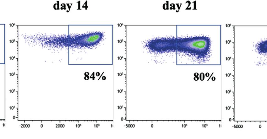
Hejazi et al.                                                                                                                      Characterization of CD33+ NK Cells

CAR NK cells for therapy of myeloid leukemia. To this end,                               CD33 (but otherwise identical features), no such effects on the
PBMC-derived and magnetically enriched NK cells were                                     frequency of CD33+ NK cells were noted, and no overt inhibition
transduced with a Baboon envelope (BaEV)-pseudotyped                                     of NK cell expansion was observed for CD19-CAR-transduced
lentiviral vector (LV) carrying a CD33-specific CAR. The                                  compared to non-transduced NK cells (Figure 1D). The data
construct consisted of an antigen-specific scFv antibody                                  suggested that CD33+ NK cells arising in the cultures were
domain derived from the CD33-specific mAb Gemtuzumab,                                     targets for CD33-CAR NK cells. To more closely look into this
CD8a hinge and transmembrane (TM) domains, and 4-1BB                                     possibility, we analyzed the cytotoxic activity of CD33-CAR NK
and CD3z intracellular (IC) domains (Figure 1A). Following                               cells against autologous NK cells, labeled with a cell tracing
lentiviral transduction, CD33-CAR NK cells were stimulated                               fluorescent reagent. Indeed, CD33-CAR NK cells exhibited
using NK MACS medium, supplemented with IL-2 and IL-15,                                  substantial cytotoxicity against autologous NK cells that were
for 2 weeks. As shown in Figure 1B, NK cells expanded much                               stimulated for 2 weeks in order to induce CD33 expression,
less in the transduced setting compared to non-transduced NK                             whereas non-transduced NK cells did not show cytotoxicity
cells. Flow cytometric analyses revealed that this was due to                            against the same autologous NK cell targets (Figure 1E).
selective depletion of CD33+ NK cells, whereas CD33− NK cells                            Finally, the specificity of CD33-CAR NK cells was tested
remained unaffected (Figure 1C). Depletion of CD33+ NK cells                             against CD33+ tumor cells: the RS4;11 tumor cell line was
was dependent on the presence of CD33-CAR NK cells since in                              killed by CD33-CAR NK cells with high efficiency when
non-transduced controls, CD33+ NK cells were present at high                             expressing CD33, whereas a variant that did not express CD33
frequencies (>50%) (Figure S1). Furthermore, when employing                              (GFP+RS4;11) was not recognized (Figure 1F). Similarly, non-
control CAR constructs with specificity for CD19 instead of                               transduced NK cells did not kill the tumor cells regardless of

                             A                                                    B

                                 C                                               D

                                 E                                              F

  FIGURE 1 | CD33-CAR-mediated NK cell fratricide. (A) CD33-CAR construct design. (B) In vitro proliferation of non-transduced (NT) NK cells and CD33-CAR NK
  cells after isolation. CD33 expression on (C) CD33-CAR NK cells and (D) CD19-CAR NK cells was determined by flow cytometry (day 14) compared with NT NK
  cells (E) CD33-CAR-induced NK cell fratricide with ex vivo expanded autologous NK cells as targets. NT NK cells were included as control. (F) Cytotoxicity of CD33-
  CAR NK cells against CD33-expressing RS4;11 tumor cells. NT NK cells and CD33-negative RS4;11-GFP cells were included as controls. Data were analyzed by
  two-tailed unpaired t-test ***p < 0.001; ****p < 0.0001.

Frontiers in Immunology | www.frontiersin.org                                        4                                     January 2022 | Volume 12 | Article 798087
Hejazi et al.                                                                                                                  Characterization of CD33+ NK Cells

CD33 expression due to natural resistance of RS4;11 cells to NK                     shown in Figure 2A, both subsets contributed to the expansion of
cell-mediated killing. Thus, the observed killing of NK cells by                    the CD33+ subset: whereas in CD56bright NK cells the large
other autologous NK cells, referred to as fratricide, was due to                    majority of cells maintained CD33 expression, in CD56dim NK
recognition of CD33 by the respective CD33-CAR NK cells.                            cells the frequency of CD33 increased over time from basically no
                                                                                    expression at the beginning of culture to around 50% at day 28 as
CD33 Expression in Culture Is Primarily                                             shown in the exemplary experiment in Figure 2A. The CD56dim
Due to Upregulation on CD56dim NK cells                                             data largely resembled the kinetics of CD33 expression in the
and Depends on the Stimulation Protocol                                             control setup using unseparated NK cells, which was expected
CD33 is expressed in vivo on CD56bright but not CD56dim NK                          since CD56dim NK cells typically constitute more than 90% of all
cells (8). We were thus wondering if the CD33+ NK cells                             NK cells and thus are the dominant subset contributing to the
developing in culture were also primarily due to expansion of                       increase in CD33+ cells observed in the cultures (Figure 2A).
CD56bright NK cells. We thus flow cytometrically sorted the two                          Of note, in about 10% of samples, CD33 expression remained
respective CD56 subsets to high purity from peripheral blood and                    low during stimulation, and this was accompanied by a lack of
cultured them separately for 4 weeks in NK MACS medium. As                          CD33 expression on CD56bright NK cells in vivo (Figure S2A).

                                A

                                           B

  FIGURE 2 | Upregulation of CD33 on CD56dim NK cells depends on stimulation protocol. (A) CD56dim and CD56bright NK cells were enriched by flow cytometric cell
  sorting and subsequently cultured in NK MACS medium for 28 days. Unsorted NK cells served as control. Flow cytometric dot plots of one representative donor of
  three show CD33 expression on CD56dim, CD56bright, and unseparated NK cells cultured in NK MACS medium on days 0, 8, 14, 21, and 28. (B) CD33 expression
  on NK cells stimulated with different stimulatory protocols using NK MACS medium (n=10), K562-mb15-41BBL (n=10), or K562-mb15-mb21-41BBL (n=8) cells and
  combination of K562-mb15-41BBL and NK MACS medium (n=3) on days 0, 8, 14, 21, and 28. Data were analyzed by 1-way ANOVA, *p < 0.05; ***p < 0.001.

Frontiers in Immunology | www.frontiersin.org                                   5                                      January 2022 | Volume 12 | Article 798087
Hejazi et al.                                                                                                      Characterization of CD33+ NK Cells

Subsequent search for a putative underlying genetic                          differentially expressed between the two subsets (Figure S3).
polymorphism by targeted sequencing of CD33 (Figures S2B,                    Among the most significant differences were the transcription
C) revealed that all samples, which did not express CD33 on the              factors RORA, encoding the RAR-related orphan receptor a
CD56bright subset and failed to upregulate CD33 during culture,              (RORa), which was upregulated in CD33− NK cells, and RORC,
carried a previously described single nucleotide polymorphism                encoding RORgT, which was more abundant in CD33+ NK cells
(SNP, rs12459419 C>T) associated with skipping of exon 2                     (Figure 3B). RORa and RORgt are well-known for their
encoding the IgV domain of CD33 due to alternative splicing                  involvement in regulation of innate lymphoid cell (ILC) 2 and
(19). The SNP occurred either in homozygous configuration or                  ILC3 development, respectively, but their role in NK cell
in one case in combination with a known, more rarely occurring               development is currently unknown (21). The third highly
null allele (Figure S2C). Since the IgV domain encodes the                   significant difference in TF expression was represented by the
epitope recognized by the CD33-specific antibody used in the                  Ikaros family member Aiolos (IKZF3), which was downregulated
CAR (and other commercially available mAbs), failure to detect               in CD33+ NK cells. Other significantly overexpressed genes in
CD33 expression in culture seems to be largely due to this                   CD33+ NK cells are receptors involved in cell-cell interaction
splicing polymorphism (20). This notion is also compatible                   such as the integrins ITGAX (CD11c), ITGAD (CD11d), and
with the allelic frequency of the SNP (rs12459419), which is                 ITGB7. This is compatible with analysis of the underlying
found in homozygous configuration in approximately 10% of the                 biological pathways showing highest significance for differences
Caucasoid population (https://gnomad.broadinstitute.org/                     in the adhesion pathway (Figure S4).
variant/19-51728477-C-T?dataset=gnomad_r2_1).                                    The expression differences between CD33+ and CD33− NK
    Besides NK MACS medium, another established method for                   cells for several genes encoding cell surface receptors such as
expansion of primary NK cells utilizes stimulator cells expressing           abundant transcripts for CD117 (c-kit) and low transcripts of
the ligand for 4.1BB (CD137) together with a membrane-bound                  KLRG1 and CD16 (FCGR3A) (Figures 3A, B) were reminiscent
version of IL-15, IL-21, or both (10, 11). In order to understand how        of the differences between CD56bright and CD56dim NK cells (22),
far the particular stimulatory protocol influences the upregulation           suggesting that CD33+ NK cells might be more similar to
of CD33, we comparatively analyzed the different NK cell                     CD56bright NK cells. However, within principal component
stimulation protocols side by side. As shown in Figure 2B,                   analysis (PCA), CD33+ NK cells were not closer related to
upregulation of CD33 was significantly stronger when using NK                 CD56bright NK cells than CD33− NK cells (data not shown).
MACS medium compared to protocols using K562 stimulator cells                Moreover, this similarity did not extend to key cytotoxic
expressing 4.1BB and membrane-bound IL-15 (K562-mb15-                        molecules such as perforin and granzyme B, which were
41BBL) or additionally membrane-bound IL-21 (K562-mb15-                      strongly expressed in both subsets on the mRNA (data not
mb21-41BBL). Analysis of the kinetics revealed that whereas NK               shown) and protein level (Figure 3C). Next, we analyzed
MACS medium led to a continuous increase in CD33 expression                  surface expression of a panel of typical NK cell-related
over time as already outlined above, the K562-based protocols                molecules: besides verifying the transcriptional differences in
exhibited an initial moderate increase to 10–20% CD33+ cells before          CD16 and CD117 expression, we noted a higher expression of
significantly decreasing to
Hejazi et al. Characterization of CD33+ NK Cells
A B
C D
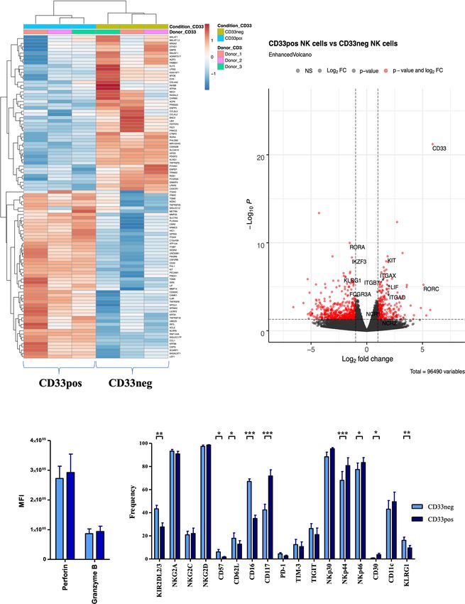
FIGURE 3 | Different transcriptional and phenotypical characteristics of CD33− and CD33+ NK cells. (A) CD33− and CD33+ NK cells were sorted on day 21
following culture with NK MACS medium (n=3). RNA sequencing was performed on the Illumina platform. The heatmap illustrates the top 100 differentially
expressed genes between CD33− and CD33+ NK cells. (B) The volcano plot indicates the RNA sequencing data based on 55,394 genes by plotting the
logarithm of the fold change between CD33− and CD33+ NK cells on the x axis and the negative logarithm of the p value on the y axis. The dashed lines
indicate p values equaling 0.05. Red points represent the genes with highest statistical significance of fold change. Top genes of interest are labeled.
Stimulated NK cells with NK MACS medium (days 14–21) were analyzed for (C) intracellular perforin and granzyme B (n=5) or (D) for the cell-surface
expression of the indicated molecules (n=17) by flow cytometry. Expression was compared between CD33- and CD33+ NK cells. Statistical significance was
determined by paired t-test, *p < 0.05; **p < 0.01; ***p < 0.001.
between the two NK cell subsets. As shown in Figure 4, a a range of effector/target ratios (Figure 4D). Similar results were
significantly higher frequency of CD33+ cells produced IFNg obtained when NK cells were expanded with the CliniMACS
and TNFa in response to K562 target cells compared to the Prodigy system (Figure S5B). Finally, we assessed antibody-
CD33− subset (Figures 4A, B). Notably, CD33+ NK cells also dependent cellular cytotoxicity (ADCC) employing the NK cell-
exhibited a higher frequency of CD107+ cells, reflecting more resistant CD20+ target cell line Raji in combination with the
effective mobilization of cytotoxic granules to the cell surface therapeutic anti-CD20 reagent Rituximab, which is a human
(Figure 4C). However, when measuring direct cytotoxicity IgG1 mAb that binds with its Fc part to the CD16 receptor on
against K562 cells, both subsets showed comparable lysis over NK cells. In accordance with the significantly higher expression
Frontiers in Immunology | www.frontiersin.org 7 January 2022 | Volume 12 | Article 798087
Hejazi et al.                                                                                                                     Characterization of CD33+ NK Cells

                                    A                             B                          C

                                D

                            E                                 F                               G

  FIGURE 4 | CD33+ NK cells display strong functionality in both cytokine production and cytotoxicity. Stimulated NK cells with NK MACS medium (day 14) were
  incubated with K562 cells at an effector/target ratio of 1:1. Intracellular (A) IFN-g and (B) TNF-a production and (C) CD107a mobilization were evaluated by flow
  cytometry (n=13). (D) CFDA-SE stained K562 cells were incubated with CD33+ and CD33− NK cells in effector/target ratios of 10:1, 5:1, and 1:1 (n=10). For ADCC,
  NK cells stimulated in NK MACS medium (day 14) were incubated with/without Raji and Rituximab, and (E) CD107a expression and intracellular (F) IFN-g and (G)
  TNF-a production were measured by flow cytometry (n=8). Statistical significance was determined by paired t-test, *p < 0.05; **p < 0.01; ***p < 0.001.

of CD16, CD33− NK cells exhibited stronger mobilization of                            malignancies, initially with the antibody-drug conjugate
cytotoxic granules and higher cytokine responses to Raji                              Gemtuzumab Ozagamicin, followed by bispecific antibody
compared to CD33+ NK cells, with IFNg showing more                                    conjugates, antibody-cytokine conjugates, and lately CD33-
significant differences than TNFa (Figures 4E–G).                                      CAR T cells (9, 24–26). Unexpectedly, we found that CD33 is
                                                                                      upregulated in vitro on a significant subset of NK cells. Although
                                                                                      in vivo CD33 expression is restricted to CD56bright NK cells,
DISCUSSION                                                                            which are a subset of non-cytotoxic, rather immature NK cells,
                                                                                      this was not the case in vitro: CD33 could be efficiently induced
The expression of CD33 is largely restricted to the myeloid                           on purified CD56dim NK cells leading to frequencies of CD33+
lineage and recently gained much interest as a target for                             NK cells up and above 50% with NK MACS medium. The
immunotherapy of AML and other CD33-expressing                                        present work suggests that CD33 constitutes a marker to

Frontiers in Immunology | www.frontiersin.org                                     8                                      January 2022 | Volume 12 | Article 798087
Hejazi et al.                                                                                                     Characterization of CD33+ NK Cells

distinguish between functionally divergent NK cell subsets in              ITIM motifs, and it was previously shown that crosslinking of
vitro. Expression of CD33 defines a polyfunctional NK cell subset           CD33 leads to inhibition of effector functions (30). Notably,
combining cytotoxic and cytokine effector functions. On the                Siglecs were shown to bind not only in trans but also in cis to cell-
other hand, due to lower expression of CD16, CD33+ NK cells                bound ligands, which might lead to a constitutive inhibitory state
are less efficient in ADCC compared to CD33− NK cells, which in             (30). However, our study clearly shows that CD33+ NK cells have
turn produce significantly less cytokines. Interestingly, CD33              polyfunctional characteristics marked by strong cytokine
seems to demarcate two separate cell states, which are either              production and efficient killing. The association of ITIM-
positive or negative with very few NK cells expressing                     containing inhibitory receptors with gain of function is not
intermediate levels of CD33 (see also Figures 2, 3).                       without precedent in NK cells, since expression of ITIM-
Comparative transcriptional analysis by RNAseq revealed that               containing inhibitory KIR receptors mediate licensing of NK
the transcriptional programs of the two subsets are not simply             cells, an educational process associated with a strong gain of
mirroring CD56bright and CD56dim subsets but constitute quite              function (31). Similarly to CD33, inhibitory KIRs are providing a
independent cellular entities compatible with their unique                 constitutive inhibitory state since they bind to ubiquitous HLA
functional properties, making both subsets potentially                     class I molecules present on all healthy nucleated cells. Moreover,
interesting tools for cancer therapy. It remains to be                     both KIR and the Siglec family are not only structurally related
determined which of the differentially expressed TFs are                   but are also genetically linked in the extended leukocyte receptor
involved in orchestrating these transcriptional changes, e.g.,             complex (LRC) on chromosome 19q13.4 (32). At this time, it can
RORa and RORgT, which are so far described as master                       only be speculated whether these similarities including the
regulators of ILC2 and ILC3 development, respectively (21).                common signal transduction via SHP1 and SHP2 could be an
Furthermore, downregulation of the Ikaros family member                    indicator for a similar licensing-like function of CD33.
Aiolos in CD33+ NK cells is interesting in this context since it               Altogether, expression of CD33 delineates a novel
was previously reported to be involved in shaping of the final NK           transcriptional and functional dichotomy that arises during in
cell maturation program in an Aiolos-deficient mouse                        vitro expansion of NK cells. In terms of clinical translation,
model (27).                                                                analysis of CD33 expression might give simple guidance on the
    The upregulation of CD33 on NK cells in vitro is significant            composition of NK cell products during the in vitro expansion
and strong enough to mediate recognition and subsequent                    process for clinical use, with increased cytokine production and
fratricide by CD33-CAR NK cells. Whereas this principally                  mobilization of cytotoxic granula on CD33+ cells on the one
hampers their expansion for cell therapeutic purposes, our                 hand and superior CD16-mediated functions on CD33− NK cells
study suggests that the problem can be circumvented in several             on the other hand.
ways: firstly, about 10% of the Caucasoid population are
homozygous for an SNP (rs12459419) that leads to efficient
alternative splicing and skipping of the IgV domain of CD33,
which is the binding site for the CAR used in this study (19, 20).
                                                                           DATA AVAILABILITY STATEMENT
Due to the lack of the CD33 target site, those donors could be
selected for expansion of CD33-CAR NK cells without inhibition             The data presented in the study are deposited in NCBI Project ID:
by fratricide and stored for further clinical use such as allogeneic       PRJNA777044 (http://www.ncbi.nlm.nih.gov/bioproject/777044).
therapy of AML. Of note, due to the lack of GvH disease,
application of CAR NK cells in the allogeneic setting constitutes
a major advantage compared to CAR T cell therapies, which are
presently only applicable in the autologous setting (28).
Moreover, upregulation of CD33 was largely restricted to NK                ETHICS STATEMENT
MACS medium, whereas an alternative protocol for expansion of
                                                                           The studies involving human participants were reviewed and
NK cells based on K562 stimulator cells, which is already part of
                                                                           approved by Ethikkommission Düsseldorf, Medical Faculty
NK cell-based clinical protocols (29), did only lead to a transient
                                                                           (study number 2019-383). The patients/participants provided
wave of CD33 expression. Whether fading of the CD33 subset
                                                                           their written informed consent to participate in this study.
with stimulator cell-based protocols is due to downregulation of
CD33 or possibly outperformance by CD33− NK cells is currently
unknown. In any case, by adding NK MACS medium to
stimulator cell cultures, expression of CD33 is again induced,
suggesting a yet undefined component in the medium that                     AUTHOR CONTRIBUTIONS
supports expansion of the CD33+ subset.
    The biological role of CD33 expression on CD56bright NK cells          MH, CZ, MU, and NM conceived and planned the experiments.
is currently unclear. Generally, as a member of the Siglec family,         MH, CZ, KH, SO, and VB carried out the experiments. MH and
CD33 is able to recognize sialylated ligands, but so far, no specific       SBB performed calculations. MH, CZ, SB, and VB conducted
cell-bound glycoproteins or other glycosylated ligands were                data analysis and interpretation. MU supervised the project. MH
identified that are preferentially recognized by NK cells. In               and MU wrote the manuscript. MU, SB, WW, and PH edited the
functional terms, the CD33 receptor contains intracytoplasmic              manuscript. MN, SC, and RP provided data. SR, MQ, SH, HH,

Frontiers in Immunology | www.frontiersin.org                          9                                  January 2022 | Volume 12 | Article 798087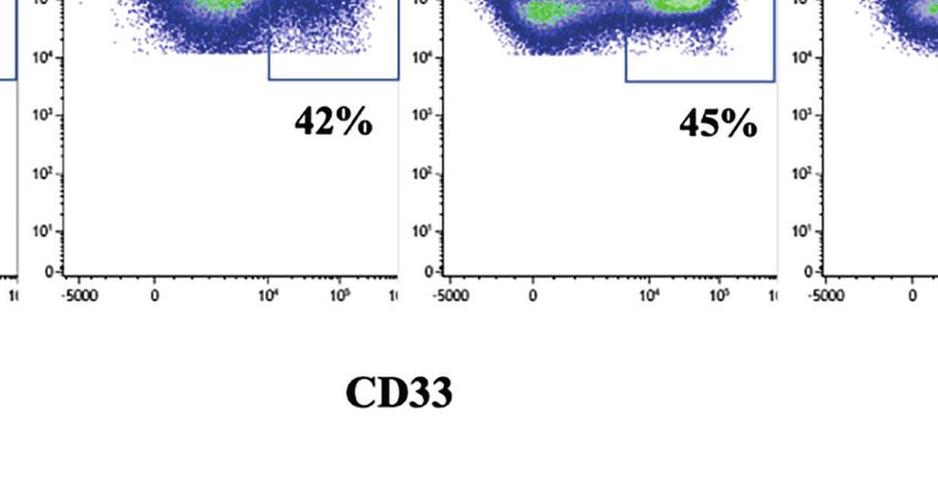
Hejazi et al.                                                                                                                           Characterization of CD33+ NK Cells

PO, HR, JO, PB, PH, FB, WW, and JF provided critical feedback                             ACKNOWLEDGMENTS
and commented on the manuscript. All authors contributed to
the article and approved the submitted version.                                           The authors thank Dario Campana for providing the K562-
                                                                                          mb15-41BBL cell line.

FUNDING                                                                                   SUPPLEMENTARY MATERIAL
This work was conducted in the framework of the iCAN33                                    The Supplementary Material for this article can be found online
project, funded by the European Regional Development Fund                                 at: https://www.frontiersin.org/articles/10.3389/fimmu.2021.
NRW (ERDF, German EFRE) 2014-2020.                                                        798087/full#supplementary-material

                                                                                          16. Kolde R. Pretty Heatmaps (2015). Available at: https://CRANR-projectorg/
REFERENCES                                                                                    package=pheatmap.
 1. Miller JS, Lanier LL. Natural Killer Cells in Cancer Immunotherapy. Annu              17. Glighe K, Rana S, Lewis M. EnhancedVolcano: Publication-Ready Volcano
    Rev Cancer Biol (2019) 3:77–103. doi: 10.1146/annurev-cancerbio-030518-                   Plots With Enhanced Colouring and Labeling (2019). Available at: https://
    055653                                                                                    githubcom/kevinblighe/EnhancedVolcano.
 2. Caligiuri MA. Human Natural Killer Cells. Blood (2008) 112(3):461–9. doi:             18. Hejazi M, Manser AR, Frobel J, Kundgen A, Zhao X, Schonberg K, et al.
    10.1182/blood-2007-09-077438                                                              Impaired Cytotoxicity Associated With Defective Natural Killer Cell
 3. Freud AG, Mundy-Bosse BL, Yu J, Caligiuri MA. The Broad Spectrum of                       Differentiation in Myelodysplastic Syndromes. Haematologica (2015) 100
    Human Natural Killer Cell Diversity. Immunity (2017) 47(5):820–33. doi:                   (5):643–52. doi: 10.3324/haematol.2014.118679
    10.1016/j.immuni.2017.10.008                                                          19. Lamba JK, Chauhan L, Shin M, Loken MR, Pollard JA, Wang YC, et al. CD33
 4. Romee R, Foley B, Lenvik T, Wang Y, Zhang B, Ankarlo D, et al. NK Cell                    Splicing Polymorphism Determines Gemtuzumab Ozogamicin Response in
    CD16 Surface Expression and Function is Regulated by a Disintegrin and                    De Novo Acute Myeloid Leukemia: Report From Randomized Phase III
    Metalloprotease-17 (ADAM17). Blood (2013) 121(18):3599–608. doi:                          Children's Oncology Group Trial Aaml0531. J Clin Oncol (2017) 35
    10.1182/blood-2012-04-425397                                                              (23):2674–82. doi: 10.1200/JCO.2016.71.2513
 5. Moustaki A, Argyropoulos KV, Baxevanis CN, Papamichail M, Perez SA.                   20. Gbadamosi MO, Shastri VM, Hylkema T, Papageorgiou I, Pardo L, Cogle CR,
    Effect of the Simultaneous Administration of Glucocorticoids and IL-15 on                 et al. Novel CD33 Antibodies Unravel Localization, Biology and Therapeutic
    Human NK Cell Phenotype, Proliferation and Function. Cancer Immunol                       Implications of CD33 Isoforms. Future Oncol (2021) 17(3):263–77. doi:
    Immunother (2011) 60(12):1683–95. doi: 10.1007/s00262-011-1067-6                          10.2217/fon-2020-0746
 6. Crocker PR, Paulson JC, Varki A. Siglecs and Their Roles in the Immune                21. Vivier E, Artis D, Colonna M, Diefenbach A, Di Santo JP, Eberl G, et al. Innate
    System. Nat Rev Immunol (2007) 7(4):255–66. doi: 10.1038/nri2056                          Lymphoid Cells: 10 Years on. Cell (2018) 174(5):1054–66. doi: 10.1016/
 7. Son M, Diamond B, Volpe BT, Aranow CB, Mackay MC, Santiago-Schwarz F.                     j.cell.2018.07.017
    Evidence for C1q-Mediated Crosslinking of CD33/LAIR-1 Inhibitory                      22. Michel T, Poli A, Cuapio A, Briquemont B, Iserentant G, Ollert M, et al.
    Immunoreceptors and Biological Control of CD33/LAIR-1 Expression. Sci                     Human CD56bright NK Cells: An Update. J Immunol (2016) 196(7):2923–31.
    Rep (2017) 7(1):270. doi: 10.1038/s41598-017-00290-w                                      doi: 10.4049/jimmunol.1502570
 8. Handgretinger R, Schafer HJ, Baur F, Frank D, Ottenlinger C, Buhring HJ,              23. Bjorkstrom NK, Riese P, Heuts F, Andersson S, Fauriat C, Ivarsson MA, et al.
    et al. Expression of an Early Myelopoietic Antigen (CD33) on a Subset of                  Expression Patterns of NKG2A, KIR, and CD57 Define a Process of CD56dim
    Human Umbilical Cord Blood-Derived Natural Killer Cells. Immunol Lett                     NK-Cell Differentiation Uncoupled From NK-Cell Education. Blood (2010)
    (1993) 37(2-3):223–8. doi: 10.1016/0165-2478(93)90034-Y                                   116(19):3853–64. doi: 10.1182/blood-2010-04-281675
 9. Lichtenegger FS, Krupka C, Haubner S, Kohnke T, Subklewe M. Recent                    24. Sarhan D, Brandt L, Felices M, Guldevall K, Lenvik T, Hinderlie P, et al.
    Developments in Immunotherapy of Acute Myeloid Leukemia. J Hematol                        161533 TriKE Stimulates NK-Cell Function to Overcome Myeloid-Derived
    Oncol (2017) 10(1):142. doi: 10.1186/s13045-017-0505-0                                    Suppressor Cells in MDS. Blood Adv (2018) 2(12):1459–69. doi: 10.1182/
10. Fujisaki H, Kakuda H, Shimasaki N, Imai C, Ma J, Lockey T, et al. Expansion               bloodadvances.2017012369
    of Highly Cytotoxic Human Natural Killer Cells for Cancer Cell Therapy.               25. Kenderian SS, Ruella M, Shestova O, Klichinsky M, Aikawa V, Morrissette JJ,
    Cancer Res (2009) 69(9):4010–7. doi: 10.1158/0008-5472.CAN-08-3712                        et al. CD33-Specific Chimeric Antigen Receptor T Cells Exhibit Potent
11. Oberoi P, Kamenjarin K, Ossa JFV, Uherek B, Bonig H, Wels WS. Directed                    Preclinical Activity Against Human Acute Myeloid Leukemia. Leukemia
    Differentiation of Mobilized Hematopoietic Stem and Progenitor Cells Into                 (2015) 29(8):1637–47. doi: 10.1038/leu.2015.52
    Functional NK Cells With Enhanced Antitumor Activity. Cells (2020) 9                  26. Reusing SB, Vallera DA, Manser AR, Vatrin T, Bhatia S, Felices M, et al.
    (4):811. doi: 10.3390/cells9040811                                                        CD16xCD33 Bispecific Killer Cell Engager (BiKE) as Potential
12. Girard-Gagnepain A, Amirache F, Costa C, Levy C, Frecha C, Fusil F, et al.                Immunotherapeutic in Pediatric Patients With AML and Biphenotypic
    Baboon Envelope Pseudotyped LVs Outperform VSV-G-LVs for Gene                             ALL. Cancer Immunol Immunother (2021) 70:3701–8. doi: 10.1007/s00262-
    Transfer Into Early-Cytokine-Stimulated and Resting HSCs. Blood (2014)                    021-03008-0
    124(8):1221–31. doi: 10.1182/blood-2014-02-558163                                     27. Holmes ML, Huntington ND, Thong RP, Brady J, Hayakawa Y, Andoniou
13. Bari R, Granzin M, Tsang KS, Roy A, Krueger W, Orentas R, et al. A Distinct               CE, et al. Peripheral Natural Killer Cell Maturation Depends on the
    Subset of Highly Proliferative and Lentiviral Vector (LV)-Transducible NK                 Transcription Factor Aiolos. EMBO J (2014) 33(22):2721–34. doi: 10.15252/
    Cells Define a Readily Engineered Subset for Adoptive Cellular Therapy. Front              embj.201487900
    Immunol (2019) 10:2001. doi: 10.3389/fimmu.2019.02001                                  28. Basar R, Daher M, Rezvani K. Next-Generation Cell Therapies: The Emerging
14. Bennstein SB, Weinhold S, Manser AR, Scherenschlich N, Noll A, Raba K,                    Role of CAR-NK Cells. Blood Adv (2020) 4(22):5868–76. doi: 10.1182/
    et al. Umbilical Cord Blood-Derived ILC1-Like Cells Constitute a Novel                    bloodadvances.2020002547
    Precursor for Mature KIR(+)NKG2A(-) NK Cells. Elife (2020) 9:e55232. doi:             29. Liu E, Marin D, Banerjee P, Macapinlac HA, Thompson P, Basar R, et al. Use
    10.7554/eLife.55232                                                                       of CAR-Transduced Natural Killer Cells in CD19-Positive Lymphoid Tumors.
15. Bennstein SB, Scherenschlich N, Weinhold S, Manser AR, Noll A, Raba K,                    N Engl J Med (2020) 382(6):545–53. doi: 10.1056/NEJMoa1910607
    et al. Transcriptional and Functional Characterization of Neonatal Circulating        30. Hernandez-Caselles T, Miguel RC, Ruiz-Alcaraz AJ, Garcia-Penarrubia P.
    ILCs. Stem Cells Transl Med (2021) 10:867–82. doi: 10.1002/sctm.20-0300                   CD33 (Siglec-3) Inhibitory Function: Role in the NKG2D/DAP10

Frontiers in Immunology | www.frontiersin.org                                        10                                        January 2022 | Volume 12 | Article 798087
Hejazi et al. Characterization of CD33+ NK Cells
Activating Pathway. J Immunol Res (2019) 2019:6032141. doi: 10.1155/2019/ Publisher’s Note: All claims expressed in this article are solely those of the authors
6032141 and do not necessarily represent those of their affiliated organizations, or those of
31. Manser AR, Weinhold S, Uhrberg M. Human KIR Repertoires: Shaped by the publisher, the editors and the reviewers. Any product that may be evaluated in
Genetic Diversity and Evolution. Immunol Rev (2015) 267(1):178–96. doi: this article, or claim that may be made by its manufacturer, is not guaranteed or
10.1111/imr.12316 endorsed by the publisher.
32. Barrow AD, Trowsdale J. The Extended Human Leukocyte Receptor
Complex: Diverse Ways of Modulating Immune Responses. Immunol Rev Copyright © 2022 Hejazi, Zhang, Bennstein, Balz, Reusing, Quadflieg, Hoerster,
(2008) 224:98–123. doi: 10.1111/j.1600-065X.2008.00653.x Heinrichs, Hanenberg, Oberbeck, Nitsche, Cramer, Pfeifer, Oberoi, Rühl, Oldenburg,
Brossart, Horn, Babor, Wels, Fischer, Möker and Uhrberg. This is an open-access
article distributed under the terms of the Creative Commons Attribution License
Conflict of Interest: CZ, MQ, MN, SC, RP, and NM are employees of Miltenyi Biotec.
(CC BY). The use, distribution or reproduction in other forums is permitted, provided
The remaining authors declare that the research was conducted in the absence of the original author(s) and the copyright owner(s) are credited and that the original
any commercial or financial relationships that could be construed as a potential publication in this journal is cited, in accordance with accepted academic practice. No
conflict of interest. use, distribution or reproduction is permitted which does not comply with these terms.
Frontiers in Immunology | www.frontiersin.org 11 January 2022 | Volume 12 | Article 798087You can also read