Superior efficacy of co-targeting GFI1/KDM1A and BRD4 against AML and post-MPN secondary AML cells
←
→
Page content transcription
If your browser does not render page correctly, please read the page content below
Fiskus et al. Blood Cancer Journal (2021)11:98
                                    https://doi.org/10.1038/s41408-021-00487-3                                                                                                          Blood Cancer Journal

                                     ARTICLE                                                                                                                                                    Open Access

                                    Superior efficacy of co-targeting GFI1/KDM1A and
                                    BRD4 against AML and post-MPN secondary AML
                                    cells
                                    Warren Fiskus 1, Christopher P. Mill1, Behnam Nabet 2, Dimuthu Perera3, Christine Birdwell1, Taghi Manshouri1,
                                    Bernardo Lara1, Tapan M. Kadia 1, Courtney DiNardo 1, Koichi Takahashi 1, Naval Daver 1, Prithviraj Bose 1,
                                    Lucia Masarova1, Naveen Pemmaraju 1, Steven Kornblau 1, Gautam Borthakur 1, Guillermo Montalban-Bravo1,
                                    Guillermo Garcia Manero 1, Sunil Sharma4, Matthew Stubbs5, Xiaoping Su1, Michael R. Green 1, Cristian Coarfa 3,
                                    Srdan Verstovsek1, Joseph D. Khoury 1, Christopher R. Vakoc6 and Kapil N. Bhalla 1

                                      Abstract
                                      There is an unmet need to overcome nongenetic therapy-resistance to improve outcomes in AML, especially post-
                                      myeloproliferative neoplasm (MPN) secondary (s) AML. Studies presented describe effects of genetic knockout,
                                      degradation or small molecule targeted-inhibition of GFI1/LSD1 on active enhancers, altering gene-expressions and
                                      inducing differentiation and lethality in AML and (MPN) sAML cells. A protein domain-focused CRISPR screen in LSD1
                                      (KDM1A) inhibitor (i) treated AML cells, identified BRD4, MOZ, HDAC3 and DOT1L among the codependencies. Our
                                      findings demonstrate that co-targeting LSD1 and one of these co-dependencies exerted synergistic in vitro lethality in
                  1234567890():,;
1234567890():,;
                  1234567890():,;
1234567890():,;

                                      AML and post-MPN sAML cells. Co-treatment with LSD1i and the JAKi ruxolitinib was also synergistically lethal against
                                      post-MPN sAML cells. LSD1i pre-treatment induced GFI1, PU.1 and CEBPα but depleted c-Myc, overcoming nongenetic
                                      resistance to ruxolitinib, or to BETi in post-MPN sAML cells. Co-treatment with LSD1i and BETi or ruxolitinib exerted
                                      superior in vivo efficacy against post-MPN sAML cells. These findings highlight LSD1i-based combinations that merit
                                      testing for clinical efficacy, especially to overcome nongenetic therapy-resistance in AML and post-MPN sAML.

                                    Introduction                                                                                        Es of myeloid lineage transcription factors (TFs) and of
                                      During complete remission in AML, AML stem/pro-                                                   their target genes in AML LSCs are characterized by his-
                                    genitor cells (LSCs) in the measurable residual disease                                             tone H3 lysine (K) 27 acetyl (H3K27Ac) and H3K4 mono-
                                    (MRD) resist differentiation, retain leukemia-initiating                                            methylation (H3K4Me1) marks, as well as by high occu-
                                    potential and mediate relapse of AML1–3. This pheno-                                                pancy with BRD4 and histone acetyl transferase CBP/
                                    type of LSCs is mainly orchestrated by dysregulated super-                                          p3007–9. Among the key LSC-specific TFs that regulate
                                    enhancers (SEs)/enhancers (Es) and the ensuing dysregu-                                             cell growth, differentiation and survival of AML stem cell
                                    lated transcriptome/proteome, which directly results from                                           differentiation are RUNX1, PU.1, CEBPα, c-Myb, and c-
                                    genetic alterations and/or perturbed levels and activity of                                         Myc10–13. LSD1 (lysine specific demethylase 1; KDM1A) is
                                    epigenetic and transcriptional regulators in AML4–6. SEs/                                           a FAD-dependent amine-oxidase family member and a
                                                                                                                                        component of the co-repressor complexes involving
                                                                                                                                        HDAC1/2 and CoREST (RCOR1) or NuRD (MTA2)14.
                                    Correspondence: Kapil N. Bhalla (kbhalla@mdanderson.org)                                            LSD1 contains a conserved N-terminal SWIRM domain
                                    1
                                    2
                                     The University of Texas M.D. Anderson Cancer Center, Houston, TX, USA                              (100 AA) and C-terminal amine oxidase-like (AOL) cata-
                                     Department of Cancer Biology, Dana-Farber Cancer Institute and Department
                                                                                                                                        lytic domain14,15. The AOL domain binds to FAD and the
                                    of Biological Chemistry and Molecular Pharmacology, Harvard Medical School,
                                    Boston, MA, USA                                                                                     methylated H3K4 substrate, causing demethylation of
                                    Full list of author information is available at the end of the article

                                    © The Author(s) 2021
                                                        Open Access This article is licensed under a Creative Commons Attribution 4.0 International License, which permits use, sharing, adaptation, distribution and reproduction
                                                        in any medium or format, as long as you give appropriate credit to the original author(s) and the source, provide a link to the Creative Commons license, and indicate if
                                    changes were made. The images or other third party material in this article are included in the article’s Creative Commons license, unless indicated otherwise in a credit line to the material. If
                                    material is not included in the article’s Creative Commons license and your intended use is not permitted by statutory regulation or exceeds the permitted use, you will need to obtain
                                    permission directly from the copyright holder. To view a copy of this license, visit http://creativecommons.org/licenses/by/4.0/.

                                    Blood Cancer Journal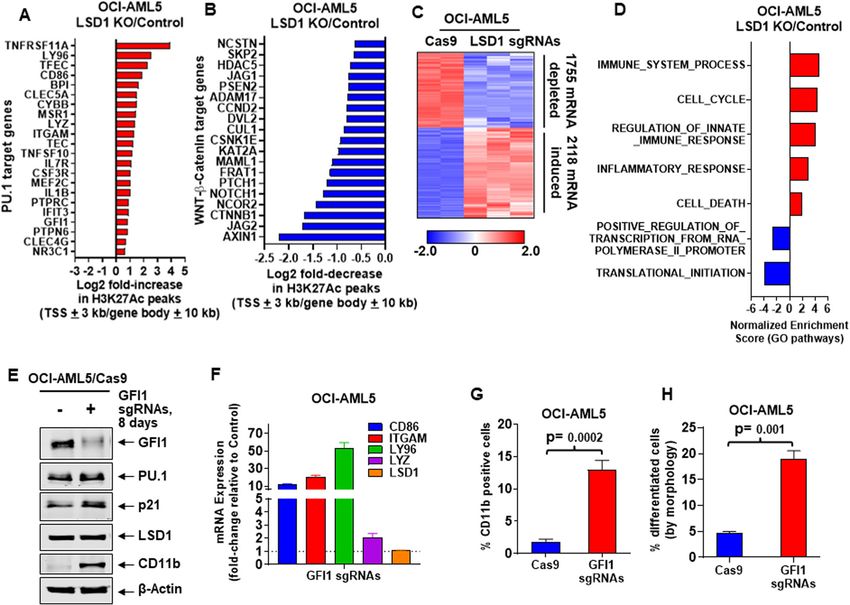
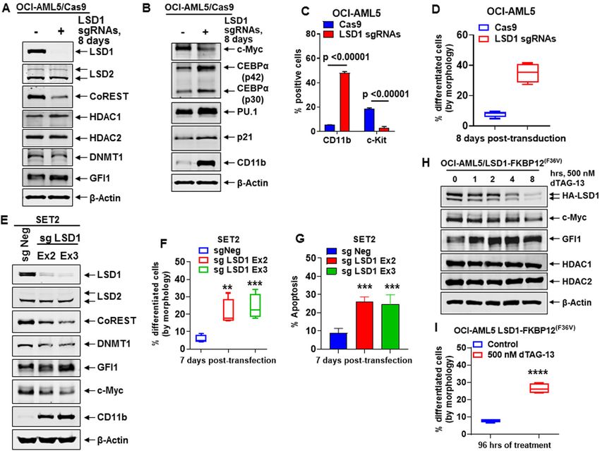
Fiskus et al. Blood Cancer Journal (2021)11:98                                                                 Page 2 of 16

mono and dimethyl histone H3 lysine 4 (H3K4Me1 and              Myc levels, but induced expressions of GFI1, PU.1, p21,
H3K4Me2)15,16. Two antiparallel α-helices divide and            and CD11b, inhibiting in vitro growth, inducing differ-
project away from the AOL domain as the Tower domain,           entiation/cell lethality, as well as extending survival in
which provides the binding interface with CoREST                immune-depleted mice engrafted with AML or sAML.
(RCOR1 and HDAC1/2) and allosterically regulates the            Utilizing a domain-focused CRISPR-Cas9 sgRNA screen
catalytic activity and stability of LSD114–16. In addition to   followed by LSD1i treatment, present studies also
H3K4, LSD1 also demethylates TP53, E2F1, and                    demonstrate co-dependencies, including BRD4, in AML
DNMT117,18. LSD1 interacts with GFI1/1B, a zinc finger           cells. Whereas co-treatment with an LSD1i and BETi
transcriptional repressor and master regulator of normal        (OTX015) synergistically induced lethality in PD AML
and malignant lineage development and differentiation in        including sAML BPCs, pre-treatment with LSD1i sensi-
hematopoiesis18–21. GFI1/1B contains an N-terminal, 20          tized JAKi-resistant post-MPN sAML cells to ruxolitinib
AA SNAG domain and six C-terminal Zn fingers22. LSD1             and BETi P/R cells to BETi-induced apoptosis33. Co-
binds to the methylated lysine in the 8KSKK11 motif in the      treatment with LSD1i and BETi (OTX015) also inhibited
SNAG (Snail/GFI1) domain, and thereby helps recruit             AML cell burden and improved median survival of NSG
RCOR1-HDAC1/2 co-repressor complex to mediate                   mice engrafted with AML or post-MPN sAML cells.
transcriptional repression and differentiation block due to
GFI1 activity in AML stem/progenitor cells19–22. GFI1/1B        Results
can be co-immunoprecipitated with LSD1 and CoREST               Biologic effects of CRISPR-Cas9-mediated LSD1 knockout
and exhibits overlapping DNA-binding sites at enhancers         or dTAG-13-mediated degradation of LSD1 in AML cells
(by ChIP-Seq analyses) of key myeloid/monocyte                    We first determined effects of CRISPR-Cas9-mediated
differentiation–regulatory genes21,23. LSD1 inhibitors          knockout (KO) of LSD1 on the CoREST complex and on
(LSD1i) disrupt GFI1/1B interaction with LSD1-CoREST,           GFI1 and its targets in AML OCI-AML5 and post-MPN
inducing differentiation of AML blast progenitor                sAML SET-2 cells. Fig. 1A demonstrates that 8-days fol-
cells17,21,23–25. Importantly, CRISPR-suppressor scanning       lowing transduction of two gRNAs targeting exon 2 and 3
revealed that enzymatic activity of LSD1 was not required       of LSD1 into OCI-AML5 cells, stably transduced with
for blocked AML differentiation and survival26. GFI1/1B         Cas9, LSD1 was profoundly depleted, associated with
interaction with LSD1 and CoREST also causes deme-              reduction in protein levels of CoREST and slightly of
thylation of K372 on TP53, thereby inactivating TP5317,21.      DNMT1, increased GFI1, and unaltered HDAC1/2 and
Notably, LSD1 is over-expressed in the stem/progenitor          LSD2 levels. LSD1-KO also attenuated c-Myc levels, while
versus differentiated sub-types of AML17,21, and GFI1           increasing protein levels of PU.1, CEBPα, p21 and CD11b
expression is a documented prognostic factor in MDS/            (Fig. 1B). Alterations in the levels of these proteins were
AML27. LSD1i treatment also increased chromatin acces-          associated with decline in % of cells in cell cycle S-phase
sibility and binding of SPI1 and CEBPα at their target Es/      and increase in % of G1-phase cells, accompanied by
promoters28. Knockdown by shRNA or treatment with               augmentation in % of cells expressing CD11b that also
either the irreversible tranylcypromine (TCP)-derivative        displayed morphologic features of differentiation (%
LSD1i or the reversible LSD1i SP2509 disrupted LSD1-            myelocytes and metamyelocytes or bands by morphologic
binding to CoREST and GFI1/1B, induce differentiation           features of hematoxylin & eosin-stained cytospun cells),
markers (CD86 and CD11b) and morphologic differ-                while reducing % of cells expressing c-KIT (Figs. 1C, D
entiation, repress colony growth, as well as sensitize AML      and S1A). LSD1-KO via CRISPR-Cas9 in post-MPN
blast progenitor cells (BPCs) to all-trans retinoic acid        sAML SET-2 cells, was also accompanied by decline in
(ATRA)23,25,29–32. Co-treatment with LSD1i and cytar-           the protein levels of LSD1, CoREST, c-Myc, and DNMT1,
abine, DNA hypomethylating agents, or inhibitor of              but increased protein levels of GF11, PU.1 and CEBPα
HDACs, FLT3, DOT1L or BCL2, was shown to exert                  (Figs. 1E and S1B). LSD1-KO also inhibited in vitro
synergistic lethality in AML expressing MLL fusion pro-         growth and increased % of morphologically-differentiated
tein25,30. However, these studies did not interrogate the       (% myelocytes and metamyelocytes or bands by mor-
activity of LSD1i and LSD1i-based combinations in post-         phologic features of hematoxylin & eosin-stained cytos-
MPN sAML blast progenitor cells (BPCs). In present              pun cells) SET-2 cells with increased CD11b but reduced
studies, utilizing for the first time CRISPR-Cas9, or LSD1-      c-KIT expression (Figs. 1F, S1C, D), along with significant
FKBP12F36V and dTAG-13, we demonstrate that knockout            increase in % apoptotic cells (Fig. 1G). Following CRISPR-
(KO) or degradation of LSD1 inhibits growth and induces         Cas9-mediated LSD1-KO, perturbed protein expressions
differentiation of AML BPCs with or without expression of       could be documented only 5–8 days later. Therefore, we
MLL fusion oncoproteins, and of post-MPN sAML BPCs.             employed the dTAG system34 to degrade LSD1-
While disrupting the binding of LSD1 to CoREST and              FKBP12(F36V) in OCI-AML5 cells following knockout of
GFI1, treatment with irreversible LSD1i also attenuated c-      the endogenous LSD1 to assess within hours loss of LSD1

Blood Cancer Journal
Fiskus et al. Blood Cancer Journal (2021)11:98                                                                                          Page 3 of 16

  Fig. 1 Knockout of LSD1 by CRISPR/Cas9 depletes c-Myc, and derepresses myeloid differentiation gene CD11b in AML and sAML cells. A, B
  Representative immunoblot analysis of OCI-AML5 Cas9- expressing cells transduced with two lentiviral sgRNAs against LSD1 and incubated for
  8 days. C Expression of c-Kit and CD11b (assessed by flow cytometry) in OCI-AML5 Cas9 and LSD1 knockout cells 8 days post-transduction. Mean of
  two independent experiments performed in duplicate + S.D. Significance determined by a two-tailed, unpaired t-test. D Morphologic differentiation
  (% myelocytes and metamyelocytes or bands by morphologic features of hematoxylin & eosin-stained cytospun cells) of OCI-AML5 Cas9 and LSD1
  knockout cells 8 days post-transduction. Mean of two independent experiments performed in duplicate + S.D. E Representative immunoblot analysis
  of SET2 cells transfected with two sgRNAs against LSD1 and incubated for 7 days. F, G Induction of morphologic differentiation (% myelocytes and
  metamyelocytes or bands by morphologic features of hematoxylin & eosin-stained cytospun cells) and apoptosis in SET-2 LSD1 knockout cells 7 days
  post-transfection. Mean of two independent experiments performed in duplicate + S.D. **p < 0.01, ***p < 0.005 compared to sgNeg-transfected SET-
  2 cells (determined by a two-tailed, unpaired t-test). H Immunoblot analysis of OCI-AML5/LSD1-FKBP12(F36V) cells treated with 500 nM of dTAG-13 for
  the indicated times. I OCI-AML5/LSD1-FKBP12(F36V) cells were treated with 500 nM of dTAG-13 for 96 h. At the end of treatment, differentiation was
  assessed by cell morphology, as described above. Mean of three experiments + S.D. ****p < 0.001 compared to untreated control cells (determined
  by a two-tailed, unpaired t-test).

and the downstream biological consequences. As shown                         Effect of LSD1 knockout on enhancer activity and
in Figs. 1H, S1E–H, 4–24 h after adding dTAG-13, pro-                        transcriptome in AML cells
tein levels of LSD1, CoREST, and c-Myc declined, while                         Utilizing ChIP-Seq analysis, we next determined the
GFI1, PU.1, RUNX1, p21, p27, and CD11b levels were                           H3K27Ac signal-density at specific loci in LSD1-KO com-
induced in OCI-AML5 cells. dTAG-13 treatment did not                         pared to the control OCI-AML5 cells. Increases in peak-
affect LSD2, HOXA9, and Meis1 levels in OCI-AML5                             densities were seen in PU.1 targets, while the peak-densities
cells (Figs. S1G, H). Four days after dTAG-13 addition, an                   decreased on WNT-β-catenin targets (Figs. 2A, B).
increase in the % of cells expressing CD11b and displaying                   H3K27Ac signal-density also decreased at the MYC and
morphologic features of differentiation was observed                         CDK6 loci, whereas ITGAM, LY96, and LYZ gene-loci
(Figs. 1I and S1I).                                                          demonstrated increased H3K27Ac occupancy (Fig. S2A).

Blood Cancer Journal
Fiskus et al. Blood Cancer Journal (2021)11:98                                                                                         Page 4 of 16

  Fig. 2 Knockout of LSD1 alters chromatin accessibility, augments H3K27Ac occupancy on PU.1 target and WNT-β-Catenin target genes,
  and knockout of GFI1 induces myeloid differentiation genes, LY96 and ITGAM (CD11b) and morphologic differentiation of AML cells. A
  Log2 fold-change in H3K27Ac ChIP-Seq peaks (TSS + 3 kb or gene body + 10 kb) for PU.1 target genes (from the MSigDB database) in LSD1 knockout
  compared to Cas9-only-expressing OCI-AML5 cells. B Log2 fold-change in H3K27Ac ChIP-Seq peaks (TSS + 3 kb or gene body + 10 kb) in WNT-
  β-Catenin target genes (from HALLMARK datasets in the MSigDB database) in LSD1 knockout compared to Cas9-only-expressing OCI-AML5 cells. C
  RNA-Seq analysis was performed on OCI-AML5 LSD1 knockout (biologic triplicates) and Cas9-only-expressing (biologic duplicates) cells 8 days post-
  transduction. Heat map shows the number of mRNAs depleted or induced > 1.25-fold and p-value < 0.05 in the LSD1 knockout versus Cas9-only-
  expressing cells. D Gene set enrichment analysis was performed with RNA expression changes in OCI-AML5 LSD1-KO cells compared to GO
  pathways. The q-values are < 0.1 in all comparisons. E Representative immunoblot analysis of OCI-AML5 Cas9-expressing cells transduced with two
  lentiviral sgRNAs against GFI1 and incubated for 8 days. F Relative mRNA expression analysis of OCI-AML5 GFI1 knockout cells compared to Cas9-
  only-expressing cells. GAPDH expression was utilized as the normalization control. G Expression of CD11b (assessed by flow cytometry) in OCI-AML5-
  Cas9 and GFI1 knockout cells 8 days post-transduction. Mean of two independent experiments performed in duplicate + S.D. Significance
  determined by a two-tailed, unpaired t-test. H Morphologic differentiation (% myelocytes and metamyelocytes or bands by morphologic features of
  hematoxylin & eosin-stained cytospun cells) of OCI-AML5 GFI1 knockout cells compared to Cas9-only-expressing cells 8 days post-transduction. Mean
  of two independent experiments performed in duplicate + S.D. Significance determined by a two-tailed, unpaired t-test in GraphPad V8.

Consistent with this, H3K27Ac peaks also declined on the                    mRNAs, according to GO and HALLMARK pathways,
chromatin of many c-Myc target genes (Figs. S2B, C). ChIP-                  demonstrated significant positive normalized enrichment
Seq analysis following LSD1-KO also increased H3K27Ac                       scores (NES) for gene-sets, including those of innate
and BRD4 peaks at the enhancer-promoter regions of GFI1                     immune and inflammatory responses, apoptosis pathways,
gene (Fig. S2D), accompanied by increased GFI1 levels in                    but negative NES for c-Myc, WNT-β-catenin and
OCI-AML5 cells (Fig. 1A). RNA-Seq analysis following                        translation–initiation pathway genes (Figs. 2D and S2G).
LSD1-KO revealed the heat map of the up- or down-                           Utilizing two custom-designed, nonoverlapping primer sets
regulated mRNAs (Fig. 2C), which included perturbations                     and qPCR analysis, we also determined enhancer RNA
of PU.1 and CEBPα target-gene expressions (Fig. S2E, F)33.                  (eRNA) abundance within the known enhancer regions in
Gene set enrichment analyses (GSEA) of the perturbed                        the MYC SE33. LSD1-KO in OCI-AML5 cells depleted

Blood Cancer Journal
Fiskus et al. Blood Cancer Journal (2021)11:98                                                                    Page 5 of 16

eRNA expression from enhancers 3, 4, and 5 but not from         GFI1 shRNA: 30.4%) and morphologic features of myeloid
enhancer 2 of MYC (Fig. S2H).                                   differentiation (LSD1 shRNA: 71.7% and GFI1 shRNA:
                                                                33.9%). This suggests that the amount of differentiation
Biologic outcome following depletion of GFI1 versus LSD1        induced by knockdown or knockout of LSD1 or GFI1 in
in AML cells                                                    AML cells best correlated with increased expression of PU.1
   Next, we compared effects of LSD1-KO with those of           and p21 but depletion of c-Myc. Notably, 96 h post treat-
GFI1 knockout in OCI-AML5 cells. Utilizing two gRNAs            ment with shRNA to LSD1 or GFI1, a small (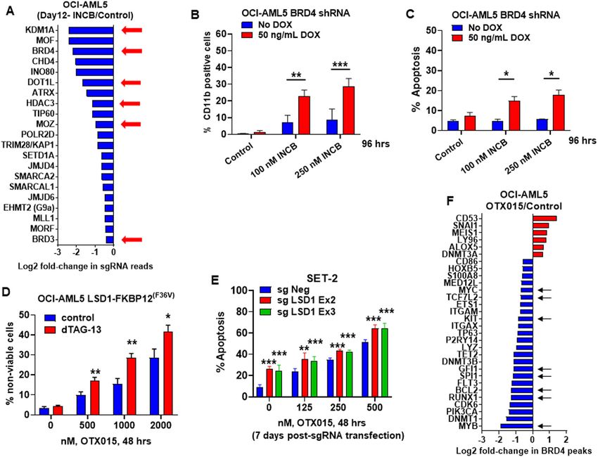
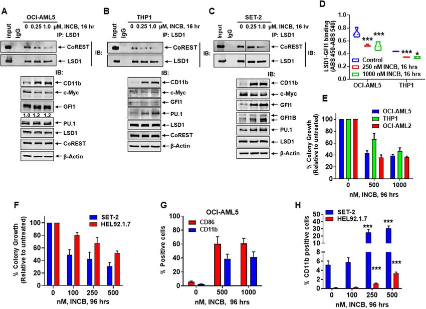
Fiskus et al. Blood Cancer Journal (2021)11:98                                                                                          Page 6 of 16

  Fig. 3 Treatment with LSD1 inhibitor (LSD1i) disrupts binding of LSD1 to CoREST and GFI1, attenuates colony growth, and induces
  differentiation in AML cells. A–C Immunoprecipitation and immunoblot analysis of OCI-AML5, THP1, and SET-2 cells treated with INCB059872 for
  16 h. D Reduced binding of LSD1 and GFI1 (determined by sandwich ELISA assay) following 16 h of treatment with INCB059872. Mean of two
  independent experiments performed in duplicate + S.D. *p < 0.05, ***p < 0.005 (determined by a two-tailed, unpaired t-test). E, F OCI-AML5, OCI-
  AML2, THP1, SET-2, and HEL92.1.7 cells were treated with the indicated concentrations of INCB059872 for 96 h. Then, cells were plated in Methocult
  media and incubated at 37 °C. Colony growth was assessed after 7–10 days. Mean of two independent experiments + S.D. G, H The % of CD86 and/
  or CD11b-positive OCI-AML5, SET2 and HEL92.1.7 cells was determined by flow cytometry following treatment with INCB059872 for 96 h. Mean of
  three experiments + S.E.M. ***p < 0.005 in INCB059872-treated cells compared to untreated cells (determined by a two-tailed, unpaired t-test in
  GraphPad V8).

LSD1 inhibitor-mediated early alterations in epigenome/                      target genes (Fig. S8C–F). Similar gain of ATAC-seq
transcriptome in AML and post-MPN sAML cells                                 peaks were also noted in INCB-treated SET-2 cells (Fig.
  Since effects of LSD1-KD (utilizing shRNA) or LSD1-                        S8G, H). Although H3K27Ac ChIP-seq analysis showed
KO (via CRISPR-Cas9) on epigenome/transcriptome can                          only a slight increase in H3K27Ac signal-tag density at
only be evaluated several days after gene-editing, we next                   active enhancers, ROSE plot highlighted active SEs of
determined whether these effects also occur soon after a                     RUNX1, GFI1, BCL2, PU.1, IRF8 and SMYD3 in INCB-
16-h exposure of AML and post-MPN sAML cells to                              treated compared to untreated OCI-AML5 cells, accom-
INCB. Following INCB treatment, ATAC-Seq analysis                            panied by increased H3K27Ac occupancy at the chro-
demonstrated large numbers of lost and gained peaks,                         matin of GFI1 and PU.1 target genes (Fig. S8I–K).
including those gained in the bivalent poised enhancers,                     Notably, INCB treatment also increased BRD4 occupancy
enhancers, active TSS (transcription start sites) and                        at +/− 3 kb of TSSs, and especially at the GFI1 and PU.1-
polycomb-repressed chromatin of OCI-AML5 cells (Fig.                         target genes (Fig. S8L–N). RNA-Seq analysis showed that
S8A, B)39,40. TF motifs in the open chromatin included                       INCB treatment up- and downregulated large numbers of
those of CTCF, FOSL1, PU.1, RUNX1, IRF8 and c-Myc,                           mRNA expressions, with positive NESs for gene-sets
and the gained peaks included those in GFI1 and PU.1-                        including those for interferon α, inflammatory response

Blood Cancer Journal
Fiskus et al. Blood Cancer Journal (2021)11:98                                                                 Page 7 of 16

and E2F-target genes, and negative NES for c-Myc-targets       compared to control, OTX015 treatment caused down-
and oxidative phosphorylation gene-sets (Fig. S9A, B).         regulation of BRD4 occupancy at enhancers/promoters of
INCB treatment also positively enriched for GFI1-targets,      genes, including MYC, CDK6, BCL2, MYB, PU.1, RUNX1,
while negatively enriching for c-Myc targets (Fig. S9C, D).    TCF7L2, and GFI1 (Fig. 4F). Consistent with this, OTX015
Similar positive or negative NESs for these gene sets were     treatment depleted mRNA expressions of these genes, but
also noted for INCB-treated SET-2 cells (Fig. S9E).            induced mRNA levels of p21, ITGAM, LYZ, and LY96 (Fig.
Compared to untreated, RNA-Seq analyses of INCB-               S11A, and data not shown). Exposure to OTX015 also
treated OCI-AML5 and SET-2 cells also demonstrated             repressed LSD1, GFI1, c-Myc, c-Myb, and PU.1, while
increased mRNA expressions of GFI1, PU.1, and CEBPα            inducing p21 and p27 protein levels in OCI-AML5 and
target-genes (Fig. S9F–K). The target genes induced were       SET-2 cells (Fig. S11B, D, E). OTX015 represses c-Myc and
different in OCI-AML5 versus SET2 cells, most likely           as a result represses LSD1––a target of c-Myc42. Co-
because of their disparate cell biology due to their de novo   treatment with INCB enhanced effects of OTX015 on
AML versus post-MPN sAML derivation. Notably,                  specific mRNAs, decreasing CDK6 and MYC but increasing
although LSD1i treatment induced GFI1 expression,              LY96, LYZ, ITGAM, and p21, as well as augmented effects
because it disrupted binding of CoREST-LSD1 to GFI1,           on protein-expressions, reducing c-Myc, c-Myb, GFI1, and
LSD1i treatment prevented GFI1-mediated repression,            PU.1 but increasing levels of p21 and p27 in OCI-AML5
causing derepression of GFI1 target genes associated with      and SET-2 cells (Fig. S11A, D, E).
AML cell differentiation.
                                                               Co-inhibition of GFI1/LSD1 and BRD4, HDAC3, MOZ, or
Targetable dependencies detected by protein domain-            DOT1L induces synergistic lethality in AML and post-MPN
focused CRISPR-Cas9 screen in LSD1i-treated versus             sAML cells
untreated AML cells                                               We next determined in vitro lethal effects of co-
  Utilizing a previously validated, GFP-tagged gRNA library    inhibition of LSD1 and the codependencies discovered by
(1390 gRNAs and ~8 gRNA per gene) (Table S1) targeting         the CRISPR-Cas9 screen. Co-treatment with INCB and
chromatin regulators transduced into OCI-AML5 cells that       OTX015 or ABBV-075, a potent BETi43, synergistically
stably expressed Cas9, we next conducted a protein             induced apoptosis in the indicated AML and sAML cell
domain-focused CRISPR-Cas9 screen to nominate potential        lines with diverse genetic alterations documented by NGS
drug targets in INCB-treated and untreated OCI-AML5            (Figs. S11C, S12A, B). Differentiation due to the combi-
cells41. Eight days after transduction of gRNAs, cells were    nation was not observed (data not shown). Similarly,
treated or untreated with INCB (250 nM) for 4 days. NGS        ORY1001 and OTX015 treatment also exerted synergistic
was performed to determine negative selection of the           lethality (Fig. S11F). Whereas modestly effective alone, co-
gRNAs identified on day 12 in INCB-treated versus               treatment with the HDAC3 inhibitor RGFP96644
untreated OCI-AML5 cells, compared to the gRNA profile          (≥2.5 µM) and LSD1 depletion by dTAG-13 induced more
sequenced on day-2 following transduction of gRNAs41. Fig.     differentiation, not apoptosis, than either drug-treatment
S10 demonstrates the decline in % of GFP-positive cells        alone (Fig. S13A and data not shown). Combined treat-
noted on day-12 compared to day-2, with the fold               ment with INCB and RGFP966 induced synergistic leth-
sequencing coverage of guide RNAs maintained above             ality in sAML SET2 while enhancing differentiation in
~400X (data not shown). Fig. 4A demonstrates the loss in       OCI-AML5 cells (Bliss synergy scores >5) (Fig. S13B–E).
gRNA reads targeting specific genes, including LSD1,            Treatment with the MOZ inhibitor WM1119
BRD4, DOT1L, HDAC3, and MOZ. Based on this, we next            (100–1000 nM) alone was also only modestly effective
determined the lethal effect of doxycycline-inducible BRD4     (Fig. S14A)45, but co-treatment with dTAG-13 induced
shRNA-mediated BRD4 KD in AML cells that were co-              significantly more differentiation (Fig. S14A) (p < 0.05).
treated with INCB. Figure 4B, C demonstrate that BRD4          Co-treatment with INCB and WM1119 exerted similar
KD significantly increased INCB-induced CD11b and               effects as INCB and RGFP966 in the AML and sAML cells
apoptosis of OCI-AML5 cells. As previously reported,           (Bliss synergy score >5.0 or CI values
Fiskus et al. Blood Cancer Journal (2021)11:98 Page 8 of 16 Fig. 4 Synthetic lethal activity of treatment with INCB059872 and a CRISPR-Cas9-mediated domain-focused epigenetically targeted sgRNA library in AML cells. A OCI-AML5 Cas9 cells were transduced with a library of domain-specific sgRNAs against chromatin modifying proteins and incubated for 8 days. Then, 250 nM of INCB059872 was added and the cells were incubated for an additional 96 h. Live cells were harvested; genomic DNA was isolated and minimally amplified with primers flanking the sgRNA sequences. Sequencing libraries were generated and amplicon-seq was performed. The graph shows log2 fold-changes in selected sgRNAs which dropped out more due to treatment with INCB059872 than control cells at day 12 post-transduction. B OCI-AML5 cells with stable expression of DOX-inducible BRD4 shRNA were induced with DOX and treated with the indicated concentrations of INCB059872 for 96 h. Following this, the % of CD11b cells was determined by flow cytometry. Mean of two independent experiments + S.D. **p < 0.01, ***p < 0.005 as determined by a two-tailed, unpaired t-test. C OCI-AML5 cells with stable expression of DOX-inducible BRD4 shRNA were induced with DOX and then treated with the indicated concentrations of INCB059872 for 96 h. Following this, the % of annexin V- positive, apoptotic cells were determined by flow cytometry. Mean of two independent experiments + S.D. *p < 0.05 as determined by a two-tailed, unpaired t-test. D OCI-AML5 LSD1-FKBP12(F36V) cells were treated with the indicated concentrations of OTX015 with and without 500 nM of dTAG-13 for 48 h. Then, the % of To-Pro-3 iodide positive cells were determined by flow cytometry. Mean of three experiments + S. E. M. *p < 0.05, **p < 0.01 compared to cells with no dTAG-13 treatment (determined by a two-tailed, unpaired t-test). E SET-2 cells were transfected with sgNeg or LSD1 sgRNAs and incubated for 5 days. Then, cells were treated with the indicated concentrations of OTX015 for 48 h and the % of apoptotic cells was determined by flow cytometry. Mean of two independent experiments performed in duplicate + S.D. **p < 0.01, ***p < 0.005 relative to sgNeg- transfected cells (determined by a two-tailed, unpaired t-test). F Log2 fold-changes in BRD4 peaks (determined by DiffReps analysis of BRD4 ChIP-Seq data) on LSD1 target genes and selected AML-relevant genes in OCI-AML5 cells treated with 1000 nM of OTX015 for 8 h. Next, we evaluated the lethal activity of LSD1i and BETi inducing levels of GFI1, PU.1, ITGAM, (and LY96 in patient-derived (PD) AML blasts with genetic altera- mRNA) in PD AML blasts (Fig. 5B, C). In contrast, tions documented by NGS (Fig. S12C). We determined treatment with OTX015 did not induce mRNA or protein that co-treatment with INCB and OTX015 for 48 h levels encoded by these genes, but attenuated MYC induced synergistic loss of viability in 14 samples of AML expression in the PD AML blasts (Fig. 5B, C). Co- blasts (Fig. 5A). Exposure to INCB (250 nM for 16 h) treatment with INCB and OTX015 induced synergistic reduced mRNA and protein levels of c-Myc, while loss of viability in 22 samples of PD, post-MPN sAML Blood Cancer Journal
Fiskus et al. Blood Cancer Journal (2021)11:98                                                                                              Page 9 of 16

  Fig. 5 Co-treatment with LSD1i and BET inhibitor (BETi) or ruxolitinib exerts synergistic lethal activity in patient-derived AML and sAML
  cells. A PD CD34+ AML blast cells (n = 14) were treated with INCB059872 (100–1000 nM) and/or OTX015 (dose range 250–1000 nM) for 48 h. The %
  of To-Pro-3 iodide-positive, nonviable cells was determined by flow cytometry. Combination index values were calculated with CompuSyn.
  Combination index values
Fiskus et al. Blood Cancer Journal (2021)11:98                                                                Page 10 of 16

blasts (combination index values < 1.0) (Figs. 5D and           expressions GFI1, TCF7L2, JMJD6, MYC, ITGAM, LY96,
S12D). We also determined the effect of ruxolitinib in          BCL2, and BCL2L1 but increased p21 mRNA levels (Fig.
sAML SET-2 cells with CRISPR-Cas9 mediated KO of                S16E). Western analyses showed that pretreatment with
LSD1. As shown in Fig. 5E, ruxolitinib-induced apoptosis        INCB followed by OTX015 caused abrogation of GFI1,
was significantly augmented in SET-2 cells with LSD1-KO          TCF7L2, and PU.1 induced by INCB treatment alone,
(p < 0.005). Co-treatment with INCB and ruxolitinib also        with significant decline in c-Myc and JMJD6 but increase
synergistically induced lethality in HEL92.1.7 and SET-2,       in p27 levels (Fig. 6D). A significant increase in OTX015-
as well as in samples of PD post-MPN sAML blasts in             induced lethality was also observed following pretreat-
which genetic mutations were documented by NGS (CI              ment with INCB of PD AML and sAML blasts (Fig. 6E, F).
values < 1.0) (Figs. 5F and S12D). Notably, in previously       Similar to a recent report, we also determined the effects
described      nongenetic     ruxolitinib-persister/resistant   of INCB pretreatment on chromatin accessibility and
HEL92.1.7-RuxP and SET-2-RuxP cells, unlike in parental         potential enhancer reprogramming that could explain
SET-2 or HEL92.1.7 cells, pre-treatment with INCB for           sensitization to BETi49, by conducting ATAC-Seq and
48 h, markedly enhanced ruxolitinib-induced apoptosis in        RNA-Seq analyses in INCB-treated versus -untreated
HEL-RuxP and SET-2-RuxP cells (p < 0.001) (Fig. 5G)47.          SET-2 OTX P/R cells. INCB treatment caused increases
Pre-treatment with INCB or ORY1001 for 48 h, sensitized         in ATAC-Seq peaks in the chromatin of GFI1, PU.1, and
ruxolitinib-resistant PD CD34+ blast progenitor cells           IRF8 target genes (Fig. S17A–C). Consistent with this,
(3 samples) to ruxolitinib-induced loss of cell viability       RNA-Seq analysis also showed increases in mRNA
(Fig. 5H). Additionally, as shown in Fig. 5I, in a short-term   expressions of GFI1, PU.1, and CEBPα target genes, with
in vitro culture of PD, post-MPN sAML progenitor cells          concomitant decline in mRNAs of several MYC-target
(sAML#20), LSD1-KO via CRISPR-Cas9 significantly                 genes (Fig. S17D–G). These findings highlight that INCB
enhanced ruxolitinib-induced lethality (p < 0.01). In           pretreatment potentially commissions enhancers of GFI1,
sAML#20, CRISPR-Cas9-mediated LSD1-KO depleted                  PU.1, IRF8, and MYC genes and their targets to sensitize
protein levels of LSD1, CoREST and c-Myc, but increased         BETi-P/R sAML cells to BETi-induced lethality.
GFI1, GATA2, CEBPα, p21, and CD11b, without altering
protein levels of HDACs1/2 (Fig. 5J). These studies sug-        In vivo efficacy of combination of LSD1i with ruxolitinib or
gest that co-treatment with LSD1i and ruxolitinib might         BETi against post-MPN sAML cells
overcome development of nongenetic ruxolitinib resis-             Given the marked in vitro synergy noted above, we
tance in post-MPN sAML cells48. Co-treatment with               determined in vivo antileukemia efficacy of INCB alone or
INCB and the BETi OTX015 also exerted synergistic               co-treatment with ruxolitinib or OTX015 in NSG mice
lethality in HEL-RuxP and SET-2-RuxP cells (CI values           engrafted with AML or post-MPN sAML cells. Treatment
Fiskus et al. Blood Cancer Journal (2021)11:98 Page 11 of 16
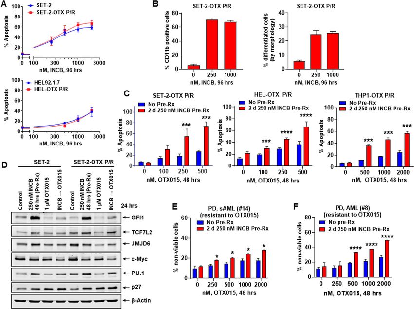
Fig. 6 LSD1i pre-treatment sensitizes BETi persister/resistant post-MPN sAML and de novo AML cells to BETi. A HEL92.1.7, HEL-OTX P/R, SET-2
and SET-2-OTX P/R cells were treated with INCB059872 for 96 h. The % of annexin V-positive, To-Pro-3 iodide-positive, apoptotic cells was determined
by flow cytometry. Mean of three experiments + S.D. B SET-2-OTX P/R cells were treated with the indicated concentrations of INCB059872 for 96 h.
Differentiated cells were assessed by CD11b flow cytometry and by cell morphology. Mean of three experiments + S.D. C SET-2-OTX P/R, HEL-OTX P/
R, and THP1-OTX P/R cells were treated with the indicated concentrations of OTX015 with or without 2 days of pre-treatment with 250 nM of
INCB059872. The % of annexin V-positive, apoptotic cells was determined by flow cytometry. Mean of three experiments + S.D. ***p < 0.005, ****p <
0.001 compared to cells treated with OTX015 without INCB059872 pre-treatment (determined by a two-tailed, unpaired t-test). D Immunoblot
analyses conducted on total cell lysates from SET-2 and SET-2-OTX P/R cells pre-treated with INCB059872 (48 h) and/or OTX015 (24 h), as indicated.
E PD, sAML (#14) cells (ex vivo resistant to OTX015) were treated (in duplicate) with the indicated concentrations of OTX015 with or without 2 days of
pre-treatment with 250 nM of INCB059872. The % of To-Pro-3 iodide-positive, nonviable cells was determined by flow cytometry. *p < 0.05 compared
to cells treated with OTX015 without INCB059872 pre-treatment (determined by a two-tailed, unpaired t-test). F PD, AML (#8) blast cells (ex vivo
resistant to OTX015) were treated (in duplicate) with the indicated concentrations of OTX015 with or without 2 days of pre-treatment with 250 nM of
INCB059872. The % of To-Pro-3 iodide-positive, nonviable cells was determined by flow cytometry. ****p < 0.001 versus cells treated with OTX015
without INCB059872 pretreatment (determined by a two-tailed, unpaired t-test).
BETi may exert in vivo efficacy and overcome nongenetic repressed enhancers and transcription factors, including
BETi-P/R in post-MPN sAML cells. PU.1, CEBPα and IRF8 while repressing c-Myc, and their
target gene-expressions. This induces cell cycle inhibitors
Discussion including p21 and p27, up-regulates differentiation-asso-
Utilizing the dTAG system to degrade LSD1 and ciated gene-expressions such as CD11b, CD86, LY96, and
CRISPR-Cas9 to edit and KO LSD1, findings presented LYZ, and induces morphologic features of AML cell dif-
here highlight gene-expression perturbations that lead to ferentiation. Although LSD1i inhibits both the scaffold and
differentiation of AML and post-MPN sAML blasts. Also, enzymatic functions of LSD1, AML cell-differentiation
disruption of LSD1 interactions with CoREST-HDAC1/2 occurs independent of inhibiting enzymatic demethylase
complex and with GFI1/1B by LSD1i activates GFI1/1B- function of LSD117,23,25,26,50. CRISPR-suppressor scanning
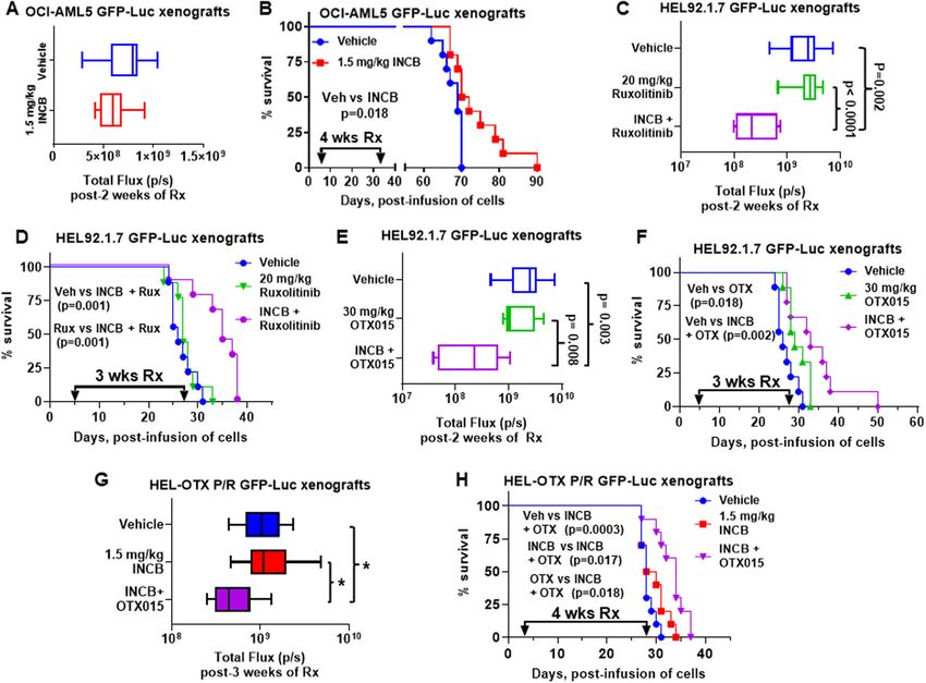
Blood Cancer JournalFiskus et al. Blood Cancer Journal (2021)11:98 Page 12 of 16 Fig. 7 Treatment with INCB059872 and ruxolitinib or OTX015 reduced leukemia burden and improved survival of NSG mice engrafted with sAML xenografts. A Total photon counts [flux] (determined by bioluminescent imaging) in NSG mice engrafted with OCI-AML5 GFP-Luc cells and treated for 2 weeks with INCB059872. B Kaplan–Meier survival plot of NSG mice engrafted with OCI-AML5 GFP-Luc cells and treated with 1.5 mg/ kg of INCB059872 (daily x 5 days, P.O.) for 4 weeks. Significance was calculated by a Mantel–Cox log-rank test. C Total photon counts [flux] in NSG mice engrafted with HEL92.1.7 GFP-Luc cells and treated for 2 weeks with INCB059872 and/or ruxolitinib. D Kaplan–Meier survival plot of NSG mice engrafted with HEL92.1.7 GFP-Luc cells and treated with 1.5 mg/kg of INCB059872 (daily x 5 days, P.O.) and/or 20 mg/kg of ruxolitinib (daily x 5 days, P.O.) for 3 weeks. Significance calculated by Mantel–Cox log-rank test. E Total photon counts [flux] in NSG mice engrafted with HEL92.1.7 GFP-Luc cells and treated for 2 weeks with INCB059872 and/or OTX015. F Kaplan–Meier survival plot of NSG mice engrafted with HEL92.1.7 GFP-Luc cells and treated with 1.5 mg/kg of INCB059872 (daily x 5 days, P.O.) and/or 30 mg/kg of OTX015 (daily x 5 days, P.O.) for 3 weeks. Significance calculated by Mantel–Cox log-rank test. G Total photon counts [flux] in NSG mice engrafted with HEL-OTX P/R GFP-Luc cells and treated for 1 week with INCB059872 followed by two weeks of INCB059872 and/or OTX015. *p < 0.05 (determined by a two-tailed, unpaired t-test). H Kaplan–Meier survival plot of NSG mice engrafted with HEL-OTX P/R GFP-Luc cells and treated with 1.5 mg/kg of INCB059872 (daily x 5 days, P.O.) alone for 1 week, followed by 3 weeks of 1.5 mg/kg of INCB059872 (daily x 5 days, P.O.) and/or 50 mg/kg OTX015 (daily x 5 days, P.O.). Significance was calculated by a Mantel–Cox log-rank test. was utilized to show that LSD1 enzyme activity is not not correlate with LSD1i-mediated changes in H3K4 required for AML survival26. Instead, LSD1 inhibitors mono- and dimethyl-chromatin marks23,50. disrupt interaction between LSD1 and transcription Additional findings highlighted here are that disruption repressor GFI1/1B on the chromatin. Also, in a knock- of LSD1-CoREST-HDAC1/2 complex derepresses GFI1/ down and rescue experiment, the LSD1 K661A catalytic 1B SE/Es to cause early induction of GFI1/1B47, which mutant was as capable of rescuing the clonogenic potential could serve as a predictive early biomarker of achieving of LSD1 knockdown cells as the wild type LSD1 pro- biologically effective intracellular levels of LSD1i in AML tein23,50. Additionally, LSD1 inhibitor treatment induces and post-MPN sAML cells. LSD1-KO also increased rapid and extensive transcriptional alterations, which do H3K27Ac and BRD4 occupancy at the GFI1 locus Blood Cancer Journal
Fiskus et al. Blood Cancer Journal (2021)11:98                                                                  Page 13 of 16

inducing GFI1 in AML cells. However, it is noteworthy          observed greater decline in c-Myc and increased p21 and
that without intact LSD1-CoREST-HDAC1/2 complex,               p27 protein levels. Additionally, in previous reports, we
upregulated GFI1/1B is unable to repress differentiation       have highlighted that inclusion of BETi enhances lethal
in AML/sAML cells19,21. Studies presented here also            activity of BETi-based combinations due to greater
involved CRISPR-Cas9-mediated KO of LSD1 or shRNA-             depletion of c-Myc, cell cycle dependent kinases CDK4
mediated KD of LSD1, which showed induction of GFI1/           and CDK6, as well as concomitant induction of CDK
1B, as well as of PU.1, CEBPα, and p21, but repression of      inhibitor p21, pro-apoptotic (BIM) and downregulation of
c-Myc in both post-MPN-sAML and non-MPN asso-                  anti-apoptotic (MCL1, BCL-xL, and BCL2) pro-
ciated AML cells. Notably, reduced H3K27Ac occupancy           teins.33,43,54–56. Synergistic in vitro lethality was also
at the MYC, but increased at the ITGAM, LY96, and LYZ          observed due to co-treatment with LSD1i and agents
loci, induced ITGAM, LY96, and LYZ expressions, dif-           targeting HDAC3, MOZ or DOT1L. However, whether
ferentiation and cell lethality of AML and post-MPN            these LSD1i-based combinations would also exert super-
sAML blasts. Our findings also confirmed that CRISPR-            ior in vivo anti-AML or anti-sAML efficacy remains to be
Cas9-mediated GFI1-KO or shRNA-mediated GFI1-KD                determined. Notably, a dual class I HDACi and LSD1
also induced differentiation markers and morphologic           inhibitor was shown to exert synergistic lethality in AML
features of differentiation in AML cells, albeit to a lesser   as well as in tumor models other than AML30,57,58.
extent than knockout or knockdown of LSD1. However,            CRISPR-Cas9 screens in AML cells expressing MLL-
these findings also demonstrated that LSD1i treatment           fusion gene showed superior efficacy of co-targeting
augmented differentiation due to GFI1-KD. This suggests        mTORC1 with LSD1, which was not revealed by our
that disruption of LSD1 binding to repressive complexes        screen59. This difference is because our domain-focused
not involving GFI1/1B may also be contributing to LSD1i-       CRISPR-Cas9 screen involved AML cells without MLL
induced differentiation in AML cells17,18,51. Alternatively,   fusion gene-expression, and utilized gRNAs targeting
it also suggests that both scaffold and enzymatic functions    chromatin regulators not mTORC1. Our findings also
of LSD1 (inhibited by INCB) may be involved in AML cell        demonstrate that co-treatment with LSD1i and the JAK1/
differentiation induced by INCB38. Again, LSD1i-induced        2 inhibitor ruxolitinib exerts synergistic in vitro lethality
differentiation was also associated with induction of GFI1,    and exhibited superior in vivo efficacy than LSD1i
PU.1, and p21, but depletion of c-Myc in both post-MPN-        monotherapy in a mouse xenograft model of post-MPN
sAML and non-MPN associated AML cells. This high-              sAML. A recent report highlighted that LSD1i treatment
lights targeted degradation and depletion of LSD1, which       exhibited clinical efficacy by reducing spleen size and
would disrupt both scaffolding and enzymatic activities of     constitutional symptoms in patients with advanced MPN,
LSD1, as an attractive therapeutic approach. Notably,          thus combination therapy with LSD1i and JAKi merits
LSD1-KO decreased H3K27Ac occupancy at enhancers/              further in vivo evaluation60. Our studies demonstrating
promoters and mRNA expressions of MYC and WNT-                 synergistic in vitro lethality due to combination of LSD1i
β-catenin-target genes, associated with positive enrich-       with decitabine also merits further verification through
ment of HALLMARK gene-sets of innate immunity,                 in vivo AML xenograft studies.
inflammatory response and apoptosis in AML cells.                  There is a growing evidence and recognition of epige-
LSD1-KD by shRNA also positively enriched similar              netic heterogeneity and plasticity in cancer cells, which
HALLMARK gene-sets in AML cells. These findings are             under selection pressure of targeted therapies facilitates
consistent with recent reports that LSD1i treatment sti-       emergence of drug-tolerant persister/resistant cancer cells
mulates antitumor immunity by inhibiting Foxp3+ Treg           displaying nongenetic therapy-resistance61–63. Agents that
cell function, thus enabling and enhancing immune              target signaling kinases (e.g., JAK1/2) and epigenetic
checkpoint blockade52,53. Collectively, these findings          mechanisms (e.g., BET proteins) were shown to result in
highlight testing potential utility of including LSD1i co-     enhancer reprogramming via newly-marshaled activities of
treatment with immune therapies in AML.                        lineage-specific transcriptional regulators which creates
   Utilizing a protein domain-focused CRISPR-                  the transcriptome/proteome conferring drug tolerant
Cas9 screen, our studies revealed that BRD4, DOT1L,            persister/resistance in AML cells33,48,49. We previously
HDAC3 and MOZ could serve as effective co-targets to           reported that increased c-Myc expression due to increased
achieve synergistic efficacy with LSD1i against AML and         levels and activity of β-catenin-TCF7L2-JMJD6-MYC axis
sAML cells. Consistent with this, LSD1i and BETi co-           induced nongenetic BETi-persister/resistance in AML and
treatment exhibited synergistic loss of viability in PD        sAML blasts33. This was due to enhancer reprogramming
AML and post-MPN sAML blasts displaying diverse                and increased transcriptional activities of MYC, RUNX1,
genetic alterations. In addition to abrogating LSD1i-          and TCF7L2, based on H3K27Ac and BRD4 occupancy at
induced GFI1 and PU.1, BRD4 inhibition by BETi                 their Es/promoters33. Our findings demonstrate, for the
increased lethal activity of LSD1i most likely due to          first time, that LSD1i is as active in inducing differentiation

Blood Cancer Journal
Fiskus et al. Blood Cancer Journal (2021)11:98                                                                                     Page 14 of 16

and lethality in the BETi-P/R as in BETi-sensitive sAML        RNA isolation and quantitative polymerase chain reaction
cells. Notably, sensitization of BETi-P/R sAML and AML           Following the designated treatments, total RNA was
cells to BETi-induced apoptosis by pre-treatment with          isolated from AML or sAML cells utilizing a PureLink
LSD1i was associated with increased chromatin accessi-         RNA Mini kit from Ambion, Inc. (Austin, TX) and reverse
bility and mRNA expressions of GFI1, PU.1, and IRF8-           transcribed. Quantitative real time PCR analysis was
target genes. However, mRNA expressions of c-Myc tar-          performed on cDNA using TaqMan probes. Relative
gets were mostly downregulated. Importantly, our findings       mRNA expression was normalized to GAPDH and com-
also show that LSD1i pre-treatment similarly sensitized        pared to the untreated cells.
sAML-RuxP cells that display nongenetic resistance to
ruxolitinib48. Taken together, these findings highlight that,   Statistical analysis
by reprogramming of SEs/Es and modifying gene-                   Significant differences between AML or sAML cells
expressions involved in conferring nongenetic persister/       treated with different experimental conditions compared
resistance, LSD1i treatment could sensitize AML to BETi,       to control cells were determined using the two-tailed,
and sAML cells to ruxolitinib or BETi, thereby high-           unpaired t-test. For the in vivo mouse models, a two-tailed,
lighting novel differentiation and lethal therapies for AML    unpaired t-test was utilized for comparing total biolumi-
or post-MPN sAML. Findings presented here also beg the         nescent flux. For survival analysis, a Kaplan–Meier plot
question where in AML or post-MPN sAML therapy                 and a Mantel–Cox log rank test were utilized for com-
LSD1i-based combinations should best be clinically tested      parisons of different cohorts. P values of
Fiskus et al. Blood Cancer Journal (2021)11:98                                                                                                                Page 15 of 16

Supplementary information The online version contains supplementary                       28. Cusan, M. et al. LSD1 inhibition exerts its antileukemic effect by recommis-
material available at https://doi.org/10.1038/s41408-021-00487-3.                             sioning PU.1- and C/EBPalpha-dependent enhancers in AML. Blood 131,
                                                                                              1730–1742 (2018).
Received: 11 March 2021 Revised: 20 April 2021 Accepted: 30 April 2021                    29. Moroy, T. & Khandanpour, C. Role of GFI1 in epigenetic regulation of MDS and
                                                                                              AML pathogenesis: mechanisms and therapeutic implications. Front. Oncol. 9,
                                                                                              824 (2019).
                                                                                          30. Fiskus, W. et al. Highly effective combination of LSD1 (KDM1A) antagonist and
                                                                                              pan-histone deacetylase inhibitor against human AML cells. Leukemia 28,
References                                                                                    2155–2164 (2014).
 1. Jongen-Lavrencic, M. et al. Molecular minimal residual disease in acute               31. Lynch, J. T., Cockerill, M. J., Hitchin, J. R., Wiseman, D. H. & Somervaille, T. C.
     myeloid leukemia. N. Engl. J. Med. 378, 1189–1199 (2018).                                CD86 expression as a surrogate cellular biomarker for pharmacological inhi-
 2. Ng, S. W. et al. A 17-gene stemness score for rapid determination of risk in              bition of the histone demethylase lysine-specific demethylase 1. Anal. Bio-
     acute leukaemia. Nature 540, 433–437 (2016).                                             chem. 442, 104–106 (2013).
 3. Thomas, D. & Majeti, R. Biology and relevance of human acute myeloid                  32. Schenk, T. et al. Inhibition of the LSD1 (KDM1A) demethylase reactivates the
     leukemia stem cells. Blood 129, 1577–1585 (2017).                                        all-trans-retinoic acid differentiation pathway in acute myeloid leukemia. Nat.
 4. Luskin, M. R., Murakami, M. A., Manalis, S. R. & Weinstock, D. M. Targeting               Med. 18, 605–611 (2012).
     minimal residual disease: a path to cure? Nat. Rev. Cancer 18, 255–263 (2018).       33. Saenz, D. T. et al. Mechanistic basis and efficacy of targeting the beta-catenin-
 5. Bradner, J. E., Hnisz, D. & Young, R. A. Transcriptional addiction in cancer. Cell        TCF7L2-JMJD6-c-Myc axis to overcome resistance to BET inhibitors. Blood 135,
     168, 629–643 (2017).                                                                     1255–1269 (2020).
 6. Hnisz, D. et al. Convergence of developmental and oncogenic signaling                 34. Nabet, B. et al. The dTAG system for immediate and target-specific protein
     pathways at transcriptional super-enhancers. Mol. Cell 58, 362–370 (2015).               degradation. Nat. Chem. Biol. 14, 431–441 (2018).
 7. Duy, C. et al. Rational targeting of cooperating layers of the epigenome yields       35. Fang, Y., Liao, G. & Yu, B. LSD1/KDM1A inhibitors in clinical trials: advances and
     enhanced therapeutic efficacy against AML. Cancer Discov. 9, 872–889 (2019).              prospects. J. Hematol. Oncol. 12, 129 (2019).
 8. McKeown, M. R. et al. Superenhancer analysis defines novel epigenomic                  36. Pandey, M. R. & Wang, E. S. What potential is there for LSD1 inhibitors to reach
     subtypes of non-APL AML, including an RARalpha dependency targetable by                  approval for AML? Expert Opin. Emerg. Drugs 24, 205–212 (2019).
     SY-1425, a potent and selective RARalpha agonist. Cancer Discov. 7, 1136–1153        37. Wu, L. et al. Cyclopropylamines as LSD1 inhibitors WO/2015/123465. US Patent
     (2017).                                                                                  No. 20190211014 (2019).
 9. Raisner, R. et al. Enhancer activity requires CBP/P300 bromodomain-                   38. Barth, J. et al. LSD1 inhibition by tranylcypromine derivatives interferes with
     dependent histone H3K27 acetylation. Cell Rep. 24, 1722–1729 (2018).                     GFI1-mediated repression of PU.1 target genes and induces differentiation in
10. Mill, C. P. et al. RUNX1-targeted therapy for AML expressing somatic or                   AML. Leukemia 33, 1411–1426 (2019).
    germline mutation in RUNX1. Blood 134, 59–73 (2019).                                  39. Morgan, M. A. & Shilatifard, A. Chromatin signatures of cancer. Genes Dev. 29,
11. Roe, J. S. & Vakoc, C. R. C/EBPalpha: critical at the origin of leukemic trans-           238–249 (2015).
    formation. J. Exp. Med. 211, 1–4 (2014).                                              40. Shema, E. et al. Single-molecule decoding of combinatorially modified
12. Sood, R., Kamikubo, Y. & Liu, P. Role of RUNX1 in hematological malignancies.             nucleosomes. Science 352, 717–721 (2016).
    Blood 129, 2070–2082 (2017).                                                          41. Shi, J. et al. Discovery of cancer drug targets by CRISPR-Cas9 screening of
13. Will, B. et al. Minimal PU.1 reduction induces a preleukemic state and pro-               protein domains. Nat. Biotechnol. 33, 661–667 (2015).
    motes development of acute myeloid leukemia. Nat. Med. 21, 1172–1181                  42. Nagasaka, M. et al. Lysine-specific demethylase 1 (LSD1/KDM1A) is a novel
    (2015).                                                                                   target gene of c-Myc. Biol. Pharm. Bull. 42, 481–488 (2019).
14. Shi, Y. J. et al. Regulation of LSD1 histone demethylase activity by its associated   43. Fiskus, W. et al. Superior efficacy of cotreatment with BET protein inhibitor and
    factors. Mol. Cell 19, 857–864 (2005).                                                    BCL2 or MCL1 inhibitor against AML blast progenitor cells. Blood Cancer J. 9, 4
15. Stavropoulos, P., Blobel, G. & Hoelz, A. Crystal structure and mechanism of               (2019).
    human lysine-specific demethylase-1. Nat. Struct. Mol. Biol. 13, 626–632 (2006).       44. Beyer, M. et al. HDAC3 activity is essential for human leukemic cell growth and
16. Yang, M. et al. Structural basis for CoREST-dependent demethylation of                    the expression of beta-catenin, MYC, and WT1. Cancers 11, 1436 (2019).
    nucleosomes by the human LSD1 histone demethylase. Mol. Cell 23, 377–387              45. Baell, J. B. et al. Inhibitors of histone acetyltransferases KAT6A/B induce
    (2006).                                                                                   senescence and arrest tumour growth. Nature 560, 253–257 (2018).
17. Magliulo, D., Bernardi, R. & Messina, S. Lysine-specific demethylase 1A as a           46. Daigle, S. R. et al. Potent inhibition of DOT1L as treatment of MLL-fusion
    promising target in acute myeloid leukemia. Front. Oncol. 8, 255 (2018).                  leukemia. Blood 122, 1017–1025 (2013).
18. Majello, B., Gorini, F., Sacca, C. D. & Amente, S. Expanding the role of the          47. Tatsumi, G. et al. LSD1-mediated repression of GFI1 super-enhancer plays an
    histone lysine-specific demethylase LSD1 in cancer. Cancers 11, 324 (2019).                essential role in erythroleukemia. Leukemia 34, 746–758 (2020).
19. Moroy, T., Vassen, L., Wilkes, B. & Khandanpour, C. From cytopenia to leukemia:       48. Saenz, D. T. et al. Targeting nuclear beta-catenin as therapy for post-
    the role of Gfi1 and Gfi1b in blood formation. Blood 126, 2561–2569 (2015).                 myeloproliferative neoplasm secondary AML. Leukemia 33, 1373–1386 (2019).
20. Thambyrajah, R. et al. GFI1 proteins orchestrate the emergence of haemato-            49. Bell, C. C. et al. Targeting enhancer switching overcomes non-genetic drug
    poietic stem cells through recruitment of LSD1. Nat. Cell Biol. 18, 21–32 (2016).         resistance in acute myeloid leukaemia. Nat Commun 10, 2723 (2019).
21. van Bergen, M. & van der Reijden, B. A. Targeting the GFI1/1B-CoREST complex          50. Maiques-Diaz, A., Lynch, J. T., Spencer, G. J. & Somervaille, T. C. P. LSD1 inhi-
    in acute myeloid leukemia. Front. Oncol. 9, 1027 (2019).                                  bitors disrupt the GFI1 transcription repressor complex. Mol. Cell Oncol. 5,
22. Velinder, M. et al. GFI1 functions in transcriptional control and cell fate               e1481813 (2018).
    determination require SNAG domain methylation to recruit LSD1. Biochem. J.            51. Amente, S., Lania, L. & Majello, B. The histone LSD1 demethylase in stemness
    473, 3355–3369 (2016).                                                                    and cancer transcription programs. Biochim. Biophys. Acta 1829, 981–986
23. Maiques-Diaz, A. et al. Enhancer activation by pharmacologic displacement of              (2013).
    LSD1 from GFI1 induces differentiation in acute myeloid leukemia. Cell Rep. 22,       52. Sheng, W. et al. LSD1 ablation stimulates anti-tumor immunity and enables
    3641–3659 (2018).                                                                         checkpoint blockade. Cell 174, 549–563 e519 (2018).
24. Ishikawa, Y. et al. A novel LSD1 inhibitor T-3775440 disrupts GFI1B-containing        53. Xiong, Y. et al. Inhibiting the coregulator CoREST impairs Foxp3+ Treg
    complex leading to transdifferentiation and impaired growth of AML cells.                 function and promotes antitumor immunity. J. Clin. Invest. 130, 1830–1842
    Mol. Cancer Ther. 16, 273–284 (2017).                                                     (2020).
25. Maes, T. et al. ORY-1001, a potent and selective covalent KDM1A inhibitor, for        54. Fiskus, W. et al. Highly active combination of BRD4 antagonist and histone
    the treatment of acute leukemia. Cancer Cell 33, 495–511 e412 (2018).                     deacetylase inhibitor against human acute myelogenous leukemia cells. Mol.
26. Vinyard, M. E. et al. CRISPR-suppressor scanning reveals a nonenzymatic role of           Cancer Ther. 13, 1142–1154 (2014).
    LSD1 in AML. Nat. Chem. Biol. 15, 529–539 (2019).                                     55. Fiskus, W. et al. BET protein antagonist JQ1 is synergistically lethal with FLT3
27. Hones, J. M. et al. GFI1 as a novel prognostic and therapeutic factor for AML/            tyrosine kinase inhibitor (TKI) and overcomes resistance to FLT3-TKI in AML
    MDS. Leukemia 30, 1237–1245 (2016).                                                       cells expressing FLT-ITD. Mol. Cancer Ther. 13, 2315–2327 (2014).

Blood Cancer Journal
Fiskus et al. Blood Cancer Journal (2021)11:98 Page 16 of 16
56. Saenz, D. T. et al. BET protein bromodomain inhibitor-based combinations are 60. Pettit, K. G. et al. A phase 2a study of the LSD1 inhibitor Img-7289
highly active against post-myeloproliferative neoplasm secondary AML cells. (bomedemstat) for the treatment of myelofibrosis. Blood 134, 556
Leukemia 31, 678–687 (2017). (2019).
57. Anastas, J. N. et al. Re-programing chromatin with a bifunctional LSD1/HDAC 61. Angus, S. P., Zawistowski, J. S. & Johnson, G. L. Epigenetic mechanisms reg-
inhibitor induces therapeutic differentiation in DIPG. Cancer Cell 36, 528–544 ulating adaptive responses to targeted kinase inhibitors in cancer. Annu. Rev.
e510 (2019). Pharmacol. Toxicol. 58, 209–229 (2018).
58. Kalin, J. H. et al. Targeting the CoREST complex with dual histone deacetylase 62. Bell, C. C. & Gilan, O. Principles and mechanisms of non-genetic resistance in
and demethylase inhibitors. Nat. Commun. 9, 53 (2018). cancer. Br. J. Cancer 122, 465–472 (2020).
59. Deb, G. et al. Pre-clinical activity of combined LSD1 and mTORC1 inhibition in 63. Shaffer, S. M. et al. Rare cell variability and drug-induced reprogramming as a
MLL-translocated acute myeloid leukaemia. Leukemia 34, 1266–1277 (2020). mode of cancer drug resistance. Nature 546, 431–435 (2017).
Blood Cancer JournalYou can also read