Irisin Enhances Angiogenesis of Mesenchymal Stem Cells to Promote Cardiac Function in Myocardial Infarction via PI3k/Akt Activation
←
→
Page content transcription
If your browser does not render page correctly, please read the page content below
eISSN 2005-5447
 International Journal of Stem Cells Vol. 14, No. 4, 2021                                                   https://doi.org/10.15283/ijsc21005

                                                                    ORIGINAL ARTICLE

  Irisin Enhances Angiogenesis of Mesenchymal Stem Cells to
     Promote Cardiac Function in Myocardial Infarction via
                     PI3k/Akt Activation
              1,2                 3               1,2                 1,2                   1,2             1,2             4                  1,2
Fan Yang , Zhi Wang , Bing Li , Youfu He , Fawang Du , Shui Tian , Yu Zhang , Yongyao Yang

                            1
                             Department of Cardiology, Guizhou Provincial People’s Hospital, Guiyang, China
                            2
                             Department of Cardiology, Guizhou University People’s Hospital, Guiyang, China
                                         3
                                          Qingdao Municipal Hospital (Group), Qingdao, China
                               4
                                Department of Cardiology, Xixiu District People’s Hospital, Anshun, China

  Background and Objectives: With the growing incidence of acute myocardial infarction (MI), angiogenesis is vital for
  cardiac function post-MI. The role of bone marrow mesenchymal stem cells (BMSCs) in angiogenesis has been pre-
  viously confirmed. Irisin is considered a potential vector for angiogenesis. The objective of the present study was to
  investigate the potential role of irisin in the angiogenesis of BMSCs.
  Methods and Results: In vivo, irisin-treated BMSCs (BMSCs+irisin) were transplanted into an MI mouse model. On
  day 28 post-MI, blood vessel markers were detected, and cardiac function and infarct areas of mice were evaluated.
  In vitro, paracrine effects were assessed by examining tube formation in human umbilical vein endothelial cells
  (HUVECs) co-cultured with the BMSCs+irisin supernatant. The scratch wound-healing assay was performed to eval-
  uate HUVEC migration. Western blotting was performed to determine PI3k/Akt pathway activation in the BMSCs+iri-
  sin group. Transplantation of BMSCs+irisin promoted greater angiogenesis, resulting in better cardiac function in
  the MI mouse model than in controls. In the BMSC+irisin group, HUVECs demonstrated enhanced tube formation
  and migration. Activation of the PI3k/Akt pathway was found to be involved in mediating the role of irisin in the
  angiogenesis of BMSCs.
  Conclusions: In cardiovascular diseases such as MI, irisin administration can enhance angiogenesis of BMSCs and pro-
  mote cardiac function via the PI3k/Akt pathway, optimizing the therapeutic effect based on BMSCs transplantation.

  Keywords: Irisin, Angiogenesis, Mesenchymal stem cells, PI3k/Akt pathway

                                                                                      Introduction

Received: January 6, 2021, Revised: May 25, 2021,                                       In acute myocardial infarction (MI), atherosclerotic vas-
Accepted: July 1, 2021, Published online: August 31, 2021                             cular occlusions trigger pathological injury owing to my-
Correspondence to Yongyao Yang                                                        ocardial ischemia and hypoxia. Angiogenesis and vascular
  Department of Cardiology, Guizhou Provincial People’s Hospital,                     remodeling play essential roles in affording protection
  NO. 83 Zhongshan East Road, Guiyang 550002, China
  Tel: +86-851-8592-5127, Fax: +86-851-8592-5127                                      against myocardial ischemia (1). Notably, angiogenesis
  E-mail: 729142921@qq.com                                                            primarily decelerates the development of heart failure (2).
   This is an open-access article distributed under the terms of the Creative         Percutaneous coronary intervention (PCI) and coronary
   Commons Attribution Non-Commercial License (http://creativecommons.org/            artery bypass grafting have failed to rescue microvascular
   licenses/by-nc/4.0/), which permits unrestricted non-commercial use, dis-
   tribution, and reproduction in any medium, provided the original work is           injury and reportedly result in PCI-related microvascular
   properly cited.                                                                    damage (3). Thus, new medical approaches have revealed
Copyright ⓒ 2021 by the Korean Society for Stem Cell Research                         a critical role for angiogenesis against progressive cardiac

                                                                                455
456 International Journal of Stem Cells 2021;14:455-464

deterioration during cardiovascular diseases. In previous       BMSCs were isolated and cultured from patients who un-
studies, as well as in our present study, human bone mar-       derwent hip replacement upon receipt of informed consent
row mesenchymal stem cells (BMSCs) improved angio-              and approval by the Human Ethics Committee of Guizhou
genesis through a paracrine effect, primarily rescuing car-     Provincial People’s Hospital.
diac function in the MI mouse model (4).                          BMSCs at 4-8 passage were treated with irisin (200 ng/ml;
   Irisin, a new multifunctional myokine related to meta-       Phoenix Pharmaceuticals, USA) and LY294002 (50 μM;
bolic factors, is predominantly released from skeletal mus-     Selleck Chemicals, USA), as previously reported (10), for
cle and myocardium. Irisin is known to be involved in cell      24 h under normoxic conditions. Dimethyl sulfoxide
physiology and pathology, reportedly associated with the        (DMSO), the solvent employed for irisin, was used as a
browning of white adipocytes and increased energy ex-           control.
penditure (5). Irisin is cleaved from fibronectin type-III
domain-containing protein 5 (FNDC5) and released into           Immunostaining
the circulation, which is reportedly involved in energy me-        Twenty-eight days post-MI, mice were sacrificed, and
tabolism in skeletal muscle, adipose tissue, and pancreatic     their hearts were harvested and embedded in O.C.T. com-
islets (6). In a transverse aortic constriction (TAC)-induced   pound (Sakura Finetek, USA). Heart tissue sections (7-μm
cardiac hypertrophy mouse model, irisin reportedly alle-        thick) were fixed using 4% paraformaldehyde. After per-
viated cardiac fibrosis and hypertrophy, thus improving         meabilization with 0.2% Triton, tissue slices were blocked
cardiac function through 5’-adenosine monophosphate-acti-       with 3% bovine serum albumin (BSA) and incubated with
vated protein kinase (AMPK)-mammalian target of rapa-           primary antibody at 4℃ overnight. Sections were in-
mycin (mTOR) signaling (7). Similarly, irisin attenuates        cubated with the secondary antibody at room temperature
cardiac fibrosis in TAC-induced hypertrophy by increas-         for 1 h and then observed using a fluorescence microscope
ing autophagy (8). Furthermore, irisin is involved in the       (Leica, Germany). The vascular density at border areas
angiogenesis of human umbilical vein endothelial cells          was calculated as the positive number of vascular markers
(HUVECs), thus protecting cardiac function after MI (9,         per area for one high-power field (34.68 μm2), with meas-
10). Recent studies have reported that irisin regulates the     urements performed in at least five random high-power
crosstalk between adipose tissue and muscle via paracrine       fields for each section.
signaling (10). However, the role of irisin in the paracrine
signaling of BMSCs needs to be elucidated. In the present       Sirius Red staining
study, we hypothesized that irisin could enhance the para-         Twenty-eight days post-MI, mice were euthanized, and
crine effect of BMSCs on angiogenesis to improve cardiac        hearts were harvested and embedded in paraffin. After
function post-MI injury.                                        sectioning (3-μm thick sections), the cardiac tissue was
                                                                stained using Sirius Red (Solaribio, Beijing, China), ac-
Materials and Methods                                           cording to the manufacturer’s instructions. Infarct areas
                                                                were measured using Image-Pro software (Media Cybernetics,
Animal experiment                                               USA) and assessed according to the sum of the endocardial
  All animal procedures were conducted in accordance            and epicardial length of the infarct zone in proportion to
with the guidelines of the Animal Use Committee of the          the sum of the endocardial and epicardial length of the
Guizhou Provincial People’s Hospital. To establish the MI       entire left ventricle.
mouse model, the left anterior descending coronary artery
of 8∼12-week old male C57BL/6J mice (Guizhou Medical            Tube formation assay
University) was ligated as previously described (11). BMSCs       Tube formation was assessed using HUVECs, co-cul-
with/without irisin treatment (1.5×10⁵ cells suspended in       tured with the supernatant of BMSCs treated with irisin,
20 μl Dulbecco’s Modified Eagle Medium [DMEM] per               LY294002, irisin+LY294002, or control. HUVECs were
mouse) were immediately transplanted into the border            seeded at a density of 5×10⁴ cells per well and co-cultured
zone at four different sites. The DMEM group received           with the BMSC supernatant. Tube formation images were
DMEM without BMSCs (mice received an equivalent vol-            acquired 4 h after transplantation, and the number of bran-
ume of DMEM).                                                   ch points was quantified using the Image-Pro software.

Human BMSCs isolation and culture                               Scratch wound-healing assay
  Based on a previously reported protocol (11), human             The wound-healing assay was performed as previously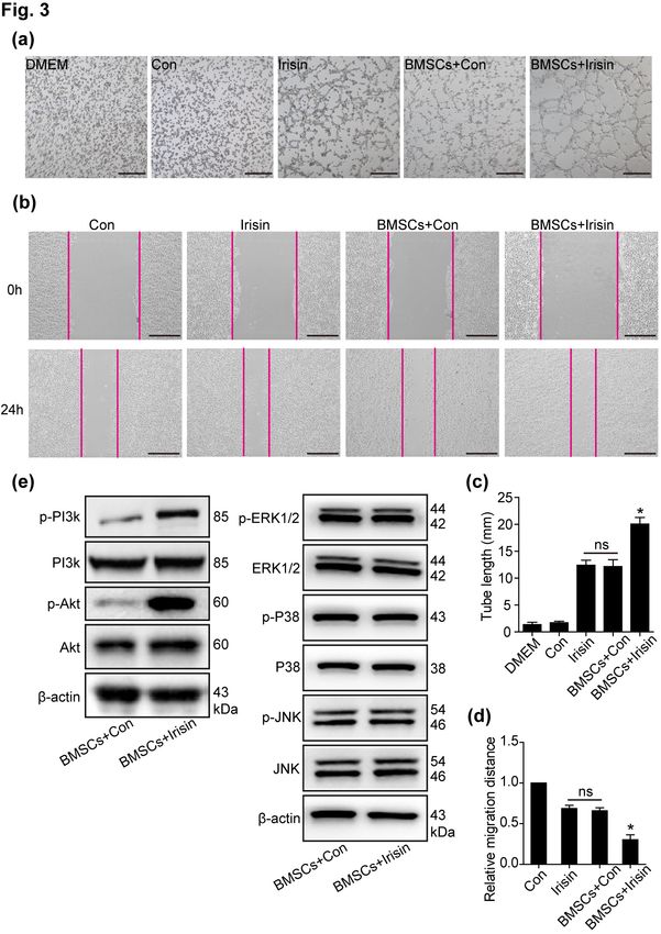
Fan Yang, et al: Irisin Promoted the Angiogenesis and Paracrine Effects of BMSCs via the PI3k/Akt Pathway 457

described (10). Briefly, HUVECs were cultured in a 6-well         compared using one-way analysis of variance. *p<0.05
plate (1×10⁶ cells/well) for 48 h under normal conditions.        was deemed statistically significant.
After scratching the surface with a sterile 10-μl pipette
tip, HUVECs were washed twice and cultured in DMEM                Results
(containing 1% fetal bovine serum [FBS]) for 24 h, con-
taining an equal volume of BMSC supernatant treated               Irisin enhanced the protection of BMSCs in the MI
with irisin, LY294002, irisin+LY294002, or an equivalent          mouse model
volume of DMSO. The wound areas were photographed                    The therapeutic efficacy of BMSCs in ischemic heart
with an inverted microscope (Leica, Germany), and cell            disease has been clarified in several animal studies and
migration was calculated as the average wound area in five        clinical trials (13, 14). To confirm the protective role of
random fields, compared with the zero-time point, using           irisin toward BMSCs, we treated BMSCs with irisin (200
Image-Pro software.                                               ng/ml) for 24 h under normoxic conditions (BMSCs+iri-
                                                                  sin). DMSO, the solvent used for irisin, was used as the
Cell apoptosis assay                                              control (BMSCs+Con). In the MI mouse model, we trans-
  Following treatment with irisin for 24 h under normal           planted BMSCs treated with irisin into the border zone
conditions, BMSCs were cultured in DMEM with glucose              immediately after MI surgery. Twenty-eight days post-MI,
and FBS deprivation under hypoxia (95% air/5% CO2, at             improved heart function was noted, with an increased
37℃) for 24 h.                                                    ejection fraction (EF) and fractional shortening (FS), us-
                                                                  ing two-dimensional echocardiography in the BMSCs+
TUNEL staining assay                                              irisin group when compared with the BMSCs+Con and
  Apoptosis was detected using the TUNEL Apoptosis                DMEM groups (Fig. 1a and 1b). Moreover, the infarct
Assay Kit (Beyotime Biotechnology, Shanghai, China) ac-           areas were examined by Sirius Red staining, presenting re-
cording to the manufacturer’s instructions. After fixation        duced infarct areas in the BMSCs+irisin group when
with 4% paraformaldehyde for 30 min, BMSCs were per-              compared with the BMSCs+Con and DMEM groups
meabilized with 0.2% Triton for 5 min at room temper-             (Fig. 1c and 1d). These results revealed that irisin pro-
ature, followed by incubation with TUNEL reaction re-             tected BMSCs in response to ischemic insults in vivo.
agent for 1 h at 37℃ in the dark. Images were captured
using a fluorescence microscope. Apoptotic cells were cal-        Irisin regulated angiogenesis of BMSCs in vivo
culated as TUNEL-positive cells in proportion to the total           The angiogenic efficacy of BMSCs mediated via para-
number of cells.                                                  crine signaling has been reported (4). Additionally, the
                                                                  role of irisin in angiogenesis was previously verified (9,
Western blotting                                                  10). For further evaluation, we investigated the ability of
   Western blotting was performed to detect protein levels        irisin-regulated BMSC paracrine signaling to induce
of p-PI3K, PI3K, p-Akt, and Akt. In brief, the proteins           angiogenesis. On day 28 post-MI modeling, alpha-smooth
were obtained from cells using RIPA lysis buffer on ice,          muscle actin (α-SMA), a marker of vascular smooth mus-
followed by centrifugation for clarification. Western blot-       cle cells, was significantly elevated in the BMSCs+irisin
ting was performed as previously described (12).                  group when compared with the BMSCs+Con and DMEM
                                                                  groups (Fig. 2a). Similarly, vascular endothelial cells
Echocardiography                                                  markers, including von Willebrand factor (vWF) and
   Echocardiography was performed to assess cardiac func-         CD31, were considerably enhanced in the BMSCs+irisin
tion on day 28 post-MI. After isoflurane inhalation, the mice     group when compared with the BMSCs+Con and DMEM
were anesthetized and bound. Then, two-dimensional im-            groups (Fig. 2b and 2c). Interestingly, these vascular
ages were obtained using a Vevo 2100 system (VisualSonics,        markers were marginally increased in the BMSCs+Con
Inc., Toronto, Canada).                                           group when compared with the DMEM group. Blood ves-
                                                                  sels in the border zone were quantified (Fig. 2d), indicat-
Statistical analysis                                              ing that irisin further promoted the angiogenic efficiency
   All experiments were independently performed at least          in BMSCs, despite the moderate efficiency of BMSCs in
three times, and data values are presented as the mean±           angiogenesis.
standard error of the mean. Student’s t test was used to
compare any two groups, and more than three groups were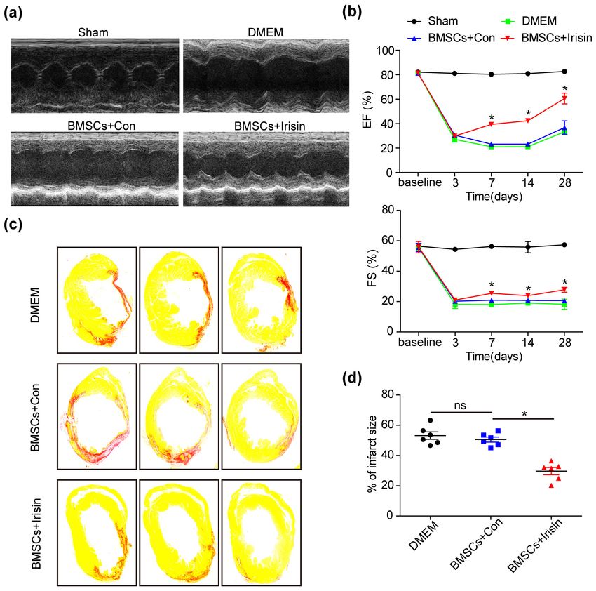
458 International Journal of Stem Cells 2021;14:455-464
Fig. 1. Irisin-treated BMSCs rescued
the cardiac function in MI mouse in
vivo. (a) Representative images of
echocardiography showing the im-
proved cardiac function in BMSCs
treated with irisin. (b) The ejection
fraction (EF) and fractional short-
ening (FS) are gradually recovered in
the BMSCs+irisin group when com-
pared with other groups (n=8 for
Sham, 5 for DMEM, 6 for BMSCs+
irisin and BMSCs+Con). (c) The in-
farct area was examined by Sirius
Red staining 28 days post-MI (n=6
for every group). (d) The infarct size
in hearts was quantified using
Image-Pro. All data were measured
as mean±standard error of the mean
(SEM). *p<0.05. BMSCs, bone mar-
row mesenchymal stem cells; MI, my-
ocardial infarction; DMEM, Dulbecco’s
Modified Eagle Medium.
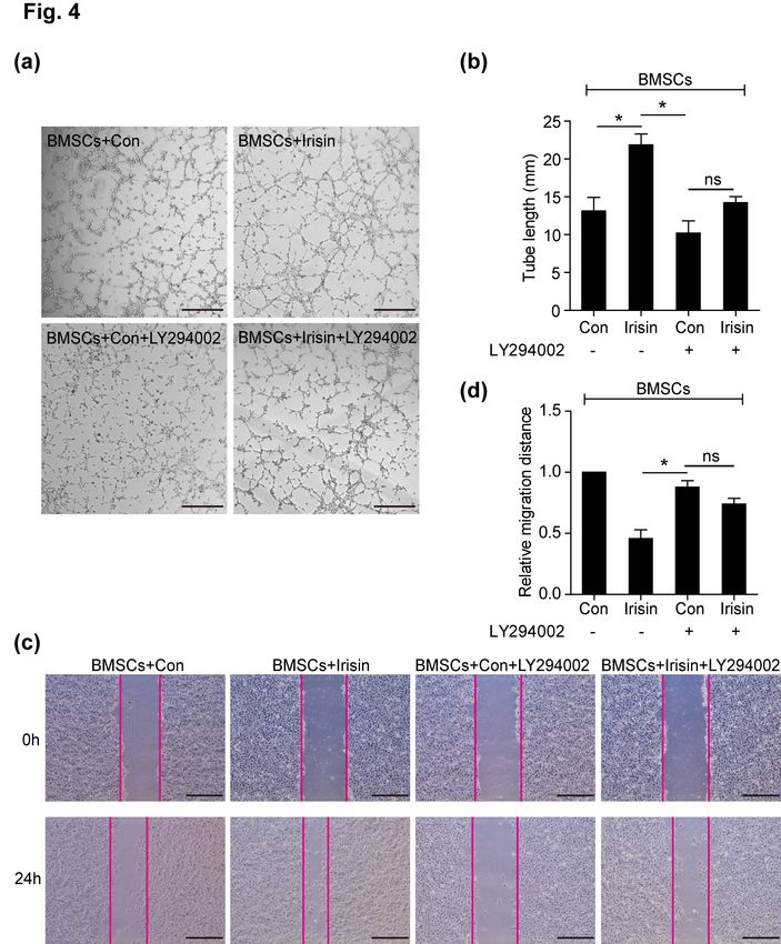
Irisin mediated the paracrine effects of BMSCs by in irisin-regulated BMSC paracrine signaling. In the pres-
primarily regulating the PI3K/Akt pathway ent study, p-PI3k/PI3k and p-Akt/Akt were increased in
Previous studies have shown that the paracrine effects irisin-mediated BMSCs when compared with the control
of BMSCs influence cellular fate (4, 15). To determine the group, with no differences in the p-ERK1/2/ERK1/2,
role of irisin in mediating paracrine effects of BMSCs, p-P38/P38, and p-JNK/JNK pathways, revealing that
conditioned media of BMSCs, with or without irisin, were PI3k/Akt was involved in irisin-regulated BMSC paracrine
collected; then, tube formation of HUVECs was assessed signaling (Fig. 3e). Furthermore, inhibition of the PI3k/
using the Matrigel assay, with cell migration examined us- Akt pathway (LY294002) was performed to suppress iri-
ing a wound-healing assay. In the BMSCs+irisin group, sin-induced tube formation (Fig. 4a and 4b). In the
tube formation (Fig. 3a and 3c) and migration (Fig. 3b wound-healing assay, the increased HUVEC migration,
and 3d) of HUVECs were significantly increased when regulated by irisin-treated BMSCs, was blocked by
compared with the BMSCs+Con and irisin alone groups, LY294002 (Fig. 4c and 4d). However, a slight increased
but not in control alone; no difference was observed be- effect of tube formation and HUVEC migration by irisin
tween the BMSCs+Con group and irisin alone group. The despite LY294002 administration, suggesting that alter-
results indicated that irisin enhanced the paracrine effects nate signaling pathways are involved in irisin-regulated
of BMSCs, consistent with a previous study (16). BMSC paracrine signaling (Fig. 4c and 4d). These data
In MI, the PI3k/Akt pathway is deemed the primary revealed that irisin promoted BMSC-mediated paracrine
signal transduction pathway associated with cardioprotec- signaling via the PI3k/Akt pathway.
tion (17). In a cardiotoxicity model, irisin was found to
be involved in cardiomyocyte apoptosis via Akt activation
(6). Herein, we sought to monitor the level of PI3k/AktFan Yang, et al: Irisin Promoted the Angiogenesis and Paracrine Effects of BMSCs via the PI3k/Akt Pathway 459
Fig. 2. Irisin-treated BMSCs enhance
angiogenesis. (a∼c) Representative
images of vascular markers includ-
ing α-SMA, vWF and CD31 were
captured in the border zone of irisin-
treated BMSCs, control, and DMEM
groups. (d) The vascular density was
measured in at least 5 high-power
fields per section (n=8 for DMEM,
7 for BMSCs+irisin and BMSCs+
Con). Scale bar, 100 μm. Data are
presented as mean±standard error
of the mean (SEM). *p<0.05. BMSCs,
bone marrow mesenchymal stem cells;
DMEM, Dulbecco’s Modified Eagle
Medium; α-SMA, alpha-smooth mus-
cle actin; vWF, von Willebrand factor.
Discussion crine effect of MSCs is reportedly considered the principal
mechanism underlying MSC-based therapy, including
In the present study, we observed that the irisin admin- exosomes and other paracrine factors such as basic fibro-
istration increased the angiogenesis of BMSCs in the bor- blast growth factor (bFGF) and vascular endothelial
der zone to protect cardiac function post-MI in vivo. growth factor (VEGF) (13, 20). Based on the paracrine ac-
Consistent with this phenomenon in vivo, irisin treatment tion, MSCs enhance angiogenesis via HIF-1α, miR-200b,
promoted the paracrine efficacy of BMSCs in vitro. and the leptin signaling pathway (17). Similarly, the para-
Mechanistically, we observed that irisin activated the crine function was observed in the present study, as re-
PI3k/Akt signaling pathway, primarily improving the vealed by the Matrigel assay.
paracrine efficiency of BMSCs. Combined with these data, In skeletal muscles, irisin is a myokine cleaved from
it was speculated that irisin could be a potential novel FNDC5, especially after exercise. The role of irisin is mul-
therapeutic agent for BMSC transplantation in tissue en- tifunctional, with activities such as anti-inflammation,
gineering and regenerative medicine for cardiovascular neuroprotection, bone formation, and metabolic regulation
diseases. (21, 22). As a hormone secreted into circulation, irisin has
Mesenchymal stem cells (MSCs) are a promising ther- been shown to afford protection against cardiovascular in-
apeutic option for immune-mediated diseases, cardiova- jury, reduce cardiac fibrosis, and improve cardiac function
scular diseases, diabetes, and cancer (13, 18, 19). The para- (23, 24). Moreover, irisin is considered crosstalk in muscle460 International Journal of Stem Cells 2021;14:455-464
Fig. 3. Irisin promotes the paracrine
efficacy of BMSCs through the
PI3K/AKT pathway. (a) Conditioned
media of equivalent cell numbers
6
(1×10 cells) were collected for iri-
sin-treated BMSCs and control groups.
By employing HUVECs, tube forma-
tion was assessed using the super-
natant of BMSCs treated with irisin
and control, irisin alone, and control
alone. Scale bar, 50 μm. (b) The
wound-healing assay was performed
to demonstrate HUVEC migration
using the above conditioned media,
respectively. Scale bar, 243.2 μm.
(c) The number of branch points was
quantified by Image-Pro software
and is shown in bar graphs (n=3
separate studies). (d) Quantification
of HUVEC migration by Image-Pro
software (n=3). (e) Western blot
analyses of pPI3K, PI3K, p-AKT,
AKT, p-ERK1/2, ERK1/2, p-P38, P38,
p-JNK, and JNK pathway were de-
tected in irisin-treated BMSCs and
control groups. All data were meas-
ured as mean±standard error of the
mean (SEM). *p<0.05. BMSCs, bone
marrow mesenchymal stem cells;
HUVECs, human umbilical vein en-
dothelial cells.
–adipose–bone–neuron connectivity mediated by paracrine the development of BMSC-mediated treatment strategies;
or endocrine activity (25, 26). However, the functional role therefore, numerous studies have attempted to improve
of irisin in mediating the paracrine effects of MSCs re- the ability of BMSCs, including genetic modification and
mains elusive. In the present study, we further demon- target tissue modification (28, 29). Although we failed to
strated that irisin treatment enhanced the paracrine func- demonstrate the cardioprotection afforded by BMSCs, iri-
tion of MSCs. Despite irisin functions discovered in sev- sin showed a trend toward improvement when compared
eral previous studies, our data revealed a new aspect of with DMEM. Meanwhile, we observed that irisin en-
irisin-related paracrine effects in MSCs. hanced not only the angiogenesis of BMSCs but also their
The therapeutic potential of BMSCs has been reported survival (Supplementary Fig. S1), which was consistent
in several studies (27). However, the lower viability, in- with a previous study demonstrating irisin-mediated im-
efficient homing, and migration remain major hurdles in provements on BMSCs in an MI model (16).Fan Yang, et al: Irisin Promoted the Angiogenesis and Paracrine Effects of BMSCs via the PI3k/Akt Pathway 461
Fig. 4. The PI3K/AKT pathway is re-
quired to mediate the paracrine effi-
cacy of BMSCs. (a) Representative
images of tube formation were ana-
lyzed using HUVECs co-cultured
with the supernatant of BMSCs treat-
ed with irisin, LY294002, irisin+
LY294002, or control, respectively.
Scale bar, 50 μm. (b) The number
of branch points in the above groups
was quantified by Image-Pro soft-
ware and is shown in bar graphs
(n=3). (c) The wound-healing assay
assessed HUVEC migration using the
above conditioned media, respec-
tively. Scale bar, 243.2 μm. (d)
Quantification of HUVEC migration
(n=3) by Image-Pro software. Data
are presented as mean±standard er-
ror of the mean (SEM). *p<0.05.
BMSCs, bone marrow mesenchymal
stem cells; HUVECs, human um-
bilical vein endothelial cells.
The mechanism via which irisin exerts its paracrine growth, proliferation, metabolism, and survival (31).
function remains unclear. Mitogen-activated protein kin- Reportedly, improved MSC survival and paracrine signal-
ases (MAPKs), as a major signaling pathway of irisin, play ing have been associated with the activation of the
a critical role in cellular mitosis, differentiation, metabo- PI3k/Akt pathway in a mixture of moringin and cannabi-
lism, and apoptosis (5). However, we failed to determine diol-induced apoptosis and heat stress-induced skin injury
the influence of irisin on the ERK1/2, P38, and JNK path- models (32, 33). Moreover, inhibition of the PI3k/Akt
ways in BMSCs. As irisin regulates neural differentiation pathway could trigger cancer stem cell death and alleviate
and endothelial cell proliferation via the ERK1/2 pathway ovarian cancer (34). However, irisin regulation via the
and mediates browning of white adipocytes via the P38 PI3k/Akt pathway needs to be comprehensively investi-
pathway (30), we speculated that irisin mainly activated gated. Zhang et al. (35) have observed that irisin modu-
these pathways during cell proliferation; however, we de- lated apoptosis of pancreatic cancer cells by inactivating
tected that irisin had no impact on BMSC proliferation the PI3K/AKT pathway. Liu et al. (36) have reported that
as examined by the Cell Counting Kit 8 (CCK-8) assay irisin inhibits doxorubicin-induced apoptosis in pancre-
(data not shown). atic cancer. Meanwhile, in liver cancer, irisin increased
The regulation of the PI3k/Akt signaling pathway is in- cell proliferation and invasion via activation of the PI3K/
volved in a large spectrum of cellular fates, including AKT pathway (37). In a mouse model of type 2 diabetes,462 International Journal of Stem Cells 2021;14:455-464
Liu et al. (38) have demonstrated that irisin improves in- 2. Oka T, Akazawa H, Naito AT, Komuro I. Angiogenesis and
sulin resistance and insulin sensitivity via PI3K/AKT cardiac hypertrophy: maintenance of cardiac function and
pathway activity. It has been suggested that irisin may causative roles in heart failure. Circ Res 2014;114:565-571
3. Boström P, Wu J, Jedrychowski MP, Korde A, Ye L, Lo
bind to different cellular receptors to activate or inhibit
JC, Rasbach KA, Boström EA, Choi JH, Long JZ, Kajimura
the PI3K/AKT pathway in diverse cells. Therefore, our S, Zingaretti MC, Vind BF, Tu H, Cinti S, Højlund K,
study determined the effect of irisin on the PI3K/AKT Gygi SP, Spiegelman BM. A PGC1-α-dependent myokine
pathway following MSC transplantation in an MI mouse that drives brown-fat-like development of white fat and
model. PI3K kinases are a family of enzymes that con- thermogenesis. Nature 2012;481:463-468
stitute three classes, Class I (p85/p110 and p101/p120), 4. Tachibana A, Santoso MR, Mahmoudi M, Shukla P, Wang
Class II (PI3KC2α, PI3KC2β, and PI3KC2γ), and L, Bennett M, Goldstone AB, Wang M, Fukushi M, Ebert
AD, Woo YJ, Rulifson E, Yang PC. Paracrine effects of the
Class III (VPS34), and p85 is the primary and canonical
pluripotent stem cell-derived cardiac myocytes salvage the
subunit of PI3K (39). We observed that the PI3K/AKT
injured myocardium. Circ Res 2017;121:e22-e36
pathway was activated following irisin administration us- 5. Wang H, Zhao YT, Zhang S, Dubielecka PM, Du J, Yano
ing the PI3K p85 antibody. However, irisin demonstrated N, Chin YE, Zhuang S, Qin G, Zhao TC. Irisin plays a
marginal paracrine effects following administration of a pivotal role to protect the heart against ischemia and re-
PI3k/Akt pathway inhibitor, indicating other minor sig- perfusion injury. J Cell Physiol 2017;232:3775-3785
naling pathways are involved in mediating the effects of 6. Zhang X, Hu C, Kong CY, Song P, Wu HM, Xu SC, Yuan
YP, Deng W, Ma ZG, Tang QZ. FNDC5 alleviates oxida-
irisin, such as PGC-1α and IGF-1 (40).
tive stress and cardiomyocyte apoptosis in doxorubicin-in-
In conclusion, in the present study, we demonstrated
duced cardiotoxicity via activating AKT. Cell Death Differ
that irisin promoted the angiogenesis and paracrine effects 2020;27:540-555
of BMSCs to improve cardiac function in MI model mice 7. Yu Q, Kou W, Xu X, Zhou S, Luan P, Xu X, Li H, Zhuang
through the PI3k/Akt pathway. Accordingly, we propose J, Wang J, Zhao Y, Xu Y, Peng W. FNDC5/Irisin inhibits
a novel optimized MSC strategy for regenerative medicine. pathological cardiac hypertrophy. Clin Sci (Lond) 2019;133:
611-627
Acknowledgments 8. Li RL, Wu SS, Wu Y, Wang XX, Chen HY, Xin JJ, Li
H, Lan J, Xue KY, Li X, Zhuo CL, Cai YY, He JH, Zhang
The present work was supported by National Natural
HY, Tang CS, Wang W, Jiang W. Irisin alleviates pressure
Science Foundation of China (No. 81960083), the Science overload-induced cardiac hypertrophy by inducing pro-
and Technology Fund of Guizhou Provincial Health tective autophagy via mTOR-independent activation of the
Commission (gzwjkj2019-1-094), Post-subsidy funds of the AMPK-ULK1 pathway. J Mol Cell Cardiol 2018;121:242-
National Natural Science Foundation of China 255
(GPPH-NSFC-2019-22), Guizhou Province Science and 9. Wu F, Song H, Zhang Y, Zhang Y, Mu Q, Jiang M, Wang
Technology Platform and Talent Team Planning Project F, Zhang W, Li L, Li H, Wang Y, Zhang M, Li S, Yang
L, Meng Y, Tang D. Irisin induces angiogenesis in human
(Qiankehe Platform Talent 2017-5405), and Anshun
umbilical vein endothelial cells in vitro and in zebrafish
Science and Technology Plan Project (asks 2019-07).
embryos in vivo via activation of the ERK signaling
pathway. PLoS One 2015;10:e0134662
Potential Conflict of Interest 10. Liao Q, Qu S, Tang LX, Li LP, He DF, Zeng CY, Wang
The authors have no conflicting financial interest. WE. Irisin exerts a therapeutic effect against myocardial
infarction via promoting angiogenesis. Acta Pharmacol Sin
Supplementary Materials 2019;40:1314-1321
11. Yang F, Wu R, Jiang Z, Chen J, Nan J, Su S, Zhang N,
Wang C, Zhao J, Ni C, Wang Y, Hu W, Zeng Z, Zhu K,
Supplementary data including one figure can be found
Liu X, Hu X, Zhu W, Yu H, Huang J, Wang J. Leptin in-
with this article online at https://doi.org/10.15283/ijsc21005. creases mitochondrial OPA1 via GSK3-mediated OMA1
ubiquitination to enhance therapeutic effects of mesen-
References chymal stem cell transplantation. Cell Death Dis 2018;9:
556
1. Bubb KJ, Aubdool AA, Moyes AJ, Lewis S, Drayton JP, 12. Zhang N, Ye F, Zhu W, Hu D, Xiao C, Nan J, Su S, Wang
Tang O, Mehta V, Zachary IC, Abraham DJ, Tsui J, Hobbs Y, Liu M, Gao K, Hu X, Chen J, Yu H, Xie X, Wang J.
AJ. Endothelial C-type natriuretic peptide is a critical regu- Cardiac ankyrin repeat protein attenuates cardiomyocyte
lator of angiogenesis and vascular remodeling. Circulation apoptosis by upregulation of Bcl-2 expression. Biochim
2019;139:1612-1628 Biophys Acta 2016;1863:3040-3049Fan Yang, et al: Irisin Promoted the Angiogenesis and Paracrine Effects of BMSCs via the PI3k/Akt Pathway 463
13. Golpanian S, Wolf A, Hatzistergos KE, Hare JM. connectivity. Eur Rev Med Pharmacol Sci 2017;21:4687-
Rebuilding the damaged heart: mesenchymal stem cells, 4693
cell-based therapy, and engineered heart tissue. Physiol 27. Lee CW, Hsiao WT, Lee OK. Mesenchymal stromal
Rev 2016;96:1127-1168 cell-based therapies reduce obesity and metabolic syn-
14. Ranganath SH, Levy O, Inamdar MS, Karp JM. Harnessing dromes induced by a high-fat diet. Transl Res 2017;182:
the mesenchymal stem cell secretome for the treatment of 61-74.e8
cardiovascular disease. Cell Stem Cell 2012;10:244-258 28. Yao D, Liu NN, Mo BW. Assessment of proliferation, mi-
15. Hsiao ST, Asgari A, Lokmic Z, Sinclair R, Dusting GJ, Lim gration and differentiation potentials of bone marrow mes-
SY, Dilley RJ. Comparative analysis of paracrine factor ex- enchymal stem cells labeling with silica-coated and amine-
pression in human adult mesenchymal stem cells derived modified superparamagnetic iron oxide nanoparticles.
from bone marrow, adipose, and dermal tissue. Stem Cells Cytotechnology 2020;72:513-525
Dev 2012;21:2189-2203 29. Ullah M, Liu DD, Thakor AS. Mesenchymal stromal cell
16. Deng J, Zhang N, Wang Y, Yang C, Wang Y, Xin C, Zhao homing: mechanisms and strategies for improvement.
J, Jin Z, Cao F, Zhang Z. FNDC5/irisin improves the ther- iScience 2019;15:421-438
apeutic efficacy of bone marrow-derived mesenchymal stem 30. Rabiee F, Lachinani L, Ghaedi S, Nasr-Esfahani MH,
cells for myocardial infarction. Stem Cell Res Ther 2020; Megraw TL, Ghaedi K. New insights into the cellular activ-
11:228 ities of Fndc5/Irisin and its signaling pathways. Cell Biosci
17. Mahajan UB, Chandrayan G, Patil CR, Arya DS, Suchal 2020;10:51
K, Agrawal Y, Ojha S, Goyal SN. Eplerenone attenuates 31. Ersahin T, Tuncbag N, Cetin-Atalay R. The PI3K/AKT/
myocardial infarction in diabetic rats via modulation of the mTOR interactive pathway. Mol Biosyst 2015;11:1946-1954
PI3K-Akt pathway and phosphorylation of GSK-3β. Am J 32. Lanza Cariccio V, Scionti D, Raffa A, Iori R, Pollastro F,
Transl Res 2018;10:2810-2821 Diomede F, Bramanti P, Trubiani O, Mazzon E. Treatment
18. Wollert KC, Drexler H. Cell therapy for the treatment of of periodontal ligament stem cells with MOR and CBD
coronary heart disease: a critical appraisal. Nat Rev Cardiol promotes cell survival and neuronal differentiation via the
2010;7:204-215 PI3K/Akt/mTOR pathway. Int J Mol Sci 2018;19:2341
19. Parekkadan B, Milwid JM. Mesenchymal stem cells as 33. Li JY, Ren KK, Zhang WJ, Xiao L, Wu HY, Liu QY, Ding
therapeutics. Annu Rev Biomed Eng 2010;12:87-117 T, Zhang XC, Nie WJ, Ke Y, Deng KY, Liu QW, Xin HB.
20. Park J, Lee JH, Yoon BS, Jun EK, Lee G, Kim IY, You Human amniotic mesenchymal stem cells and their para-
S. Additive effect of bFGF and selenium on expansion and crine factors promote wound healing by inhibiting heat
paracrine action of human amniotic fluid-derived mesen- stress-induced skin cell apoptosis and enhancing their pro-
chymal stem cells. Stem Cell Res Ther 2018;9:293 liferation through activating PI3K/AKT signaling pathway.
21. Korta P, Pocheć E, Mazur-Biały A. Irisin as a multifunc- Stem Cell Res Ther 2019;10:247
tional protein: implications for health and certain diseases. 34. Deng J, Bai X, Feng X, Ni J, Beretov J, Graham P, Li Y.
Medicina (Kaunas) 2019;55:485 Inhibition of PI3K/Akt/mTOR signaling pathway alleviates
22. Lourenco MV, Frozza RL, de Freitas GB, Zhang H, ovarian cancer chemoresistance through reversing epi-
Kincheski GC, Ribeiro FC, Gonçalves RA, Clarke JR, thelial-mesenchymal transition and decreasing cancer stem
Beckman D, Staniszewski A, Berman H, Guerra LA, cell marker expression. BMC Cancer 2019;19:618
Forny-Germano L, Meier S, Wilcock DM, de Souza JM, 35. Zhang D, Zhang P, Li L, Tang N, Huang F, Kong X, Tan
Alves-Leon S, Prado VF, Prado MAM, Abisambra JF, X, Shi G. Irisin functions to inhibit malignant growth of
Tovar-Moll F, Mattos P, Arancio O, Ferreira ST, De Felice human pancreatic cancer cells via downregulation of the
FG. Exercise-linked FNDC5/irisin rescues synaptic plasti- PI3K/AKT signaling pathway. Onco Targets Ther 2019;12:
city and memory defects in Alzheimer’s models. Nat Med 7243-7249
2019;25:165-175 36. Liu J, Huang Y, Liu Y, Chen Y. Irisin enhances doxor-
23. Zhou X, Xu M, Bryant JL, Ma J, Xu X. Exercise-induced ubicin-induced cell apoptosis in pancreatic cancer by in-
myokine FNDC5/irisin functions in cardiovascular pro- hibiting the PI3K/AKT/NF-κB pathway. Med Sci Monit
tection and intracerebral retrieval of synaptic plasticity. 2019;25:6085-6096
Cell Biosci 2019;9:32 37. Shi G, Tang N, Qiu J, Zhang D, Huang F, Cheng Y, Ding
24. Tan Y, Ouyang H, Xiao X, Zhong J, Dong M. Irisin amelio- K, Li W, Zhang P, Tan X. Irisin stimulates cell pro-
rates septic cardiomyopathy via inhibiting DRP1-related liferation and invasion by targeting the PI3K/AKT pathway
mitochondrial fission and normalizing the JNK-LATS2 sig- in human hepatocellular carcinoma. Biochem Biophys Res
naling pathway. Cell Stress Chaperones 2019;24:595-608 Commun 2017;493:585-591
25. Colaianni G, Mongelli T, Colucci S, Cinti S, Grano M. 38. Liu TY, Shi CX, Gao R, Sun HJ, Xiong XQ, Ding L, Chen
Crosstalk between muscle and bone via the muscle-my- Q, Li YH, Wang JJ, Kang YM, Zhu GQ. Irisin inhibits hep-
okine Irisin. Curr Osteoporos Rep 2016;14:132-137 atic gluconeogenesis and increases glycogen synthesis via
26. Grygiel-Górniak B, Puszczewicz M. A review on irisin, a the PI3K/Akt pathway in type 2 diabetic mice and
new protagonist that mediates muscle-adipose-bone-neuron hepatocytes. Clin Sci (Lond) 2015;129:839-850464 International Journal of Stem Cells 2021;14:455-464
39. Bilanges B, Posor Y, Vanhaesebroeck B. PI3K isoforms in 40. Cheng CF, Ku HC, Lin H. PGC-1α as a pivotal factor in
cell signalling and vesicle trafficking. Nat Rev Mol Cell lipid and metabolic regulation. Int J Mol Sci 2018;19:3447
Biol 2019;20:515-534You can also read