HUMAN ENDOGENOUS RETROVIRUS-ENVELOPED BACULOVIRAL DNA VACCINES AGAINST MERS-COV AND SARS-COV2 - NATURE
←
→
Page content transcription
If your browser does not render page correctly, please read the page content below
www.nature.com/npjvaccines

                  ARTICLE                  OPEN

                  Human endogenous retrovirus-enveloped baculoviral DNA
                  vaccines against MERS-CoV and SARS-CoV2
                  Hansam Cho1,6, Yuyeon Jang2,3,6, Ki-Hoon Park2,3, Hanul Choi2,3, Aleksandra Nowakowska3, Hee-Jung Lee3, Minjee Kim3,
                  Min-Hee Kang4, Jin-Hoi Kim 4, Ha Youn Shin3, Yu-Kyoung Oh5 and Young Bong Kim1,2,3 ✉

                      Here we report a recombinant baculoviral vector-based DNA vaccine system against Middle East respiratory syndrome coronavirus
                      (MERS-CoV) and the severe acute respiratory syndrome coronavirus-2 (SARS-CoV2). A non-replicating recombinant baculovirus
                      expressing the human endogenous retrovirus envelope gene (AcHERV) was constructed as a DNA vaccine vector for gene delivery
                      into human cells. For MERS-CoV vaccine construction, DNA encoding MERS-CoV S-full, S1 subunit, or receptor-binding domain
                      (RBD) was inserted into the genome of AcHERV. For COVID19 vaccine construction, DNA encoding SARS-CoV2 S-full or S1 or a
                      MERS-CoV NTD domain-fused SARS-CoV2 RBD was inserted into the genome of AcHERV. AcHERV-DNA vaccines induce high
                      humoral and cell-mediated immunity in animal models. In challenge tests, twice immunized AcHERV-MERS-S1 and AcHERV-
                      COVID19-S showed complete protection against MERS-CoV and SARS-CoV2, respectively. Unlike AcHERV-MERS vaccines, AcHERV-
                      COVID19-S provided the greatest protection against SARS-CoV2 challenge. These results support the feasibility of AcHERV-MERS or
                      AcHERV-COVID19 vaccines in preventing pandemic spreads of viral infections.
1234567890():,;

                      npj Vaccines (2021)6:37 ; https://doi.org/10.1038/s41541-021-00303-w

                  INTRODUCTION                                                                             candidates encoding full-length S, S1 subunit or RBD antigens of
                  Severe acute respiratory syndrome coronavirus-2 (SARS-CoV2), the                         MERS-CoV or COVID19. Here, we report the humoral and cellular
                  virus responsible for the unprecedented COVID19 (coronavirus                             immunogenicity and prophylactic effects of AcHERV-based
                  disease 2019) pandemic, has prompted an urgent global effort to                          vaccines in challenge models of MERS-CoV and SARS-CoV2 viruses.
                  develop effective vaccines. Currently, at least 321 vaccine
                  candidates are in preclinical or clinical development1–3. Middle
                  East Respiratory Syndrome Coronavirus (MERS-CoV), which ulti-                            RESULTS
                  mately spread to 27 countries, is the cause of a lethal (~35%                            Cellular expression of MERS-CoV and SARS-CoV2 antigens
                  mortality rate) acute respiratory infection in humans4. Both SARS-                       delivered via recombinant baculoviral vectors
                  CoV2 and MERS-CoV are zoonotic viral pathogens belonging to                              A schematic diagram of recombinant baculoviruses encoding
                  the family Coronaviridae, genus ß-coronavirus. The S protein of                          MERS-CoV antigens is shown in Fig. 1a. All transfer plasmids were
                  coronaviruses is known to play a critical role in inducing                               constructed to express the HERV env under the polyhedron
                  neutralizing antibodies and antiviral T-cell responses5,6.                               promoter. AcHERVs encoding MERS-CoV full-length S, S1 subunit,
                     Various viral vectors have been studied for delivery of DNA                           or RBD were constructed to express antigens under control of the
                  encoding S protein antigen7–22. Because they target human                                CMV promoter. Immunofluorescence analyses revealed expression
                  cellular receptors and are capable of translocating the target gene                      of MERS S protein in Huh7 cells following transduction with
                  from the cytoplasm to the nucleus, DNA viral vector vaccines                             AcHERV-MERS-S (Fig. 1b), and western blotting showed that
                  provide the most efficient gene transfer function.                                        293T cells transduced with AcHERV-MERS-S, AcHERV-MERS-S1 or
                     Few studies have tested baculoviruses as DNA vaccine delivery                         AcHERV-MERS-RBD expressed MERS S (150 kDa), S1 (100 kDa) or
                  systems. Baculoviruses possess a nuclear transport signal, which                         RBD (20 kDa) protein, respectively (Fig. 1c).
                  can lead to efficient gene expression of inserted DNA, and the                               A schematic diagram of recombinant baculoviruses encoding
                  inability of baculoviruses to replicate in mammalian cells reduces                       SARS-CoV2 is shown in Fig. 1d. AcHERV-encoding SARS-CoV2 full-
                  pathogenicity concerns. To enhance the delivery of genes into                            length S, S1 subunit, or MERS NTD-SARS RBD (N-RBD) were
                  human cells, researchers have constructed a recombinant                                  constructed to express the indicated antigens under the control of
                  baculovirus expressing the envelope glycoprotein of human                                the CMV promoter. Immunofluorescence assays revealed expres-
                  endogenous retrovirus (HERV). The resulting AcHERV system                                sion of SARS-CoV2 S protein in 293T cells following transduction
                  efficiently delivers vaccine genes into human cells through type                          with AcHERV-COVID19-S (Fig. 1e), and western blotting showed
                  D retrovirus receptor (RDR) binding-dependent endocytosis with                           that 293T cells transduced with AcHERV-COVID19-S, AcHERV-
                  multiple boosting23–27.                                                                  COVID19-S1, or AcHERV-COVID19-N-RBD expressed SARS-CoV2 S,
                     In this study, we tested the ability of AcHERV to serve as a vector                   S1, or MERS NTD-SARS RBD protein, respectively. The molecular
                  system for delivery of DNA encoding MERS-CoV or SARS-CoV2                                weights of SARS-CoV2 S, S1, and MERS N-RBD were ~180 kDa,
                  antigens. To this end, we constructed various AcHERV vaccine                             100 kDa, and 100 kDa, respectively (Fig. 1f).

                  1
                   KR BioTech, Seoul, Republic of Korea. 2Department of Bio-industrial Technologies, Konkuk University, Seoul, Republic of Korea. 3Department of Biomedical Science and
                  Engineering, Konkuk University, Seoul, Republic of Korea. 4Department of Stem Cell and Regenerative Biotechnology, Konkuk University, Seoul, Republic of Korea. 5College of
                  Pharmacy and Research Institute of Pharmaceutical Sciences, Seoul National University, Seoul, Republic of Korea. 6These authors contributed equally: Hansam Cho, Yuyeon Jang.
                  ✉email: kimera@konkuk.ac.kr

                  Published in partnership with the Sealy Institute for Vaccine Sciences
H. Cho et al.
                  2
1234567890():,;

                      Fig. 1 Characterization of recombinant baculoviruses encoding the MERS-CoV or SARS-CoV2 S gene. a Schematic diagrams of
                      recombinant MERS baculoviruses. b Expression of MERS S in Huh7 cells, detected by immunofluorescence. c Expression of MERS S protein in
                      293T cells, analyzed by western blotting on 10% SDS PAGE. Lane 1: MERS pseudovirus, Lane 2: AcHERV-MERS S, Lane 3: AcHERV-MERS S1, Lane
                      4: uninfected 293T cells. d Expression of MERS S RBD protein in 293T cells, analyzed by western blotting on 15% SDS PAGE. Lane 1: AcHERV-
                      MERS S, Lane 2: AcHERV-MERS S1, Lane 3: AcHERV-MERS RBD, Lane 4: uninfected 293T cells. e Schematic diagrams of recombinant COVID19
                      baculoviruses. f Expression of SARS-CoV2 S protein, detected by immunofluorescence in 293T cells. g Expression of COVID19 S protein in
                      293T cells, normalized to β-actin expression. Lane 1: uninfected cells, Lane 2: AcHERV-MERS-N-RBD, Lane 3: AcHERV-COVID19-S1, Lane 4:
                      AcHERV-COVID19-S, Lane 5: inactivated SARS-CoV2 lysate. Scale bar = 10 μm.

                      npj Vaccines (2021) 37                                                    Published in partnership with the Sealy Institute for Vaccine Sciences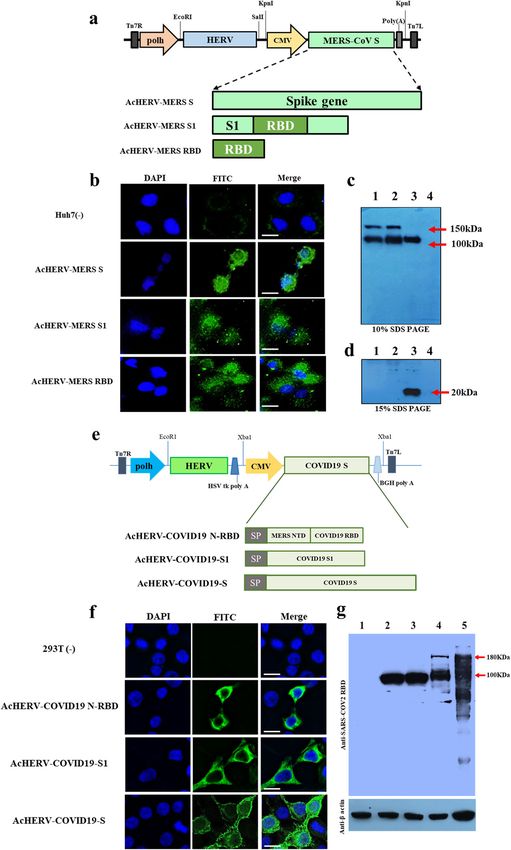
H. Cho et al.
3
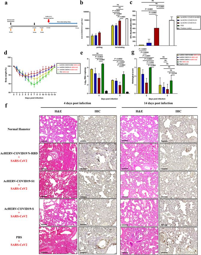
Immunogenicity and MERS-CoV challenge test in hDPP4 b). By day 9 post-MERS-CoV challenge, only one of six mice in the
transgenic mice vaccinated with AcHERV-MERS variants AcHERV-GFP group (16.7%) survived (Fig. 3b). In contrast, mice in
hDPP4 knock-in transgenic (Tg) mice develop severe lung disease the AcHERV-MERS-RBD (3×), AcHERV-MERS-RBD (2×) and AcHERV-
in association with acute respiratory symptoms after challenge MERS-S (2×) groups showed significant decreases in body weight,
with MERS-CoV. To perform various immunization tests, we mass- but partially recovered, exhibiting survival rates of 33.3%, 50%,
produced hDPP4 Tg mice of the same age using an in vitro and 83.3%, respectively (Fig. 3a, c). Notably, mice in AcHERV-MERS-
fertilization (IVF) approach (SI 1). Based on results of previous Ad5- S1 (3×), AcHERV-MERS-S1 (2×) and AcHERV-MERS-S (3×) groups
hDPP4-transduced mice tests (Supplementary Table 1, Supple- showed limited body weight loss and complete protection against
mentary Fig. 1), we performed vaccine experiments on hDPP4 Tg virus lethality (100% survival) by day 14 post challenge (Fig. 3b, d).
mice using two different immunization schedules: three times at Lungs harvested 10 days after infection showed no detectable
levels of MERS-CoV in AcHERV-MERS-S1 (3×) or AcHERV-MERS S
3-week interval (3×) and two times at a 4-week interval (2×)
(3×) groups, determined by RT-qPCR analysis of the gene
(Supplementary Table 2 and Fig. 2a). Under both immunization
encoding the N protein. In contrast, high viral titers were observed
schedules, AcHERV-MERS-S1 induced significantly higher levels of
in lungs of mice in AcHERV-MERS-RBD, AcHERV-GFP, and PBS
IgG and neutralizing antibodies than AcHERV-MERS-S or AcHERV- groups (Fig. 3e). Lung tissues from mice in PBS control and
MERS-RBD (Fig. 2b–d). After the first boost, mice in the 2× group AcHERV-MERS-RBD groups showed severe lesions, including the
showed higher IgG titers than those in the 3× group (Fig. 2b and loss of pulmonary alveoli and diffuse mononuclear cell infiltration.
d). One week after the last immunization, the titers of MERS-CoV AcHERV-MERS-S1 and AcHERV-MERS-S groups exhibited a sig-
neutralizing antibodies were higher in groups treated with nificantly milder lung pathology following MERS-CoV infection
AcHERV-MERS-S1 compared with those in other groups (Fig. 2c, e). (Fig. 3f). These results indicate that vaccination with AcHERV-
Changes in body weight and survival of mice treated with MERS-S1 reduced respiratory pathologies in hDPP4-Tg mice after
AcHERV-MERS vaccines were tested in hDPP4-Tg mice after MERS-CoV challenge. Thus, AcHERV-MERS-S1 can be considered a
challenge with MERS-CoV (Fig. 3a–d). After MERS‐CoV challenge, vaccine candidate against MERS-CoV, which is likely to re-emerge
PBS-injected mice showed profound clinical signs on days 4–6, as a pandemic in the future.
rapid weight loss on days 6–9, and 0% survival by day 11 (Fig. 3a,
Immunogenicity of AcHERV-COVID19 vaccines in BALB/c mice
To evaluate immune responses to AcHERV-based COVID19
vaccines, we immunized BALB/c mice intramuscularly with each
recombinant baculovirus at a concentration of 2 × 10⁷ FFU/ml
(Supplementary Table 3 and Supplementary Fig. 2a). Two weeks
after the last vaccination, AcHERV-COVID19-S, AcHERV-COVID19-
S1, and AcHERV-COVID19-N-RBD induced increases in SARS-CoV2-
specific serum IgG levels in mice (Supplementary Fig. 2b).
However, only AcHERV-COVID19-S elicited neutralizing antibodies
specific to SARS-CoV2 (Supplementary Fig. 2c). The failure of
AcHERV-COVID19-S1 to induce neutralizing antibodies contrasts
with the efficacy of AcHERV-MERS-S1 and is consistent with the
idea that SARS-CoV2 S1 adopts a more complex three-dimensional
structure than MERS-CoV S1. To evaluate cell-mediated immune
responses, we performed ELISPOT assays on splenocytes isolated
4 weeks after the last immunization. These assays revealed
significantly higher levels of IFN-γ in AcHERV-COVID19-vaccinated
mice compared with PBS-treated mice (P < 0.0001; Supplementary
Fig. 2d). The levels of splenocytes-secreted IFN-γ did not
significantly differ among mice treated with AcHERV-COVID19-S,
AcHERV-COVID19-S1, and AcHERV-COVID19-N-RBD.
Immunogenicity and SARS-CoV2 challenge test in Syrian
golden hamsters vaccinated with AcHERV-COVID19 variants
To test the prophylactic effect of AcHERV-COVID19 vaccines, we
first used BALB/c mice. In particular, mice vaccinated with AcHERV-
COVID19 showed a high cell-mediated immune response. Based
on the results of BALB/c mice test (Supplementary Table 3, E
Supplementary Fig. 2), we also used Syrian golden hamsters as an
infectious animal model for SARS-CoV2. Hamsters were immu-
nized twice at a 4-week interval with AcHERV-COVID19 vaccines
Fig. 2 Immunogenicity of AcHERV-MERS vaccines in hDPP4 by intramuscular injection (Supplementary Table 4) and chal-
transgenic mice. a Vaccination schedule. hDPP4 Tg mice used for lenged with intranasal SARS-CoV2 (Fig. 4a). AcHERV-COVID19-S
challenge experiments (Extended Table 2) were immunized three induced significantly higher levels of IgG than other AcHERV-
times at 3-week intervals (3×, prime-boost-boost group) or two COVID19 vaccines (P < 0.0001; Fig. 4b). Neutralization assays
times at a 4-week interval (2×, prime-boost group). b MERS-CoV-S- revealed that vaccination with AcHERV-COVID19-S also elicited
specific total IgG antibody responses in 3× immunization groups, median neutralizing antibody titers that were more than 20-fold
determined by ELISA. c MERS-CoV-S-specific neutralizing antibody in
3× immunization groups. d MERS-CoV-S-specific total IgG antibody higher than those induced by vaccination with AcHERV-COVID19-
responses in 2× immunization groups, determined by ELISA. S1 or AcHERV-COVID19- N-RBD (Fig. 4c).
e MERS-CoV S-specific neutralizing antibody in 2× immunization After SARS-CoV2 challenge, the symptoms of SARS-CoV2 in
groups. P-value determined by one-way ANOVA followed by hamsters were measured by decreased mobility and weight loss,
Tukey–Kramer post hoc tests; NS, P > 0.05. but all recovered at 2 weeks without death. The hamsters of the
Published in partnership with the Sealy Institute for Vaccine Sciences npj Vaccines (2021) 37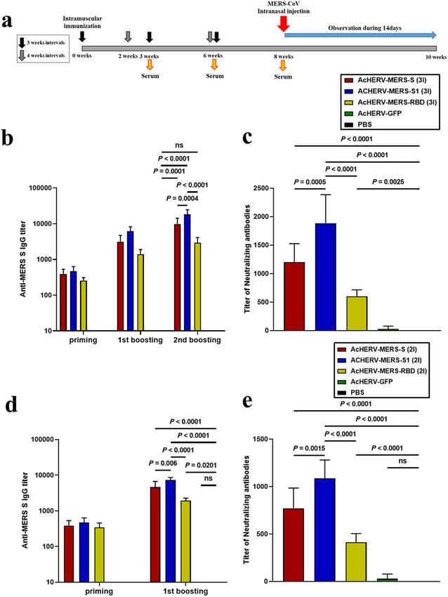
H. Cho et al.
4

    Fig. 3 MERS-CoV challenge test in hDPP4 mice. Immunized hDPP4 mice (2× and 3× groups) were intranasally challenged with MERS-CoV
    and monitored for 14 days. a Body weight in 3× immunization groups. Results are presented as average change in body weight ± standard
    deviation. b The mouse survival rate in 3× immunization groups. c Body weight in 2× immunization groups. d The mouse survival rate in 2×
    immunization groups. e Viral lung titer in vaccinated mice (MERS full-length S, S1 subunit, and RBD), GFP, and non-vaccinated control (PBS)
    mice was determined by qPCR detection of the MERS-CoV N gene. f Assessment of H&E staining in lungs. g Histological evaluation of MERS-
    CoV vaccine groups using hDPP4 mouse. For each of group a score from 0 to 3 was given (i.e., 0 = absent, 1 = minimal, 2 = moderate or 3 =
    severe). Scale bar = 100 μm. P value determined by one-way ANOVA followed by Tukey–Kramer post hoc tests; NS, P > 0.05.

    npj Vaccines (2021) 37                                                     Published in partnership with the Sealy Institute for Vaccine Sciences
H. Cho et al.
5
Fig. 4 Humoral immune responses to AcHERV-COVID19 and SARS-CoV2 challenge test in hamsters. a Vaccination schedule. b SARS-CoV2-
S-specific total IgG antibody responses, determined by ELISA. c SARS-CoV-2–specific neutralizing antibody, determined using SARS-CoV2 virus.
Positive control is serum from recovered SARS-CoV2-infected patients. d General effects of viral infection were assessed by monitoring
changes in body weight daily for 14 days. Data are presented as the average change in weight ± SD. e Viral lung titers of vaccinated hamsters
(COVID19 full-length S, S1 subunit and MERS-N-RBD), non-vaccinated controls (PBS + SARS-CoV2 infection) and negative controls (PBS
without SARS-CoV2 infection) were measured by qPCR detection of the SARS-CoV2 N gene. f H&E staining and immunohistochemistry.
g Histological evaluation of SARS-CoV2 vaccine groups using hamster. For each of group a score from 0 to 3 was given (i.e., 0 = absent, 1 =
minimal, 2 = moderate or 3 = severe) Scale bar = 100 μm. P values were determined by one-way ANOVA followed by Tukey–Kramer post hoc
tests; NS, P > 0.05.
PBS-treated group lost 10.4% of their weight by day 7 and COVID19-S1 and AcHERV-COVID19-N-RBD groups, the body
recovered gradually. On the other hand, body weights of the weight decreased 6.1% by day 5 and 7.1% by day 7, respectively
AcHERV-COVID19-S group were 94.4% of their initial body weights (Fig. 4d). On days 4 and 6 after SARS-CoV2 challenge, viral titers
by day 3 and recovered rapidly. In hamsters of the AcHERV- were measured in lung tissue from all groups by RT-qPCR.
Published in partnership with the Sealy Institute for Vaccine Sciences npj Vaccines (2021) 37H. Cho et al.
6
Although AcHERV-COVID19-S did not provide complete protec- In conclusion, we have provided evidence that AcHERV can
tion, this group showed the lowest viral titer compared with other serve as a vaccine platform of MERS-CoV as well as SARS-CoV2.
groups (Fig. 4e). Animals vaccinated with AcHERV-COVID19-S also The AcHERV system offers several advantages as a DNA vaccine
exhibited diminished pathology compared with controls (Fig. 4f). delivery vector. One major advantage is versatility. Given the
Lung tissues in the PBS group and AcHERV-COVID19-N-RBD group propensity of infectious viruses to mutate, replacement of viral
showed severe lesions, including loss of pulmonary alveoli and antigen S protein-encoding genes in the AcHERV genome would
diffuse mononuclear cell infiltration. Unlike these groups, AcHERV- be a fast and effective way to adapt vaccines to emerging new
COVID19-S and AcHERV-COVID19-S1 groups showed significantly pandemics in the future. Another advantage of the AcHERV
milder pathologies upon SARS-CoV2 infection. These results system is enhanced cellular uptake of AcHERV mediated by HERV
indicate that AcHERV-COVID19-S vaccination reduced respiratory envelope proteins present on the virus surface (Supplementary
pathologies in hamsters after SARS-CoV2 challenge. When looking Fig. 4). The other advantage of AcHERV is safety. Baculoviral genes
at the lung tissue 2 weeks after the challenge, the overall recovery are mostly silent in mammals27; the AcHERV-COVID19 S is
was clearly in the AcHERV-COVID19 S group compared to the PBS expected to solve the safety issues of existing adenovirus vector
control group. vaccines.
These results indicate that the AcHERV-COVID19 S vaccine
candidate can be applied to future vaccine seed viruses.
MATERIALS AND METHODS
Cells
DISCUSSION Spodoptera frugiperda 9 (Sf9) insect cells were propagated in Sf-900 II
In this study, we demonstrated that the AcHERV system could be medium (Invitrogen, USA) supplemented with 3% fetal bovine serum (FBS;
used to induce both humoral and cellular immunogenicity. Gibco, USA) and 1% antibiotic-antimycotic (Invitrogen) at 28 °C. Human
AcHERV expressing the MERS S1 or COVID19 S gene elicited embryonic kidney 293 T (293T) cells, hepatocellular carcinoma (Huh7) and
increases in antigen-specific serum IgG levels and provided 100% Africa green monkey kidney (Vero E6) cells were cultured in Dulbecco’s
protection against the lethal effects of challenge. The complete Modified Eagle Medium supplemented with 10% FBS (Gibco) and 1%
protective effect of AcHERV vaccines in animal models supports penicillin/streptomycin (Gibco) at 37 °C.
the feasibility of AcHERV baculoviral vaccines against MERS-CoV
and SARS-CoV2. Virus preparation and titration
We found that immunization with the AcHERV-COVID19-S MERS-CoV (1-001-MER-IS-2015001) was provided by the Korean Centers for
vaccine induced serum IgG, neutralizing antibody, and antigen- Disease Control and Prevention. SARS-CoV2 (BetaCoV/Korea/LCDC03/2020,
specific IFN-γ secretion. AcHERV-MERS-S1 also elicited high levels NCCP No.43326) was provided by the National Culture Collection for
of IgG, neutralizing antibody, and T-cell immune responses (IFN-γ Pathogens. All experimental procedures were performed in a Biosafety
secretion). Unlike AcHERV-MERS-S1, AcHERV-COVID19-S1 induced Level 3 (BL3) facility with the approval of the Konkuk University
Institutional Animal Care and Use Committee. MERS-CoV was passaged
relatively low neutralizing antibody titers, but still proved effective
in Huh7 cells and the median tissue culture infectious dose (TCID50/ml) was
against SARC-CoV2 challenge. This implies that high cellular determined in the same cell line. Titration of MERS-CoV in Huh7 cells was
immunity conferred therapeutic efficacy after infection. Our carried out using the same protocol as used previously for influenza virus
findings also indicate that AcHERV-based vaccines elicited both titration30. SAR-CoV2 was propagated and titered (TCID50/ml determina-
Th1 and Th2 responses. This balanced activation of Th1 and Th2 tion) in Vero E6 cells.
responses by the AcHERV system would be beneficial for clearing
viral loads. Recent studies have reported that the survival of Construction of AcHERV-MERS and AcHERV-COVID
patients infected with SARS-CoV2 depends on both cell-mediated recombinant baculoviruses
immune responses (Th1) and neutralizing antibodies. Similarly,
A recombinant baculoviral vector expressing HERV env (pFastBac1-HERV)
patients with strong MERS-specific CD8+ T-cell responses exhibit was previously constructed by inserting a synthetic, codon-optimized
rapid virus clearance and thus a relatively brief virus exposure28. envelope gene of HERV type W (GenBank accession number NM014590;
Although the AcHERV system induced the expression of GenScript, USA) into pFastBac1 (Invitrogen). Recombinant AcHERVs
encoded antigens at the cellular level, the degree of induction encoding MERS-CoV antigens (AcHERV-MERS) were constructed by PCR-
and protection afforded by this expression differed depending on amplifying full-length S, S1 subunit, and RBD using the MERS-CoV S gene
the type of encoded antigen. We found that in vivo expression of (EMC strain; provided by Prof. Shibo Jiang, Fudan University) as a template.
the S protein of COVID19, delivered as AcHERV-COVID19-S, was These three genes were individually cloned into pcDNA3.1(+) containing a
more effective than expression of the S1 protein by the AcHERV CMV promoter (Invitrogen), resulting in pc-MERS-S, pc-MERS-S1, and pc-
system. The converse was observed in the case of AcHERV-MERS MERS-RBD, respectively. Then, pFB-HERV-MERS-S, pFB-HERV-MERS-S1, and
vaccines, where greater immunogenicity was observed for the S1 pFB-HERV-MERS-RBD were constructed by inserting pc-MERS-S, pc-MERS-
S1, and pc-MERS-RBD into the pFastBac1-HERV vector. Recombinant
protein than for the full-length S protein. baculoviruses were produced using the Bac-to-Bac baculovirus expression
A previous study similarly reported that recombinant human system according to the manufacturer’s instructions (Invitrogen). The
Ad5 vectors encoding the shorter S1 domain of MERS-CoV scheme for constructing the recombinant baculoviruses, AcHERV-MERS-S,
induced a stronger neutralizing antibody response than those AcHERV-MERS-S1 and AcHERV-MERS-RBD, is shown in Fig. 1a.
encoding full-length S. The mechanisms by which specific Recombinant AcHERVs encoding SARS-CoV2 antigens were constructed
antigens of S or S1 proteins might induce higher immunogenicity by first synthesizing a SARS-CoV2 S gene (structurally similar to the MERS-
will require further study. However, it is possible that the three- COV S gene) that was codon-optimized for optimal expression in
dimensional conformation of proteins expressed using the mammalian cells (GeneArt, USA), and then individually cloning full-
recombinant baculovirus system could affect antigen processing length S and S1 subunit genes into pcDNA3.1 (+) plasmids containing a
and interactions with MHC class proteins in antigen-presenting CMV promoter (Invitrogen). A segment of the MERS-CoV S gene
corresponding to the N-terminal domain (NTD; amino acids 1–386) was
cells. In this context, it is possible that S1 expressed from AcHERV-
fused to the SARS-CoV2 S RBD (amino acids 386–739), resulting in a MERS-
COVID19-S1 might not assemble into the trimeric conformation CoV S NTD-SARS-CoV2 S RBD fusion (MERS-N-RBD) gene. S, S1, and MERS
that mimics the natural three-dimensional S protein. Current all NTD-COVID19 RBD genes were individually cloned into the pFastBac1-
RNA or Ad COVID19 vaccines under clinical trial are encoding full HERV plasmid, and recombinant baculoviruses were produced using the
length of S12,13,21,29. This result underscores the importance of Bac-to-Bac baculovirus expression system (Invitrogen) as described by the
customized protein expression for antigenicity in vivo. manufacturer. The scheme for constructing the recombinant baculoviruses,
npj Vaccines (2021) 37 Published in partnership with the Sealy Institute for Vaccine SciencesH. Cho et al.
7
AcHERV-COVID19-S, AcHERV-COVID19-S1 and AcHERV-MERS-N-RBD, is external jugular vein was centrifuged at 13,000 rpm for 10 min, and the
shown in Fig. 1d. supernatant fraction was used for further assays.
These recombinant baculoviruses (AcHERVs) were further amplified by
propagation in Sf9 cells. The various AcHERVs were purified by first
Enzyme-linked immunosorbent assay
removing debris from virus-infected cells by centrifugation at 6000 × g at
4 °C for 10 min. Supernatants were then overlaid on a 30% sucrose cushion Induction of antibodies specific for MERS S, SARS-CoV2 S, or SARS-CoV2
and centrifuged at 40,000 rpm at 4 °C for 1.5 h in a 50.2Ti rotor (Beckman RBD was tested by enzyme-linked immunosorbent assay (ELISA). For
Coulter Inc., USA). The resulting pellet was resuspended in phosphate- detection of MERS S antibodies, a 96-well plate was coated with the MERS
buffered saline (PBS) and used for characterization and immunization. S protein (WooGene B&G, Korea) at 1 μg/ml by incubating for 16 h at 4 °C.
Baculovirus was titrated by quantitative polymerase chain reaction (qPCR) After blocking with 2% bovine serum albumin in PBS for 1 h at 37 °C, the
using the BacPAK qPCR Titration Kit (Takara Bio USA Inc., USA) according to plate was washed with PBS-T, after which 1/200 serial dilutions of mouse
the manufacturer’s instructions. sera (0.06 ml/well) were added and plates were incubated at room
temperature for 2 h. After washing with PBS-T, plates were incubated with
horseradish peroxidase (HRP)-conjugated goat anti-mouse IgG antibody
Immunofluorescence assay and western blotting (1:10000; Abcam) for 1 h at 37 °C. Next, 3,3′,5,5′-tetramethylbenzidine
The expression of MERS S, S1, and RBD proteins in mammalian cells was (TMB) substrate solution (Bio-Rad, Hercules, USA) was added and plates
tested by infecting Huh7 cells with baculovirus at a multiplicity of infection were incubated for 7 min, followed by addition of 1 N H2SO4 to terminate
(MOI) of 100. Three days after infection, immunofluorescence analyses and the reaction. Absorbance was recorded at 450 nm using a microplate
western blotting were carried out using a polyclonal antibody against spectrophotometer (BioTek Epoch, USA).
MERS-CoV S protein (SICGEN, Portugal). For detection of SARS-CoV2 S-specific antibodies, a 96-well plate was
The expression of SARS-CoV2 S, S1, and MERS N-RBD proteins in coated with the SARS-CoV2 S RBD protein (produced by our lab using an
mammalian cells was tested by infecting 293T cells with baculovirus at a Escherichia coli expression system) at 1 μg/ml by incubating for 16 h at 4 °C.
MOI of 50. Three days after infection, immunofluorescence assays and After blocking with 5% skim milk in PBS for 1 h at 37 °C, plates were
western blotting were carried out using a polyclonal antibody against the washed with PBS-T, then 1/20 serial dilutions of mouse sera (0.06 ml/well)
spike RBD of SARS-CoV2 (Elabscience, USA). were added and plates were incubated at room temperature for 2 h. Wells
The expression of the HERV gene in Sf9 cells following infection (3 MOI) were washed with PBS-T, then HRP-conjugated goat anti-mouse IgG
with each baculovirus construct was assessed by western blotting using a secondary antibody (1:10000; Abcam, UK) was added and plates were
polyclonal rabbit primary antibody specific for HERV Env (abcam, UK). incubated for 1 h at 37 °C. After washing, TMB solution (Bio-Rad) was
Immunofluorescence analyses were observed under a microscope (NIS- added and absorbance at 450 nm was measured using a microplate
Elements-BR, Nikon or THUNDER Imager Model Organism, Leica). Western spectrophotometer (BioTek Epoch).
blots were developed using X-ray films. Uncropped figures are available in
the Supplementary Information file. Marker bands have been indicated by MERS-CoV and SARS-CoV2 neutralization assay
their individual size. All western blots derive from the same experiment MERS-CoV neutralization assays were carried out using MERS-CoV
and were processed in parallel. pseudovirus (provided by Shibo Jang, Pudan University, China). Two
weeks after the last immunization, serum samples were harvested, serially
Animals diluted twofold, and mixed 1:1 with MERS pseudovirus (3 × 10³ RLU
BALB/c mice, human dipeptidyl peptidase 4-transgenic mice (hDPP4 Tg [relative light units]/well). After a 1 h incubation at 37 °C, each
mice), and hamsters were used for immunogenicity testing. Six-week-old pseudovirus–antibody mixture was added to 293T cells. After incubating
female BALB/c mice, purchased from Orient-Bio (Seungnam, Kyonggi-do, at 37 °C for 3 days, cells were lysed, and luciferase content in cell lysates
Republic of Korea), were bred under the highest filter conditions while was determined using Beetle-Juice Luciferase Assay Firefly (PJK GmbH,
Kleinblittersdorf, Germany). Neutralization titers were defined as the
allowing ad libitum access to water and food. Ten-week-old male Syrian
reciprocal of the highest serum dilution at which luciferase activity was
golden hamsters, purchased from the Central Lab Animal (Seoul, Korea),
reduced by at least 50%. Neutralization assays were performed as
were used as the SARS-CoV2 animal model. Human DPP4 mice were
previously reported31.
obtained from the Korea Center for Disease Control (Osong, Korea).
SARS-CoV2 neutralization assays were performed using SARS-CoV2 in a
Because the natural reproductive ability of hDPP4-Tg mice is significantly
BSL3 facility. One week after the last immunization, serum samples were
lower than that of wild-type mice, heterologous DPP4 mice for
harvested, serially diluted twofold, and mixed 1:1 with SARS-CoV2 virus (50
immunization and challenge tests were mass-produced through IVF. Mice TCID50). After a 1-h incubation at 37 °C, each virus–antibody mixture was
were maintained and immunized in a BL2 animal facility in conformance added to Vero E6 cells. After incubating at 37 °C for 3 days, cells were fixed
with The Guide for the Care and Use of Laboratory Animals. Challenge and stained with Crystal violet. Serum from recovered SARS-CoV2-infected
experiments were performed in a BL3 animal facility at Konkuk University. patients was used as a positive control.
All animal husbandry and experimental procedures were approved by the
Konkuk University Institutional Animal Care and Use Committee (IACUC
approval numbers: MERS-CoV, KU18144-1; SARS-CoV2, KU2007). IFN-γ ELISPOT assay
The production of interferon (IFN-γ) from splenocytes of immunized mice
Immunization of animal models was detected by enzyme-linked immune spot assay (ELISOPT; BD
Bioscience, USA), as described by the manufacturer. Briefly, a 96-well plate
For immunization with AcHERV-MERS vaccines, 6-week-old female BALB/c was coated with 0.5 μg/ml of anti-mouse IFN-γ, and then blocked by
mice and hDPP4 Tg mice were injected intramuscularly into the hind legs incubating with RPMI-1640 medium containing 10% FBS and penicillin/
with 2 × 10⁷ FFU of AcHERV-MERS-S, AcHERV-MERS-S1, or AcHERV-MERS- streptomycin/L-glutamine at 37 °C. Splenocytes were seeded at 1 × 10⁶
RBD in PBS or with PBS alone (negative control). BALB/c mice were cells per well in 100 μl of medium, and stimulated by adding MERS
immunized three times at 3-week intervals (Supplementary Table 1), pseudovirus (3 × 10³ RLU/well) or inactivated SARS-CoV2 virus (7 × 10⁵
whereas hDPP4-Tg mice, used for challenge experiments (Supplementary TCID50/well), constructed in our laboratory, and incubating for an
Table 2), were immunized three times at 3-week intervals (3× group) or additional 24 h at 37 °C. Plates were then washed with PBS-T and
two times at a 4-week interval (2× group). incubated with 0.25 μg of biotinylated anti-mouse IFN-γ detection
For immunization with AcHERV-COVID vaccines, 6-week-old female antibodies. After 2 h, streptavidin-HRP was added to the wells, and color
BALB/c mice and 10-week-old male Syrian golden hamsters were injected was developed using an AEC substrate reagent (BD Biosciences, Franklin
intramuscularly into the hind legs with 2 × 10⁷ FFU or 1 × 10⁸ FFU of Lakes, USA). The number of spots was counted using an ELISPOT reader
AcHERV-COVID19-S, AcHERV-COVID19-S1, or AcHERV-MERS-N-RBD in PBS (AID EliSpot Reader ver. 4; Straßberg, Germany).
or with PBS alone (vehicle control). BALB/c mice were immunized three
times at 3-week intervals (Supplementary Table 3), whereas hamsters were
vaccinated two times at a 4-week interval (Supplementary Table 4). Blood Coronavirus challenge models
samples were collected after anesthetizing mice by intramuscular injection MERS-CoV challenge in BALB/c mice transiently expressing the hDPP4
of 40 mg/kg of Zoletil50 (Virbac Laboratories, France) and 5 mg/kg of gene. Prior to challenge tests, BALB/c mice were sensitized to MERS-
Rompun (Bayer Korea, Republic of Korea). Blood sampled from the right CoV by transiently infecting with recombinant Ad5 expressing human
Published in partnership with the Sealy Institute for Vaccine Sciences npj Vaccines (2021) 37H. Cho et al.
8
DPP4 (Ad5-hDPP4; provided by Professor Jae-Hwan Nam, Catholic isolated from mouse-tail tissue using the commercial kit DNeasy® Blood &
University, Korea). Specifically, 2 weeks after the last immunization, mice Tissue (Qiagen) following the protocol provided by the manufacturer. The
were intranasally transduced with 60 μl of Ad5-hDPP4 (3 × 10¹⁰ IFU/ml), hDPP4 gene was identified by PCR using a specific primer set (forward
titrated using an Adeno-X Rapid Titer Kit (Clontech, USA). Five days later, primer: 5′CGC TAT TAC CAT GGT GAT GCG 3′, reverse primer: 5′AGC TGT
mice were intranasally challenged with 60 μl of MERS-CoV (1 × 10⁶ TCID50/ AGC ATC ATC TGT GCC 3′), obtaining an amplicon size of 984 bps. The
ml). Mouse weight was monitored daily for 8 days after challenge. At the following PCR conditions were used: 94 °C for 5 min followed by 35 cycles
end of this period, mice were sacrificed and lungs were harvested. Virus at 94 °C for 1 min, 55 °C for 1 min, 72 °C for 1 min, and final extension at
titer, expressed as TCID50/ml, was measured in Huh7 cells using the same 72 °C for 10 min. The single amplified product was confirmed using
methods as used for MERS-CoV titration. agarose gel electrophoresis.
MERS-CoV challenge in hDPP4-Tg mice. For MERS-CoV challenge, hDPP4- GFP fluorescence and FACS analysis
Tg mice were immunized with various recombinant vaccines or were
administered vehicle control (PBS). The challenge test was conducted To evaluate the AcHERV delivery system, 293T cells were infected with
10 days after the last immunization by intranasally infecting with MERS- AcHERV-cmvGFP or Ac-cmvGFP (MOI 50). Mammalian cells were trans-
CoV (60 μl/mouse, 1 × 10⁶ TCID50/ml). Body weights of mice were duced with recombinant baculoviruses and analyzed by measuring GFP
monitored after challenge. Mice were sacrificed 10 days post infection, fluorescence by FACS analysis at 48 h post transduction (Supplementary
and lungs were harvested for histological staining and qPCR. Fig. 4). GFP fluorescence analyses were observed under a microscope (NIS-
Elements-BR, Nikon). FACS analyses were observed FACSCalibur (Becton
SARS-CoV2 challenge in hamsters. Seven days after the final immunization, Dickinson, USA).
serum was harvested from hamsters immunized with various AcHERV-
COVID vaccines. ELISAs and neutralizing assays were performed using Statistical analysis
hamster sera. Challenge tests were conducted 14 days after the last All statistical analyses were performed using GraphPad Prism 8.0.2
immunization by intranasally infecting with SARS-CoV2 (0.1 ml/dose, 7 × (GraphPad Software), and data are presented as means ± standard
106 TCID50/ml). Body weights were monitored after challenge. Hamsters deviation (SD). Comparisons of data between groups were performed
were sacrificed at 4, 6 and 14 days post infection, and lungs were using one-way analysis of variance (ANOVA) followed by Tukey–Kramer
harvested for histological staining and qPCR. post hoc tests. P valuesH. Cho et al.
9
13. Logunov, D. Y. et al. Safety and immunogenicity of an rAd26 and rAd5 vector- ACKNOWLEDGEMENTS
based heterologous prime-boost COVID-19 vaccine in two formulations: two This research was supported by a grant of the Korea Health Technology R&D Project
open, non-randomised phase 1/2 studies from Russia. Lancet 396, 887–897 through the Korea Health Industry Development Institute (KHIDI), funded by the
(2020). Ministry of Health & Welfare (grant nos. HI15C2842, HI15C1685, HI18C2177). The
14. Li, H. et al. Establishment of replication-competent vesicular stomatitis virus- pathogenic resources (MERS-CoV, SARS-CoV2) for this study were provided by the
based recombinant viruses suitable for SARS-CoV-2 entry and neutralization National Culture Collection for Pathogens. DPP4 + TG mice were provided by
assays. Emerg. Microbes Infect. 9, 2269–2277 (2020). the KCDC.
15. Forster, R., Fleige, H. & Sutter, G. Combating COVID-19: MVA vector vaccines
applied to the respiratory tract as promising approach toward protective
immunity in the lung. Front. Immunol. 11, 1959 (2020). AUTHOR CONTRIBUTIONS
16. Kurup, D., Wirblich, C., Ramage, H. & Schnell, M. J. Rabies virus-based COVID-19
H. Cho, Y.J.: construction of the AcHERVs, data curation, these authors contributed
vaccine CORAVAX induces high levels of neutralizing antibodies against SARS-
equally to this work (co-first author), K.H.P. and H. Choi: ABL3 and BL3 experiment, A.N.:
CoV-2. NPJ Vaccines 5, 98 (2020).
in vitro assay, H.J.L. and M.K.: material supply and data analysis, M.H.K. and J.H.K.: animal
17. Hassan, A. O. et al. A single-dose intranasal ChAd vaccine protects upper and
IVF mass production, H.Y.S.; data analysis, Y.K.O.; MS writing. Y.B.K.: design, and writing.
lower respiratory tracts against SARS-CoV-2. Cell 183, 169–184 (2020). e113.
18. Feng, L. et al. An adenovirus-vectored COVID-19 vaccine confers protection from
SARS-COV-2 challenge in rhesus macaques. Nat. Commun. 11, 4207 (2020).
19. Case, J. B. et al. Replication-competent vesicular stomatitis virus vaccine vector COMPETING INTERESTS
protects against SARS-CoV-2-mediated pathogenesis in mice. Cell Host Microbe The authors declare no competing interests.
28, 465–474 (2020). e464.
20. Chiuppesi, F. et al. Development of a synthetic poxvirus-based SARS-CoV-2 vac-
cine. Preprint at bioRxiv https://doi.org/10.1101/2020.07.01.183236 (2020). ADDITIONAL INFORMATION
21. Tostanoski, L. H. et al. Ad26 vaccine protects against SARS-CoV-2 severe clinical Supplementary information The online version contains supplementary material
disease in hamsters. Nat. Med. 26, 1694–1700 (2020). available at https://doi.org/10.1038/s41541-021-00303-w.
22. Munster, V. J. et al. Protective efficacy of a novel simian adenovirus vaccine
against lethal MERS-CoV challenge in a transgenic human DPP4 mouse model. Correspondence and requests for materials should be addressed to Y.B.K.
NPJ Vaccines 2, 28 (2017).
23. Kost, T. A. & Condreay, J. P. Recombinant baculoviruses as mammalian cell gene- Reprints and permission information is available at http://www.nature.com/
delivery vectors. Trends Biotechnol. 20, 173–180 (2002). reprints
24. Lung, O., Westenberg, M., Vlak, J. M., Zuidema, D. & Blissard, G. W. Pseudotyping
Autographa californica multicapsid nucleopolyhedrovirus (AcMNPV): F proteins Publisher’s note Springer Nature remains neutral with regard to jurisdictional claims
from group II NPVs are functionally analogous to AcMNPV GP64. J. Virol. 76, in published maps and institutional affiliations.
5729–5736 (2002).
25. Wilson, S., Baird, M. & Ward, V. K. Delivery of vaccine peptides by rapid con-
jugation to baculovirus particles. Vaccine 26, 2451–2456 (2008).
26. Buck, C. B., Pastrana, D. V., Lowy, D. R. & Schiller, J. T. Efficient intracellular
assembly of papillomaviral vectors. J. Virol. 78, 751–757 (2004). Open Access This article is licensed under a Creative Commons
27. Shin, H. Y. et al. Unraveling the genome-wide impact of recombinant baculovirus Attribution 4.0 International License, which permits use, sharing,
infection in mammalian cells for gene delivery. Genes 11, https://doi.org/10.3390/ adaptation, distribution and reproduction in any medium or format, as long as you give
genes11111306 (2020). appropriate credit to the original author(s) and the source, provide a link to the Creative
28. Zhang, Y. Y., Li, B. R. & Ning, B. T. The comparative immunological characteristics Commons license, and indicate if changes were made. The images or other third party
of SARS-CoV, MERS-CoV, and SARS-CoV-2 coronavirus infections. Front. Immunol. material in this article are included in the article’s Creative Commons license, unless
11, 2033 (2020). indicated otherwise in a credit line to the material. If material is not included in the
29. Baden, L. R. et al. Efficacy and safety of the mRNA-1273 SARS-CoV-2 vaccine. N. article’s Creative Commons license and your intended use is not permitted by statutory
Engl. J. Med. https://doi.org/10.1056/NEJMoa2035389 (2020). regulation or exceeds the permitted use, you will need to obtain permission directly
30. Gwon, Y. D. et al. Immunogenicity of virus like particle forming baculoviral DNA from the copyright holder. To view a copy of this license, visit http://creativecommons.
vaccine against pandemic influenza H1N1. PLoS ONE 11, e0154824 (2016). org/licenses/by/4.0/.
31. Chun, J. et al. Effect of Fc fusion on folding and immunogenicity of middle east
respiratory syndrome coronavirus spike protein. J. Microbiol. Biotechnol. 29,
813–819 (2019). © The Author(s) 2021
Published in partnership with the Sealy Institute for Vaccine Sciences npj Vaccines (2021) 37You can also read