Functional assessment of miR 1291 in colon cancer cells
←
→
Page content transcription
If your browser does not render page correctly, please read the page content below
INTERNATIONAL JOURNAL OF ONCOLOGY 60: 13, 2022

           Functional assessment of miR‑1291 in colon cancer cells
      JIAQI WANG1, YUHKI YOKOYAMA1, HARUKA HIROSE1, YUKI SHIMOMURA1, SAKI BONKOBARA1,
          HIROAKI ITAKURA2, SHIHORI KOUDA1, YOSHIHIRO MORIMOTO2, KAZUMASA MINAMI3,
          HIDEKAZU TAKAHASHI2, SATOSHI SHIBATA1, SHOGO KOBAYASHI2, MAMORU UEMURA2,
       SUSUMU TANAKA4, XIN WU1, SHINJI TANAKA5,6, MASAKI MORI7 and HIROFUMI YAMAMOTO1,2

      1
        Department of Molecular Pathology, Division of Health Sciences, Osaka University; Departments of 2Surgery and
           Gastroenterological Surgery, and 3Radiation Oncology, Graduate School of Medicine, Osaka University;
4
  First Department of Oral and Maxillofacial Surgery, Graduate School of Dentistry, Osaka University, Suita, Osaka 565‑0871;
         Departments of 5Molecular Oncology and 6Hepato‑Billiary‑Pancreatic Surgery, Graduate School of Medicine,
           Tokyo Medical and Dental University, Tokyo 113‑8510; 7Tokai University, Graduate School of Medicine,
                                             Isehara, Kanagawa 259‑1193, Japan

                                         Received May 7, 2021; Accepted December 6, 2021

                                                      DOI: 10.3892/ijo.2022.5303

Abstract. miR‑1291 exerts an anti‑tumor effect in a subset             formation, invasion and mobility in HCT116 cells were also
of human carcinomas, including pancreatic cancer. However,             explored and verified using DCLK1 siRNAs. Furthermore,
its role in colorectal cancer (CRC) is largely unknown. In the         miR‑1291 induced CDK inhibitors p21WAF1/CIP1 and p27KIP1
present study, the expression and effect of miR‑1291 in CRC            in three CRC cell lines, and the overexpression of DCLK1
cells was investigated. It was identified that miR‑1291 signifi‑       in HCT116 cells led to a decrease of p21WAF1/CIP1 and p27KIP1.
cantly suppressed the proliferation, invasion, cell mobility           Intravenous administration of miR‑1291 loaded on the super
and colony formation of CRC cells. Additionally, miR‑1291              carbonate apatite delivery system significantly inhibited tumor
induced cell apoptosis. A luciferase reporter assay revealed           growth in the DLD‑1 xenograft mouse model. Additionally,
that miR‑1291 directly bound the 3'‑untranslated region                the resultant tumors exhibited significant upregulation of the
sequence of doublecortin‑like kinase 1 (DCLK1). miR‑1291               p21WAF1/CIP1 and p27KIP1 protein with treatment of miR‑1291.
also suppressed DCLK1 mRNA and protein expression in                   Taken together, the results indicated that miR‑1291 served an
HCT116 cells that expressed DCLK1. Furthermore, miR‑1291               anti‑tumor effect by modulating multiple functions, including
suppressed cancer stem cell markers BMI1 and CD133, and                cancer stemness and cell cycle regulation. The current data
inhibited sphere formation. The inhibitory effects on sphere           suggested that miR‑1291 may be a promising nucleic acid
                                                                       medicine against CRC.

                                                                       Introduction
Correspondence to: Dr Yuhki Yokoyama, Department of
                                                                       Colorectal cancer (CRC) was the third most widespread
Molecular Pathology, Division of Health Sciences, Osaka University,
Yamadaoka 1‑7, Suita, Osaka 565‑0871, Japan
                                                                       cancer and the second most deadly cancer in 2018, world‑
E‑mail: yyokoyama@sahs.med.osaka-u.ac.jp                               wide. There were 1,800,977 new cases of CRC and 861,663
                                                                       related deaths in 2018 (1,2). In the past few decades, improved
Abbreviations: Antagomir‑1291, Hsa‑miR‑1291 inhibitor S‑Tud;           treatment options have become available, including surgery,
BMI1, B cell‑specific Moloney murine leukemia virus integration        radiotherapy, chemotherapy and molecular‑targeted therapy
site 1; BrdU, Bromodeoxyuridine; CDK, cyclin dependent kinase;         for advanced CRC (3,4). However, the 5‑year survival rate of
CSC, cancer stem cell; CRC, colorectal cancer; Del, deleted type;      CRC is
2                                     WANG et al: ROLE OF miR-1291 IN COLORECTAL CANCER

pathways, including Wnt/β‑catenin, epidermal growth factor          that miR‑1291 levels were slightly increased in liver metas‑
receptor (EGFR), transforming growth factor‑ β (TGF‑ β),            tasis compared with primary CRC by using the NCBI Gene
tumor protein p53 and epithelial‑to‑mesenchymal transition          Expression Omnibus (GEO) database (47). Therefore, the aim
(EMT) (12‑16). Furthermore, miRNAs have been determined             of the present study was to clarify the anti‑tumor effect of
to play a pivotal role in regulating these pathways. For example,   miR‑1291 in CRC cells, partially including DCLK1 regulation.
miR‑4689 exerts an anti‑tumor effect on mutant kirsten rat
sarcoma virus (KRAS) in CRC by inhibiting the EGFR and              Materials and methods
AKT pathway (17). MIRTX, a byproduct of miR‑29b‑1‑5p
suppresses the NF‑κ B signaling pathway in KRAS‑mutated             Cell lines and cell culture. Human CRC cell lines (CACO‑2,
CRC cells by directly binding to CXC chemokine receptor 2           COLO205, DLD‑1, HCT116, LoVo, RKO and SW480), the
and phosphatidylinositol 3‑kinase regulatory subunit alpha          human pancreatic cancer cell line Panc‑1, and non‑tumor
mRNA (18). In addition, miR‑34a can inhibit cell prolif‑            cell lines (293, CCD‑18Co and MRC5) were purchased
eration and increase the expression of p21WAF1/CIP1 in HCT116       from the American Type Culture Collection. The HT29 cell
and RKO CRC cells (19). It has been recently reported that          line (cat. no. KBN0398_01) was obtained from Japanese
miR‑4711‑5p regulates cancer stemness and the cell cycle in         Collection of Research Bioresources Cell Bank. Short tandem
CRC cells by targeting Kruppel‑like factor 5, mouse double          repeat (STR) profiling for authentication indicated that the
minute 2 homolog and transcription factor Dp‑1 (20).                HT29 cell line was the same as the one registered with the
    Cancer stem cells (CSCs) have distinct capability for           American Type Culture Collection (HTB‑38 HT‑29: human
self‑renewal and multi‑potential differentiation (21). CSCs         adenocarcinoma; Colorectal). KM12SM (48) was a gift from
confer resistance to anticancer drugs and radiotherapy,             Professor Toshinari Minamoto (Cancer Research Institute,
and thus cause distant metastases and recurrence (22‑24).           Kanazawa University, Kanazawa, Japan). These cell lines were
Several CSC markers for CRC have been identified, including         authenticated by morphological inspection, STR profiling
CD133, B cell‑specific Moloney murine leukemia virus                and mycoplasma testing. Cells were cultured either in RPMI
integration site 1 (BMI1) and leucine rich repeat containing        1640 medium or in DMEM (each, Nissui Pharmaceutical Co.,
G protein‑coupled receptor 5 (23,25,26). Previously, double‑        Ltd.) supplemented with 10% FBS (Biowest SAS), 100 U/ml
cortin‑like kinase 1 (DCLK1) was demonstrated to be a CSC           penicillin and 100 µg/ml streptomycin (Nacalai Tesque, Inc.).
marker in CRC (27,28). DCLK1 belongs to the protein kinase          Cells were cultured in a humidified incubator at 37˚C in an
superfamily and is overexpressed in several human malignan‑         atmosphere containing 5% CO2.
cies, including colorectal, pancreas and kidney cancer (29‑31).
Our preliminary data also demonstrated that miR‑1291 exhib‑         Retroviral transduction of the degron reporter into pancre‑
ited a potent growth inhibitory effect on pancreatic cancer         atic cancer Panc‑1 cells. The degron sequence of ornithine
stem‑like cells in which the ornithine decarboxylase (ODC)          decarboxylase (ODC) is recognized directly by proteasomes,
degron was transduced (32‑34).                                      which leads to the destruction of the involved protein. The
    It has been reported that miR‑1291 sensitizes cells to          retroviral expression vector pQCXIN‑ZsGreen‑cODC,
doxorubicin by suppressing multidrug resistance‑associated          containing green fluorescence ZsGreen‑labeled degron ODC
protein 1 (35). Additionally, miR‑1291 is considered to be a        (Gdeg) was kindly provided by Dr Frank Pajonk (Jonsson
candidate biomarker for the diagnosis of acute myocardial           Comprehensive Cancer Center, UCLA, CA, USA). The
infarction (36) and Bullous Pemphigoid (37), and is also a          plasmid was transfected into Platinum retroviral packaging
marker for the prediction of severe symptoms in SARS‑CoV‑2          cells using Lipofectamine® 2000 (Thermo Fisher Scientific,
infection (38). In relation to human cancers, miR‑1291 has          Inc.). The plasmid and Lipofectamine® 2000 were diluted
been reportedly upregulated in liver cirrhosis and hepatocel‑       with Opti‑MEM I reduced serum medium (Thermo Fisher
lular carcinoma (39). In addition, miR‑1291 is a biologically       Scientific, Inc.) for 5 min at room temperature, separately. The
relevant regulator of glypican‑3 expression in hepatoma cells       diluted Lipofectamine® 2000 was subsequently mixed with
and acts by silencing the endoplasmic reticulum (ER) stress         the diluted plasmid and incubated for 15 min at room tempera‑
sensor, inositol‑requiring transmembrane kinase/endoribo‑           ture. The mixture was then added to cells immediately at room
nuclease 1α (40). Several studies have demonstrated that            temperature, after which the cells were incubated at 37˚C. And
miR‑1291 inhibits cell proliferation and tumorigenesis, and         the retrovirus collected from the supernatant was used for
sensitizes pancreatic cancer cells to chemotherapy (41‑43).         Panc‑1 cell infection as previously described (32‑34). Stable
Furthermore miR‑1291 is reportedly downregulated in                 transfectants were selected with G418 solution (Sigma‑Aldrich;
kidney, esophagus and prostate carcinoma, serving anti‑tumor        Merck KGaA) and maintained in 0.1 mg/ml G418 solution.
effects (44‑46). The target molecules for miR‑1291 include
solute career family 2 member 1/glucose transporter 1 in renal      Clinical tissue samples. When the tumor diameter >3 cm,
cancer cells, mucin 1 (MUC1) in human esophagus cancer              paired clinical tissue specimens (normal mucosa and
cells (44,45) and forkhead box protein A2‑anterior gradient 2       CRC tissue) were randomly collected from 20 patients
pathway in PANC‑1 pancreatic cancer cells (43). Furthermore,        (10 males and 10 females; age range, 18‑87 years) who had
miR‑1291 has been demonstrated to inhibit cell growth and           surgery for colorectal cancer at Osaka University Hospital
tumorigenesis in prostate cancer by binding to Mediator             (Osaka, Japan) between October 2016 and April 2017. All
Complex Subunit 1 (46).                                             tissue specimens were stored at ‑80˚C until RNA extraction.
    Detailed functional assessments of miR‑1291 have not yet        This study was performed in accordance with the Declaration
been conducted in CRC. However, Salehi et al (47) reported          of Helsinki. All patients provided written informed consent, in
INTERNATIONAL JOURNAL OF ONCOLOGY 60: 13, 2022 3
accordance with the guidelines approved (approval no. 08226) 30 sec; followed by 40 cycles of 95˚C for 10 sec, 60˚C for
by the Institutional Research Board of the institute. The present 10 sec and 72˚C for 30 sec. The expression of the target gene
study was conducted under the supervision of and approved by was normalized to endogenous GAPDH expression. Relative
the Ethics Board of Osaka University Hospital. expression was quantified by the 2‑ΔΔCq method (49). The PCR
primers are listed in Table SI.
miRNA, antagomir, siRNA and plasmid transfection.
Mimic‑hsa‑miR‑1291 (miR‑1291) sense (5'‑UGGCCCUGA RT‑qPCR analysis of miRNA. The TaqMan MicroRNA
CUG A AG ACC AGC AGU‑3') and antisense (5'‑ACU G CU Reverse Transcription kit (Thermo Fisher Scientific, Inc.) was
GGUC UUCAGUCAG GGC CA‑3') sequences, along with used to synthesize the complementary DNA from 25 ng of
the negative control miR (miR‑NC) sense (5'‑AUCCGCGCG total RNA according to the manufacturer's protocol. qPCR of
AUAGUACGUA‑3') and antisense (5'‑UACGUAC UAUCG miRNA was then performed using TaqMan Universal PCR
CGCG GAU‑3') sequences were designed and synthesized Master Mix, No AmpErase UNG (Thermo Fisher Scientific,
by Ajinomoto Bio‑Pharma. Hsa‑miR‑1291 inhibitor S‑Tud Inc.) with a 7900 HT Sequence Detection System (Thermo
(antagomir‑1291) sense [5'‑GACG GCG CUAGGAUCAUC Fisher Scientific, Inc.). RNU6B was used as the endogenous
AACACUG CUG GUC UUC AGU CAG GG C CAC AAG UA control. The primers for miR‑1291 and RNU6B were designed
UUCUGGU‑3'] and antisense [5'‑ACCAGAAUACAACAC by Thermo Fisher Scientific, Inc. (miR‑1291, Assay ID:
UGCU GGU CU U CAG UCAGG G CCACA AGAU GAU CC 002838; RNU6B, Assay ID: 001093; cat. no. 4427975). The
UAGCGCCGUC‑3'] were 2‑OMethyl modified, and were qPCR conditions were as follows: 95˚C for 10 min; followed
also designed and synthesized by Ajinomoto Bio‑Pharma. by 45 cycles of 95˚C for 15 sec, and 60˚C for 1 min and 72˚C
Three small interfering RNAs (siRNAs) targeting DCLK1 for 1 sec; cooling to 40˚C for 30 sec. Relative expression was
were obtained from Thermo Fisher Scientific, Inc. (Assay quantified with the 2‑ΔΔCq method.
IDs: s17584, s17585 and s17586). Cells were transfected
with miRNAs, antagomir, and siRNAs at final concentra‑ Cell viability assay. Cells were seeded in 96‑well plates at a
tion of 30‑50 nM and plasmids at concentration of 50 ng per density of 4,000‑8,000 cells per well and were transfected with
well (96‑well plate) or 1 µg per well (six‑well plate) using miR‑NC or miR‑1291 or antagomir‑1291 or DCLK1‑siRNA at
Lipofectamine® 2000 or Lipofectamine® RNAiMAX (both a final concentration of 30 nM as aforementioned, the second
from Thermo Fisher Scientific, Inc.) The nucleotide (miRNAs, day after seeding. A total of 24, 48 and 72 h after transfection,
antagomir, siRNAs and plasmids) and transfection reagent 10 µl Cell Counting Kit‑8 (Dojindo Molecular Technologies,
(Lipofectamine® 2000 or Lipofectamine® RNAiMAX) were Inc.) solution was added to each well, after which the 96‑well
diluted with serum free medium (RPMI‑1640 or DMEM) for plates were kept in dark for 2 h at 37˚C. The absorbance was
5 min at room temperature, separately. The diluted transfection then detected using a Multiskan Go plate reader (Thermo
reagent was subsequently mixed with the diluted nucleotide Fisher Scientific, Inc.). The differences in absorption
and incubated for 15 min at room temperature. The mixture at 630 and 450 nm wavelengths were then subtracted and used
was then added to cells immediately at room temperature, to determine cell viability.
after which the cells were incubated at 37˚C. The subsequent
experiments were performed at 4 and 24 h after transfection Matrigel invasion assay. Corning BioCoat Matrigel Invasion
for miRNA uptake into cells and from 24 to 72 h for various Chambers (pore size: 8.0 µm; Corning, Inc.; cat. no. 354480)
anti‑tumor feature assessments. were used. The upper chamber, which was pre‑coated with
Matrigel, was rehydrated with culture medium (RPMI‑1640
RNA isolation. Total RNA was collected from each cell or DMEM) for 2 h at 37˚C in 5% CO2 before seeding the cells.
line using TRIzol® Reagent (Thermo Fisher Scientific, The medium (RPMI‑1640 or DMEM) was then removed,
Inc.) followed by phenol‑chloroform extraction and ethanol and DLD‑1, HT29 and HCT116 cells were seeded into the
precipitation. Subsequently, miRNA was collected from upper chambers at a density of 1‑2x10⁵ cells per chamber with
tissue specimens and cultured cells using the miRNeasy kit medium (RPMI‑1640 or DMEM) containing 0.1% bovine
(Qiagen GmbH) according to the manufacturer's protocol. serum albumin. Medium (RPMI‑1640 or DMEM) containing
Total RNA concentration and purity were measured using a 10% FBS were put in the lower wells. The cells were trans‑
NanoDrop one spectrophotometer (Thermo Fisher Scientific, fected with the miRNAs or antagomir or DCLK1‑siRNA at
Inc.) at 260 and 280 nm (A260/280) wavelengths. a final concentration of 50 nM and incubated at 37˚C after
transfection. After incubation for 48, 72 and 96 h, cells
Reverse transcription‑quantitative (RT‑q) PCR analysis passing through the Matrigel were fixed with 10% formalin
of mRNA. A High Capacity cDNA Reverse Transcription for 1 h at room temperature and then stained with hematoxylin
kit (Thermo Fisher Scientific, Inc.) was used to synthesize for 1 h at room temperature. To count cells passing through
complementary DNA from 2.5 µg of total RNA according the Matrigel, images were captured using a bright field light
to the manufacturer's protocol. qPCR for DCLK1, BMI1 and microscope (CKX53; Olympus Corporation) with Visualix
CD133 RNA was performed using oligonucleotide primers camera (Visualix, Corporation) at a magnification of x200.
and the LightCycler 480 Real‑Time PCR system (Roche
Diagnostics). The amplification products were detected Gap closure assay. Ibidi culture inserts (8.4 width x8.4 length
using the THUNDERBIRD SYBR qPCR Mix (Toyobo x5 mm height; Ibidi GmbH) with two 70 µl wells were put
Life Science), and the level of target gene expression was on the 24‑well plates before cell seeding. DLD‑1, HT29 and
calculated. The qPCR conditions were as follows: 95˚C for HCT116 cell suspensions (70 µl) were then added into each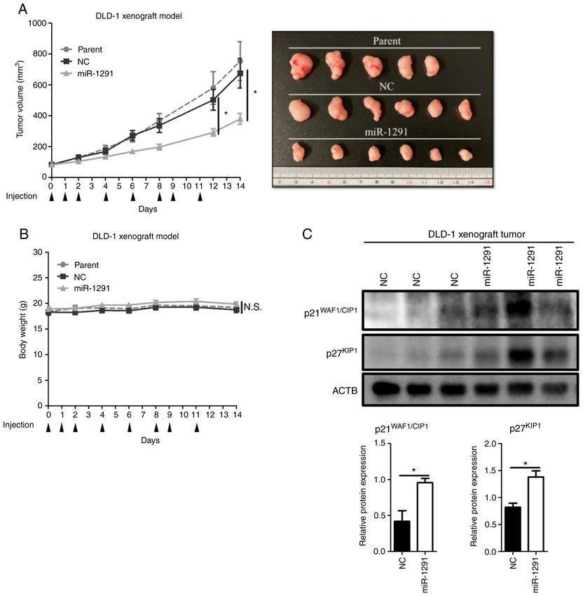
4 WANG et al: ROLE OF miR-1291 IN COLORECTAL CANCER
70 µl well at a density of 2x10⁵, 2.5x10⁵ and 8x10⁵ cells per 5 µl Alexa Flour 488 Annexin V and 1 µl 100 µg/ml PI was
ml, respectively. The inserts were removed after 24 h to create added. Samples were subsequently incubated for 15 min at
gap with an area of 6x105 pixel 2. After that, the miRNAs room temperature. A total of 400 µl 1X annexin‑binding buffer
or antagomir or DCLK1‑siRNA were transfected at a final was added to each cell suspension and apoptotic cells were
concentration of 30 nM. To enhance gap closure, cells were counted by flow cytometry using Spectral Analyzer SA3800
cultured in the medium (RPMI‑1640 or DMEM) supplemented (Sony Biotechnology, Inc.) with SA3800 2.0 software (Sony
with 10% FBS as previously described (50,51). At 24 and 48 h Biotechnology, Inc.).
after transfection, images were captured by a bright field light
microscope (CKX53; Olympus Corporation) with Visualix Western blot analysis. Cells were seeded in six‑well plates at a
camera (Visualix, Corporation) at a magnification of x100. density of 1x10⁵‑2x10⁵ per well and transfected with miR‑NC,
The areas of the gaps were measured using ImageJ 1.52v soft‑ miR‑1291 or DCLK1‑siRNA at a final concentration of
ware (National Institutes of Health). 30‑50 nM. After 48 and 72 h, cells were rinsed twice with PBS
and lysed by RIPA buffer (0.05 M Tris‑HCl, pH 7.6, 0.15 M
Colony formation assay. Cells were seeded in a six‑well NaCl, 1% Nonidet P40, 0.5% sodium deoxycholate, 0.1%
plate at a density of 1x10⁵ cells per well, incubated at 37˚C SDS) with 1% proteinase inhibitor cocktail (Nacalai Tesque,
overnight and transfected with miR‑NC or miR‑1291 at a final Inc.). The protein samples (30 µg/lane) were electrophoresed
concentration of 30 nM for 8 h. Samples were then reseeded in by SDS‑PAGE using 10, 13 or 15% acrylamide gel and trans‑
six‑well plates at a density of 500 cells per well. A colony was ferred to PVDF transfer membranes (Bio‑Rad Laboratories,
considered to consist of ≥50 cells. Cells were incubated for Inc.). The membranes were blocked with 5% non‑fat dry
10 days at 37˚C after transfection, after which cells were fixed milk (Cell Signaling Technology, Inc.) in TBS with Tween‑20
with 100% methanol for 30 min at room temperature, and (TBS‑T; 50 mM Tris, 158 mM NaCl, 2.7 mM KCl, pH 7.5,
stained by 0.5% crystal violet for 10 min at room temperature 0.1% Tween‑20) for 1 h at room temperature and incubated
for counting. The images of each well were scanned using an with the following primary antibodies overnight at 4˚C:
Epson scanner GT‑X970 (Seiko Epson Corporation), and the Anti‑ACTB (1:4,000; Rabbit mAb, cat. no. 4970; Cell Signaling
colonies were counted using ImageJ 1.52v software (National Technology, Inc.), and anti‑DCLK1 (1:2,000; cat. no. ab31704;
Institutes of Health). Abcam) anti‑p21WAF1/CIP1 (1:1,000; cat. no. ab80633; Abcam),
anti‑p27 KIP1 (1:1,000; sc‑528, Santa Cruz Biotechnology,
Cell cycle assay. DLD‑1, HT29 and HCT116 cells were seeded Inc.), anti‑CD133 (1:1,000; ab216323; Abcam), anti‑CDC25A
to six‑well plates at a density of 3x10⁵, 4x10⁵ and 3.5x10⁵ cells (1:1,000; cat. no. 3652; Cell Signaling Technology, Inc.),
per well, respectively. Cells were starved in serum‑free anti‑CDC25B (1:1,000; cat. no. 9525; Cell Signaling
medium (RPMI‑1640 or DMEM) for 48 h. A total of 24 h Technology, Inc.), anti‑CDC25C Rabbit mAb (1:1,000;
before the end of starvation, miR‑NC or miR‑1291 was trans‑ cat. no. 4688; Cell Signaling Technology, Inc.), anti‑CDK4
fected at a final concentration of 30 nM. Cells were collected (1:1,000; cat. no. MAB8879; MilliporeSigma), anti‑CDK6
at the indicated times (0, 12, 24 and 48 h) and fixed in 70% (1:1,000; cat. no. SAB4300596; Sigma‑Aldrich; Merck
ethanol for 30 min at 4˚C. After fixation, cells were washed KGaA), anti‑Cyclin D1 (1:1,000; cat. no. 2922; Cell Signaling
twice with PBS and incubated with RNase (Sigma Aldrich; Technology, Inc.), anti‑Cyclin E1 (1:1,000; cat. no. sc‑247; Santa
Merck KGaA) for 20 min at 37˚C. Cells were treated with Cruz Biotechnology, Inc.), anti‑retinoblastoma (Rb; 1:1,000;
propidium iodide (PI; Dojindo Molecular Technologies, Inc.) cat. no. ab24; Abcam), anti‑cdc2 (1:1,000; cat. no. 77055; Cell
for 20 min on ice and analyzed by flow cytometry (Spectral Signaling Technology, Inc.) and anti‑phosphorylated (p)‑cdc2
Analyzer SA3800; Sony Biotechnology, Inc.) with SA3800 [(Tyr15); 1:1,000; cat. no. 9111; Cell Signaling Technology,
2.0 software (Sony Biotechnology, Inc.). Inc.)]. Subsequently, the membranes were incubated with
secondary antibodies, including HRP anti‑mouse IgG (1:3,000;
Bromodeoxyuridine (BrdU) Proliferation assay. Cell prolif‑ cat. no. NA931; GE Health Care Life Sciences) or anti‑rabbit
eration assays were performed using CysLex Cellular BrdU IgG antibodies (1:3,000; cat. no. NA934; GE Healthcare
ELISA kit Ver.2. (Medical & Biological Laboratories Co., Life Sciences) for 1 h at room temperature. The bands were
Ltd.). Cells were cultured with BrdU labeling reagent at 10 µM visualized by the ECL Detection System (GE Healthcare Life
for 2 h at 37˚C, then incubated with 50 µl per well of anti‑BrdU Sciences) and analyzed using ImageJ 1.52v software (National
monoclonal antibody (provided in the kit) for 1 h at room Institutes of Health).
temperature, followed by 50 µl per well of secondary antibody
reaction with HRP‑conjugated anti‑mouse IgG (provided in pmirGLO plasmid vector construction. The 3'UTR of
the kit), for 1 h at room temperature. After the addition of DCLK1 mRNA was amplified by PCR using the following
the substrate reagent (provided in the kit), the absorbance in primer sequences (amplified product size, 211 bp): forward,
each well was measured using a Multiskan Go microplate 5'‑GCTCGCTAGCCTCGAGCTAGTGTACTGAGCCTGCG
spectrophotometer (Thermo Fisher Scientific, Inc.) at dual G‑3' and reverse, 5'‑ATG C CT G CA G GT C GA C TG ACT
wavelengths of 450/540 nm. GGTCACATTCCAC TG‑3'. The amplified products were
subcloned and ligated into the multicloning site between
Annexin V apoptosis assay. Apoptotic cells were assessed SalI and XhoI in the pmirGLO Dual‑Luciferase miRNA
using an Alexa Fluor 488. Annexin V/Dead Cell Apoptosis Target Expression Vector (Promega Corporation) using the
kit (Thermo Fisher Scientific, Inc.). A total of 2x10⁵ cells were In‑Fusion HD Cloning kit (Clontech Laboratories, Inc.) The
diluted with 100 µl 1X annexin‑binding buffer, after which vectors with mismatched 3'UTR sequences were constructed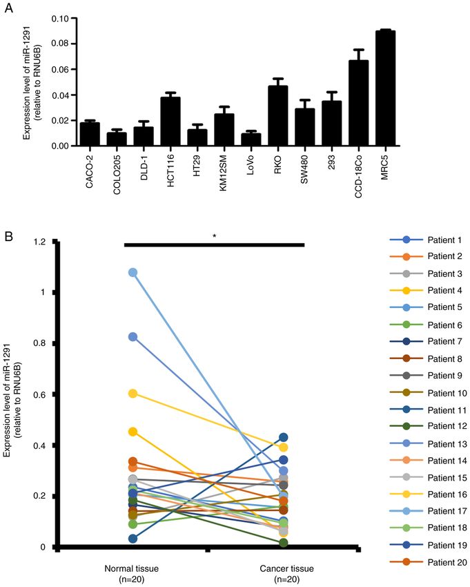
INTERNATIONAL JOURNAL OF ONCOLOGY 60: 13, 2022 5
using the QuikChange Site Directed Mutagenesis kit (Agilent overnight, and transfected with miR‑NC or miR‑1291 or
Technologies, Inc.) according to the manufacturer's protocol. DCLK1‑siRNA at a final concentration of 50 nM. After 24 h
The PCR primers for mutated type and deleted type plas‑ of transfection, single cells were reseeded in 96‑Well Clear
mids are listed in Table SI. The entire sequence (insert and Ultra Low Attachment Microplates (Corning, Inc.) at a density
vector) was confirmed by Sanger sequencing (outsourced to of 1,000 cells per well. The cells were cultured in DMEM/
Genome Information Research Center, Osaka University, F‑12 serum‑free medium (Thermo Fisher Scientific, Inc.)
Suita, Osaka). supplemented with 20 ng/ml epithelial growth factor, 10 ng/ml
basic fibroblast growth factor‑2 (PeproTech, Inc.), 100 U/ml
Luciferase reporter assay. Cells were seeded in 96‑well penicillin and 100 µg/ml streptomycin. Cells were cultured
plates at a density of 1x10 4 cells per well and transfected in a humidified incubator at 37˚C and 5% CO2. Images were
with 50 ng of DCLK1 wild type, 2‑nucleotide mutated type captured by a bright field light microscope (CKX53; Olympus
(Mut) or 3‑nucleotide deleted type (Del) 3'UTR containing Corporation) with Visualix camera (Visualix, Corporation).
pmirGLO Dual‑Luciferase miRNA Target Expression Vector The number of spheres was counted manually on days 4 or 7
(aforementioned) using Lipofectamine® 2000 (Thermo Fisher after reseeding.
Scientific, Inc.). Additionally, 50 nM of either miR‑NC sense
(5'‑AUCCGCGCGAUAGUACGUA‑3') and antisense (5'‑UAC Flow cytometric analysis. HCT116 cells were seeded in
GUACUAUCGCGCGGAU‑3') sequences or miR‑1291 sense six‑well plates at a density of 1x10⁵ cells per well, incubated
(5'‑UGGCCCUGACUGAAGACCAGCAGU‑3') and antisense at 37˚C overnight and transfected with miR‑NC or miR‑1291
(5'‑ACUG CUG GUCUUCAGUCAG GGCCA‑3') sequences, at a final concentration of 50 nM. For CD133 marker expres‑
which were designed and synthesized by Ajinomoto sion, after 48 h of transfection, cells were resuspended and one
Bio‑Pharma, were transfected using Lipofectamine ® million cells were incubated with antibodies against human
RNAiMAX (Thermo Fisher Scientific, Inc.). After 24 h of CD133 (1:50; APC‑conjugated; cat. no. 130‑113‑106; Miltenyi
transfection, cells were assayed for both firefly and Renilla Biotec GmbH) on ice for 20 min in the dark. Samples were
luciferase using the Dual‑Luciferase Reporter Assay System then washed twice with PBS containing 2% FBS. For CD166
(Promega Corporation). marker expression, after 72 and 96 h of transfection, the cells
were resuspended and one million cells were incubated with PE
Transient overexpression of DCLK1 in HCT116. Total Mouse Anti‑Human CD166 antibody (1:6.7; cat. no. 559263;
complementary DNA (cDNA) was synthesized using the BD Biosciences on ice for 20 min in the dark. Samples were
High Capacity cDNA Reverse Transcription kit (Thermo then washed twice with PBS containing 2% FBS. The Spectral
Fisher Scientific, Inc.) from total RNA, which was obtained Analyzer SA3800 (Sony Biotechnology, Inc.) with SA3800
from the SW480 CRC cell line as aforementioned, and 2.0 software (Sony Biotechnology, Inc.) was used for flow
performed in accordance with the manufacturers protocol. cytometric analyses. Dead cells were excluded by utilizing
The coding sequence of DCLK1 (NCBI Reference Sequence: forward and side scatter.
NM_001195416.2) was amplified from total cDNA via PCR
using the KOD FX Neo (Toyobo Life Science). The PCR In vivo experiments. DLD‑1 cells were mixed with Matrigel
conditions were as follows: 94˚C for 2 min; followed by (Corning, Inc.) and RPMI‑1640 at a 1:1 ratio (vol:vol).
40 cycles of 60˚C for 10 sec, 62˚C for 30 sec and 68˚C for Subsequently, ~2x10⁶ cells in 100 µl RPMI‑1640/Matrigel solu‑
60 sec. The concentration and purity of DCLK1 cDNA were tion were injected subcutaneously into both sides of the lower
measured using a NanoDrop one spectrophotometer (Thermo back regions of 17 4‑week‑old female nude mice (CLEA Japan,
Fisher Scientific, Inc.) at 260 and 280 nm (A260/280) Inc.). The mice were divided randomly into a parent group
wavelengths. The primer sequences (amplified product size, (n=5), a miR‑NC group (n=6) and a miR‑1291 group (n=6) for
1302 bp) were as follows: BamHI_DCLK1_forward, 5'‑TAC the evaluation of anti‑tumor growth effects and safety. After
CGAGCTCGGATCCATGTTAGAACTCATAGAAGTTA‑3' tumor volumes reached 80 mm3, miRNA was formulated with
and reverse, 5'‑GATATCTGCAGAATTCTTA AAAGGGCG super carbonate apatite (sCA), which was intravenously admin‑
AGTTAGG G‑3'. The amplified product was subcloned and istered as the vehicle via the tail vein at a dose of 40 µg per
ligated into the multicloning site between BamHI and EcoRI injection as previously described (17,18,20,52‑55). Mice were
in the pcDNA3.1 plasmid (Thermo Fisher Scientific, Inc.) treated eight times with formulated miR‑NC or miR‑1291 over
using the In‑Fusion HD Cloning kit (Clontech Laboratories, 2 weeks. The tumors were resected on day 14. Tumor volumes
Inc.). The DCLK1‑inserted vector or empty vector was trans‑ were determined as previously described (17,18). The animal
fected to HCT116 cells by Lipofectamine® 2000. facility was specific pathogen free and was kept at 20‑24˚C
with 40‑60% humidity. The dark/light cycle was 12/12 h. All
ShDCLK1 HCT116 clones. Sh (short hairpin)‑DCLK1 animals could access food and water ad libitum. All animal
HCT116 clones were generated as previously described (29). experiments were performed in accordance with currently
ShDCLK1 #1 (Clone ID: TRCN0000002145) targeted prescribed guidelines and the Animal (Scientific Procedures)
the sequence 5'‑GAA C TG TAT C TT G TC ATG G AA‑3'. 1986 Act. Regarding the tumor burden, a marked increase in
ShDCLK1 #2 (Clone ID: TRCN0000002146) targets the tumor size (≥10% body weight) was applied as one a humane
sequence 5'‑CAGGTATCTTTGTAGCGGTTT‑3'. endpoints according to Guidelines for Proper Conduct of
Animal Experiments by Science Council of Japan in 2006 (56).
Sphere formation assay. HCT116 cells were seeded in six‑well The present study was approved (approval no. 13377‑5) by the
plates at a density of 1x10⁵ cells per well, incubated at 37˚C Ethics Board of Osaka University (Osaka, Japan). Physical6 WANG et al: ROLE OF miR-1291 IN COLORECTAL CANCER
methods of euthanasia were applied. Thus, mice were anes‑ in the present study whether miR‑1291 would be a therapeutic
thetized by isoflurane (4‑5%), followed by immediate incision option against CRC.
in the abdomen, and successive cut of diaphragm and the
post caval vein. After that, the death of mice was verified Expression of miR‑1291 in CRC cells and clinical tissue speci‑
by assessing the observation of respiratory arrest, cessation mens. The expression of miR‑1291 was evaluated in nine CRC
of heart beat (lack of activity for ≥5 min) (56) and pupillary cell lines and three non‑tumor cell lines (293, CCD‑18Co and
response to light (57). The weight of the animals ranged from MRC5) by RT‑qPCR (Fig. 1A). The expression of miR‑1291 in
15.6 to 20.3 g/mouse on day 0, and at the time of sacrifice the nine CRC cell lines was generally lower when compared
(on day 14) ranged from 16.2 to 21.8 g/mouse. with the normal colon tissue cell lines. According to the tissue
atlas database, miR‑1291 levels in normal colon tissues is
In silico analysis. TargetScan human version 7.2 (http://www. 1/50 and 1/1,500 of putative anti‑oncomiR miR‑34a‑5p and
targetscan.org/vert_72/), miRwalk (http://mirwalk.umm. oncomiR miR‑21‑5p, respectively (Fig. S2). The expression
uni‑heidelberg.de/), miRabel (http://bioinfo.univ‑rouen. levels of miR‑1291 in normal mucosa and CRC tissues from
fr/mirabel/view/result.php?page=mir), and miRmap 20 paired clinical tissue specimens were examined in the
(https://mirmap.ezlab.org/app/) were utilized to search present study. The results revealed that miR‑1291 expression
and crosscheck the target candidates of miR‑1291. The was significantly lower in tumor tissues compared with normal
tissue atlas database (https://ccb‑web.cs.uni‑saarland. mucosa (Fig. 1B). When the clinicopathological characteristics
de/tissueatlas/patterns) was used to examine basal expression of tumors were assessed, there was no significant difference of
of miRNAs in normal tissues. the clinicopathological characteristics between high and low
miR‑1291 levels (Table SII).
Statistical analysis. Data are presented as the mean ± SEM.
Statistical analyses were performed using GraphPad miR‑1291 overexpression after transfection. At 4 or 24 h after
Prism 5 (GraphPad Software, Inc.) and Microsoft Excel miRNA transfection, miR‑1291‑transfected cells presented
(Microsoft Corporation). Statistical differences between markedly higher miR‑1291 expressions compared with the
the miR‑NC and miR‑1291 groups were analyzed by miR‑NC transfection group in DLD‑1, HT29 and HCT116
Student's t‑test (two‑tailed, unpaired). The expression levels cells (Fig. 2A).
of miRNAs in normal and cancer colorectal tissues were
analyzed using the Wilcoxon signed‑rank test (two‑tailed, In vitro tumor inhibitory effects of miR‑1291. miR‑1291 signif‑
paired). In Table SII, Student's t‑test (two‑tailed, unpaired) icantly suppressed cell viability compared with the miR‑NC
was used for analyzing the statistical differences of age transfection group in DLD‑1, HT29 and HCT116 cells after
and tumor size, and Fisher's exact test was used for other 48 and 72 h of transfection (Fig. 2B).
clinicopathological characteristics of tumors. PINTERNATIONAL JOURNAL OF ONCOLOGY 60: 13, 2022 7 Figure 1. Expression levels of miR‑1291. (A) CRC cell lines and non‑tumor cell lines. Reverse transcription‑quantitative PCR was used to detect the expression of miR‑1291 in CRC cell lines (CACO‑2, COLO205, DLD‑1, HCT116, HT29, KM12SM, LoVo, RKO and SW480) and human non‑tumor cell lines (293, CCD‑18Co and MRC5). RNU6B was used as a loading control. All experiments were performed in triplicate. All data are presented as the mean ± SEM. (B) Expression levels of miR‑1291 were detected in CRC tissues and corresponding normal colon mucosa (n=20). Different colored lines represent the individual patient analyzed. *P
8                                            WANG et al: ROLE OF miR-1291 IN COLORECTAL CANCER

Figure 2. Inhibition of colorectal cancer cell malignancy by miR‑1291. (A) miR‑1291 levels were recorded at 4 or 24 h after miR‑1291 and miR‑NC transfection.
RNU6B was used as an internal control. (B) The viability of DLD‑1, HT29, and HCT116 cells was detected following miR‑1291 overexpression. (C) Invasion
was assessed using the Matrigel Invasion Chambers. For DLD‑1 and HCT116 cells, 1x10⁵ cells/chamber were seeded and 2x10⁵ cells/chamber were seeded for
HT29 cells. Invasive cell numbers were counted at 48, 72 and 96 h after transfection. All experiments were performed in triplicate. All data are presented as
the mean ± SEM. *P
INTERNATIONAL JOURNAL OF ONCOLOGY 60: 13, 2022                                                              9

Figure 3. Inhibition of colorectal cancer cell gap closure and colony formation by miR‑1291. (A) Gap closure assay in DLD‑1, HT29 and HCT116 cells treated
with miR‑NC or miR‑1291. The area of gap (unit, pixel2) was assessed 0, 24, and 48 h by ImageJ 1.52v software. (B) The colony formation of DLD‑1, HT29 and
HCT116 cells was subsequently assessed 10 days after transfection with miR‑1291. All data are presented as mean ± SEM. *P
10                                          WANG et al: ROLE OF miR-1291 IN COLORECTAL CANCER

Figure 4. Treatment of miR‑1291 inhibits cell proliferation and induces apoptosis. (A) The relative absorbance of BrdU (450/540 nm) was measured in DLD‑1,
HT29 and HCT116 cell lines at 24 and 48 h after transfection All experiments were performed in triplicate. All data represent the mean ± SEM. (B) The
percentage of apoptotic cells (in bold font in the center of the charts) were calculated by combining both the percentage of early apoptosis (Annexin V+,
PI‑; shown in B‑Q2) and late apoptosis (Annexin V+, PI+; shown in B‑Q4). *P
INTERNATIONAL JOURNAL OF ONCOLOGY 60: 13, 2022                                                            11

Figure 5. miR‑1291 directly targets the 3'UTR of DCLK1 in HCT116 cells. (A) TargetScan (http://www.targetscan.org/vert_72/) was used to identify a binding
site at position 2,459‑2,465 of the DCLK1 mRNA 3'UTR that was complementary to the seed sequence of miR‑1291 (WT). The binding sequence of DCLK1
was mutated by changing two nucleotides (Mut) or deleting three nucleotides (Del). (B) A luciferase reporter assay was performed in HCT116 cells following
transfection with miR‑1291. (C) DCLK1 expression in HCT116 cells was assessed using an anti‑human rabbit polyclonal antibody against DCAMKL1.
(D) The effect of miR‑1291 on the expression of DCLK1 was assessed by RT‑qPCR. (E) A repeat RT‑qPCR experiment was performed using antagomir‑1291.
GAPDH was utilized as an endogenous control. All experiments were performed in triplicate. All data are presented as the mean ± SEM. (F) Western
blotting was performed to determine the expression of DCLK1 protein after 48 h of transfection in HCT116 cells. Anti‑human rabbit polyclonal antibodies
against DCAMKL1 were used. ACTB was used as a loading control and two independent experiments were performed. *P
12                                          WANG et al: ROLE OF miR-1291 IN COLORECTAL CANCER

Figure 6. miR‑1291 suppresses the stemness of HCT116 cells. (A) Reverse transcription‑quantitative PCR was performed to evaluate stemness. GAPDH was
utilized as an endogenous control. (B) Flow cytometric analysis was performed to determine the ratio of stem cell surface marker CD133 following miR‑1291
and miR‑NC transfection. (C) Western blotting was performed to determine the protein expression of CD133 following miR‑1291 and miR‑NC transfec‑
tion. (D) Sphere formation ability was assessed in miR‑1291‑transfected HCT116 cells. The number of spheres >40 µm was counted 4 days after seeding.
Representative images are presented. (E) The effects of three siRNAs targeting DCLK1 were assessed via western blotting in HCT116 cells. Anti‑human rabbit
polyclonal antibodies against DCAMKL1 were used with ACTB as a loading control. (F) The number of spheroids (>100 µm) following DCLK1‑siRNA trans‑
fection was counted at day 7 after seeding. (G) Spheroid formation of shDCLK1 clones. All experiments were performed in triplicate. All data are presented
as the mean ± SEM. ShDCLK1 clones of HCT116 were generated in our previous study (29). *P
INTERNATIONAL JOURNAL OF ONCOLOGY 60: 13, 2022                                                             13

Figure 7. Effect of DCLK1‑siRNAs on the viability, invasion and gap closure of HCT116 cells. (A) HCT116 cells were treated with miR‑1291 or DCLK1‑siRNAs
at a concentration of 30 nM for 48 and 72 h. Cell viability was assessed by Cell Counting Kit‑8 assay. (B) HCT116 cell invasion was assessed following 50 nM
DCLK1‑siRNAs transfection at 72 h. BD BioCoat Matrigel invasion chambers (BD Biosciences) were used. (C) The gap closure ability of HCT116 cells was
observed by transfection of DCLK1‑siRNA1 and DCLK1‑siRNA2 at concentration of 30 nM. The area of gap (unit, pixel2) was assessed at 0, 24 and 48 h
by ImageJ 1.52v software. All experiments were performed in triplicate. All data represent the mean ± SEM. *P
14                                            WANG et al: ROLE OF miR-1291 IN COLORECTAL CANCER

Figure 8. Cell cycle analysis. (A) The expression of cell cycle‑related proteins was evaluated by western blotting. (B) Two independent experiments using rapid
growing cultures evaluated the expression of p21WAF1/CIP1 and p27KIP1 in DLD‑1, HT29 and HCT116 cells at 48 h. ACTB was used as a loading control. (C) Western
blotting for p21WAF1/CIP1 and p27KIP1 in DCLK1‑OE HCT116 clones. DCLK1 cDNA was subcloned into the pcDNA3.1 plasmid, after which the DCLK1 vector
or empty vector was transfected into HCT116 cells by Lipofectamine® 2000. Cells were collected at 48 h. *P
INTERNATIONAL JOURNAL OF ONCOLOGY 60: 13, 2022                                                                     15

Figure 9. Systemic administration of formulated sCA‑miR‑1291 in vivo. (A) Tumor volume and images of resected tumors. When the mean tumor volume
reached 80 mm3 (day 0), tumor xenograft mouse models received intravenous administrations of miR‑1291 or miR‑NC using sCA as a vehicle. This was admin‑
istered on days 0, 1, 2, 4, 6, 8, 9 and 11 via a tail vein injection (arrow heads indicate days of injections). Each injection contained 40 µg of formulated oligo. Data
are presented as the mean ± SEM. (B) Body weight did not significantly differ between the groups. (C) Protein expression of cell cycle components in the tumors
resected on day 14. ACTB was used as a loading control. *P
16                                      WANG et al: ROLE OF miR-1291 IN COLORECTAL CANCER

according to TargetScan predictions, it is postulated that the         may be a candidate for next generation nucleic acid medicine.
decrease in BMI1 and CD133 may reflect certain diminution              Furthermore, it is also expected to serve as a CSC‑targeted
of the stem‑like properties of HCT116 cells, which could be an         therapeutic strategy for DCLK1‑expressing CRC when the
indirect effect of miR‑1291 through DCLK1.                             practical drug delivery systems are equipped (62‑65,81,82).
    DCLK1 siRNAs displayed relatively weak inhibition of cell
viability, but exhibited potent anti‑tumor effects in invasion         Acknowledgements
and cell mobility assays. Additionally, DCLK1 siRNAs inhib‑
ited sphere formation, which was subsequently confirmed by             The authors would like to thank Dr Masaaki Miyo (Department
shDCLK1 clones in HCT116 cells. The results regarding sphere           of Surgery, Gastroenterological Surgery, Graduate School
formation are consistent with other studies, which demonstrated        of Medicine, Osaka University, Osaka, Japan.) for editing
that DCLK1 levels were closely associated with spheroid forma‑         this manuscript, Dr Koki Takeda (Department of Surgery,
tion in HCT116 cells (75,76). Taken together, it was suggested         Gastroenterological Surgery, Graduate School of Medicine,
that miR‑1291 may be involved in the regulation of stem cell           Osaka University, Osaka, Japan.) for technical advice and
properties through the inhibition of DCLK1 in HCT116 cells.            Professor Toshinari Minamoto (Cancer Research Institute,
    DLD‑1 and HT29 cells, which express no or scarce DCLK1,            Kanazawa University, Kanazawa, Japan) for the generous
exerted potent tumor inhibitory effects, suggesting that an addi‑      gift of the KM12SM cell line. The authors would also like
tional mechanism may be operating in these CRC cells. In this          to thank Dr Frank Pajonk (Jonsson Comprehensive Cancer
regard, it is reported that miR‑1291 regulates MUC1 in human           Center, UCLA, CA, USA) for providing the retroviral expres‑
esophagus cancer EC9706 and EC‑1 cells (45). However, when             sion vector pQCXIN‑ZsGreen‑cODC containing green
MUC1 expression was examined with treatment of miR‑1291                fluorescence ZsGreen‑labeled degron ODC (Gdeg).
in CRC cells, its level was not decreased (unpublished data),
suggesting that the target molecule may differ between tumor           Funding
types. Instead, one of the major effects observed following
miR‑1291 treatment was the drastic increase in the cell cycle          The present study was supported by a grant from Kagoshima
components p21WAF1/CIP1 and p27KIP1. These CDK inhibitors bind         Shinsangyo Sousei Investment Limited Partnership (its general
to and block the G1‑S accelerators Cyclin D1‑CDK4/6 complex            partner is Kagoshima Development Co., Ltd.) and by Grant‑in‑Aid
and Cyclin E1‑CDK2 complex, leading to arrest from the G1 to           for Young Scientists (B), JSPS KAKENHI (grant no. 18K16361).
S phase (77‑79). In addition, p21WAF1/CIP1 has an ability to inhibit
G2‑M transition by inhibiting Cyclin B and cdc2 (80). Under            Availability of data and materials
conditions supplemented with FBS, no obvious change could
be observed in the cell cycle distribution between miR‑NC and          The datasets used and/or analyzed during the current study are
miR‑1291 treatment (unpublished data). However, following              available from the corresponding author on reasonable request.
detailed cell cycle distribution analysis after cells were
serum‑starved, it was found that the G1‑S population increased         Authors' contributions
in DLD‑1 cells at 12 h and that the G2‑M fraction increased
in HT29 cells at 48 h with treatment of miR‑1291 compared              HY and MM designed the study. HY, MM, YY and MU
with NC treatment. Accordingly, western blotting indicated             supervised the study. HT and YM are responsible for meth‑
downregulation of G1‑S accelerators CDK4 in DLD‑1 cells,               odology. SS and SKou analyzed and interpreted of data and
HT29 cells and HCT116 cells. G2‑M accelerators CDC25B,                 also confirmed the authenticity of all the raw data. JW, SB,
along with CDC25C, and phosphorylated‑cdc2/cdc2 ratio                  YS, HH, KM and SuT performed the experiments. SKob and
decreased in HT29 cells. In vivo experiments also revealed that        HI are responsible for statistical analysis. MU collected and
systemic administrations of miR‑1291 led to an upregulation            provided the normal and colorectal cancer tissue samples and
of p21WAF1/CIP1 and p27KIP1 in DLD‑1 derived tumors. Although          their clinical data. JW wrote the original draft. HY and MU
p27KIP1 protein levels differed among the treated tumors, this         reviewed and edited the manuscript. ShT and XW performed
is not unique, as the cell cycle during in vivo treatment could        ODC degron‑related experiments. All authors have read and
be affected by several factors such as tumor size, tumor vascu‑        approved the final manuscript.
larization and stromal development. Collectively, these findings
suggested that miR‑1291 could cause dysregulation of cell cycle        Ethics approval and consent to participate
control, which may partially account for mechanism underlying
the anti‑tumor effect of DLD‑1 and HT29 cells. Although the            The present study was performed in accordance with
precise mechanism is yet to be fully elucidated, DCLK1 in              the Declaration of Helsinki and all animal procedures
HCT116 cells may also be involved in the regulation of the CDK         (approval no. 13377‑5) and patient‑derived tissue experiments
inhibitors, since it was determined that the overexpression of         were approved by the Ethics Board of Osaka University (Osaka,
DCLK1 decreased p21WAF1/CIP1 and p27KIP1 protein expression.           Japan). All patients provided written informed consent, in
    In conclusion, miR‑1291 presented a potent anti‑tumor effect       accordance with the guidelines approved (approval no. 08226)
in CRC cells. Furthermore, miR‑1291 could suppress cancer              by the Institutional Research Board of the institute.
stemness in HCT116 cells. To the best of our knowledge, this
is the first study that conducted detailed functional assessments      Patient consent for publication
of miR‑1291 in CRC. Considering the anti‑tumor effect of
miR‑1291 in the broad range of cancer types (39‑46), this miRNA        Not applicable.
INTERNATIONAL JOURNAL OF ONCOLOGY 60: 13, 2022 17
Competing interests 22. Zhao J: Cancer stem cells and chemoresistance: The smartest
survives the raid. Pharmacol Ther 160: 145‑58, 2016.
23. de Sousa e Melo F, Kurtova AV, Harnoss JM, Kljavin N, Hoeck JD,
The authors declare that they have no competing interests. Hung J, Anderson JE, Storm EE, Modrusan Z, Koeppen H, et al:
A distinct role for Lgr5+ stem cells in primary and metastatic
colon cancer. Nature 543: 676‑680, 2017.
References 24. Vermeulen L, Todaro M, de Sousa Mello F, Sprick MR, Kemper K,
Perez Alea M, Richel DJ, Stassi G and Medema JP: Single‑cell
1. Bray F, Ferlay J, Soerjomataram I, Siegel RL, Torre LA and cloning of colon cancer stem cells reveals a multi‑lineage differ‑
Jemal A: Global cancer statistics 2018: GLOBOCAN esti‑ entiation capacity. Proc Natl Acad Sci USA 105: 13427‑1332,
mates of incidence and mortality worldwide for 36 cancers in 2008.
185 countries. CA Cancer J Clin 68: 394‑424, 2018. 25. Ren F, Sheng WQ and Du X: CD133: A cancer stem cells
2. Rawla P, Sunkara T and Barsouk A: Epidemiology of colorectal Marker, is used in colorectal cancers. World J Gastroenterol 19:
cancer: Incidence, mortality, survival, and risk factors. Prz 2603‑2611, 2013.
Gastroenterol 14: 89‑103, 2019. 26. Soheilifar MH, Moshtaghian A, Maadi H, Izadi F and
3. Brenner H, Kloor M and Pox CP: Colorectal cancer. Lancet 383: Saidijam M: BMI1 roles in cancer stem cells and its asso‑
1490‑1502, 2014. ciation with MicroRNAs dysregulation in cancer: Emphasis on
4. Graham JS and Cassidy J: Adjuvant therapy in colon cancer. colorectal cancer. Int J Cancer Manag 11: 9, 2018.
Expert Rev Anticancer Ther 12: 99‑109, 2012. 27. Nakanishi Y, Seno H, Fukuoka A, Ueo T, Yamaga Y, Maruno T,
5. Rupaimoole R and Slack FJ: MicroRNA therapeutics: Towards Nakanishi N, Kanda K, Komekado H, Kawada M, et al: Dclk1
a new era for the management of cancer and other diseases. Nat distinguishes between tumor and normal stem cells in the intes‑
Rev Drug Discov 16: 203‑222, 2017. tine. Nat Genet 45: 98‑103, 2013.
6. Fischer SE: RNA Interference and MicroRNA‑mediated 28. Chandrakesan P, Weygant N, May R, Qu D, Chinthalapally HR,
silencing. Curr Protoc Mol Biol: Oct 1, 201 (Epub ahead of print). Sureban SM, Ali N, Lightfoot SA, Umar S and Houchen CW:
doi: 0.1002/0471142727.mb2601s112. DCLK1 facilitates intestinal tumor growth via enhancing pluri‑
7. Mehta A and Baltimore D: MicroRNAs as regulatory elements potency and epithelial mesenchymal transition. Oncotarget 5:
in immune system logic. Nat Rev Immunol 16: 279‑294, 2016. 9269‑9280, 2014.
8. Pons‑Espinal M, de Luca E, Marzi MJ, Beckervordersandforth R, 29. Makino S, Takahashi H, Okuzaki D, Miyoshi N, Haraguchi N,
Armirotti A, Nicassio F, Fabel K, Kempermann G and Hata T, Matsuda C, Yamamoto H, Mizushima T, Mori M, et al:
De Pietri Tonelli D: Synergic functions of miRNAs determine DCLK1 integrates induction of TRIB3, EMT, drug resistance
neuronal fate of adult neural stem cells. Stem Cell Reports 8: and poor prognosis in colorectal cancer. Carcinogenesis 41:
1046‑1061, 2017. 303‑312, 2020.
9. Aghaei M, Khodadadian A, Elham KN, Nazari M and 30. Westphalen CB, Takemoto Y, Tanaka T, Macchini M, Jiang Z,
Babakhanzadeh E: Major miRNA involved in insulin secretion Renz BW, Chen X, Ormanns S, Nagar K, Tailor Y, et al:
and production in beta‑cells. Int J Gen Med 13: 89‑97, 2020. Dclk1 defines quiescent pancreatic progenitors that promote
10. Ali Syeda Z, Langden SSS, Munkhzul C, Lee M and Song SJ: injury‑induced regeneration and tumorigenesis. Cell Stem
Regulatory mechanism of MicroRNA expression in cancer. Int Cell 18: 441‑455, 2016.
J Mol Sci 21: 1723, 2020. 31. Weygant N, Qu D, May R, Tierney RM, Berry WL,
11. Lee YS and Dutta A: MicroRNAs in cancer. Annu Rev Pathol 4: Zhao L, Agarwal S, Chandrakesan P, Chinthalapally HR,
199‑227, 2009. Murphy NT, et al: DCLK1 is a broadly dysregulated target against
12. Li VS, Ng SS, Boersema PJ, Low TY, Karthaus WR, Gerlach JP, epithelial‑mesenchymal transition, focal adhesion, and stemness
Mohammed S, Heck AJ, Maurice MM, Mahmoudi T and in clear cell renal carcinoma. Oncotarget 6: 2193‑2205, 2015.
Clevers H: Wnt signaling through inhibition of β‑catenin degra‑ 32. Adikrisna R, Tanaka S, Muramatsu S, Aihara A, Ban D,
dation in an intact Axin1 complex. Cell 149: 1245‑1256, 2012. Ochiai T, Irie T, Kudo A, Nakamura N, Yamaoka S and Arii S:
13. Spano JP, Lagorce C, Atlan D, Milano G, Domont J, Identification of pancreatic cancer stem cells and selective
Benamouzig R, Attar A, Benichou J, Martin A, Morere JF, et al: toxicity of chemotherapeutic agents. Gastroenterology 143:
Impact of EGFR expression on colorectal cancer patient prog‑ 234-245.e7, 2012.
nosis and survival. Ann Oncol 16: 102‑108, 2005. 33. Munakata K, Uemura M, Tanaka S, Kawai K, Kitahara T,
14. Markowitz S, Wang J, Myeroff L, Parsons R, Sun L, Lutterbaugh J, Miyo M, Kano Y, Nishikawa S, Fukusumi T, Takahashi Y, et al:
Fan RS, Zborowska E, Kinzler KW, Vogelstein B, et al: Cancer stem‑like properties in colorectal cancer cells with low
Inactivation of the type II TGF‑beta receptor in colon cancer cells proteasome activity. Clin Cancer Res 22: 5277‑5286, 2016.
with microsatellite instability. Science 268: 1336‑1338, 1995. 34. Qian Y, Wu X, Yokoyama Y, Okuzaki D, Taguchi M, Hirose H,
15. Baker SJ, Fearon ER, Nigro JM, Hamilton SR, Preisinger AC, Wang J, Hata T, Inoue A, Hiraki M, et al: E‑cadherin‑Fc chimera
Jessup J M, vanTuinen P, Ledbetter DH, Ba rker DF, protein matrix enhances cancer stem‑like properties and induces
Nakamura Y, et al: Chromosome 17 deletions and p53 gene mesenchymal features in colon cancer cells. Cancer Sci 110:
mutations in colorectal carcinomas. Science 244: 217‑221, 1989. 3520‑3532, 2019.
16. Thiery JP, Acloque H, Huang RY and Nieto MA: Epithelial‑ 35. Pan YZ, Zhou A, Hu Z and Yu AM: Small nucleolar RNA‑derived
mesenchymal transitions in development and disease. Cell 139: microRNA hsa‑miR‑1291 modulates cellular drug disposition
871‑890, 2009. through direct targeting of ABC transporter ABCC1. Drug
17. Hiraki M, Nishimura J, Takahashi H, Wu X, Takahashi Y, Metab Dispos 41: 1744‑1751, 2013.
Miyo M, Nishida N, Uemura M, Hata T, Takemasa I, et al: 36. Peng L, Chun‑Guang Q, Bei‑Fang L, Xue‑Zhi D, Zi‑Hao W,
Concurrent Targeting of KRAS and AKT by MiR‑4689 is a Yun‑Fu L, Yan‑Ping D, Yang‑Gui L, Wei‑Guo L, Tian‑Yong H
novel treatment against mutant KRAS Colorectal cancer. Mol and Zhen‑Wen H: Clinical impact of circulating miR‑133,
Ther Nucleic Acids 4: e231, 2015. miR‑1291 and miR‑663b in plasma of patients with acute
18. Inoue A, Mizushima T, Wu X, Okuzaki D, Kambara N, myocardial infarction. Diagn Pathol 9: 89, 2014.
Ishikawa S, Wang J, Qian Y, Hirose H, Yokoyama Y, et al: 37. Qiu L, Zhang L, Qi R, Gao X, Chen H and Xiao T: miR‑1291
miR‑29b byproduct sequence exhibits potent tumour‑suppressive Functions as a potential serum biomarker for bullous pemphi‑
activities via inhibition of NF‑κ B signaling in KRAS‑mutant goid. Dis Markers 2020: 9505312, 2020.
colon cancer cells. Mol Cancer Ther 17: 977‑987, 2018. 38. Chen Z, Wang X, Li L, Han M, Wang M, Li Z, Xie X, Du H,
19. Tazawa H, Tsuchiya N, Izumiya M and Nakagama H: Xie Z and Zhang H: Construction of an autophagy interaction
Tumor‑suppressive miR‑34a induces senescence‑like growth network based on competitive endogenous RNA reveals the key
arrest through modulation of the E2F pathway in human colon pathways and central genes of SARS‑CoV‑2 infection in vivo.
cancer cells. Proc Natl Acad Sci USA 104: 15472‑15477, 2007. Microb Pathog 158: 105051, 2021.
20. Morimoto Y, Mizushima T, Wu X, Okuzaki D, Yokoyama Y, 39. Hagag NA, Ali YB, Elsharawy AA and Talaat RM: Clinical impact
Inoue A, Hata T, Hirose H, Qian Y, Wang J, et al: miR‑4711‑5p of circulated miR‑1291 in plasma of patients with liver cirrhosis
regulates cancer stemness and cell cycle progression via KLF5, (LC) and Hepatocellular Carcinoma (HCC): Implication on
MDM2 and TFDP1 in colon cancer cells. Br J Cancer 122: Glypican‑3 Expression. J Gastrointest Cancer 51: 234‑241, 2020.
1037‑1049, 2020. 40. Maurel M, Dejeans N, Taouji S, Chevet E and Grosset CF:
21. Ayob AZ and Ramasamy TS: Cancer stem cells as key drivers of MicroRNA‑1291‑mediated silencing of IRE1alpha enhances
tumour progression. J Biomed Sci 25: 20, 2018. Glypican‑3 expression. RNA 19: 778‑788, 2013.18 WANG et al: ROLE OF miR-1291 IN COLORECTAL CANCER
41. Tu MJ, Duan Z, Liu Z, Zhang C, Bold RJ, Gonzalez FJ, Kim EJ and 62. Forterre A, Komuro H, Aminova S and Harada M: A
Yu AM: MicroRNA‑1291‑5p sensitizes pancreatic carcinoma cells Comprehensive review of cancer MicroRNA therapeutic delivery
to arginine deprivation and chemotherapy through the regulation strategies. Cancers (Basel) 12: 1852, 2020.
of arginolysis and glycolysis. Mol Pharmacol 98: 686‑694, 2020. 63. Abd‑Aziz N, Kamaruzman NI and Poh CL: Development
42. Tu MJ, Ho PY, Zhang QY, Jian C, Qiu JX, Kim EJ, Bold RJ, of MicroRNAs as Potential Therapeutics against Cancer.
Gonzalez FJ, Bi H and Yu AM: Bioengineered miRNA‑1291 J Oncol 2020: 8029721, 2020.
prodrug therapy in pancreatic cancer cells and patient‑derived 64. Merhautova J, Demlova R and Slaby O: MicroRNA‑Based
xenograft mouse models. Cancer Lett 442: 82‑90, 2019. therapy in animal models of selected gastrointestinal cancers.
43. Tu MJ, Pan YZ, Qiu JX, Kim EJ and Yu AM: MicroRNA‑1291 Front Pharmacol 7: 329, 2016.
targets the FOXA2‑AGR2 pathway to suppress pancreatic cancer 65. Takahashi RU, Prieto‑Vila M, Kohama I and Ochiya T:
cell proliferation and tumorigenesis. Oncotarget 7: 45547‑45561, Development of miRNA‑based therapeutic approaches for
2016. cancer patients. Cancer Sci 110: 1140‑1147, 2019.
44. Yamasaki T, Seki N, Yoshino H, Itesako T, Yamada Y, 66. Sadanandam A, Lyssiotis CA, Homicsko K, Collisson EA,
Tatarano S, Hidaka H, Yonezawa T, Nakagawa M and Enokida H: Gibb WJ, Wullschleger S, Ostos LC, Lannon WA, Grotzinger C,
Tumor‑suppressive microRNA‑1291 directly regulates glucose Del Rio M, et al: A colorectal cancer classification system that
transporter 1 in renal cell carcinoma. Cancer Sci 104: 1411‑149, 2013. associates cellular phenotype and responses to therapy. Nat
45. Luo H, Guo W, Wang F, You Y, Wang J, Chen X, Wang J, Wang Y, Med 19: 619‑625 2013.
Du Y, Chen X, et al: miR‑1291 targets mucin 1 inhibiting cell 67. Yeung TM, Gandhi SC, Wilding JL, Muschel R and Bodmer WF:
proliferation and invasion to promote cell apoptosis in esopha‑ Cancer stem cells from colorectal cancer‑derived cell lines. Proc
geal squamous cell carcinoma. Oncol Rep 34: 2665‑2673, 2015. Natl Acad Sci USA 107: 3722‑3727, 2010.
46. Cai Q, Zhao A, Ren L, Chen J, Liao K, Wang Z and Zhang W: 68. Chen KL, Pan F, Jiang H, Chen JF, Pei L, Xie FW and Liang HJ:
MicroRNA‑1291 mediates cell proliferation and tumorigenesis Highly enriched CD133(+)CD44(+) stem‑like cells with CD133(+)
by downregulating MED1 in prostate cancer. Oncol Lett 17: CD44(high) metastatic subset in HCT116 colon cancer cells. Clin
3253‑3260, 2019. Exp Metastasis 28: 751‑763, 2011.
47. Salehi Z, Hadadi P and Tavallaei O: Prediction of biomarker 69. Dexter DL, Barbosa JA and Calabresi P: N,N‑dimethylformamide‑
miRNAs signature in colorectal cancer metastasis to liver cancer. induced alteration of cell culture characteristics and loss of
Electron J Gen Med 16: em100, 2019. tumorigenicity in cultured human colon carcinoma cells. Cancer
48. Morikawa K, Walker SM, Nakajima M, Pathak S, Jessup JM Res 39: 1020‑1025, 1979.
and Fidler IJ: Influence of organ environment on the growth, 70. Miyamoto M, Sawada K, Nakamura K, Yoshimura A,
selection, and metastasis of human colon carcinoma cells in nude Ishida K, Kobayashi M, Shimizu A, Yamamoto M, Kodama M,
mice. Cancer Res 48: 6863‑6871, 1988. Hashimoto K and Kimura T: Paclitaxel exposure downregulates
49. Livak KJ and Schmittgen TD: Analysis of relative gene expres‑ miR‑522 expression and its downregulation induces paclitaxel
sion data using real‑time quantitative PCR and the 2(‑Delta Delta resistance in ovarian cancer cells. Sci Rep 10: 16755, 2020.
C(T)) method. Methods 25: 402‑408, 2001. 71. Seki Y, Yamamoto H, Ngan CY, Yasui M, Tomita N, Kitani K,
50. Jin H, Li XJ, Park MH and Kim SM: FOXM1‑mediated down‑ Takemasa I, Ikeda M, Sekimoto M, Matsuura N, et al: Construction
regulation of uPA and MMP9 by 3,3'‑diindolylmethane inhibits of a novel DNA decoy that inhibits the oncogenic beta‑catenin/
migration and invasion of human colorectal cancer cells. Oncol T‑cell factor pathway. Mol Cancer Ther 5: 985‑994, 2006.
Rep 33: 3171‑3177, 2015. 72. O'Connell MR, Sarkar S, Luthra GK, Okugawa Y, Toiyama Y,
51. Jung H, Kim HS, Lee JH, Lee JJ and Park HS: Wound Gajjar AH, Qiu S, Goel A and Singh P: Epigenetic changes and
healing promoting activity of tonsil‑derived stem cells on alternate promoter usage by human colon cancers for expressing
5‑Fluorouracil‑Induced oral mucositis model. Tissue Eng Regen DCLK1‑isoforms: Clinical implications. Sci Rep 5: 14983, 2015.
Med 17: 105‑119, 2020. 73. Sarkar S, O'Connell MR, Okugawa Y, Lee BS, Toiyama Y,
52. Wu X, Yamamoto H, Nakanishi H, Yamamoto Y, Inoue A, Tei M, Kusunoki M, Daboval RD, Goel A and Singh P: FOXD3
Hirose H, Uemura M, Nishimura J, Hata T, et al: Innovative Regulates CSC Marker, DCLK1‑S, and invasive potential:
delivery of siRNA to solid tumors by super carbonate apatite. Prognostic implications in colon cancer. Mol Cancer Res 15:
PLoS One 10: e0116022, 2015. 1678‑1691, 2017.
53. Ogawa H, Wu X, Kawamoto K, Nishida N, Konno M, Koseki J, 74. Manhas J, Bhattacharya A, Agrawal SK, Gupta B, Das P, Deo SV,
Matsui H, Noguchi K, Gotoh N, Yamamoto T, et al: MicroRNAs Pal S and Sen S: Characterization of cancer stem cells from
induce epigenetic reprogramming and suppress malignant pheno‑ different grades of human colorectal cancer. Tumour Biol 37:
types of human colon cancer cells. PLoS One 10: e0127119, 2015. 14069‑14081 2016.
54. Takeyama H, Yamamoto H, Yamashita S, Wu X, Takahashi H, 75. Kantara C, O'Connell M, Sarkar S, Moya S, Ullrich R and
Nishimura J, Haraguchi N, Miyake Y, Suzuki R, Murata K, et al: Singh P: Curcumin promotes autophagic survival of a subset of
Decreased miR‑340 expression in bone marrow is associated colon cancer stem cells, which are ablated by DCLK1‑siRNA.
with liver metastasis of colorectal cancer. Mol Cancer Ther 13: Cancer Res 74: 2487‑2498, 2014.
976‑985, 2014. 76. Ji D, Zhan T, Li M, Yao Y, Jia J, Yi H, Qiao M, Xia J, Zhang Z,
55. Fukata T, Mizushima T, Nishimura J, Okuzaki D, Wu X, Ding H, et al: Enhancement of sensitivity to Chemo/Radiation
Hirose H, Yokoyama Y, Kubota Y, Nagata K, Tsujimura N, et al: therapy by using miR‑15b against DCLK1 in colorectal cancer.
The Supercarbonate Apatite‑MicroRNA complex inhibits Stem Cell Reports 11: 1506‑1522, 2018.
dextran sodium sulfate‑induced colitis. Mol Ther Nucleic 77. Abukhdeir AM and Park BH: P21 and p27: Roles in carcino‑
Acids 12: 658‑671, 2018. genesis and drug resistance. Expert Rev Mol Med 10: e19, 2008.
56. SCJ (Science Council of Japan): Guidelines for Proper Conduct 78. Slingerland J and Pagano M: Regulation of the Cdk Inhibitor p27
of Animal Experiments, 2006. and Its Deregulation in Cancer J Cell Physiol 183: 10‑17, 2000.
57. UCI Office of Research: Euthanasia of Research Animals. 79. LaBaer J, Garrett MD, Stevenson LF, Slingerland JM, Sandhu C,
Assessment Criteria for Confirmation of Death, 2014. Chou HS, Fattaey A and Harlow E: New functional activities for
58. Mogavero A, Maiorana MV, Zanutto S, Varinelli L, Bozzi F, the p21 family of CDK inhibitors. Genes Dev 11: 847‑862, 1997.
Belfiore A, Volpi CC, Gloghini A, Pierotti MA and Gariboldi M: 80. Wang Y, Ji P, Liu J, Broaddus RR, Xue F and Zhang W:
Metformin transiently inhibits colorectal cancer cell prolif‑ Centrosome‑associated regulators of the G(2)/M checkpoint as
eration as a result of either AMPK activation or increased ROS targets for cancer therapy. Mol Cancer 8: 8, 2009.
production. Sci Rep 7: 15992, 2017. 81. To KK, Tong CW, Wu M and Cho WC: MicroRNAs in the prog‑
59. Gharib E, Nasri Nasrabadi P and Reza Zali M: miR‑497‑5p medi‑ nosis and therapy of colorectal cancer: From bench to bedside.
ates starvation‑induced death in colon cancer cells by targeting World J Gastroenterol 24: 2949‑2973, 2018.
acyl‑CoA synthetase‑5 and modulation of lipid metabolism. 82. Chi Y and Zhou D: MicroRNAs in colorectal carcinoma‑from
J Cell Physiol 235: 5570‑5589, 2020. pathogenesis to therapy. J Exp Clin Cancer Res 35: 43, 2016.
60. Zou P, Zhu M, Lian C, Wang J, Chen Z, Zhang X, Yang Y, Chen X,
Cui X and Liu J: miR‑192‑5p suppresses the progression of lung
cancer bone metastasis by targeting TRIM44. Sci Rep 9: 19619, 2019. This work is licensed under a Creative Commons
61. Weng J, Zhang H, Wang C, Liang J, Chen G, Li W, Tang H and
Attribution-NonCommercial-NoDerivatives 4.0
Hou J: miR‑373‑3p targets DKK1 to promote EMT‑Induced
metastasis via the Wnt/β‑Catenin pathway in tongue squamous International (CC BY-NC-ND 4.0) License.
cell carcinoma. Biomed Res Int 2017: 6010926, 2017.You can also read