Retrospective study of small pet tumors treated with Artemisia annua and iron
←
→
Page content transcription
If your browser does not render page correctly, please read the page content below

INTERNATIONAL JOURNAL OF ONCOLOGY 56: 123-138, 2020

                            Retrospective study of small pet tumors
                            treated with Artemisia annua and iron
      MOHAMED E.M. SAEED1*, ELMAR BREUER2*, MOHAMED‑ELAMIR F. HEGAZY1 and THOMAS EFFERTH1

      1
      Department of Pharmaceutical Biology, Institute of Pharmacy and Biochemistry, Johannes Gutenberg University, Mainz,
     D‑55128 Rhineland‑Palatinate; 2Veterinary Clinic for Small Animals, ‘Alte Ziegelei’ Müllheim, D‑79379 Baden, Germany

                                       Received February 26, 2019; Accepted October 7, 2019

                                                    DOI: 10.3892/ijo.2019.4921

Abstract. Artemisinin from Artemisia annua L. and its deriv-       the expression of these two biomarkers and the IC50 values
atives are well‑known antimalarial drugs. In addition, in vitro    for artemisinin in National Cancer Institute tumor cell lines
studies, in vivo studies and clinical trials have demonstrated     in vitro. Both markers were inversely associated with artemis-
that these drugs exhibit anticancer activity in human patients     inin response (P18 months compared with animals without A. annua treat-           are neither maintained by transplantation nor are they induced
ment (P=0.0331). Using a second set of small pet tumors, a         by chemical carcinogens (2). Therefore, spontaneous veteri-
significant correlation was identified between TfR and Ki‑67       nary tumors represent an attractive, although underestimated,
expression by immunohistochemistry (P=0.025). To further           opportunity to study novel treatment strategies prior to clinical
assess the association of transferrin and Ki‑67 expression with    application in human patients with cancer. As the treatment
cellular response to artemisinin, the present study compared       of spontaneous tumors in animals is also of importance in
                                                                   veterinary medicine, studies on veterinary tumors are also of
                                                                   great impact for veterinary oncology. The survival prognosis
                                                                   of malignant tumors in small animals is far from satisfactory
Correspondence to: Professor Thomas Efferth, Department of         and the majority succumb to the disease even after application
Pharmaceutical Biology, Institute of Pharmacy and Biochemistry,    of surgical, radio‑ or chemotherapeutic interventions (3,4). The
Johannes Gutenberg University, Staudinger Weg 5, Mainz,            clinical prognosis and clinical, pathological and biochemical
D‑55128 Rhineland‑Palatinate, Germany                              factors influencing the survival of pets have been reported for
E‑mail: efferth@uni‑mainz.de                                       dogs (5‑9), cats (10‑12), canines (13) and other species (14,15).
                                                                   Despite considerable progress in treating both veterinary
*
    Contributed equally
                                                                   and human tumors, the situation remains poor and numerous
Abbreviations: NMR, nuclear magnetic resonance; TfR, transferrin   patients succumb to their disease. Therefore, novel treatment
receptor; TIL, tumor‑infiltrating lymphocyte                       options are urgently required.
                                                                       The majority of clinically established drugs are derived
Key words: artemisinin, Asteraceae, chemotherapy, natural          from natural products (16). Therefore, the search for novel
products, phytotherapy, prognostic factors                         treatments appears to be most promising when using natural
                                                                   sources. A recent example for the validity of this concept is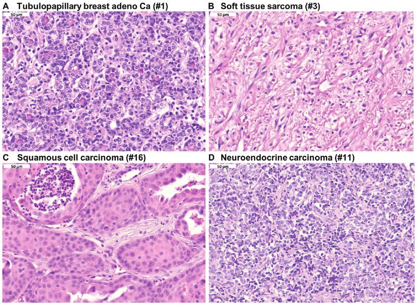
124                                   SAEED et al: Artemisia annua AGAINST SMALL PET TUMORS

artemisinin. Artemisinin is a bioactive terpenoid isolated            of water (A) and acetonitrile (B). The samples were eluted
from Artemisia annua L., which is a medicinal herb that               using the following gradient: 0 min, 98.0% A; 0‑8 min, linear
has been used for about two millennia in traditional Chinese          increase to 100% B; 100% A held for 2 min, 11‑12 min, return
medicine (17). The isolation of artemisinin from A. annua             to 98.0% A. The final optimization of ESI source operation
led to a novel treatment option of eminent importance for             parameters were a nitrogen gas flow of 11 l/min, nebulizer of
malaria. Artemisinin and its derivatives have been responsible        30 psi, capillary voltage of ±2.4 kV and a drying gas tempera-
for the survival of millions of patients with malaria (18,19).        ture of 250˚C (N2). The Quattro Ultima triple quadrupole MS
This achievement was appreciated in 2015 with the confer-             operated in multiple reaction monitoring mode with a resolu-
ment of the Nobel Prize for Medicine or Physiology to the             tion of 0.7 m/z. Electrospray ionization (positive mode) and
Chinese scientist Youyou Tu (20). While artemisinin deriva-           photodiode array detection at 254 nm were performed.
tives, such as artemether and artesunate, are well‑established             Different batches of A. annua (Luparte®) were determined
as anti‑malarial drugs, herbal preparations of A. annua also          by the Department of Pharmaceutical Biology, Johannes
inhibit Plasmodia infections in patients with malaria (21).           Gutenberg University (Mainz, Germany). This blinded
Notably, the bioactivity of artemisinin is not restricted to          approach was used as a quality measure to independently
malaria, and other diseases are also susceptible to artemisinin       guarantee the presence of artemisinin in Luparte®. Each batch
and A. annua treatment, such as schistosomiasis and trypano-          of A. annua powder (2x10 g) was extracted with two different
somiasis (22‑24), diverse viral infections (25) and diseases          polarity solvents, methylene chloride and methanol, at room
related to the metabolic syndromes, including obesity, diabetes       temperature for 24 h. The extract was concentrated in vacuo
and atherosclerosis (26‑28).                                          to obtain a residue of 0.8 and 1.2 g for methylene chloride and
    Artemisinin derivatives also inhibit human tumor cell             methanol, respectively. Each extract was transferred to a small
growth in vitro and in vivo (29‑31). This is relevant not only        vial and kept dry at room temperature. Small crystals were
for cancer therapy, but also for cancer prevention (32,33).           formed, which were collected and washed with n‑hexane and
Artemisinin derivatives exert additive or synergistic interac-        methylene chloride to remove extract residues. Then, 1H and
tions in combination with a wide array of clinically established      13
                                                                         C NMR analyses were performed.
drugs (34‑36). This has also been demonstrated in veterinarian
tumor cell lines in vitro and veterinarian clinical trials (37‑39).   Tumors. The animals were treated between 2010 and 2017.
Based on the anticancer activity in experimental tumor                Briefly, 16 dogs (10 males and 6 females) and 4 cats (2 females,
models, it has been possible to investigate the anticancer            1 male and 1 N/A) were treated with A. annua. The mean age
activity in human cancer patients in the form of compassionate        of the dogs was 10.31 years and the mean age of the cats was
uses (40,41) and even to perform clinical phase I/II trials in        12.25 years. The mean weight of the dogs was 30.125 kg and
human cancer patients (42‑44). Another previous clinical              the mean weight of the cats was 5.10 kg. Tissue samples were
phase I/II trial indicated the anticancer activity in a number        taken from all animals and initially fixed at room temperature
of dogs with tumors (39). Preliminary results in three dogs           (~22˚C) in 4% formaldehyde solution for 48 h. Following
and one cat revealed that a herbal preparation of A. annua            fixation, the organs were trimmed, processed and embedded
(Luparte®) may exhibit the potential to prolong the survival          in paraffin wax at approximately 60˚C. Sections were cut to
time of animals with tumors (45). To substantiate these               a thickness of 4 µm and routinely stained with hematoxylin
preliminary results from the compassionate use of A. annua,           and eosin at room temperature. Histopathological exami-
the present study has performed a retrospective analysis by           nation of the hematoxylin and eosin‑stained sections was
evaluating survival times of 25 pets treated with A. annua            performed under a light microscope in immersion oil with
compared with 11 animals without A. annua treatment.                  a Zeiss Axioskop. After routine pathological diagnosis, the
Independent of A. annua food supplementation, all animals             paraffin blocks were used as excess material for subsequent
were subjected to standard treatment protocols. In addition,          immunohistochemical studies. Furthermore, the histological
the expression of two biomarkers, transferrin receptor (TfR)          hematoxylin and eosin stained tumor sections were assessed
and the proliferation marker Ki‑67, was determined by immu-           for the presence of the tumor‑infiltrating lymphocytes (TILs).
nohistochemistry analysis of tumor biopsies.                          The clinical data are presented in Tables I and II. The owners
                                                                      of the animals provided written informed consent for this
Materials and methods                                                 retrospective study. The signed written consent forms are
                                                                      deposited at the Department of Pharmaceutical Biology,
Artemisinin determination in A. annua. Nuclear magnetic               Johannes Gutenberg University (Mainz, Germany) and can be
resonance (NMR) spectra were recorded on a Bruker 300                 inspected upon reasonable demand.
NMR spectrometer (Bruker Corporation). Reversed phase
high‑performance liquid chromatography (HPLC)‑mass                    Treatment protocol. The serum iron content was determined
spectrometry (MS) analysis was performed on a Waters                  following collection of blood samples; a normal range is
Alliance 2695 LC (Waters Corporation) coupled to a Quattro            between 140 and 170 µg/dl (47). After initial iron content
Ultima triple quadrupole MS (Waters Corporation) using the            determination using ferrozine color test (48), the tumors
same separation conditions as described previously (46). The          were surgically removed with safety margins by a standard
separation conditions were follow: Chromatogram column,               protocol after visual inspection and where required after lung
XBridge™ column (4.6x150 mm, 5 µm); column temperature,               radiography. Between blood collection and determination
20˚C; and injection volume, 1 µl. Elution was performed at a          of the serum iron results from the clinical diagnosis labora-
flow rate of 1 ml/min, using as the mobile phase a mixture            tory, iron was administered orally [Ferrosanol® capsules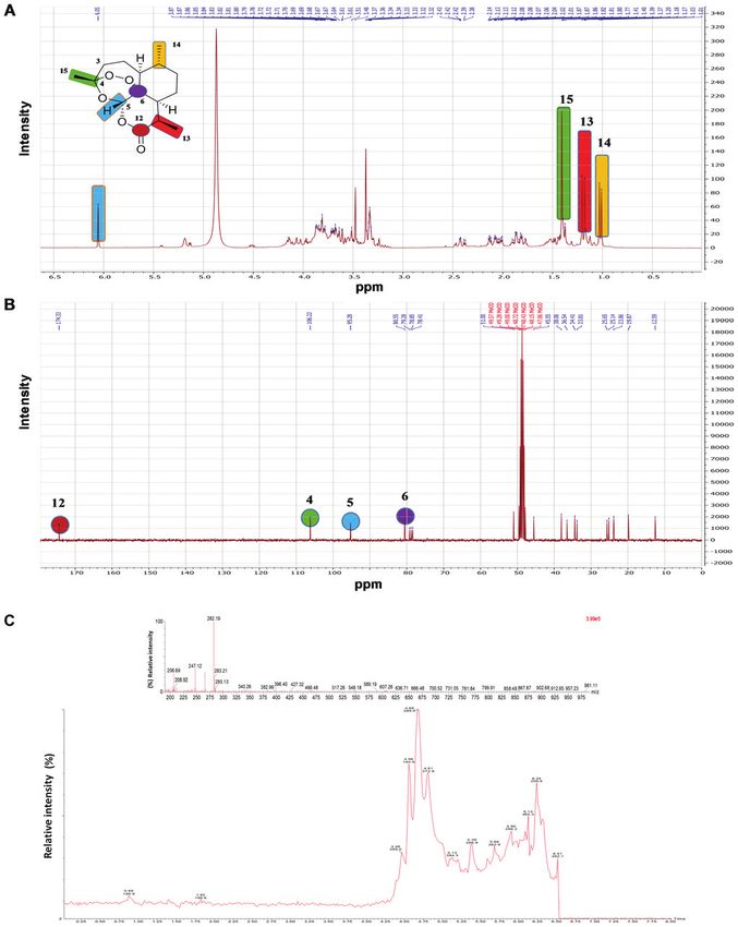
Table I. Clinical data of animals treated with Artemisia annua after standard treatment.

A, Carcinoma

				                                                               Date of diagnosis		                                      Survival      Initial blood     Treatment
			               Weight,                                              and start      Tumor                                  time,        iron content,       before         Age,        Source of
Species Breed Sex  kg                                                of treatment      type                                 months            µg/dl         A. annua         years       the tumor

Dog         Doberman                      F              33          May 2, 2014           Adenocarcinoma                      1.0              74               a             8      Subcutis
Dog         White Swiss Shepherd          M              32          Jan 1, 2015           Adenocarcinoma                      5.0             111               a             9      Mammary glands
Dog         Pyrenean Shepherd             F              11          Apr 28, 2015          Adenocarcinoma                      3.0              95               a            13      Subcutis
Dog         Poodle                        Fcastr          9          Mar 17, 2011          carcinoma                          48.0             134               c            14      Mammary glands
Dog         Mix                           F              55          N/A                   carcinoma                           1.0             223               a             9      Lung
Dog         Border Collie                 M              16          Oct 17, 2016          carcinoma                           2.5             106               a             1      Lung
Cat         European Shorthair            Fcastr          4          Dec 21, 2012          Squamous carcinoma                  5.0              45               b            16      Gingiva
Cat         European Shorthair            N/A            N/A         Sep 14, 2010          Non-specified carcinoma            38.0             222               b            12      Abdominal skin
Dog         Labrador                      M              42          Aug 23, 2012          Keratinizing carcinoma              4.0              93               b             9      Nose skin
Dog         Mix                           Mcastr         35          Jul 22, 2011          Hepatoma                            8.0             164               a            13      Liver

B, Sarcoma

				                                                               Date of diagnosis		                                      Survival      Initial blood     Treatment
			               Weight,                                              and start      Tumor                                  time,        iron content,       before         Age,        Source of
Species Breed Sex  kg                                                of treatment      type                                 months            µg/dl         A. annua         years       the tumor

Cat European Shorthair Mcastr 5.5 Feb 21, 2014 Fibrosarcoma     0.5  66 c 10                                                                                                         Abdominal subcutis
Dog Gordon Setter      Fcastr 26  Aug 24, 2010 Fibrosarcoma    72.0  85 c 10                                                                                                         Subcutis
Dog Mix                M      38  Feb 04, 2010 Fibrosarcoma    13.0 107 c 13                                                                                                         Subcutis
                                                                                                                                                                                                          INTERNATIONAL JOURNAL OF ONCOLOGY 56: 123-138, 2020

Cat European Shorthair Fcastr 5.8 Jul 11, 2011 Fibrosarcoma    >68   84 c 11                                                                                                         Abdominal subcutis
Dog Rottweiler         M      51  Nov 16, 2011 Fibrosarcoma     1.0  97 c  8                                                                                                         Subcutis
Dog Mix                M      26  Jan 25, 2011 Hemangiosarcoma >36  124 c 10                                                                                                         Spleen
Dog Mix                Fcastr 27  Jan 31, 2012 Hemangiosarcoma >18  131 c 14                                                                                                         Spleen
Dog Labrador           Mcastr 33  Apr 05, 2013 Hemangiosarcoma 41.0  81 c 10                                                                                                         Spleen
Dog Mix                M      22  Jan 22, 2014 Hemangiosarcoma 12.0 354 c 13                                                                                                         Spleen
Dog Mix                Mcastr 26  Apr 02, 2014 Lymphosarcoma   13.0 116 c 11                                                                                                         Lymph node,
										                                                                                                                                                                           left scapula

a, no surgical treatment done/possible; b, only incomplete/palliative surgery possible; c, macroscopic complete surgery. N/A, not available; F, female; M, male; castr, castrated.
                                                                                                                                                                                                          125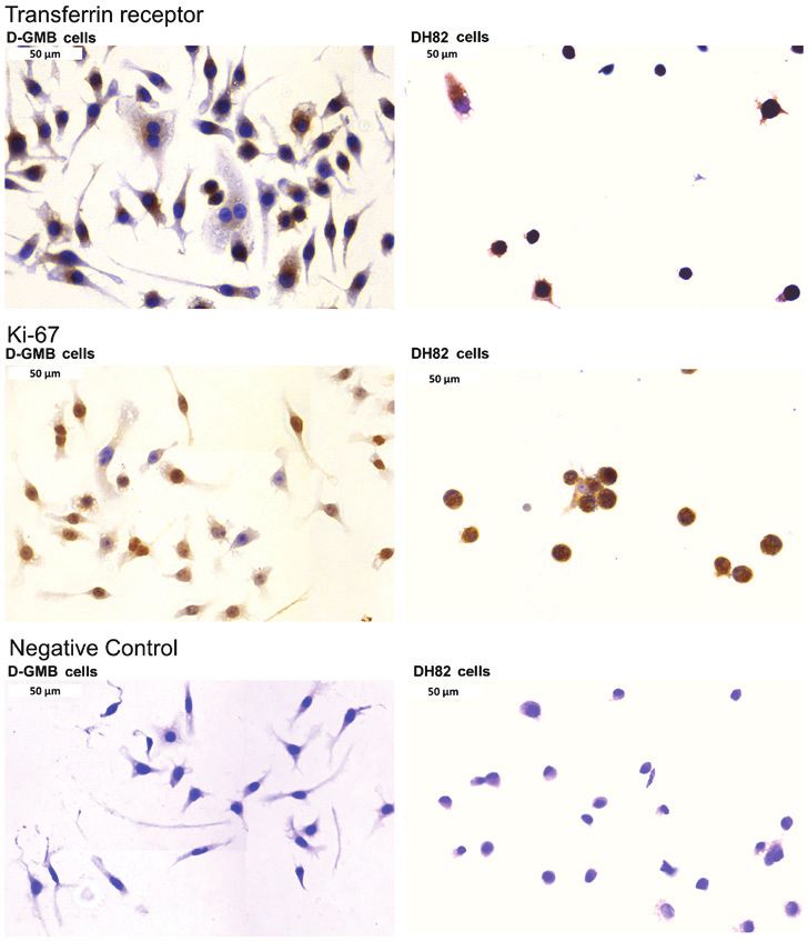
126

Table II. Clinical data of animals without Artemisia annua treatment after standard treatment.

A, Carcinoma

				                                                               Date of diagnosis		                                 Survival       Initial blood       Treatment
			               Weight,                                              and start     Tumor                              time,         iron content,         before         Age,         Source of
Species Breed Sex  kg                                                of treatment     type                             months             µg/dl           A. annua         years        the tumor

Dog          Mix                        F              6.8           Jul 22, 2011           Adenocarcinoma                N/A                6                 c            12       Mammary glands
Cat          European Shorthair         Fcastr         3.8           Aug 22, 2012           Adenocarcinoma                84.0               4                 c            16       Mammary glands
Dog          Labrador                   Fcastr         36            Jan 21, 2011           Adenocarnoma                 112.0               3                 a             9       Mammary glands
Dog          Mix                        Fcastr         8.6           Dec 29, 2014           Hepatoma                     107.0               2                 a             7       Liver
Cat          European Shorthair         Mcastr         4.2           Nov 07, 2014           Kidney carcinoma              69.0               4                 c            13       Kidney

B, Sarcoma

				                                                               Date of diagnosis		                                 Survival       Initial blood       Treatment
			               Weight,                                              and start     Tumor                              time,         iron content,         before         Age,         Source of
Species Breed Sex  kg                                                of treatment     type                             months             µg/dl           A. annua         years        the tumor

Cat European Shorthair Mcastr   6.0 Aug 06, 2012 Fibrosarcoma        62.0   7 c 14 Abdominal subcutis
Dog Mix                Mcastr  11.8 May 27, 2014 Hemangiosarcoma    220.0   7 c 13 Spleen
Dog Mix                Mcastr  38.6 Sep 18, 2012 Hemangiosarcoma    136.0 10  c 13 Spleen
Dog Dachshund          Fcastr  14.2 Jan 31, 2012 Liposarcoma         N/A  17  c 11 Eye lid
Dog Mix                F       42.0 Aug 27, 2012 Rhabdomyosarcoma    83.0 0.1 a 11 Thigh skeletal
                                                                                                                                                                                                      SAEED et al: Artemisia annua AGAINST SMALL PET TUMORS

										                                                                         muscles with
										                                                                         metastases
										                                                                         in liver and
										                                                                         lymph nodes
Cat European Shorthair N/A    N/A   Apr 16, 2015 Hemangiopericytoma  80.0 16  c 13 Abdominal subcutis

a, no surgical treatment done/possible; b, only incomplete/palliative surgery possible; c, macroscopic complete surgery; N/A, not available; F, female; M, male; castr, castrated.
INTERNATIONAL JOURNAL OF ONCOLOGY 56: 123-138, 2020 127
(100 mg/capsule or 40 mg/capsule)]. The dosage was Each assay was performed thrice independently, with six
100 mg/30 kg BW twice daily mixed into vegetable‑poor replicates each.
food to avoid iron binding plant molecules, such as phytane,
oxalates and/or phosphates. Alternatively, iron was subcu- Immunohistochemistry. A total of 17 tumor tissues repre-
taneously injected (Myofer ®/Ursoferran®; 100 mg/ml) at a senting different tumor types in pets were collected to evaluate
dose of 100 mg/10 kg BW daily, until the serum iron results the expression of Ki‑67 and TfR in animal tumors (Table V).
were obtained back from the clinical laboratory. The iron Briefly, tumor tissues were obtained from 15 dogs (7 males
substitution was individually continued until an iron content and 8 females) and 2 cats (both female). The mean age of the
of 250±30 µg/dl (~43±5 µmol/l) was stably reached in blood dogs was 9.33 years and the mean age of the cats was 4 years.
serum. From the fourth day of the initial iron substitution The mean weight of the dogs was 27.266 kg and the mean
onward, the animals were orally treated with Luparte® capsules weight of the cats was 4.00 kg. All the chosen animals had
at a dosage of 1,400 mg/m2 body surface divided to three frac- been kept in pet animal housing conditions. The immuno-
tions per day (Table III). The capsules were administered 1‑2 h histochemical staining of tumor tissues was performed as
before the next meal. The serum iron content was regularly described previously (55). The slides were washed twice with
monitored and where necessary adapted to 250±30 µg/dl. xylene (98.5% xylene for 5 min each at room temperature) to
The treatment details and the follow‑up information, remove paraffin. Then, sample tissues were rehydrated through
including survival times, have been recorded in the clinical graded washes with isopropanol in water. Heat‑induced
documentation files. All ethical issues have been discussed epitope retrieval was performed using a pressure cooker as
prior to evaluation of clinical and experimental data with a heating device. Ultra‑vision protein block and UltraVision
the Regierungspräsidium (Government Presidium) Freiburg Hydrogen Peroxide Block (catalog nos. TA‑060‑UB and
(Germany). According to the design of this retrospective study, TA‑060‑H2O2Q, respectively; Thermo Fisher Scientific, Inc.)
the Regierungspräsidium (Government Presidium) Freiburg were added to block endogenous proteins and endogenous
gave written permission for the present study (Az. 35‑9185.81/1, peroxidase activity, respectively, to avoid non‑specific back-
dated from February 4th, 2019). ground staining. Overnight incubation at 4˚C was performed
following the addition of monoclonal primary antibodies. For
Cell lines. The D‑GBM cell line was generated by Dr George the detection of Ki‑67, the SP6 clone (catalog no. ab16667;
Stoica (Department of Veterinary Pathobiology, Texas A&M Abcam) was used at a dilution of 1:200. For the determina-
University, College Station, TX, USA) from a brain tumor of an tion of TfR expression, the H68.4 clone (catalog. no. 136800;
8‑year‑old male Boxer dog (49). The D‑GBM cells used in the Thermo Fisher Scientific, Inc.) was applied at a dilution
present study were obtained from Dr Pablo Steinberg (Institute of 1:100. Subsequently, horseradish peroxidase‑labeled poly-
for Food Toxicology and Analytical Chemistry, University of mers conjugated with secondary antibodies specific for both
Veterinary Medicine Hannover, Hannover, Germany), who mouse and rabbit primary antibodies were added (catalog nos.
obtained it from Dr G. Stoica. TL‑060‑QPH and TL‑060‑QPB; Thermo Fisher Scientific,
The histiocytic DH82 cell line was generated by Dr M.L. Inc.) at room temperature for 1 h, according to the manufac-
Wellman (Department of Veterinary Pathobiology, College of turer's protocol. The final staining reaction was performed
Veterinary Medicine, Ohio State University, CO, USA) (50). with diaminobenzidine and slides were counterstained with
The DH82 cell line used in the present study was also obtained hematoxylin for 3 min at room temperature.
from Dr Pablo Steinberg, who purchased it from the European The immunostaining of DH82 and DGBM cell lines was
Tissue Culture Collection (catalog no. 94062922). performed without the rehydration or epitope retrieval steps.
A panel of 54 human tumor cell lines of the Developmental Briefly, six cover slips were placed in a 6‑well plate, then
Therapeutics Program (DTP) of the National Cancer Institute 5x10⁵ cells/well were seeded over the cover slips and incu-
consisted of cell lines derived from leukemia, melanoma, bated in humidified 5% CO2 atmosphere at 37˚C overnight to
non‑small cell lung cancer, colon cancer, renal cancer, ovarian let the cells attach to coverslips. Subsequently, the cells were
cancer, breast cancer and prostate carcinoma, as well as central fixed in 4% paraformaldehyde for 15 min at room tempera-
nervous system tumors (51). Previously, the cytotoxicity of cells ture, and the blocking and staining steps were performed as
treated with artemisinin for 48 h was evaluated using a sulforho- described for the tissue section. However, the Ki‑67 and TfR
damine B assay (52). The log10 IC50 values for artemisinin and antibodies were diluted at 1:500 and 1:1,000, respectively.
mRNA expressions for Ki‑67 and TfR were available publicly The immunostained slides were scanned using Pannoramic
in the DTP database (https://dtp.cancer.gov/default.htm). Desk (3DHISTECH Ltd.) and the amount of Ki‑67‑ or
TfR‑expressing cells was quantified by Pannoramic Viewer
Cytotoxicity assay. A resazurin reduction assay (Promega software version 1.15 (3DHISTECH Ltd.).
Corporation) was performed as previously described (53) to
assess the cytotoxicity of artemisinin, artesunate and dihydro- Statistical analysis. Significance values and correlation coef-
artemisinin towards the dog tumor cell lines DH82 and DGBM. ficients were calculated using Pearson's correlation coefficient
The concept of the assay is based on the metabolic reduction and Fisher's exact test with the WinSTAT software program
of the non‑fluorescent dye by living cells to the strongly‑fluo- version 2012.1 (www.winstat.com/). Linear regression was
rescent dye resorufin (54). IC50 values were calculated from performed using Excel 2016 (Microsoft Corporation) for the
dose‑response curves using a non‑linear regression analysis log10 IC50 values for artemisinin and mRNA expressions for
tool in GraphPad Prism 7 software (GraphPad Prism, Inc.). All Ki‑67 and TfR. P128 SAEED et al: Artemisia annua AGAINST SMALL PET TUMORS Table III. Conversion table for Luparte® treatment. Table IV. Correlation of Luparte® treatment and survival time. Body weight, kg Body surface, m² Survival time, (months) Treatment No treatment 0.5 0.06
INTERNATIONAL JOURNAL OF ONCOLOGY 56: 123-138, 2020                                                     129

Figure 1. NMR spectra of A. annua chloroform extract demonstrating the presence of artemisinin. (A) 1H NMR (300 MHz). (B) 13C NMR (75 MHz).
(C) RP‑HPLC/MS chromatogram of A. annua (Luparte®) extract. The top figure shows the MS profile, while the bottom figure represents the HPLC measure-
ments. NMR, nuclear magnetic resonance; RP‑HPLC/MS, reversed phase high‑performance liquid chromatography‑mass spectrometry.
130                                         SAEED et al: Artemisia annua AGAINST SMALL PET TUMORS

Figure 2. Hematoxylin and eosin staining of diverse pet tumors. (A) Tubulopapillary breast adenocarcinoma. (B) Soft tissue sarcoma. (C) Squamous cell
carcinoma. (D) Neuroendocrine carcinoma. Magnification, x40.

Figure 3. Quantification of immunohistochemical staining of veterinary tumors and correlation to clinical parameters. (A) TfR expression. (B) Ki‑67 expres-
sion. (C) Correlation of TfR expression to Ki‑67 expression. (D) Correlation of blood iron content to survival time. (E) Survival time of treated and untreated
animals (P=0.033). Regression analyses were performed using Excel (Microsoft Corporation). TfR, transferrin receptor.
INTERNATIONAL JOURNAL OF ONCOLOGY 56: 123-138, 2020                                             131

Table V. Expression of TfR and the proliferation marker Ki-67 in dog tumors according to immunohistochemistry.

						                                                                            Percentage of      Percentage-
				                   Weight, Age,                                               TfR-positive         of Ki-76
Case Species Breed Sex  kg     years                                                  cells          postive cells         Histology

 1 Cat European shorthair     Fcastr  4  3 94.3±9.0  85.7±4.7                                                        Tubulopapillary breast
								                                                                                                             adenocarcinoma
 2 Dog Mix                    Fcastr 17 13 95.1±3.6  84.8±5.9                                                        Tubulopapillary breast
								                                                                                                             adenocarcinoma
 3 Dog Weimarian              M      28  8 99.7±0.1  72.5±4.4                                                        Soft tissue sarcoma,
								                                                                                                             Grade 1
 4 Dog Deutsch-Adrahthaar     Fcastr 28  9 87.2±9.6  71.6±9.7                                                        Tubulopapillary breast
								                                                                                                             adenocarcinoma with
								                                                                                                             lymph node metastasis
 5 Dog French bulldog         Mcastr 11  7 83.9±6.8  53.5±18.7                                                       Soft tissue sarcoma,
								                                                                                                             Grade 1
 6 Dog American Staffordshire Mcastr 33  3 98.6±0.5  49.1±4.9                                                        Mastocytoma, Type 2
		Terrier
 7 Dog Mix                    M      21 12 52.1±32.1 43.5±3.1                                                        Hemangiosarcoma
 8 Dog Podenco                Fcastr 24 12 99.9±0.1  41.1±16.3                                                       Osteosarcoma
 9 Dog Blue lacy              M      26 11 99.8±0.2  33.9±5.0                                                        Mastocytoma, Type 2
10 Dog Mix                    F      14  2 43.5±37.9 33.2±13.8                                                       Mastocytoma, Type 2
11 Cat European shorthair     Fcastr  4  5 97.4±1.8  31.3±8.3                                                        Neuroendocrine
								                                                                                                             carcinoma
12 Dog Podenco                Fcastr 25 12 84.6±9.5  30.5±10.7                                                       Osteosarcoma, highly
								                                                                                                             malignant
13 Dog Rottweiler             Fcastr 51  8 99.0±0.1  30.1±14.6                                                       Adenocarcinoma
								                                                                                                             metastasis
14 Dog Mix                    Mcastr 27 10 99.1±1.4  29.5±8.4                                                        Teleangiectatic
								                                                                                                             osteosarcoma
15 Dog Labrador mix           F      37 12 95.2±3.9  28.6±7.4                                                        Carcinoma
16 Dog Labrador               M      43  9 99.2±0.6  21.3±9.7                                                        Squamous cell
								                                                                                                             carcinoma
17 Dog Podenco                Fcastr 24 12 53.3±17.8  6.6±1.8                                                        Breast carcinoma,
								                                                                                                             complex/myxoid

Data are present as the mean ± standard deviation. F, female; M, male; castr, castrate; TfR, transferrin receptor.

Table VI. Correlation of artemisinin (log10 IC50 values)                  54 human tumor lines consisted of cell lines derived from
with microarray-based TfR and Ki-67 mRNA expression in                    eight tumor types (leukemia, melanoma and brain tumor, and
54 human tumor cell lines.                                                carcinoma of colon, breast, ovary, kidney and prostate). The
                                                                          number of cell lines of each tumor type (n=1‑9) was too low
                                Artemisinin                 Ki-67         to reveal significant correlations except for the three following
Marker                          cytotoxicity              expression      results. In lung cancer cell lines, the log10 IC50 values of arte-
                                                                          misinin were inversely correlated with the Ki‑67 expression
TfR expression                                                            (P=0.009; r=‑0.759). In melanoma cell lines, the expression
 r value                           -0.282                    0.317        levels of Ki‑67 and TfR were significantly correlated (P=0.010;
 P-value                            0.019                    0.008        r=0.787). In renal cancer cell lines, the expression levels of
Ki-67 expression                                                          Ki‑67 and TfR were also significantly correlated (P= 0.019;
                                                                          r=0.781) (data not shown). Since all other associations between
 r value                           -0.239
                                                                          artemisinin response and the expression of Ki‑67 and TfR
 P-value                            0.041
                                                                          were not statistically significant, reliable conclusions on the
TfR, transferrin receptor.
                                                                          role of Ki‑67 and TfR may be drawn from the cell line panel
                                                                          as a whole, but not from tumor type‑specific subsets.
132 SAEED et al: Artemisia annua AGAINST SMALL PET TUMORS
Figure 4. Detection of TfR and Ki‑67 expression by immunohistochemistry. (A) TfR in tubulopapillary breast adenocarcinoma. (B) TfR in soft tissue sarcoma.
(C) TfR in squamous cell carcinoma. (D) TfR in the negative control. (E) Ki‑67 in tubulopapillary breast adenocarcinoma. (F) Ki‑67 in soft tissue sarcoma.
(G) Ki‑67 in squamous cell carcinoma. (H) Ki‑67 in neuroendocrine carcinoma. Magnification, x40. TfR, transferrin receptor.
Representative images of immunostaining for TfR expres- procedure. Furthermore, examples of Ki‑67 expression in
sion are presented in Fig. 4A‑D. Strong TfR expression was tubulopapillary breast adenocarcinoma, soft tissue sarcoma,
identified in biopsies of tubulopapillary breast adenocarci- squamous cell carcinoma and neuroendocrine carcinoma are
noma, soft tissue sarcoma and squamous cell carcinoma. presented in Fig. 4E‑H. Furthermore, assessment of the pres-
By contrast, the negative control did not demonstrate any ence of TILs in hematoxylin and eosin stained tumor tissues
reactivity, indicating the specificity of the immunostaining revealed an absence of TILs in all tumor slides.INTERNATIONAL JOURNAL OF ONCOLOGY 56: 123-138, 2020 133
Figure 5. Detection of TfR and Ki‑67 expression in dog DH82 histiocytic sarcoma and dog DGMB glioblastoma cell lines. Magnification, x40. TfR, transferrin
receptor.
For comparison, the Ki‑67 and TfR expression was analyzed leukemia and astrocytoma cells up to 10‑fold. These effects
in two dog cell lines. DH82 histiocytic sarcoma and DGMB were reversed by anti‑TfR monoclonal antibody RVS10,
glioblastoma cells were immunostained for both markers. which competes with transferrin for binding to TfR. While
Indeed, Ki‑67 and TfR were overexpressed in both cell lines as the TfR expression ranged between 48 and 95% in tumor
presented in Fig. 5. Furthermore, the present study investigated cell lines, normal peripheral mononuclear blood leukocytes
the cytotoxicity of artemisinin, artesunate and dihydroartemis- revealed ≤1.3% TfR positivity, indicating that artesunate may
inin towards the dog cell lines. As expected, artemisinin and exert tumor specific effects at least to some extent, because of
its derivatives were also active in these tumor cell lines. The the preferential TfR expression in tumor cells (59).
IC50 values for artemisinin, artesunate and dihydroartemisinin In a subsequent study, we investigated a total of 36 cell
towards DH82 cells and DGBM cells are presented in Fig. 6. lines of different tumor types for their response to treatment
with artesunate alone or in combination with Ferrosanol® (60).
Discussion This revealed that artesunate plus Ferrosanol® enhances
cytotoxicity compared with artesunate alone in the majority
It has been a matter of discussion among veterinarian physicians of cell lines; however, 11 cell lines did not demonstrate
and alternative practitioners, whether or not supplementation increased apoptosis and nine lines exhibited a decrease in
of iron is beneficial for the activity of artemisinin. The role apoptosis following the combined drug treatment compared
of heme has been discussed for the antimalarial activity of with artesunate‑treatment alone. It is understood that iron acts
artemisinin and its derivatives (57,58). In the context of cancer, as co‑factor for proliferation‑related enzymes (61). Therefore,
we previously reported that Ferrosanol® and holotransferrin Ferrosanol® may induce rather than suppress the prolifera-
increase artesunate‑induced cytotoxicity and apoptosis in tion of these nine cell lines. Based on these in vitro data, it134 SAEED et al: Artemisia annua AGAINST SMALL PET TUMORS
Figure 6. Cytotoxicity towards DH82 and DGMB cells as determined by a resazurin assay. (A) Artemisinin, (B) artesunate and (C) dihydroartemisinin.
cannot be reliably recommended that iron should be added experimental therapy strategies have been proposed to address
as a supplement to artemisinin‑type treatment in veterinary TfR as a treatment target to improve tumor‑specific killing and
or human cancer therapy. The results of the present study spare toxic side effects to normal tissues at the same time, for
revealed that the blood iron content was not associated with example by antibody‑mediated targeting of TfR or the genera-
survival times after A. annua therapy, while the TfR content tion of TfR‑directed immunotoxins (69‑71). The generation
inside the tumor was. For practical reasons, it can be assumed of transferrin‑artemisinin conjugates revealed enhanced
that the normal iron content of the body should be sufficient for cytotoxicity towards tumor cells compared with uncoupled
the activity of artesunate. Only in patients with iron‑deficient artemisinin (72‑74).
conditions may iron supplementation be considered. However, Previously, we performed a clinical phase I/II trial with
in this case it may be important that iron and artesunate reach artesunate in 23 dogs with cancer, which revealed 1 case of
the tumor at the same time so that a possible enhancing effect complete remission and 7 cases of stable disease following
of the drug combination can develop. Otherwise, Ferrosanol® artesunate‑treatment (39). The results of the present retro-
may exhibit worse effects and lead to an enhancement rather spective study with A. annua appear to be improved. The
than a suppression of proliferative tumor activity. present study identified an increase in survival time above
The effect of iron has also been demonstrated in other the threshold of 18 months in 9 of 25 dogs (36%). Although
previous studies. It has been reported that the addition of this trend needs to be confirmed in future studies, it could be
transferrin can inhibit cross‑resistance of multidrug‑resistant speculated that the anticancer effect of the whole plant extract
H69VP small cell lung cancer cells to artemisinin (62). In is better than that of isolated artemisinin or the semisynthetic
addition, retinoblastoma cells with high TfR expression are derivative, artesunate. Indeed, the plant extract contains many
more susceptible to artesunate compared with normal retina more cytotoxic compounds in addition to artemisinin (75‑78),
cells. This activity is specific, as RNA interference‑mediated including arteanuin B, artemisitene, scopoletin and 1,8‑cineole.
TfR‑knockdown increased the sensitivity of retinoblastoma Therefore, A. annua preparations alone may be used as a
cells to artesunate (63). In accordance with these findings, combination therapy. Several cytotoxic compounds may act at
supplementation with holotransferrin increases the cytotox- the same time against the tumor, which leads to an improved
icity of dihydroartemisinin in T‑cell lymphoma cells (64). tumor inhibition.
It is well documented that TfR is more highly expressed in It is well understood that rapidly proliferating tumors
tumor cells compared with normal cells (61,65‑68). Therefore, respond better to standard chemotherapy compared withINTERNATIONAL JOURNAL OF ONCOLOGY 56: 123-138, 2020 135
slowly growing tumors (79,80). Therefore, the present study is unclear whether these reports may reflect the pet owners
also included Ki‑67 as proliferation marker in the immunohis- psychological condition after successful treatment of their
tochemical analysis. Ki‑67 is closely associated with the cell pets. Nevertheless, this observation should not be neglected,
cycle. Its role as a predictive factor for the success of chemo- as it cannot be excluded that there may be an unexpected
therapy and as a prognostic factor for the survival of cancer and unintentional positive side effect of A. annua‑treatment.
patients has been discussed (81,82). Therefore, the significant Indeed, there are some previous studies that support these
association between Ki‑67 expression and cytotoxicity to arte- observations. The serotonin serum levels in the brains of
misinin in the panel of 54 human cell lines from eight different artesunate‑treated rabbits has been reported to be significantly
tumor types indicated that Ki‑67 expression may also be a higher compared with in untreated control animals (95).
prognostic marker for tumor response to artemisinin. Zhu et al (96) reported significant remissions in nociceptive,
The present study revealed a significant correlation between anxiety and depressive behaviors by dihydroartemisinin,
TfR and Ki‑67 expression in veterinary tumors and human artesunate or artemether. Amos et al (97) suggested sedative
tumor cell lines, indicating that TfR expression is associated properties of artemisinin mediated by artemisinin's effect on
with high proliferative rates and that artemisinin is more active postsynaptic dopamine D2 receptors in the brain. This novel
in fast growing tumors than in slowly growing ones. This result aspect of artemisinin's possible activity deserves further
supports previous data from human tumors and also speaks detailed investigation in the future.
for the comparability of veterinary and human tumors in this The present investigation also raises the more general
respect. The significant correlation between TfR and Ki‑67 question about the comparability of data raised in veterinary
expression has been reported in biopsies of diverse tumor tumors to the clinical situation in human tumors and thereby
types, including leukemia, melanoma, breast carcinoma, brain the suitability of veterinary tumors as models of human cancer
tumors, head and neck cancer and esophageal cancer (83‑88). biology and treatment. The present study determined that the
Although a significant association was identified between role of TfR and Ki‑67 was comparable between veterinary
TfR and Ki‑67 in the entire panel of 54 tumor cell lines, this tumors and human tumor cell lines. TfR and Ki‑67 have also
association was not seen in the majority of the subsets with been associated with response to artemisinin and artesunate
different tumor types. This may simply be explained by the in tumors of human patients (41‑43). Although these data in
limited number of cell lines per tumor entity. The previously human tumors are preliminary, they indicate that veterinary
published data on the significant correlation between TfR and tumors may represent a suitable model for clinical human
Ki‑67 expression were obtained in larger collectives of tumor tumors in the context of artemisinin therapy.
biopsies (83,89). Independent of this situation, there are more arguments
In human tumor pathology, hematoxylin and eosin that support the suitability of veterinary tumors for the
staining is also useful for the detection of tumor‑infiltrating investigation of human cancer biology. Veterinary tumors
lymphocytes (TILs) (90‑92). Therefore, in the present study are spontaneously developing, which may qualify them as
we also screened the hematoxylin and eosin‑stained slides better models compared with other tumors in mice and rats.
of the pet tumors for the presence of TILs, but TILs could Frequently tumors are maintained by transplanting syngeneic
not be detected in the tumor tissues. Since the tumors were tumors to rodents or human xenograft tumors to immunosup-
generally large and already progressed at diagnosis, it was pressed nude mice. Another possibility is to induce tumor
assumed that the immune system of the animals was weak- development in mice or rats by chemical carcinogens. Although
ened and TIL‑mediated immune defenses against the tumor these tumor models are indispensable and of high value in
were largely destroyed. preclinical oncology, they are to some degree artificial. Here,
Furthermore, it is important to mention that the present tumors in dogs and cats may be advantageous, because their
study did not observe considerable adverse side effects among spontaneous development is closer to the situation of human
the 25 A. annua‑treated dogs and cats. In comparison to tumor tumors. Veterinary tumors have not received as much attention
treatment in human cancer patients with the semi‑synthetic as the aforementioned rodent tumor models as of yet. Further
drug artesunate, A. annua appeared to be even safer. Among investigations are required to investigate the full potential
the 23 dogs with cancer treated with artesunate, fever and tran- of veterinary tumors as attractive in vivo models to develop
sient hematological and gastrointestinal toxicity were observed strategies for human tumor treatment.
in 16 dogs and 1 dog died from pneumonia (39). In human In conclusion, the current retrospective study involving
cancer patients, the compassionate use of A. annua in 1 patient 20 dogs and cats treated with standard therapy plus A. annua
with prostate carcinoma was well tolerated in a previous and 11 dogs treated with standard therapy alone clearly demon-
study (41). Treatment with artesunate has been reported to lead strated that additional food supplementation of A. annua to
to occasional and transient side effects, including hematolog- veterinary cancer patients resulted in an improved survival
ical toxicity, gastrointestinal toxicity, asthenia and thrombosis, prognosis. The activity of A. annua may be dependent on
in colon, cervix and breast carcinoma (42‑44). Rare cases of the iron content in the tumor, but not in the blood, since TfR
hepatotoxicity have been reported with artesunate (93,94). expression in the tumors was significantly correlated with
Whether phytotherapeutic approaches with A. annua are safer survival time and artemisinin cytotoxicity in a control panel of
than treatment with artesunate requires further investigation. human tumor cell lines. The same was true for Ki‑67 expres-
Notably, the majority of pet owners reported that the sion. Tumors with high Ki‑67 expression, indicating high
animals appeared to feel better after A. annua treatment; some proliferative activity, were more susceptible to artemisinin in
were more active, while others were more relaxed. Of course, the human cell line panel. The data presented in the present
these observations are subjective and non‑quantifiable, and it study should provide guidance for the activity of A. annua136 SAEED et al: Artemisia annua AGAINST SMALL PET TUMORS
against veterinary tumors. Prospective trials are required to 3. Choi JW, Yoon HY and Jeong SW: Clinical outcomes of
surgically managed spontaneous tumors in 114 client‑owned
deliver convincing evidence for this hypothesis. dogs. Immune Netw 16: 116‑125, 2016.
4. Hellmén E, Bergström R, Holmberg L, Spångberg IB, Hansson K
Acknowledgements and Lindgren A: Prognostic factors in canine mammary tumors: A
multivariate study of 202 consecutive cases. Vet Pathol 30: 20‑27,
1993.
The authors wish to thank Mrs. Doris Rohr (Department 5. Marconato L: The staging and treatment of multicentric high‑grade
of Pharmaceutical Biology, Institute of Pharmacy and lymphoma in dogs: A review of recent developments and future
prospects. Vet J 188: 34‑38, 2011.
Biochemistry, Johannes Gutenberg University, Mainz, 6. Tuohy JL, Selmic LE, Worley DR, Ehrhart NP and Withrow SJ:
Germany) for technical assistance with immunohistochem- Outcome following curative‑intent surgery for oral melanoma in
istry staining. dogs: 70 cases (1998‑2011). J Am Vet Med Assoc 245: 1266‑1273,
2014.
7. Miller RL, Van Lelyveld S, Warland J, Dobson JM and Foale RD:
Funding A retrospective review of treatment and response of high‑risk
mast cell tumours in dogs. Vet Comp Oncol 14: 361‑370, 2016.
8. Romano FR, Heinze CR, Barber LG, Mason JB and Freeman LM:
This study was supported by intramural funding from Johannes Association between body condition score and cancer prognosis
Gutenberg University (Mainz, Germany). in dogs with lymphoma and osteosarcoma. J Vet Intern Med 30:
1179‑1186, 2016.
9. Sarowitz BN, Davis GJ and Kim S: Outcome and prognostic
Availability of data and materials factors following curative‑intent surgery for oral tumours in
dogs: 234 cases (2004 to 2014). J Small Anim Pract 58: 146‑153,
The datasets generated and/or analyzed during the current 2017.
10. Ettinger SN: Principles of treatment for feline lymphoma. Clin
study are not publicly available due to restrictions of the avail- Tech Small Anim Pract 18: 98‑102, 2003.
ability of these data but are available from the corresponding 11. Morris J: Mammary tumours in the cat: Size matters, so early
author on reasonable request. intervention saves lives. J Feline Med Surg 15: 391‑400, 2013.
12. Zabielska‑Koczywąs K, Wojtalewicz A and Lechowski R:
Current knowledge on feline injection‑site sarcoma treatment.
Authors' contributions Acta Vet Scand 59: 47, 2017.
13. Martano M, Iussich S, Morello E and Buracco P: Canine oral
fibrosarcoma: Changes in prognosis over the last 30 years?
TE and EB designed the study. EB treated the animals, Vet J 241: 1‑7, 2018.
provided the material and collected the clinical data. MEMS 14. Zhang D, Hedlund EM, Lim S, Chen F, Zhang Y, Sun B and
performed the immunostaining. MEFH performed NMR and Cao Y: Antiangiogenic agents significantly improve survival
in tumor‑bearing mice by increasing tolerance to chemo-
HPLC‑MS. TE performed the statistical analysis, supervised therapy‑induced toxicity. Proc Natl Acad Sci USA 108: 4117‑4122,
the work and provided the facilities for the study. TE and 2011.
MEMS wrote the manuscript. All authors read the manuscript 15. Tiwari A, Hadley JA, Hendricks GL III, Elkin RG, Cooper T and
Ramachandran R: Characterization of ascites‑derived ovarian
and approved the final version. tumor cells from spontaneously occurring ovarian tumors of the
chicken: Evidence for E‑cadherin upregulation. PLoS One 8:
Ethics approval and consent to participate e57582, 2013.
16. Newman DJ and Cragg GM: Natural products as sources of new
drugs from 1981 to 2014. J Nat Prod 79: 629‑661, 2016.
Written permission for this retrospective study was obtained 17. Tu Y: The discovery of artemisinin (qinghaosu) and gifts from
from the Regierungspräsidium (Government Presidium) Chinese medicine. Nat Med 17: 1217‑1220, 2011.
18. Bridgford JL, Xie SC, Cobbold SA, Pasaje CFA, Herrmann S,
Freiburg, Germany (Az. 35‑9185.81/1, dated from February 4th, Yang T, Gillett DL, Dick LR, Ralph SA, Dogovski C, et al:
2019). Written informed consent for experimental work was Artemisinin kills malaria parasites by damaging proteins and
obtained from all pet owners. inhibiting the proteasome. Nat Commun 9: 3801, 2018.
19. Su XZ and Miller LH: The discovery of artemisinin and the
Nobel Prize in Physiology or Medicine. Sci China Life Sci 58:
Patient consent for publication 1175‑1179, 2015.
20. Tu Y: Artemisinin‑a gift from traditional chinese medicine
to the world (Nobel Lecture). Angew Chem Int Ed Engl 55:
No applicable. 10210‑10226, 2016.
21. Daddy NB, Kalisya LM, Bagire PG, Watt RL, Towler MJ and
Competing interests Weathers PJ: Artemisia annua dried leaf tablets treated malaria
resistant to ACT and i.v. artesunate: Case reports. Phytomedicine 32:
37‑40, 2017.
EB commercially trades Luparte®. No part of the experimental 22. Saeed ME, Krishna S, Greten HJ, Kremsner PG and Efferth T:
work at the Department of Pharmacological Biology (Johannes Antischistosomal activity of artemisinin derivatives in vivo and
in patients. Pharmacol Res 110: 216‑226, 2016.
Gutenberg University, Mainz, Germany) was funded by EB. 23. Pérez del Villar L, Burguillo FJ, López‑Abán J and Muro A:
All other authors declare that they have no competing interests. Systematic review and meta‑analysis of artemisinin based
therapies for the treatment and prevention of schistosomiasis.
PLoS One 7: e45867, 2012.
References 24. Naß J and Efferth T: The activity of Artemisia spp. and their
constituents against Trypanosomiasis. Phytomedicine 47: 184‑191,
2018.
1. Talmadge JE, Singh RK, Fidler IJ and Raz A: Murine models to 25. Efferth T: Beyond malaria: The inhibition of viruses by arte-
evaluate novel and conventional therapeutic strategies for cancer. misinin‑type compounds. Biotechnol Adv 36: 1730‑1737, 2018.
Am J Pathol 170: 793‑804, 2007. 26. Jiang W, Cen Y, Song Y, Li P, Qin R, Liu C, Zhao Y, Zheng J and
2. Jantscheff P, Beshay J, Lemarchand T, Obodozie C, Schächtele C Zhou H: Artesunate attenuated progression of atherosclerosis
and Weber H: Mouse‑derived isograft (MDI) in vivo tumor lesion formation alone or combined with rosuvastatin through
models I. Spontaneous sMDI models: Characterization and inhibition of pro‑inflammatory cytokines and pro‑inflammatory
cancer therapeutic approaches. Cancers (Basel) 11: 11, 2019. chemokines. Phytomedicine 23: 1259‑1266, 2016.INTERNATIONAL JOURNAL OF ONCOLOGY 56: 123-138, 2020 137
27. Li J, Casteels T, Frogne T, Ingvorsen C, Honore C, Courtney M, 49. Stoica G, Lungu G, Martini‑Stoica H, Waghela S, Levine J and
Huber KV, Schmitner N, Kimmel RA, Romanov RA, et al: Smith R III: Identification of cancer stem cells in dog glio-
Artemisinins target GABAA receptor signaling and impair alpha blastoma. Vet Pathol 46: 391‑406, 2009.
cell Identity. Cell 168: 86‑100.e15, 2017. 50. Wellman ML, Krakowka S, Jacobs RM and Kociba GJ: A
28. Guo Y, Fu W, Xin Y, Bai J, Peng H, Fu L, Liu J, Li L, Ma Y and macrophage‑monocyte cell line from a dog with malignant
Jiang H: Antidiabetic and antiobesity effects of artemether in histiocytosis. In Vitro Cell Dev Biol 24: 223‑229, 1988.
db/db mice. BioMed Res Int 2018: 8639523, 2018. 51. Alley MC, Scudiero DA, Monks A, Hursey ML, Czerwinski MJ,
29. Efferth T, Dunstan H, Sauerbrey A, Miyachi H and Chitambar CR: Fine DL, Abbott BJ, Mayo JG, Shoemaker RH and Boyd MR:
The anti‑malarial artesunate is also active against cancer. Int J Feasibility of drug screening with panels of human tumor cell
Oncol 18: 767‑773, 2001. lines using a microculture tetrazolium assay. Cancer Res 48:
30. Efferth T, Sauerbrey A, Olbrich A, Gebhart E, Rauch P, 589‑601, 1988.
Weber HO, Hengstler JG, Halatsch ME, Volm M, Tew KD, et al: 52. Rubinstein LV, Shoemaker RH, Paull KD, Simon RM, Tosini S,
Molecular modes of action of artesunate in tumor cell lines. Mol Skehan P, Scudiero DA, Monks A and Boyd MR: Comparison of
Pharmacol 64: 382‑394, 2003. in vitro anticancer‑drug‑screening data generated with a tetra-
31. Dell'Eva R, Pfeffer U, Vené R, Anfosso L, Forlani A, Albini A zolium assay versus a protein assay against a diverse panel of
and Efferth T: Inhibition of angiogenesis in vivo and growth of human tumor cell lines. J Natl Cancer Inst 82: 1113‑1118, 1990.
Kaposi's sarcoma xenograft tumors by the anti‑malarial arte- 53. Kuete V, Mbaveng AT, Sandjo LP, Zeino M and Efferth T: Cytotoxicity
sunate. Biochem Pharmacol 68: 2359‑2366, 2004. and mode of action of a naturally occurring naphthoquinone,
32. Efferth T: From ancient herb to modern drug: Artemisia annua 2‑acetyl‑7‑methoxynaphtho[2,3‑b]furan‑4,9‑quinone towards
and artemisinin for cancer therapy. Semin Cancer Biol 46: 65‑83, multi‑factorial drug‑resistant cancer cells. Phytomedicine 33: 62‑68,
2017. 2017.
33. Abba ML, Patil N, Leupold JH, Saeed ME, Efferth T and 54. O'Brien J, Wilson I, Orton T and Pognan F: Investigation of the
Allgayer H: Prevention of carcinogenesis and metastasis by Alamar Blue (resazurin) fluorescent dye for the assessment of
Artemisinin‑type drugs. Cancer Lett 429: 11‑18, 2018. mammalian cell cytotoxicity. Eur J Biochem 267: 5421‑5426, 2000.
34. Krusche B, Arend J and Efferth T: Synergistic inhibition of 55. Saeed ME, Mertens R, Handgretinger R and Efferth T: Identification
angiogenesis by artesunate and captopril in vitro and in vivo. of fatal outcome in a childhood nasopharyngeal carcinoma patient
Evid Based Complement Alternat Med 2013: 454783, 2013. by protein expression profiling. Int J Oncol 53: 1721‑1731, 2018.
35. Efferth T: Cancer combination therapy of the sesquiterpenoid 56. Blaskó G, Cordell GA and Lankin DC: Definitive 1H‑and
artesunate and the selective EGFR‑tyrosine kinase inhibitor 13
C‑NMR assignments of artemisinin (Qinghaosu). J Nat Prod 51:
erlotinib. Phytomedicine 37: 58‑61, 2017. 1273‑1276, 1988.
36. Efferth T: Cancer combination therapies with artemisinin‑type 57. Haynes RK, Cheu KW, N'Da D, Coghi P and Monti D:
drugs. Biochem Pharmacol 139: 56‑70, 2017. Considerations on the mechanism of action of artemisinin anti-
37. Hosoya K, Murahari S, Laio A, London CA, Couto CG and malarials: Part 1 - the ‘carbon radical’ and ‘heme’ hypotheses.
Kisseberth WC: Biological activity of dihydroartemisinin in Infect Disord Drug Targets 13: 217‑277, 2013.
canine osteosarcoma cell lines. Am J Vet Res 69: 519‑526, 2008. 58. Klonis N, Creek DJ and Tilley L: Iron and heme metabolism in
38. Hosoya K, Couto CG, London CA, Kisseberth WC, Phelps MA Plasmodium falciparum and the mechanism of action of arte-
and Dalton JT: Comparison of high‑dose intermittent and misinins. Curr Opin Microbiol 16: 722‑727, 2013.
low‑dose continuous oral artemisinin in dogs with naturally 59. Efferth T, Benakis A, Romero MR, Tomicic M, Rauh R,
occurring tumors. J Am Anim Hosp Assoc 50: 390‑395, 2014. Steinbach D, Häfer R, Stamminger T, Oesch F, Kaina B, et al:
39. Rutteman GR, Erich SA, Mol JA, Spee B, Grinwis GC, Enhancement of cytotoxicity of artemisinins toward cancer cells
Fleckenstein L, London CA and Efferth T: Safety and efficacy by ferrous iron. Free Radic Biol Med 37: 998‑1009, 2004.
field study of artesunate for dogs with non‑resectable tumours. 60. Kelter G, Steinbach D, Konkimalla VB, Tahara T, Taketani S,
Anticancer Res 33: 1819‑1827, 2013. Fiebig HH and Efferth T: Role of transferrin receptor and the
40. Berger TG, Dieckmann D, Efferth T, Schultz ES, Funk JO, ABC transporters ABCB6 and ABCB7 for resistance and differ-
Baur A and Schuler G: Artesunate in the treatment of metastatic entiation of tumor cells towards artesunate. PLoS One 2: e798,
uveal melanoma - first experiences. Oncol Rep 14: 1599‑1603, 2007.
2005. 61. Aulbert E, Disselhoff W, Sörje H, Schulz E and Gericke D:
41. Michaelsen FW, Saeed ME, Schwarzkopf J and Efferth T: Lysosomal accumulation of 67Ga - transferrin in malignant
Activity of Artemisia annua and artemisinin derivatives, in tumors in relation to their growth rate. Eur J Cancer 16:
prostate carcinoma. Phytomedicine 22: 1223‑1231, 2015. 1217‑1232, 1980.
42. Jansen FH, Adoubi I, JC KC, DE Cnodder T, Jansen N, 62. Sadava D, Phillips T, Lin C and Kane SE: Transferrin overcomes
Tschulakow A and Efferth T: First study of oral Artenimol‑R in drug resistance to artemisinin in human small‑cell lung
advanced cervical cancer: Clinical benefit, tolerability and tumor carcinoma cells. Cancer Lett 179: 151‑156, 2002.
markers. Anticancer Res 31: 4417‑4422, 2011. 63. Zhao F, Wang H, Kunda P, Chen X, Liu QL and Liu T: Artesunate
43. Krishna S, Ganapathi S, Ster IC, Saeed ME, Cowan M, exerts specific cytotoxicity in retinoblastoma cells via CD71.
Finlayson C, Kovacsevics H, Jansen H, Kremsner PG, Oncol Rep 30: 1473‑1482, 2013.
Efferth T, et al: A randomised, double blind, placebo‑controlled 64. Wang Q, Wu S, Zhao X, Zhao C, Zhao H and Huo L: Mechanisms
pilot study of oral artesunate therapy for colorectal cancer. of dihydroartemisinin and dihydroartemisinin/holotransferrin
EBioMedicine 2: 82‑90, 2014. cytotoxicity in t‑cell lymphoma cells. PLoS One 10: e0137331,
44. von Hagens C, Walter‑Sack I, Goeckenjan M, Osburg J, 2015.
Storch‑Hagenlocher B, Sertel S, Elsässer M, Remppis BA, 65. Judd W, Poodry CA and Strominger JL: Novel surface antigen
Edler L, Munzinger J, et al: Prospective open uncontrolled phase expressed on dividing cells but absent from nondividing cells.
I study to define a well‑tolerated dose of oral artesunate as add‑on J Exp Med 152: 1430‑1435, 1980.
therapy in patients with metastatic breast cancer (ARTIC M33/2). 66. Sutherland R, Delia D, Schneider C, Newman R, Kemshead J and
Breast Cancer Res Treat 164: 359‑369, 2017. Greaves M: Ubiquitous cell‑surface glycoprotein on tumor cells
45. Breuer E and Efferth T: Treatment of iron‑loaded veterinary is proliferation‑associated receptor for transferrin. Proc Natl
sarcoma by Artemisia annua. Nat Prod Bioprospect 4: 113‑118, Acad Sci USA 78: 4515‑4519, 1981.
2014. 67. Trowbridge IS and Omary MB: Human cell surface glycoprotein
46. Hegazy MF, Abdelfatah S, Hamed AR, Mohamed TA, related to cell proliferation is the receptor for transferrin. Proc
Elshamy AA, Saleh IA, Reda EH, Abdel‑Azim NS, Shams KA, Natl Acad Sci USA 78: 3039‑3043, 1981.
Sakr M, et al: Cytotoxicity of 40 Egyptian plant extracts targeting 68. Gatter KC, Brown G, Trowbridge IS, Woolston RE and
mechanisms of drug‑resistant cancer cells. Phytomedicine 59: Mason DY: Transferrin receptors in human tissues: Their distri-
152771, 2019. bution and possible clinical relevance. J Clin Pathol 36: 539‑545,
47. Chikazawa S, Hori Y, Kanai K, Ito N, Hoshi F, Orino K, 1983.
Watanabe K and Higuchi S: Factors influencing measurement of 69. Tortorella S and Karagiannis TC: Transferrin receptor‑mediated
serum iron concentration in dogs: Diurnal variation and hyper- endocytosis: A useful target for cancer therapy. J Membr
ferritinemia. J Vet Med Sci 75: 1615‑1618, 2013. Biol 247: 291‑307, 2014.
48. Carpenter CE and Ward RE: Iron determination by Ferrozine 70. Luria‑Pérez R, Helguera G and Rodríguez JA: Antibody‑mediated
method. In: Food Analysis Laboratory Manual. Springer, targeting of the transferrin receptor in cancer cells. Bol Méd
Luxembourg, pp157‑159, 2017. Hosp Infant México 73: 372‑379, 2016.138 SAEED et al: Artemisia annua AGAINST SMALL PET TUMORS
71. Akbari B, Farajnia S, Ahdi Khosroshahi S, Safari F, Yousefi M, 86. Prior R, Reifenberger G and Wechsler W: Transferrin receptor
Dariushnejad H and Rahbarnia L: Immunotoxins in cancer expression in tumours of the human nervous system: Relation
therapy: Review and update. Int Rev Immunol 36: 207‑219, 2017. to tumour type, grading and tumour growth fraction. Virchows
72. Lai H, Sasaki T, Singh NP and Messay A: Effects of arte- Arch A Pathol Anat Histopathol 416: 491‑496, 1990.
misinin‑tagged holotransferrin on cancer cells. Life Sci 76: 87. Kearsley JH, Furlong KL, Cooke RA and Waters MJ: An immuno-
1267‑1279, 2005. histochemical assessment of cellular proliferation markers in head
73. Nakase I, Gallis B, Takatani‑Nakase T, Oh S, Lacoste E, Singh NP, and neck squamous cell cancers. Br J Cancer 61: 821‑827, 1990.
Goodlett DR, Tanaka S, Futaki S, Lai H, et al: Transferrin 88. Chan KT, Choi MY, Lai KK, Tan W, Tung LN, Lam HY,
receptor‑dependent cytotoxicity of artemisinin‑transferrin Tong DK, Lee NP and Law S: Overexpression of transferrin
conjugates on prostate cancer cells and induction of apoptosis. receptor CD71 and its tumorigenic properties in esophageal
Cancer Lett 274: 290‑298, 2009. squamous cell carcinoma. Oncol Rep 31: 1296‑1304, 2014.
74. Gong Y, Gallis BM, Goodlett DR, Yang Y, Lu H, Lacoste E, Lai H 89. Motamedi M, Xu L and Elahi S: Correlation of transferrin
and Sasaki T: Effects of transferrin conjugates of artemisinin and receptor (CD71) with Ki67 expression on stimulated human and
artemisinin dimer on breast cancer cell lines. Anticancer Res 33: mouse T cells: The kinetics of expression of T cell activation
123‑132, 2013. markers. J Immunol Methods 437: 43‑52, 2016.
75. Zhong YR: Chemical constituents of volatile oils of Artemisia 90. Darb‑Esfahani S, Kolaschinski I, Trillsch F, Mahner S, Concin N,
annua. Zhong Yao Tong Bao 8: 31‑32, 1983 (In Chinese). Vergote I, Van Nieuwenhuysen E, Achimas‑Cadariu P, Glajzer J,
76. Liao HW, Wang DY and Li XM: Studies on the chemical Woopen H, et al: Morphology and tumour‑infiltrating lymphocytes
constituents of essential oil of Hunan Artemisia annua. Zhong in high‑stage, high‑grade serous ovarian carcinoma correlated
Yao Cai 29: 562‑564, 2006 (In Chinese). with long‑term survival. Histopathology 73: 1002‑1012, 2018.
77. Efferth T, Herrmann F, Tahrani A and Wink M: Cytotoxic activity 91. Lee JS, Won HS, Sun S, Hong JH and Ko YH: Prognostic role
of secondary metabolites derived from Artemisia annua L. towards of tumor‑infiltrating lymphocytes in gastric cancer: A systematic
cancer cells in comparison to its designated active constituent review and meta‑analysis. Medicine (Baltimore) 97: e11769, 2018.
artemisinin. Phytomedicine 18: 959‑969, 2011. 92. Shen M, Wang J and Ren X: New insights into tumor‑infiltrating
78. Zhang X, Zhao Y, Guo L, Qiu Z, Huang L and Qu X: Differences B lymphocytes in breast cancer: Clinical impacts and regulatory
in chemical constituents of Artemisia annua L. from different mechanisms. Front Immunol 9: 470, 2018.
geographical regions in China. PLoS One 12: e0183047, 2017. 93. Uhl M, Schwab S and Efferth T: Fatal liver and bone marrow
79. Efferth T, Konkimalla VB, Wang YF, Sauerbrey A, Meinhardt S, toxicity by combination treatment of dichloroacetate and arte-
Zintl F, Mattern J and Volm M: Prediction of broad spectrum sunate in a glioblastoma multiforme patient: Case report and
resistance of tumors towards anticancer drugs. Clin Cancer review of the literature. Front Oncol 6: 204, 2016.
Res 14: 2405‑2412, 2008. 94. Efferth T, Schöttler U, Krishna S, Schmiedek P, Wenz F and
80. Volm M and Efferth T: Prediction of cancer drug resistance and Giordano FA: Answer to the comment of Hai Lu et al. regarding
implications for personalized medicine. Front Oncol 5: 282, 2015. ‘Hepatotoxicity by combination treatment of temozolomide,
81. Duffy MJ, Harbeck N, Nap M, Molina R, Nicolini A, Senkus E artesunate and Chinese herbs in a glioblastoma multiforme
and Cardoso F: Clinical use of biomarkers in breast cancer: patient: Case report and review of the literature. Arch Toxicol
Updated guidelines from the European Group on Tumor Markers (2016)’. Arch Toxicol 91: 2491‑2492, 2017.
(EGTM). Eur J Cancer 75: 284‑298, 2017. 95. Eigbibhalu UG, Albert Taiwo EO, Douglass IA and Abimbola EA:
82. Sun X and Kaufman PD: Ki‑67: More than a proliferation Effect of selected anti‑malarial drugs on the blood chemistry
marker. Chromosoma 127: 175‑186, 2018. and brain serotonin levels in male rabbits. Pak J Pharm Sci 26:
83. Scott CS, Ramsden W, Limbert HJ, Master PS and Roberts BE: 125‑129, 2013.
Membrane transferrin receptor (TfR) and nuclear prolif- 96. Zhu CY, Xu QH, Mao ZY and Lin N: Application of three arte-
eration‑associated Ki‑67 expression in hemopoietic malignancies. misinin derivatives in neuropathic pain: Evaluating co‑curation of
Leukemia 2: 438‑442, 1988. nociceptive and emotional syndromes in spinal cord ligation mice.
84. Soyer HP, Smolle J, Smolle‑Juettner FM and Kerl H: Proliferation Zhongguo Zhong Yao Za Zhi 43: 3058‑3063, 2018 (In Chinese).
antigens in cutaneous melanocytic tumors - an immunohisto- 97. Amos S, Chindo BA, Abbah J, Vongtau HO, Edmond I, Binda L,
chemical study comparing the transferrin receptor and the Ki‑67 Akah PA, Wambebe C and Gamaniel KS: Postsynaptic dopamine
antigen. Dermatologica 179: 3‑9, 1989. (D(2))‑mediated behavioural effects of high acute doses of arte-
85. Wrba F, Chott A, Reiner A, Reiner G, Markis‑Ritzinger E and misinin in rodents. Brain Res Bull 62: 255‑260, 2003.
Holzner JH: Ki‑67 immunoreactivity in breast carcinomas in
relation to transferrin receptor expression, estrogen receptor This work is licensed under a Creative Commons
status and morphological criteria. An immunohistochemical Attribution-NonCommercial-NoDerivatives 4.0
study. Oncology 46: 255‑259, 1989.
International (CC BY-NC-ND 4.0) License.You can also read