Extremely Low-Frequency Magnetic Field as a Stress Factor-Really Detrimental?-Insight into Literature from the Last Decade
←
→
Page content transcription
If your browser does not render page correctly, please read the page content below
brain
              sciences
Review
Extremely Low-Frequency Magnetic Field as a Stress
Factor—Really Detrimental?—Insight into Literature from the
Last Decade
Angelika Klimek and Justyna Rogalska *

                                          Department of Animal Physiology and Neurobiology, Faculty of Biological and Veterinary Sciences,
                                          Nicolaus Copernicus University, 87-100 Torun, Poland; klimek@doktorant.umk.pl
                                          * Correspondence: rogal@umk.pl; Tel.: +48-56-611-26-31

                                          Abstract: Biological effects of extremely low-frequency magnetic field (ELF-MF) and its consequences
                                          on human health have become the subject of important and recurrent public debate. ELF-MF evokes
                                          cell/organism responses that are characteristic to a general stress reaction, thus it can be regarded
                                          as a stress factor. Exposure to ELF-MF “turns on” different intracellular mechanisms into both
                                          directions: compensatory or deleterious ones. ELF-MF can provoke morphological and physiological
                                          changes in stress-related systems, mainly nervous, hormonal, and immunological ones. This review
                                          summarizes the ELF-MF-mediated changes at various levels of the organism organization. Special
                                          attention is placed on the review of literature from the last decade. Most studies on ELF-MF effects
                                          concentrate on its negative influence, e.g., impairment of behavior towards depressive and anxiety
                                          disorders; however, in the last decade there was an increase in the number of research studies
                                          showing stimulating impact of ELF-MF on neuroplasticity and neurorehabilitation. In the face of
                                          numerous studies on the ELF-MF action, it is necessary to systematize the knowledge for a better

                                   understanding of the phenomenon, in order to reduce the risk associated with the exposure to this
Citation: Klimek, A.; Rogalska, J.
                                          factor and to recognize the possibility of using it as a therapeutic agent.
Extremely Low-Frequency Magnetic
Field as a Stress Factor—Really           Keywords: magnetic field; stress; HPA axis; catecholamines; cytokines; hormones; behavior; anxiety;
Detrimental?—Insight into Literature      neuroplasticity; cell survival
from the Last Decade. Brain Sci. 2021,
11, 174. https://doi.org/10.3390/
brainsci11020174
                                          1. Introduction
Academic Editor: Ulrich Palm
                                               Many studies have suggested an association between extremely low-frequency mag-
Received: 22 December 2020
                                          netic field (ELF-MF) exposure and anxiety and/or depression. On the other hand, the ELF-
Accepted: 27 January 2021
                                          MF-induced improvement of brain function has also been found. The mechanism of
Published: 31 January 2021
                                          these effects is assumed to be a stress response induced by ELF-MF exposure. Extremely
                                          low-frequency MF is natural physical phenomenon in our environment. The rapid de-
Publisher’s Note: MDPI stays neutral
                                          velopment of science and technology resulted in the introduction of many new devices
with regard to jurisdictional claims in
                                          and technologies in industry, agriculture, and everyday life. We are continuously exposed
published maps and institutional affil-
iations.
                                          in our environment to ELF-MF (range of 0–300 Hz) [1]. MFs are either of natural ori-
                                          gin (geomagnetic field, intense solar activity, thunderstorms) or human-made (factories,
                                          transmission lines, electric appliances at work and home, magnetic resonance imaging,
                                          medical treatment, etc.) [2]. Common used frequencies of electric and magnetic fields of
                                          the electric power supply and of electric and magnetic fields generated by electricity power
Copyright: © 2021 by the authors.
                                          lines and electric/electronic devices are 50 Hz in Europe and 60 Hz in North America [2].
Licensee MDPI, Basel, Switzerland.
                                          Biological effects of ELF-MF and their consequences on human health have become the
This article is an open access article
                                          subject of important and recurrent public debate. Until now the reported studies are
distributed under the terms and
                                          largely contradictory with regard to epidemiologic studies (some of the research studies
conditions of the Creative Commons
Attribution (CC BY) license (https://
                                          found a relationship with development of diseases while the others failed to find any [3–8]
creativecommons.org/licenses/by/
                                          (Table S1). Whether or not ELF-MF exposure is related to increased health risks, it has led
4.0/).                                    many scientists to examine the potential mechanisms by which ELF-MF might affect human

Brain Sci. 2021, 11, 174. https://doi.org/10.3390/brainsci11020174                                         https://www.mdpi.com/journal/brainsci
Whether or not ELF-MF exposure is related to increased health risks, it has led many sci-
entists to examine the potential mechanisms by which ELF-MF might affect human health.
Special attention is paid to the adverse impact of both low- and high-frequency MF (radio
waves) due to many possible pathological effects and numerous reports on MF-induced
Brain Sci. 2021, 11, 174 carcinogenicity [9]. ELF-MF was proved to be a stress factor and as a consequence, it2 of can
20
provoke morphological and physiological changes in stress-related systems [10]. Some
authors argue that ELF-MF evokes cell/organism responses that are characteristic to gen-
eral stress reaction. ELF-MF exposure “turns on” different intracellular—compensatory
health. Special attention is paid to the adverse impact of both low- and high-frequency
or deleterious—mechanisms and modifies stress-related function of nervous, hormonal
MF (radio waves) due to many possible pathological effects and numerous reports on MF-
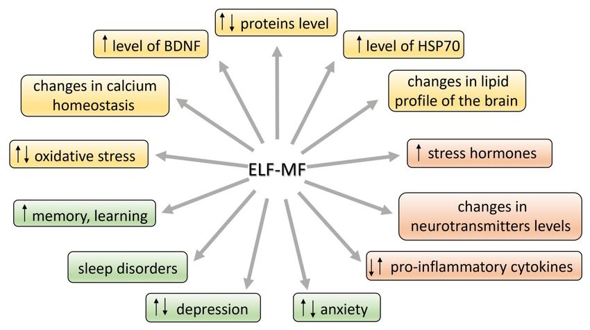
and immunological systems (Figure 1). ELF-MF influence on living matter can cause a
induced carcinogenicity [9]. ELF-MF was proved to be a stress factor and as a consequence,
detrimental increase in free radicals levels and radical-evoked damages in macromole-
it can provoke morphological and physiological changes in stress-related systems [10].
cules [11]. Most studies on ELF-MF effects concentrate on its negative influence; however,
Some authors argue that ELF-MF evokes cell/organism responses that are characteristic to
in the last
general decade
stress thereELF-MF
reaction. was an increase
exposurein“turns
the number of research
on” different studies showing stim-
intracellular—compensatory
ulating impact of ELF-MF on brain plasticity processes (the production
or deleterious—mechanisms and modifies stress-related function of nervous, of protective
hormonalpro-
teins (e.g., Hsp70 or BDNF) or an increase in the activity of antioxidant
and immunological systems (Figure 1). ELF-MF influence on living matter can cause a detri- enzymes) [12].
Furthermore,
mental increase long-term exposure
in free radicals to and
levels ELF-MF can cause permanent
radical-evoked damages in changes in behavior
macromolecules [11].
(towards
Most studiesdepressive and effects
on ELF-MF anxietyconcentrate
disorders) that
on itsare relatedinfluence;
negative to exposure to chronic
however, in thestress
last
[10,13,14].
decade there was an increase in the number of research studies showing stimulating impact
In the face
of ELF-MF of numerous
on brain plasticity studies
processeson(the
the production
effects of the of ELF-MF,
protectiveitproteins
is necessary
(e.g., to sys-
Hsp70
tematize the knowledge for a better understanding of this phenomenon, in
or BDNF) or an increase in the activity of antioxidant enzymes) [12]. Furthermore, long- order to reduce
the
termrisk associated
exposure with exposure
to ELF-MF can cause topermanent
this factor,changes
but alsointobehavior
recognize the possibility
(towards depressive of
using it as a therapeutic
and anxiety agent.
disorders) that are related to exposure to chronic stress [10,13,14].
Figure 1.
Figure Effects of
1. Effects of extremely
extremely low-frequency
low-frequency magnetic
magnetic field (ELF-MF) action in the organism.
In the face
2. Stress—A of numerous
Factor studies
Determining theon the effects
Function of the ELF-MF,
of Organism it is
at All necessary
Levels to system-
of Organiza-
atize
tion the knowledge for a better understanding of this phenomenon, in order to reduce the
risk associated with exposure to this factor, but also to recognize the possibility of using it
It is accepted that ELF-MF exposure may count as a mild stress situation [10,15–17]
as a therapeutic agent.
and it could activate a wide spectrum of interacting neuronal, molecular, and neurochem-
ical systems that
2. Stress—A underpin
Factor behavioral
Determining and physiological
the Function of Organism responses.
at All LevelsChronic stress can
promote and exacerbate pathophysiology leading to allostatic overload in human body
of Organization
[18]. The brain developed
It is accepted someexposure
that ELF-MF adaptive may
mechanisms
count as in the face
a mild stressof situation
changing[10,15–17]
environ-
ments and stress factors imposed on the nervous system. Integrated response
and it could activate a wide spectrum of interacting neuronal, molecular, and neurochem- to stressful
stimuli is an essential component of adaptive processes critical for
ical systems that underpin behavioral and physiological responses. Chronic stresssurvival of the organ-
can
ism. Failure
promote andofexacerbate
this stresspathophysiology
adaptation is considered asallostatic
leading to one of theoverload
primaryin neuropathological
human body [18].
The brain developed some adaptive mechanisms in the face of changing environments and
stress factors imposed on the nervous system. Integrated response to stressful stimuli is
an essential component of adaptive processes critical for survival of the organism. Failure
of this stress adaptation is considered as one of the primary neuropathological causes of
stress-related disorders. A healthy organism is able to turn on or off effectively physiologi-
cal and psychological responses to stimuli; however, if the stress system response is not
adequate—too slow or too high, its mediators will enhance vulnerability to stress-relatedBrain Sci. 2021, 11, 174 3 of 20
disease to which the individual is predisposed. Adaptation to repeated stress is associated
with a complex cascade of molecular and cellular events, ranging from regulation of gene
expression to release of neurotransmitters [19].
The definition of stress is not precise because the process is differently understood
by people representing various fields of science. Stress can be discussed in the context of
its influence on all levels of an organism’s organization: molecular, cellular, physiologi-
cal, and behavioral as well as psychological. The term “stress” was introduced by Hans
Selye [20] and described as a result of disturbed homeostasis in the organism. Seyle [21]
stated that stress is a “nonspecific response of the body to any demand”. McEwen de-
fined stress as “experiences that are challenging emotionally and physiologically” [18].
Other authors describe stress as a process involving perception, interpretation, response,
and adaptation to harmful, threatening, or challenging events [22]. The reaction to a stress
event is necessary for the organism to cope with danger [18]. Alarming signals include
an internal, psychological, or environmental stimulus—such as ELF-MF. Some authors
postulate that the changes turned on under the influence of exposure to ELF-MF are similar
to those caused by other stress factors. The consequences of stress can be different and
are dependent mainly on the strength of the stimulus. A low dose of stress can drive
adaptive processes such as plasticity processes, e.g., the growth of postsynaptic (dendritic)
spines, production of stress-resistant proteins, e.g., BDNF (brain-derived neurotrophic
factor) and stimulation of neural stem cells to form new neurons that replace or cooperate
with the existing ones [23], whereas even one high dose of a given factor may be harmful
or even lethal [24].
In the neuroendocrine approach, stress is related to activation of the autonomic
nervous system (SAM) and hypothalamo–pituitary–adrenal (HPA) axis. First, the auto-
nomic nervous system is activated causing the release of noradrenaline and adrenaline
from the adrenal medulla into the circulation, which—being a hormone—can rapidly
regulate the function of peripheral organs [25] as well as the immunological response,
which is supposed to adapt the organism to new, stressful conditions [26]. Acute activation
of this system leads to release of noradrenaline from an extensive network of neurons
throughout the brain, producing an enhanced state of arousal, which is critical for adap-
tive responses to stress [27]. Somewhat later, the HPA axis is activated, which causes
the secretion of corticosteroid hormones from the adrenal cortex [25]. In response to a
stressor, corticotropin-releasing hormone (CRH) is secreted in the hypothalamus. CRH is
then driven with blood to anterior pituitary, where it causes adrenocorticotropic hormone
(ACTH) release. In the next stage, ACTH reaches the adrenal glands and as a consequence
glucocorticoids (cortisol and corticosterone) are secreted. The glucocorticoids cause in-
creased arousal that ensures the organism’s readiness for action. Thus, the HPA axis
system regulates the intensity, dynamics, and termination of the stress response [28,29].
Hippocampus, which role is the inhibition the HPA axis via the negative feedback, is of
crucial importance for the dynamic of stress response [30–32]. On the other hand, corti-
costeroids can also modulate hippocampal function in the opposite directions: causing
neuron’s dysfunction or plasticity and as a result, hippocampus-related behavioral changes
can be observed. The hypothalamic–pituitary–adrenal (HPA) axis is sensitive to a broad
spectrum of experimental and environmental events [30] that may result in physiological
and behavioral changes in both directions: detrimental and compensatory ones. Sometimes
these modifications are very subtle, but in some conditions they can even underlie the
stress-related disorders or mediate the reversion of brain damage.
3. Molecular Stress Response to ELF-MF
Many studies show that stress induces the disruption in homeostasis [23,33] and as
a consequence an overcompensation response is triggered to re-establish homeostasis.
It needs gene expression and protein synthesis that progresses over time and leads to
establish a new set-point for stress response systems. As ELF-MF is able to change the stress
parameters, it is suggested that it can shift the set-point of endocrinological regulationsBrain Sci. 2021, 11, 174 4 of 20
and determine the health status of the organism as a consequence [15]. The effects of
exposure to ELF-MF are particularly prevalent in the hippocampal area of the brain [34,35].
As mentioned, the hippocampus is involved in regulating the HPA axis activity, but on
the other hand, stress hormones (mainly corticosterone and noradrenaline) are known
to modulate hippocampal function and they may determine the plasticity processes in
this area; it means an adaptive response to ELF-MF’s exposure. Targets for ELF-MF at
molecular level include the cell membrane (e.g., its permeability, inorganic ion transport,
receptor function), second messengers synthesis, chromosome structural changes and chemical
changes in DNA structure, genes expression and protein synthesis (e.g., metabolism-related),
free radicals, and neurotrophic factors. Such profound modifications have to be reflected
in neurotransmitter activity, hormone release, and metabolism of the brain [36–44]. What is
important, the effect of ELF-MF on molecular and/or cellular mechanisms is not obvious—it
can be detrimental or protective. However, the research study on these mechanisms can
shed some light on the possible metabolic pathway being possibly influenced by ELF-
MF. ELF-MF-evoked cellular stress includes the modifications of key substances in cell
metabolism—proteins and lipids. The mentioned alternations are mainly related to ELF-
MF-induced oxidative stress. The consequence of these processes can be cell death such as
apoptosis, necrosis, or autophagy [42,45–48].
3.1. Proteins and Lipids
As shown in in vivo research, ELF-MF can affect levels and function of proteins—crucial
for maintenance of cell homeostasis, e.g., proteins anchored in lipid bilayer of the cell
membrane functioning as ion channels, enzymes, and receptors, as well as the other
proteins of key importance for the response to stress, regulation of apoptosis, and a number
of metabolic processes [37,45,47,49,50]. Total protein level as well as its activity (e.g., alanine
aminotransferase (ALT), aspartate aminotransferase (AST), alkaline phosphatase (ALP),
albumin, bilirubin) was augmented in rats exposed to 1.5 mT ELF-MF [45]. Exposure to
both 0.5 and 1 mT ELF-MF altered protein pattern in rat’s hippocampus. Gene ontology
analysis showed that the most important function of the identified proteins altered after
ELF-MF exposure is to ensure the functioning of the brain. Exposure to ELF-MF caused
extreme downregulation of two proteins: Sptan1 and Dpysl2. The first is responsible for
stabilization of cell membrane and organization of intracellular organelles. The second,
Dpysl2, plays a key role in neuronal development and polarity, and additionally in neuron
projection morphogenesis. Notably, the increased intensity of ELF-MF may be associated
with more alteration in cell protein expression, and subsequent cell morphology and
proliferation rate changes [47]. The chromogranin A (CgA) is another protein that should be
mentioned as important in stress response and as a new target for electromagnetic radiation.
It is a neuroendocrine secretory protein costored and coreleased with catecholamines
from adrenal medulla, adrenergic nerve endings, and neuroendocrine cells. CgA is also
a marker of sympathoadrenal activity, so its level gives information on the course of
stress response [51]. The protein is also involved in maintaining calcium homeostasis in
the cell [52]. What is important, its level increases during a depressive mood or stress
situation [52]. The serum level of CgA in volunteer subjects chronically exposed to ELF-MF
in the range 0.1–0.3 µT did not differ from the level in control group. However, a trend
toward lower concentrations of CgA was observed in the group exposed to higher level
of ELF-MF (>0.3 µT). Suppressive effects of ELF-MF on CgA level could be recognized as
having inhibitory effects on the activity of the sympathetic nervous system [53].
In addition to proteins, the brain lipid profile is also influenced by ELF-MF exposure
and taking into account multiple roles for lipids, they can be the medium for the ELF-MF
action in the cell. Lipids are structural components of the cell membrane and they are in-
volved in transfer of signals across membranes [17]. Apart from being structural elements,
they are also required for axonal elongation and act as precursors for various secondary
messengers, including arachidonic acid, docosahexaenoic acid, or 1,2-diacylglycerol [54].
Any changes in brain lipid metabolism lead to disturbances in homeostasis and are re-Brain Sci. 2021, 11, 174 5 of 20
sponsible for altered functioning at the cell and tissue levels. It was shown that 60 Hz
2.4 mT ELF-MF induces changes in the brain lipid profile and in corticosterone concen-
tration. The level of these changes was similar to that in the positive control group of
rats exposed to stress-RS (movement restraint). After 21 days of exposure to ELF-MF or
RS or combined model (ELF-MF + RS), a general tendency to the decrease of total lipid
level in brain structures was observed in each experimental group. Total cholesterol level
was significantly increased in the cortex in the ELF-MF and RS + ELF-MF groups, and in
subcortical structures in the RS + ELF-MF group. Inversely, polar lipids level in ELF-MF
and RS + ELF-MF groups was decreased both in the cortex and in subcortical structures.
Nonesterified fatty acid levels were found to be slightly higher in subcortical structures of
the RS + ELF-MF group as compared to the control and RS groups. The analysis of fatty acid
methyl esters revealed that the level of polyunsaturated fatty acids in cerebellum of ELF-
MF-exposed rats was decreased, whereas their level in subcortical structures in the same
group was increased. In addition to the changes in the amount of different kinds of lipids,
the ELF-MF-induced lipid oxidative modifications were also noticed. The concentration of
thiobarbituric acid reactive substances (TBARS, byproduct of lipid peroxidation) in lipids
was higher, especially in the cortex and cerebellum of all treated groups [17]. Previous
research has shown that immediate changes in lipid profile and TBARS levels after 2 h of
singular exposure were visible only in the RS + ELF-MF group, whereas single exposure
to ELF-MF or RS alone did not cause any changes in reduced glutathione and nitric oxide
levels [55]. The increased level of lipid peroxidation was also noticed in rats exposed to
ELF-MF (100 µT and 500 µT) [56]. The interesting research on ELF-MF-induced (50 Hz,
3 mT) changes in lipid profile (proteomic and transcriptomic profiling) in Caenorhabditis
elegans was performed by Sun et al. [57]. In the glycerolipids (GLs) group, total triacyl-
glycerols (TGs) content was increased while diacylglycerols (DGs) level was decreased.
It should be also noted that among the most enriched proteins evaluated in this research,
there were ones involved in lipid transport [57]. These studies indicate that ELF-MF affects
the brain’s lipid balance in a similar way to physiological stressors.
3.2. Oxidative Stress and Antioxidant Status
Stress can be a factor causing an increase of the level of oxidative stress parameters in
the brain, including lipid peroxidation and on the other hand, it can activate antioxidant
response [58,59]. Oxidative stress is the result of an imbalance between reactive oxygen
species (ROS) and antioxidants [29]. Under normal conditions the synthesis of ROS is
usually balanced, but when the production of ROS increases they become harmful for
organism. The imbalance causes changes at the cellular level, which causes DNA, proteins,
and lipids damage. ROS are involved in physiological processes, for instance, in cell
signaling and respiratory chain and immune response, but some pathological factors can
contribute to their increased level [60]. Overproduction of ROS occurs, inter alia, in re-
sponse to stress (heat, anoxia, ultraviolet light, injury, environmental pollution, cigarette
smoke, psychological trauma, and many others) [61]. It has also been reported that ROS
levels increase after ELF-MF exposure and the reason for this phenomenon can be the
failure of antioxidant defense.
The disturbance of oxidative homeostasis was proved in in vitro research. Exposure
to ELF-MF of 1 mT resulted in free radical increase in mouse macrophages [62] and SH-
SY5Y neuroblastoma cells [39]. ELF-MF-induced increased ROS production was also
found in K562 human leukemia cell line (50 Hz, 0.025/0.05/0.1 mT) [63,64], and in human
osteoarthritic chondrocytes (100 Hz) [65]. The viability decrease and morphological changes
of rat hippocampal neurons concomitantly with the increase of MDA (malondialdehyde)
and ROS levels and reduction of superoxide dismutase activity were noticed after exposure
to ELF-MF (50 Hz, 8 mT) [42]. Similarly, exposure to ELF-MF (50 Hz, 25–200 µT) resulted
in increased ROS production and diminished activity of antioxidant enzymes (superoxide
dismutase (SOD), glutathione peroxidase (GPx), glutathione reductase (GR)) in the human
keratinocyte cell line NCTC 2544 [66].Brain Sci. 2021, 11, 174 6 of 20
In vitro results have been confirmed in in vivo research. The shift into oxidative
processes, presented as ROS-level elevation and significantly, the total antioxidative ca-
pacity (TAC) level decrease, were found in Caenorhabditis elegans exposed to ELF-MF
(50 Hz, 3 mT) [67]. These results, proving the ELF-MF-induced impairment of antioxi-
dant mechanisms in the organism, were also obtained from research using rodent models.
The toxic, increasing oxidative stress level effect of ELF-MF was found mainly in the brain.
Akdag et al. [68] demonstrated that the activity of antioxidant enzyme catalase (CAT) was
decreased in ELF-MF-exposed animals regardless of ELF-MF intensity (100 and 500 µT).
Moreover, in the group exposed to 500 µT, TAC was lower than in the 100 µT group. At the
same time, in the 500 µT group the levels of oxidative stress markers, MDA and MPO
(myeloperoxidase), and values of total oxidant status (TOS) and oxidative stress index
(OSI) were significantly higher. TBARS concentrations increasing concomitantly with
decreasing reduced glutathione (GSH), total free-SH group concentrations, and TAC levels
were found in rats exposed to ELF-MF (40 Hz, 7 mT and 50 Hz, 12 and 18 kV/m) [69].
The activities of antioxidant enzymes in brain homogenates were also decreased in rats
exposed to ELF-MF (50 Hz 10 kV/m, 4.3 pT) [70]. In addition, in mouse brain subjected to
ELF-MF (50 Hz, 8 mT), the levels of MDA, ROS, nitric oxide (NO), and nitric oxide synthase
(NOS) were increased, whereas activities of SOD, CAT, and GPx were decreased [71,72].
Free radical level (superoxide anion- O2 •− and NO2 − ) was increased in the hypothalamus
of rats exposed to ELF-MF (50 Hz, 10 mT) [73]. Acute exposure to ELF-MF (60 Hz 2.4 mT)
resulted in the impairment of antioxidant mechanisms in the brain as well as in other
tissues: heart, kidney, and plasma (decrease in SOD activity and reduced glutathione
level) [55,74]. The disturbance of oxidative status was also found in testes of rats (diabetic
model) exposed to ELF-MF (50 Hz, 8.2 mT): the increase in MDA and NO level, and di-
minished GSH level [75]. Many studies on the effects of ELF-MF have been conducted on
people from risk groups, occupationally and residentially (living near high voltage lines)
exposed to ELF-MF. El-Helaly and Abu-Hashem [76] carried out their research on a group
of 50 electronic equipment installers and repairers. The serum malondialdehyde (MDA)
level in the ELF-MF-exposed group was significantly higher than in control, and concomi-
tantly the melatonin level (hormone supporting the antioxidant effect) in this group was
lower. Similarly, the increment of oxidative stress and oxidative damage to DNA was also
found in other research on power plant workers (occupational exposure, 110–420 kV and
4.09 V/m, 16.27 µT) [40,77,78]. The data suggest that exposure to ELF-MF could cause the
failure of the antioxidant response and the collapse of homeostatic capability of the cell,
leading to oxidative damage and functional impairment. However, the direct connection
to the risk of disease development has not been unequivocally proved.
Subsequent studies shed light on the effects of ELF-MF on the antioxidant mecha-
nisms that can underlie the protection against neurodegeneration. The ELF-MF-induced
improvement of antioxidant protection has been evaluated in both in vitro and in vivo
research. Exposure of C2C12 cells (myoblasts) to ELF-MF of 1 mT caused a drastic decrease
in ROS level while total antioxidant status (TAS) and the activities of CAT and GPx were
elevated [79]. Ehnert et al. [80] found ELF-MF-induced (16 Hz 6–282 µT) increase of SOD2,
CAT, GPX3, and glutathione-disulfide reductase (GSR) activity concomitant with the reduc-
tion of ROS levels in human osteoblasts. Similarly, in the myelogenous leukemia cell line
K562 exposed to ELF-MF (50 Hz, 1 mT) and in human blood platelets exposed to different
sources of electromagnetic radiation (1 kHz, 0.5 mT; 50 Hz, 10 mT; or 1 kHz 220 V/m) CAT
activity was increased [81,82]. Moreover, the exposure IMR-90 human lung fibroblasts
for a total of 168 h to 6 mT ELF-MF contributed to decreased ROS level [83]. Exposure
of human neuronal cell culture SH-SY5Y to 50 Hz ELF-MF with magnetic field intensity
1 mT resulted in elevated activity of NOS. This enzyme is controlled by proinflammatory
cytokines that also activate ROS. After 1, 3, 6, and 24 h of exposure to ELF-MF, the activity
of the enzyme was significantly increased. Moreover, the augmented production of O2 −
was also found. However, CAT activity increased as the exposure time increased, possibly
indicating a gradual adaptation of cells to the conditions of oxidative stress. On the otherBrain Sci. 2021, 11, 174 7 of 20
hand, these adaptive mechanisms turn out to be insufficient when ELF-MF exposure is
combined with additional administration of H2 O2 —the oxidative effect is then exacer-
bated. These data suggest that ELF-MF may to some extent have neuroprotective effect.
The combination of ELF-MF exposure and the stressor H2 O2 prevents cells from being
effectively defended against ROS [84]. However, when H2 O2 -treated cells were exposed
to a higher value of ELF-MF (75 Hz, 2 mT), ROS level decreased and MnSOD activity
increased [85]. It definitely suggests that the protective effect of ELF-MF depends on its
intensity. The results of this in vitro research points out the beneficial effect of ELF-MF as
an upregulation of antioxidant pathways, leading to protection against oxidative damage
has been noted, reflecting an attempt to stimulate cellular response to neuronal damage.
In addition, ELF-MF as a mild stress factor activates an adaptive response that ensures
the oxidative–antioxidant balance in rodent models as well as in humans. In a rat model
of Huntington’s disease, ELF-MF (60 Hz and 0.7 mT) was found to be able to reverse the
process of neuronal degeneration and oxidative stress; it enhanced the antioxidant glu-
tathione content and reduced the oxidative stress markers, 8-hydroxy-20 -deoxyguanosine
and oxidized glutathione levels, in the whole-brain tissue [12]. Recent research evaluat-
ing the redox state in post-stroke patients demonstrated the beneficial effect of ELF-MF
on oxidative status. High magnetic intensity, 5 or 7 mT of 40 Hz ELF-MF, significantly
increased enzymatic antioxidant activity as compared to results obtained before treat-
ment. The results were correlated with the improvement in functional and mental status
of post-stroke patients [86,87]. These data show that ELF-MF is a factor that may both
increase the production of ROS and activate organisms’ antioxidant machinery in humans.
In consequence, the electromagnetic radiation may drive the mechanisms underlying cell
survival and plasticity.
3.3. Neuroprotective Proteins: Hsp70 and BDNF
Prosurvival responses include DNA repair processes and the increase in expression of
chaperone protein—70-kDa heat shock proteins (Hsp70) and neurotrophin—brain-derived
neurotrophic factor (BDNF) [88,89]. The expression of Hsp70 and BDNF appears to be
a part of the general stress response and thus it is speculated to be associated with hor-
monal response to stress [89]. The increase of expression of these proteins would indicate
the development of processes adapting neuronal networks in order to optimize circuits
responding to the external environment and to integrate the response to challenges [90].
It was shown that stress hormones (mainly corticosterone and noradrenaline) influence
via their receptors the plasticity processes in the hippocampus [31,91]. Noradrenaline can
even dictate the direction of synaptic strength change in the hippocampus [91]. Under the
influence of ELF-MF the expression of stress-response genes increases, resulting in higher
levels of molecular chaperones such as Hsp70 [92–94]. The role of Hsp proteins is to
stabilize polypeptide chains during their translocation across the cell membranes and to
prevent aggregation of proteins with abnormal structure. Moreover, the antiapoptotic
properties of Hsp70 and their role in appropriate folding and activation of proteins have
also been proved [89,95]. As there are many pathways that could be affected to upregulate
Hsp70 expression induced by stress, it is difficult to determine if any specific pathway
may be affected.
The protective value of ELF-MF mediated by its influence on Hsp70 level was proved
in in vitro research. Perez [96] showed that ELF-MF (50 MHz) leads to higher levels of
Hsp70 in human T lymphocytes and fibroblast cell lines when subjected to stress, and that
this response was of protective value. It seems to precondition and to enhance the cellular
stress response when cells are provoked by toxic stimuli. Moreover, the cell protection was
proportional to the levels of Hsp70. Exposure of human leukemia cell line K562 to ELF-MF
(less than 0.1 and 1 mT) leads to increased Hsp70 levels [63,64]. More recent in vitro studies
on ELF-MF with a density over 1 mT have shown marked effects, including an increase in
Hsp70 transcription that results in protection against chronic hypoxia-induced injury [50].Brain Sci. 2021, 11, 174 8 of 20
Interesting data were also received in in vivo research on invertebrates. According to
Gutzeit [97] exposure to 50 Hz ELF-MF with magnetic flux densities 50–150 µT enhances
the response to thermal stress in C. elegans. ELF-MF-mediated specific genes activation
could enhance transcription of an already activated set of heat shock genes by costressor
(heat stress), thus providing an adequate and optimal defense response. Exposure to
60 Hz 8 µT ELF-MF caused regeneration of the heads and tails parts of the Planarian,
Dugesia dorotocethala. This effect was accompanied by an increase in the level of Hsp70,
which is triggered by extracellular signal-regulated kinase (ERK) cascade. It is known that
ERK is activated as reaction to injury to promote regeneration [98].
In this approach, ELF-MF appears to be a mild stressor mobilizing the organism to
cope with a dangerous situation [98]. The expression of the Hsp70 in response to stress
serves to protect against the negative impact of stress. Hsp70 induction and stress systems
function were shown to be two important inter-related mechanisms in maintaining the
homeostasis under stress conditions [89]. According to the juxtaposition presented, some of
beneficial effects of ELF-MF can be due to the protective role of Hsp70.
A substance of high importance for the nervous system is also brain-derived neu-
rotrophic factor (BDNF). This neurotrophin is responsible for differentiation and survival
of neurons during development, but it is also important for the adult brain, especially
when subjected to stress conditions [90]. In a mature brain, BDNF ensures excitatory
and inhibitory synaptic transmission and neuroplasticity [99]. The mechanism of neuro-
plasticity is crucial for learning and memory processes. It includes enhancement of the
long-term potentiation (LTP), and stimulating and controlling neural growth. BDNF has a
high affinity to full length tropomyosin receptor kinase B (TrkB) and its truncated isoform,
p75 NTR. Through the activation of TrkB, the neurotrophin starts the cascade of signaling
pathways, which results in neurogenesis, neuroplasticity, cell survival, and resistance to
stress [100]. In vitro research proved that the exposure to pulsed ELF-MF (50 Hz; 1 mT for
2 h) increased the BDNF mRNA expression in cultured dorsal root ganglion neurons [101].
BDNF expression can be modulated by external, physiological, and pathological
factors proven mainly in research on both rodents and humans. In the course of some
diseases, such as Alzheimer disease, and during aging process or chronic stress, the in-
hibition of BDNF expression is noted, while exercise, enriched environment, and taking
antidepressants are related to the intensified expression of BDNF [99]. ELF-MF is used
in physical therapy due to its ability to stimulate BDNF synthesis. Several studies fo-
cused on this particular effect of ELF-MF on diseases and pathologies, like Huntington
disease or stroke [12,102,103]. In post-stroke patients subjected to ten sessions of 15 min
ELF-MF therapy (40 Hz 5 mT), plasma BDNF level was about 200% higher than before the
treatment [102]. The study undertaken on a rat model of Huntington disease indicated
that exposure to ELF-MF (60 Hz 0.7 mT 2 h in the morning and 2 h in the afternoon for
21 days) significantly elevated BDNF level in the rats with induced Huntington disease.
Moreover, changes in the rats’ behavior related to Huntington disease were neutralized by
ELF-MF [12]. Urnukhsaikhan et al. [103] showed that expression of BDNF, TrkB, and phos-
phorylated protein kinase B was increased in ELF-MF-stimulated (60 Hz, 10 mT) ischemic
mice. In vitro research proved that the exposure to pulsed EMF (50 Hz; 1 mT for 2 h) in-
creased the BDNF mRNA expression in cultured dorsal root ganglion neurons [103]. Thus,
there is evidence suggesting that the neuroprotective effect of the exposure to extremely
low-frequency MFs may be due to, at least in part, the impact of the fields on neurotrophic
factors levels, leading to an increase of cell survival.
3.4. Plasticity, Neurogenesis, Proliferation, and Differentiation
The level of activity of voltage-gated Ca2+ channels is an important factor determining
the synaptic transmission and leading to stimulation of short-term synaptic plasticity [104].
Calcium ions are involved in secretion of neurotransmitters. The influx of Ca2+ through
presynaptic voltage-gated Ca2+ (Cav) channels triggers the release of neurotransmitters
from presynaptic part of synapses. Measurements at a large glutamatergic synapse inBrain Sci. 2021, 11, 174 9 of 20
the mammalian auditory brainstem—the calyx of Held—showed that vesicle endocytosis
and synaptic transmission were enhanced in mice (8–10 postnatal days old) kept from
birth under the influence of EMF (50 Hz EMF, 1 mT). Moreover, in mice exposed to EMF,
the increase in expression of calcium channels at the presynaptic nerve terminal facilitating
the influx of calcium was found. The observed mechanism is responsible for increasing
endocytosis and synaptic plasticity [105]. In vitro research also evidenced the ELF-MF-
induced increase in intracellular Ca2+ concentration. This effect was found in C2C12 cells
(myoblasts) after 0.1 and 1 mT ELF-MF exposure [79], in human pluripotent stem cells
(iPSCs) after 1.5 mT ELF-MF exposure [105], in dorsal root ganglion neurons after 0.1, 1, 10,
and 100 mT ELF-MF application [101], and in rat hippocampal neurons exposed to 8 mT
ELF-MF [42]. The influence of ELF-MF on proliferation and apoptosis and the participation
of Ca2+ in these processes were also determined. In human neuroblastoma IMR32 and in
rat pituitary GH3-cultured cells, the exposure to 1 mT 50 Hz ELF-MF caused the increased
cell proliferation. At the same time, the increase of Ca2+ current density and of voltage-
gated Ca2+ channel expression in the cell membrane was observed. In addition, blocking
of Ca2+ channels by 15 µM Cd2+ alleviated the proliferative effect of ELF-MF. Apoptosis,
induced by H2 O2 or puromycin in IMR32 cells, was decreased after 72 h exposure to
1 mT 50 Hz ELF-MF. Blocking of L-type calcium channels by nifedipine also caused
disappearance of the antiapoptotic effect of ELF-MF [106]. It has been also shown that
ELF-MF influences calcium homeostasis in cultural entorhinal cortex neurons via calcium
channel-independent mechanism. Twenty-four hour exposure to 1 or 3 mT ELF-MF does
not affect voltage-gated calcium current and activity of calcium channels, but regulates
intracellular calcium dynamics by decreasing the high-K+ -evoked intracellular calcium
elevation [107]. In summary, this study suggests that the change in calcium currents
through voltage-gated calcium channels is the mechanism responsible for the proliferation
promotion and antiapoptotic effect of ELF-MF.
Measurements at a large glutamatergic synapse in the mammalian auditory brainstem—
the calyx of Held—showed that vesicle endocytosis and synaptic transmission was en-
hanced in mice (8–10 postnatal days old) kept from birth under the influence of ELF-MF
(50 Hz ELF-MF,1 mT). Moreover, in mice exposed to ELF-MF, the increase in expression
of calcium channels at the presynaptic nerve terminal facilitating the influx of calcium
was found. The observed mechanism is responsible for increasing endocytosis and synap-
tic plasticity [108].
Throughout the life course, new neurons are continuously formed in the hippocampus,
which is therefore a major site of structural plasticity in the adult brain. The existence of
a causal link between ELF-MF-enhanced synaptic plasticity and neurogenesis has been
shown by a number of in vivo experimental studies. ELF-MF (60 Hz, 0.7 mT, applied over
21 days) improved neurological scores, enhanced neurotrophic factor levels, and reduced
neuronal loss in a rat model of Huntington’s disease [12]. In addition, prolonged exposure
to ELF-MF (50 Hz, 100 µT; for 90 consecutive days; 2 h/day) increased LTP induction in
rat’s hippocampus [35]. In vivo exposure of adult mice to ELF-MF (50 Hz, 1 mT) produced
a marked increase in the number of newly generated neurons in the granule cell layer of
the dentate gyrus [34]. Although the ELF-MF of 1 mT (for 21 days) caused the decrease
in the dendritic spine density of neurons in hippocampus after 7 and 10 days, the effects
disappeared after 14 days [109]. Studies on the rat traumatic brain injury model [110] and
on rat Alzheimer’s disease model [49] have shown that ELF-MF reversed pathological brain
damages and learning and memory abilities impairment. Similar effects were obtained
after exposure of neurotoxin-injected mice to ELF-MF (50 Hz, 1 mT); the deficits such
as neuronal maturation impairment, neurogenesis decrease, and memory disturbance
decreased [111]. Studies on the beneficial effects of ELF-MF might yield fruitful insights
related to clinical therapy of nervous-system-related diseases.
The cell differentiation at the expense of proliferation in different tissues and increased
cell viability as an effect of exposure to low-frequency ELF-MFs is well evidenced in the
literature. Collard et al. [112] reported an acceleration of proliferation and differentiation ofBrain Sci. 2021, 11, 174 10 of 20
human epidermis cells after exposure to low frequency (40 Hz) and demonstrated that the
processes were related to a significant modification of gene expression. Falone et al. [85]
found that ELF-MF (75 Hz, 2 mT) alone did not affect the viability of the human neu-
roblastoma SH-SY5Y cell line and that ELF-MF exposure prevented reduced cell viability
after H2 O2 application. Vannoni et al. [65] concluded that ELF-MF (100 Hz) stimulation
is a useful tool to induce more divisions and thus to enhance cell proliferation of human
osteoarthritic chondrocytes. The treatment of HeLa cells IMR-90 fibroblasts with ELF-MF
(60 Hz, 6 mT) increased cell viability and activated cell cycle progression. In addition,
ELF-MF mitigated the antiproliferative effect of GOx (agent stimulating H2 O2 produc-
tion) [83]. Di Loreto et al. [113] found that ELF-MF (50 Hz, 0.1–1 mT) had a positive effect
on cell viability in primary cultures of maturing rat cortical neurons. The research of
Ardeshirylajimi and Soleimani [105] on human pluripotent stem cells (iPSCs) suggested
that ELF-MF (50 Hz, 1.5 mT) increases cell viability, division, proliferation, and mineral-
ization of extracellular matrix. These results indicated that ELF-MF would improve the
viability, proliferation, and differentiation of cells, and may be beneficial for the develop-
ment of novel therapeutic approaches in regenerative medicine. The papers pointing to
detrimental effect of ELF-MF on viability, differentiation, and proliferation should also be
mentioned. It is, however, important that this effect is caused by high values of ELF-MF
induction. Yin et al. [42] showed that the number of rat hippocampal neurons in G0/G1
phase was decreased and cells in S phase were accumulated as the effect of exposure to
ELF-MF (50 Hz, 8 mT). The exposure of mesenchymal stem cells (bone marrow or adipose
tissue derived) to ELF-MF of 20 mT (50 Hz) resulted in decreased cell proliferation [44,114].
This effect appeared to be related to the diminished expression of genes responsible for
pluripotency and neuronal differentiation [44].
4. ELF-MF-Induced Changes in Levels of Neurotransmitters, Hormones,
and Cytokines
ELF-MF-induced molecular changes modify to a certain extent some crucial neuronal
processes. As it is commonly known, the communication between main groups of signaling
substances: neurotransmitters, cytokines, and hormones, is of high importance for the
maintenance of health status of an individual. We have a lot of data confirming the effect
of ELF-MF on functioning of nervous, immune, and endocrinological systems. However,
the mechanisms by which the magnetic stimulation modulates the activity of these systems
and the interplay between them are open to be identified. Up-to-date results concluded
that the exposure of rats to ELF-MF may be sufficient to induce significant changes in the
content of neurotransmitters. The levels of major inhibitory and excitatory amino acids and
neurotransmitters: glutamate (Glu), glutamine (Gln), glycine (Gly), tyrosine (Tyr), and γ-
aminobutyric acid (GABA), were elevated in the thalamus after five days of exposure to
ELF-MF (60 Hz, 2 mT). In the striatum, higher levels of Gln, Gly and GABA were found as
well, whereas their concentrations were decreased in cortex, cerebellum, and hippocampus.
Dopamine level was increased in the thalamus [115]. Extremely low-frequency magnetic
field (10 Hz; 1.8–3.8 mT) exposure was found to alter turnover and receptor reactivity of
serotoninergic and dopaminergic systems and some behavioral disturbances induced by
these systems [116]. The rats receiving chronic (10 days) repetitive transcranial magnetic
stimulation (rTMS) treatment showed the symptoms of anxiety, and it was shown that
the rTMS-induced anxiety might involve the serotonergic system [117]. The continuous
exposure of rats to ELF-MF (50 Hz, 0.5 mT) affected cortical serotoninergic neurotrans-
mission, and intensity of these changes depended on ELF-MF exposure duration [118].
The data may indicate the ability of ELF-MF to modify the function of main neurotrans-
mitter systems and thus to modulation of some physiological processes, such as memory,
emotionality, mood changes, sleep, alertness, or stress response. The response of individual
brain tissues to exposure was varied; the level of one neurotransmitter increased in a
given tissue appeared to be decreased in another, suggesting that the radiation can induce
varying responses in the nervous system [115].Brain Sci. 2021, 11, 174 11 of 20
As noted, the existing data indicate that the exposure to ELF-MF may count as a mild
stress situation and could be a factor in the development of disturbances of brain stress
systems: hypothalamo–pituitary–adrenal (HPA) axis and sympatho–adrenal–medullary
(SAM) system [10,16,115,119,120]. Although some findings indicate the deteriorating
effects of magnetic fields on hormonal stress response, others failed to exhibit any obvious
effects. Continuous long-term (4–6 week) ELF-MF (50 Hz, 0.5 mT) treatment induced
some signs of stress: HPA-axis activation (elevated blood glucose level, elevated POMC
(the precursor protein for ACTH) mRNA level, and enhanced depression-like behavior
in a forced swimming test), although other markers of stress (elevated basal ACTH and
corticosterone secretion, adrenal gland hypertrophy, thymus involution, loss of weight gain,
and anxiety-like behavior in elevated plus maze) were not observed. This confirms that
ELF-MF of the abovementioned intensity creates a weak stress response [10]. In addition,
50 Hz ELF-MF (0.207 µT) significantly raised ACTH, cortisol, and glucose levels in guinea
pigs [15]. The concentration of plasma corticosterone level was significantly higher and
remained at a similar level in groups of rats exposed to restraint stress (RS) or ELF-MF [17].
Research by Mahdavi et al. [121] showed that exposure to both 1 and 5 Hz ELF-MF of
0.1 mT intensity caused an elevation of ACTH level in rats’ plasma, whereas corticosterone
level was reduced in both cases. In the animals exposed to 1 Hz ELF-MF, the concentration
of adrenaline increased, but in rats exposed to 5 Hz, the level of adrenaline decreased.
In rabbits exposed to ELF-MF (10 Hz ELF-MF), the level of blood corticosterone was
increased in both the normal and high-cholesterol diet groups [122]. Chronic exposure
(1 month) to 50 Hz 100/500 µT ELF-MF significantly raised corticosterone levels in rats’
plasma [123]. In mice exposed to 10 µT ELF-MF (1, 4, or 24 h/day for 1 week—short-
term exposure), no significant differences in CRH gene expression in hypothalamus were
observed, whereas ACTH plasma level was lower regardless of the daily exposure time (1, 4,
or 24 h/day for 1 week). Moreover, the expression of pituitary level of POMC was lower in
an exposure time-dependent manner, and a statistically significant decrease appeared after
24 h/day exposure [124]. Mostafa et al. [125] showed that 2- and 4-week exposure of rats to
ELF-MF (2 G, equivalent to 0.2 mT) significantly increased their plasma corticosterone level.
Other studies on mouse model showed that even a relatively low level of EMF (12 nT) can
cause corticosterone increase [126]. On the other hand, Kitaoka [16] revealed that levels
of ACTH, the hormone that regulates corticosterone secretion, and hypothalamic CRH
and pituitary POMC were not changed by ELF-MF (70 Hz, 3 mT). Significant changes
were also found in the levels of noradrenaline in various parts of rats’ brain: thalamus,
hypothalamus, cerebellum and striatum, after 2 and 5 days exposure to 2 mT ELF-MF [115].
In the group of volunteers exposed to ELF-MF (50 Hz, 62 µT, for 2 h/day for 2 days with a
6-day interval) the cortisol level was increased at the beginning of ELF-MF exposure but
later it diminished progressively [120]. The workers employed in the live-line procedures
(132 kV high-voltage) for more than two years were found to be vulnerable for EM stress
with altered adrenaline concentrations [40]. Moreover, exposure of turkey females to ELF-
MF (50 Hz, 10 µT) caused NE-activated β-adrenoceptor function decrease, which is known
to be involved in the formation of emotional disinterest and depression [126]. The data
suggest that the exposure to ELF-MF can establish a new “set-point” for stress-system
activity and the direction and dynamics of this process depend on the strength of the
field and duration of exposure. The ELF-MF-induced changes in stress hormone levels
will initiate cellular adaptation or damage by activation of intrinsic signaling pathways.
Consequently, ELF-MF can change the vulnerability of the organism to subsequent stress
factors and thus to diseases, mainly related to the nervous system.
Stress is known to strongly affect the immune system. It has been suggested that the
potential contribution of ELF-MFs to anxiety or other stress associated disorders is also
related to changes in the functioning of the immune system. Moreover, the chronic exposure
to ELF-MF appears to also lead to immune system dysfunction, chronic allergic responses,
inflammatory responses, and ill health [36]. However, as in other aspects of ELF-MF impact
in organism, this factor can also be a double-edged sword and drive the survival-promotingBrain Sci. 2021, 11, 174 12 of 20
processes. Importantly, the immune system and the stress systems—HPA axis and SAM—are
closely linked to each other. Glucocorticoids and catecholamines are known to modify the
secretion of cytokines: proteins that facilitate communication between the immune cells
and the cells of the central nervous and endocrine systems [127]. Cytokines have the ability
to modulate and activate the HPA axis. Proinflammatory cytokines: IL-1, IL-6, and TNFα,
induce corticotropin-releasing hormone (CRH) secretion and they are also involved at
every stage of stress reaction [128]. Changes in plasma proinflammatory cytokines were
observed after acute continuous exposure (24 h) to ELF-MF with magnetic intensity of
7 mT. The levels of IL-1β, IL-6, and IL-2 were elevated. The number of white and red
blood cells and lymphocytes, and the hemoglobin concentration and hematocrit level were
increased. However, the repetitive exposure to ELF-MF (1 h/day for 7 days) did not alter
either cytokines levels or blood parameters [129]. The change in cytokine production was
also noticed in stroke patients treated with ELF-MF (40 Hz, 5 mT ELF-MF) [130]. Following
the exposure to ELF-MF, the plasma levels of IL-1β and IL-2 cytokines and the level of
IL-1β mRNA expression were increased. In addition, ELF-MF exposure increases the levels
of the anti-inflammatory transforming growth factor β (TGF-β) and interleukin-18-binding
protein [84]. Thus, the ELF-MF exposure can cause deregulation of the immune system,
thereby increasing vulnerability to infectious and autoimmune diseases. Interesting results
concerning the effect of ELF-MF on lymphocytes level were obtained by de Kleijn et al. [124].
In mice exposed to 10 µT ELF-MF (1, 4, or 24 h/day for 1 week—short-term exposure, or for
15-week long-term exposure) the increase in CD3+/CD4+ T-lymphocytes was observed
only after short-term exposure to ELF-MF. The data suggest that the ELF-MF effect on
immune and stress responses may be transient, because no changes in the number of
immune cells were observed after long-term exposure. Several authors reported that the
ELF-MF-evoked neuroplasticity can be mediated by the effect of magnetic radiation on
cytokine level. Cytokines are found to influence the expression of neurotrophins and
their receptors. This may indicate the role of inflammatory cytokines in the process of
neuroplasticity [130]. The latest reports showed that 1 and 100 µT 50 Hz ELF-MF not
only downregulates proinflammatory cytokines (IL-9 and TNF-α), but also activates an
inflammation-suppressing cytokine, IL-10. In this case, the most noticeable effect was
obtained at the highest value of magnetic induction [131]. The ELF-MF (60 Hz, 10 mT) also
mitigated the deficits in ischemic mice, among others in the context of immune function:
the levels of inflammatory mediators MMP9 and IL-1β were decreased [102]. The results
demonstrate the recovery-stimulating potential of ELF-MF.
5. Association between ELF-MF Exposure and Emotional Behavior and Wellbeing
An association between ELF-MF exposure and emotional behavior has been indi-
cated in many studies. The animal studies have shown that chronic exposure to ELF-MF
may induce an anxiogenic and/or a depression-like effect. Dysfunction of stress systems
can evoke negative emotional state and can potentiate fear- and anxiety-related behav-
iors [90,132]. Therefore, it is reasonable to speculate that elevation in ELF-MF-induced
anxiety level may be attributed to the effect of ELF-MF on glucocorticoid release follow-
ing activation of HPA axis and catecholaminergic sympathetic nervous system releasing
adrenaline and noradrenaline. These pathways are key biological factors that modulate
emotional behavior [90]. Liu et al. [133] reported that ELF-MF exposure (2 mT, 4 h/day
for 25 days) had an anxiogenic effect in rats, such as anxiety-like behaviors in open field
and elevated plus maze tests. Szemerszky et al. [10] demonstrated that ELF-MF exposure
(0.5 mT, 4 weeks) in rats increased their immobility time in a forced swim test. The chronic
exposure of mice to ELF-MF (3 mT, total exposure 200 h) induced the depression- and/or
anxiety-like behavior (increase in total immobility time in a forced swim test and in the
latency to enter the light box in a light–dark transition test). These behavioral disturbances
were correlated with high corticosterone secretion [16]. Mice prenatally exposed to ELF-MF
(50 Hz, 1 mT) lacked sociability and preference of social novelty, which can be a sign
of autism-relevant social abnormalities; however, they did not show anxiety-like behav-Brain Sci. 2021, 11, 174 13 of 20
ior [134]. The continuous (21 days) exposure to extremely low-frequency magnetic field
(50 Hz, 10 mT) had no significant effect on activity and exploration activity but significantly
increased stress and anxiety-related behavior in rats [135]. Quite similar observations in
open field and elevated plus maze tests were described by Djordjevic et al. [73] after the
exposure to ELF-MF (50 Hz, 10 mT) significantly reduced activity was observed. The noted
effects of short-term ELF-MF exposure (50 Hz, 500 µT, 20 min) verified by behavioral
tests in rats (elevated plus maze, novel object exploration) appear to suggest that these
field parameters may cause some kind of discomfort, influence behavior, and increase
passivity and situational anxiety [136]. Increased level of anxiety has also been found in rats
exposed to ELF-MF of various flux density (50 Hz; 1, 100, 500, 2000 µT, and 2 mT) [56,137].
In accordance with this, Isogawa et al. [117] observed an anxiogenic effect of rTMS in rats
tested in the elevated plus maze. It has also been shown that in rat pups after 6 weeks of
exposure to ELF-MF (50 Hz, 3.5 mT, 1 h/day) behavior parameters, such as activity, motion,
and response to sound and light, were decreased during exposure, but after exposure they
settled back into normal control values [138]. However, the exposure of rats to ELF-MF
of lower flux density (100 µT, 50 Hz, for 24 weeks) did not evoke any behavioral changes.
The experimental group did not show any anxiety-like behaviors in open field or elevated
plus maze. Similarly, depression-like behavior was not detected during tail suspension
and forced swim tests [139]. Population studies paid attention to the role of ELF-MF in
the development of sleep disorders, anxiety, and depression. It was shown that residential
exposure to ELF-MF emitted by a radio–television broadcasting station could increase the
anxiety in women [140]. Similarly, power plant workers (chronically exposed to ELF-MF)
showed significantly poorer sleep quality than the unexposed group. Moreover, the level of
depression symptoms in the exposed group was also significantly higher [14]. Interestingly,
magnetic waves in the form of repetitive transcranial magnetic stimulation (rTMS) are
used in therapy of depression. Three weeks of daily treatment caused a remission in a
significant number of patients resistant to antidepressant treatment [13]. Similarly, 10-day
treatment (20 × 2 s trains of 20 Hz stimulation with 58 s intervals) administered to patients
with major depressive episodes significantly reduced scores in the Hamilton depression
rating scale [141]. Exposure of post-stroke patients to 40 Hz, 7 mT ELF-MF 15 min/day for
4 weeks improved significantly cognitive functions and decreased up to 60% of depression
syndromes [86]. As noted earlier, the possible explanation of beneficial effect of rTMS can
be the ELF-MF-induced increase of mediators of corticosterone action in the hippocampus,
i.e., neurotrophins, since these proteins appear to play a pivotal role in the structure and
function of hippocampal neurons.
Behavioral effects of the ELF-MF depend on the length, frequency, and intensity of
exposure, and on the initial balance of the brain transmitters [121,142]. Some authors noted
reduced activity of animals after ELF-MF exposure, what is known as anxiety-like behavior.
Others did not observe any changes following the exposure. Constant exposure to ELF-MF
may also cause burdensome symptoms in humans, e.g., sleep disorders or depression.
Notwithstanding, ELF-MF with high magnetic induction value (e.g., 7 mT—much higher
than average exposure on a daily basis) appears to be effective in depression therapy. ELF-
MF also improves cognitive functions in patients with a history of neurological injuries.
However, again it should be noted that a considerable variety in the values of magnetic
induction and exposure time were used in the research on ELF-MF effect.
6. Conclusions
Currently, people living in urbanized societies are exposed to the influence of various
environmental stressors, including ELF-MF. The level of exposure can be different for
individual groups and depends on the place of residence and occupation. The studies
presented in this article indicate the possibility of changes at various levels of the organism
organization as a result of exposure to ELF-MF. Effect of the field is observed in molecular
and cellular responses, complex physiological processes such as activation of HPA axis and
sympathetic system, as well as in behavioral and mood changes. As a result of exposure toYou can also read