Illuminating Human Norovirus: A Perspective on Disinfection of Water and Surfaces Using UVC, Norovirus Model Organisms, and Radiation Safety ...
←
→
Page content transcription
If your browser does not render page correctly, please read the page content below
pathogens
Perspective
Illuminating Human Norovirus: A Perspective on Disinfection
of Water and Surfaces Using UVC, Norovirus Model Organisms,
and Radiation Safety Considerations
Richard M. Mariita * , James H. Davis and Rajul V. Randive
Crystal IS Inc., an Asahi Kasei Company, 70 Cohoes Avenue, Green Island, NY 12183, USA;
James.Davis@cisuvc.com (J.H.D.); Randive@cisuvc.com (R.V.R.)
* Correspondence: richard.mariita@cisuvc.com
Abstract: Human noroviruses (HuNoVs) are a major cause of gastroenteritis and are associated
with high morbidity because of their ability to survive in the environment and small inoculum
size required for infection. Norovirus is transmitted through water, food, high touch-surfaces, and
human-to-human contact. Ultraviolet Subtype C (UVC) light-emitting diodes (LEDs) can disrupt the
norovirus transmission chain for water, food, and surfaces. Here, we illuminate considerations to be
adhered to when picking norovirus surrogates for disinfection studies and shine light on effective
use of UVC for norovirus infection control in water and air and validation for such systems and
explore the blind spot of radiation safety considerations when using UVC disinfection strategies.
This perspective also discusses the promise of UVC for norovirus mitigation to save and ease life.
Keywords: disinfection; LEDs; model organisms; human norovirus; physicochemical parameters;
Citation: Mariita, R.M.; Davis, J.H.; public health; radiation safety; UVC
Randive, R.V. Illuminating Human
Norovirus: A Perspective on
Disinfection of Water and Surfaces
Using UVC, Norovirus Model 1. Background
Organisms, and Radiation Safety
Noroviruses (NoVs), belonging to family Caliciviridae, are positive-sense, single-
Considerations. Pathogens 2022, 11,
stranded RNA viruses [1]. They are commonly spread through contaminated water and
226. https://doi.org/10.3390/
food and are a major cause of diarrheal illness [1,2]. The number of norovirus genogroups
pathogens11020226
has been expanded to 10 (GI through GX) [3], five of which (GI, GII, GIV, GVIII, and GIX)
Academic Editors: Ming Tan and comprise of human noroviruses (HuNoVs) [3]. Each genogroup is further subdivided
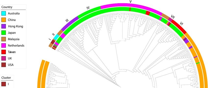
Alexis de Rougemont into genotypes. GII.2 strains (Figure 1) have been reported as dominant isolates in recent
outbreaks [4–9].
Received: 2 December 2021
Accepted: 3 February 2022
Annually, the global economic burden associated with gastroenteritis due to HuNoV
Published: 8 February 2022
is estimated to be more than USD 60 billion [10]. In the USA, norovirus gastroenteritis
caused by HuNoV accounts for an estimated 56,000–71,000 hospitalizations and an average
Publisher’s Note: MDPI stays neutral of 570–800 deaths per year [11]. To put it into perspective, in the USA, the HuNoV is asso-
with regard to jurisdictional claims in
ciated with 20 times more gastroenteritis cases than any enteric pathogen [12]. Sometimes,
published maps and institutional affil-
the effects of norovirus are overlooked because they are often self-limiting in healthy adults,
iations.
lasting a couple of days [13]. However, the effects of norovirus can be quite serious in
young children, with over a million health care visits annually [14]. Additionally, for the
elderly, HuNoV is associated with 20% of gastrointestinal (GI) deaths for those more than
Copyright: © 2022 by the authors.
65 years of age [15] as well as the immunocompromised, whose HuNoV infections can
Licensee MDPI, Basel, Switzerland.
last for years [13]. Due to a wide diversity of norovirus genotypes [16], mutations, and
This article is an open access article the lack of good indicators of immunity, vaccine design, and development has been very
distributed under the terms and difficult [2,17,18]. Therefore, prevention remains the best protection [19]. Still, the genetic,
conditions of the Creative Commons biological, antigenic, and antigen binding diversity, virulence, and stability in the environ-
Attribution (CC BY) license (https:// ment of the HuNoVs make prevention difficult even for the developed countries [20,21].
creativecommons.org/licenses/by/ Additional disinfection strategies, such as the use of UVC, will go a long way towards
4.0/). reducing infections and mortalities due to HuNoV.
Pathogens 2022, 11, 226. https://doi.org/10.3390/pathogens11020226 https://www.mdpi.com/journal/pathogens
Pathogens
Pathogens 2022, 11, 11, 226
x FOR
2022, PEER REVIEW 2 of 18
2 of 17
Figure 1. Phylogenetic
Figure 1. Phylogeneticdiversity of HuNoV
diversity of HuNoVGII.2
GII.2 strains
strains based
based on major
on major capsid
capsid proteinprotein VP1 depict-
VP1 depicting
ing lineages
lineagesiningenogroups.
genogroups. Inner circle shows geographic location, while outer cycle shows
Inner circle shows geographic location, while outer cycle shows phylogenetic phylo-
genetic clusters. Maximum likelihood was used to infer phylogeny using 519 VP1
clusters. Maximum likelihood was used to infer phylogeny using 519 VP1 gene sequences. List ofgene sequences.
ListGenBank
of GenBank accession
accession numbersnumbers haveprovided
have been been provided in supplementary
in supplementary Table S1 ofTable S1[5].
Li et al. of Figure
Li et al. [5].
Figure reused
reused from
from Li Li[5]etunder
et al. al. [5]Creative
under Commons
Creative CC Commons CChttp://creativecommons.org/
BY license BY license http://creativecom-
mons.org/publicdomain/zero/1.0/ (accessed on 11
publicdomain/zero/1.0/ (accessed on 11 January 2022). January 2022 ).
2. Annually,
Transmission
theof HuNoVs
global economic burden associated with gastroenteritis due to HuNoV
is estimated to be[22]
Norovirus more than USD 60
is transmitted viabillion
surfaces [10]. Infood
[23], the USA, norovirus
contaminated by gastroenteritis
food han-
caused by HuNoV accounts for an estimated 56,000–71,000 hospitalizations and [26].
dlers [24], contaminated vegetables, and undercooked or raw food [25], or water an aver-
agePerson-to-person
of 570–800 deaths transmission
per yearis[11].also To
unavoidable
put it into once a foodborneininfection
perspective, the USA, happens [25]. is
the HuNoV
Transmissivity is high because it only requires a small inoculum size [27,28]. HuNoVs
associated with 20 times more gastroenteritis cases than any enteric pathogen [12]. Some-
survive in the environment for a long time [27]. Disinfection has been critical to limiting
times, the effects of norovirus are overlooked because they are often self-limiting in
transmission through the application of chlorine or other disinfectants in water [29], hand
healthy adults,
washing [23],lasting a couple
and surface of days[23].
sterilization [13].However,
However, the effects
although of norovirus
norovirus can be quite
can be transmit-
serious in young children, with over a million health care visits annually
ted year-round, hospitalizations and deaths peak in late winter and early spring [30]. [14]. Addition-
It
ally, for the
mainly elderly,
spreads HuNoV
through is associated
fecal–oral with [31],
transmission 20% either
of gastrointestinal (GI) deathstrans-
through person-to-person for those
more than or
mission 65contaminated
years of age food[15] or
as water
well as theIt immunocompromised,
[31]. whosefomites
can also be transmitted through HuNoV [31].infec-
tions can last for years [13]. Due to a wide diversity of norovirus genotypes [16],tomuta-
Facilities that process food can greatly benefit by incorporating UV light technologies
control
tions, and microbial
the lack oftransmission and ensure
good indicators food safetyvaccine
of immunity, [32,33]. design, and development has
been very difficult [2,17,18]. Therefore, prevention remains the best protection [19]. Still,
the genetic, biological, antigenic, and antigen binding diversity, virulence, and stability in
the environment of the HuNoVs make prevention difficult even for the developed coun-
tries [20,21]. Additional disinfection strategies, such as the use of UVC, will go a long wayPathogens 2022, 11, 226 3 of 17
3. HuNoV Control Programs
Norovirus outbreak management and disease prevention strategies are key in the dis-
ruption of disease transmission in many setting [27]. This can be in the form of institutional
policies and development of effective hygiene programs and disinfection strategies, such
as the use of UVC. In healthcare settings, guidelines aimed at preventing and controlling
norovirus gastroenteritis outbreaks have been recommended [34]. These recommendations
rely on empirical evidence although such evidence is limited by the inability to culture
HuNoVs in laboratories [35]. Previously, there have been a few successful norovirus in-
fection control tests in hospital settings [36–38]. For environmental health and safety, the
current recommendation is physical removal of loose viral particles followed by use of
medical grade disinfectants, especially ethanol, which has been used against both feline
calicivirus (FCV) and murine norovirus (MNV). However, ethanol has limited efficacy
against noroviruses [39], whereas bleach, although more effective at concentrations greater
than 500 ppm [40], is limited to some surfaces, as it can cause corrosion of metals, craze
plastics, and remove color from fabrics [41,42]. The CDC opinion on UV irradiation in
environmental disinfection is that it remains an unresolved question [43].
There are also control programs meant to address food services [32,44] because they
are such a frequent source of outbreaks. These programs focus on training managers and
workers to identify warning signs, rigorously clean surfaces, wear gloves, and stay home
when sick, all of which are useful protective practices [45]. Similar control programs already
exist in some places for assisted living facilities and childcare facilities [45]. They generally
involve very similar procedures—wipe down and clean surfaces, inform officials of any
outbreaks, wash your hands, and wear gloves when cleaning vomit or feces.
The use of chemicals, such as bleach and ethanol, have limitations. For instance,
occupational exposure to chemical disinfectants can cause lung damage [46]. Additionally,
chemicals, such as hydrogen peroxide, peracetic acid, formaldehyde, sodium hypochlorite
(household bleach), and beta-propiolactone, can be corrosive [47,48]. Further, observations
have revealed that the disinfection performance of chemicals, such as chlorine and sodium
hypochlorite, against norovirus surrogates is reduced in the presence of feces or other
organic matter [49,50].
Minimal use of these chemicals, with the additional and safe use of UVC, will help curb
HuNoVs. Still, for us to understand the disinfection efficacy of UVC, scientific evidence
is needed. Unfortunately, at this time, model viral organisms (surrogates) are used to
investigate disinfection efficacy, as HuNoVs, although cultivable [3], are challenging to use
in this application (inactivation studies).
4. Surrogates: Considerations for Picking Norovirus Model Organisms for UVC
Disinfection Studies
Previously, HuNoVs have been especially difficult to study because they could not
be grown in tissue cultures [51]. Previous studies have involved acquiring samples from
infected people, attempting to disinfect it with varying doses of disinfectants, and then
intentionally infecting people through oral ingestion [52]. However, these studies are rare
and cannot be used as the standard for measuring UV resistance or system design. Recently,
developments on a tissue culture system for HuNoVs have been reported and used [53].
However, there are limitations to its use in inactivation studies, and the application is
currently limited to human host-pathogen studies.
Thus, surrogates are almost always required for disinfection studies. Each has pros
and cons, elaborated on in Table 1, which will be discussed further. Estimation of norovirus
sensitivity to UVC radiation is from correlating UVC exposure’s impact on transcription us-
ing RT-qPCR or qPCR and cell cultures viability. RT-qPCR or qPCR results from surrogates’
studies, such as MNV, are then assumed to be similar in relationship to HuNoVs [12,54].
Given the circumstances, these are useful measurements. However, caution should be
exercised during evaluation when using this approach, as RT-qPCR overestimates thePathogens 2022, 11, 226 4 of 17
number of infectious viral particles [55]. The viral reduction based on RT-qPCR will be
lower due the overestimation of disinfected treatments.
It is also difficulty to account for differences between viruses or the impact of peak
emission wavelengths of a UVC system.
Not all surrogates are equal. Thus, when picking the right surrogate for validation,
environmental attributes, and genetic relatedness of surrogate to HuNoVs should not be
the only attributes considered but other biological attributes as well [56,57]. For instance,
HuNoVs can bind to histo-blood group antigens (HBGAs) in addition to infection and dis-
ease severity being associated with histo-blood group type [58,59]. Previous investigation
has revealed that persons with blood group O phenotype are more likely to be infected
with HuNoV, whereas those with B histo-blood group antigen have reduced risk of infec-
tion and disease symptom progression [58]. Surrogates, such as TV, bind to HBGAs [60].
Additionally, noroviruses bind to ligands beyond the histo-blood group antigens [61].
This includes receptors/co-factors (e.g., sialic acid), which can interact with the capsids of
human norovirus [61,62]. Surrogates, such as FCV and MNV, bind to sialic acid on host
cell surface [63,64]. A detailed review on of the two commonly used surrogates (FCV and
MNV) from a food-safety perspective was published by Richards [65].
Table 1. Comparison of some human norovirus (HuNoV) model organisms (surrogates).
Surrogate Advantages Disadvantages Source
− RT-PCR results indicate FCV is
easier to inactivate (e.g compared
− Cultivable to MNV and MS2) and should be
− Stable at some temperatures used as surrogate with caution
− Frequently used as surrogate in chemical − Sensitive to low pH unlike
Feline calicivirus disinfection and irradiation studies HuNoVs [49,65–74]
(FCV) − Like HuNoVs, found to remain infective − Sensitive to drying
beyond 70 days on stainless steel and plastic at − Different transmission route (FCV
room temperature is a respiratory and not an enteric
virus link HuNoV)
− High infectivity-reduction rate
− Cultivable
− Stable across the a wide pH range.
− Genetic similarity and environmental stability
comparable to HuNoVs − Sensitive to drying
− Commonly used as surrogate in chemical − Different UVC susceptibility to
Murine disinfection and irradiation studies other surrogates [57,65,66,68–70,75,76]
norovirus (MN) − Like HuNoVs, found to remain infective − MNV environmental stability is
beyond 70 days on stainless steel and plastic at different to HuNoV
room temperature
− Conservative HuNoV surrogate for UV
disinfection
− Cultivable
− The biological features of recovirus closely
reflect HuNoVs
− Clinical manifestations and disease − Difficult to produce a high-titer
progression in infected mammalian organisms stock via tissue culture
Tulane virus (TV) reflect human norovirus − Reduction of virus titer at pH 2.5 [21,69,77,78]
− Truly intestinal pathogen, also from and 9.0
Caliciviridae family like HuNoV
− Recognizes the same ligand as HuNoVs
− More resistant at low pH than FCV
− Cultivable
− Conservative HuNoV surrogate for UV
− This is a BSL-2 organism and may
disinfection
not be used widely.
Echovirus 12 − Shares morphological similarities with [79–81]
− Long incubation period (2–5 days)
HuNoVs
− Environmental persistence
− Poses lower human health risksPathogens 2022, 11, 226 5 of 17
Table 1. Cont.
Surrogate Advantages Disadvantages Source
− Cultivable to high titers
− BSL-1 organisms (non-pathogenic), thus
possibility of widespread use
− Potential surrogate for noroviruses on fresh − Sensitive in acidic environments
produce with prolonged survival periods, − UVC resistance may lead to
MS2 and Qβ both in buffer and on fresh produce, at resource wasting, as it is too [71,82–84]
temperatures relevant to chilled foods. conservative.
− Conservative disinfection performance due to
high UVC sensitivity than most pathogenic
viruses
Although feline calicivirus (FCV) has been used as norovirus surrogate because of
its cultivability, FCV is not an ideal candidate under some circumstances (Table 1). FCV
is sensitive to low pH [72] and surface stress, including drying [85]. Therefore, if dried
for UVC disinfection studies on dry surfaces, such as stainless steel, investigators need to
consider the natural reduction due to the drying process. FCV also has different disinfection
susceptibility under chlorine exposure [72,85] in addition to being more sensitive to UVC
irradiation than HuNoVs [12].
On the other hand, murine norovirus (MNV) is an equally common surrogate for
HuNoVs [69]. MNV is stable at a wider range of pH levels, is less sensitive to tempera-
ture, and shares genetic and molecular features with HuNoVs, such as size (diameter of
28–35 nm), icosahedral shape, and buoyant density (1.36 ± 0.04 g/cm3 ) [86–88]. However,
MNV’s sensitivity to drying is like that of FCV, something that needs to be accounted
for during validation. MNV is less sensitive to UVC than FCV and genetically closer to
HuNoVs based on PCR studies [12,87].
Tulane virus (TV), the prototype recovirus strain, has been considered as a HuNoV
surrogate [89]. Recovirus (ReCV) genus belongs to the family Caliciviridae and, just like
HuNoV, is organized into three open reading frames (ORFs) [90,91]. TV recognizes the same
ligand as HuNoV in oyster tissues [77,92]. The biological diversity and features of recovirus
and clinical manifestations and disease progression in infected mammal organisms reflect
HuNoVs properties more than with any prior surrogate [21]. Echovirus is like MNV in
many ways and has been used as a surrogate [79]. MS2 has also been considered because it
is a phage and easier to cultivate and test with. MS2 seems to be much more resistant than
HuNoVs and so provides a large safety factor [12]. However, selecting a virus because of
its high resilience may waste money and resources that could have been used protecting
other pathways. Ultimately, when testing or designing new strategies for disinfection, one
must understand and clearly justify the surrogate pick.
5. UVC Disinfection Studies That Utilized Norovirus Surrogates
Using deliberately contaminated stainless-steel surfaces, a previous study utilizing
UVC at 260 nm to study the effects of 10–300 mWs/cm2 revealed that the murine norovirus-
1 (MNV-1) titer was more sensitive than hepatitis A virus (HAV) [93]. The study also
suggested that low doses of UVC can be used to decontaminated surfaces contaminated
with HuNoV [93]. The effectiveness of UVC against MNV-1 has been associated with the
disruption of the capsid protein and genomic RNA [75]. At 254 nm, with 25 mJ/cm2 UVC
dose, a 3.6-log10 reduction was reported in a study carried out by Lee et al. [94]. This study
also revealed higher MNV inactivation at high salt concentrations and temperatures (18
and 30 ◦ C) compared to lower temperatures (−20 and 4 ◦ C) [94].
Another study by Vimont et al. investigated the use of a pulsed-light device that
emitted a broadband spectrum (200–1000 nm) and obtained a 3-log10 reduction using
3.45 J cm−2 in clear suspensions [95].Pathogens 2022, 11, 226 6 of 17
6. Considerations for Effective Use of UVC in Surface Disinfection
Per recent review by Raeiszadeh et al. [96], the COVID-19 pandemic has contributed to
the increased consideration of UVC disinfection devices for disinfection. Proper use of UVC
technology is required to guarantee disinfection when using devices such as Klaran WD
array systems [97,98]. When disinfecting surfaces, considerations, such as type of surface
(microstructural properties) being illuminated [99], intensity distribution projected onto
target surface to be disinfected, and the distance between light source and target surfaces,
should be factored into design and actual application. For instance, UVC disinfection may
be more effective on plastic surfaces followed by stainless steel but lower on fabrics, such
as poly-cotton [99].
One critical path for norovirus is through the food-service industry, where sick workers,
contaminated water, or surface contact can contaminate food [25]. One strategy is to target
all the pathways in the food-service industry. Since UV light can be used to disinfect
surfaces and potentially even food surfaces, there is increasing interest for its use as a point-
of-use disinfection method [32]. Examples of areas that could benefit from implementation
of UVC technology include but are not limited to restrooms, kitchens, baggage claim areas,
security checkpoints, and meeting rooms.
As discussed, surface transmission is one important pathway for norovirus outbreaks.
However, before any research can be reasonably carried out, first, the methodology used
during the study should be reviewed. The correct unit of light to be used to model UVC
disinfection is fluence [100], or the amount of light through a sphere, divided by the cross
section of said sphere. Studies that only measure exposure time and distance of UVC
radiation from the surface being disinfected to determine disinfection efficacy of a system
make universal application and replication hard and thus are insufficient. Although both
time and distance are critical, they do not account for size, shape, reflection, temperature, or
type of lamp, and therefore, results cannot be accurately transitioned into products/designs
for real-world applications. Instead, fluence should be reported. For surfaces that absorb
UV light, this simplifies from dose (mJ/cm2 ) to intensity (mW/cm2 ) multiplied by time
(seconds) [101]. To ensure the correct measurement of the irradiance in experiments, it is
best to use an optometer, such as the X1 MD-37-SC1–4, that is calibrated for the specific
wavelength used in the experiment. Most optometers are designed for low-pressure
mercury lamps, which radiate at 254 nm. Some of these, for example, will have a cut-off
wavelength above 270 nm and are thus useless for measuring most LEDs or 222-nm filtered
krypton lamps. Alternatives to optometers include potassium iodide acitometry [102,103],
pre-existing disinfection systems with known irradiance at a well-defined point, and
dosimeters selected for the correct wavelength [104].
In addition to the effect of the amount of light, the wavelength of light has a remark-
able effect on disinfection rate. UVC LEDs have a peak wavelength between 215–280 nm,
and that peak value can have a significant germicidal impact against many pathogens [105].
For example, with the same dose, 265–268 nm can increase disinfection of pathogens, such
as SARS-CoV-2, by almost 1 log10 reduction value (1 LRV) compared to those with wave-
lengths >270 nm [106]. More studies are emphasizing that “not all wavelengths are created
equal: [107,108]. Thus, measuring and specifying the wavelength and measurement toler-
ances used in a UVC system for disinfection is of critical importance for research, design,
and implementation purposes. Spectrometers are available to perform such measurements,
but spectrometers must be calibrated to ensure accurate intensities are reported.
The type of surface is also important during disinfection, as it has an impact on
disinfection efficacy. The presence of small crevices can provide places for viruses to hide
and avoid UVC exposure. On the other hand, reflecting surfaces can create numerous
light passages, thus enhancing the disinfection efficiency [109]. For example, pathogens,
including norovirus, on UVC-reflective materials, such as polished aluminum, may see
70% more light than measured with an optometer. That is because much of the light
passes through the organism, but most will not be absorbed; it is then reflected through
again. Since pathogens on porous absorbing materials may be shielded from most UVCPathogens 2022, 11, 226 7 of 17
light, lower inactivation efficacies are obtained. In surface disinfection, such reflection or
coverage is accounted for simply by testing on different materials.
Physicochemical properties also impact UVC disinfection. Often, relative humidity is
considered and reported only for studies involving aerosolized microorganisms. However,
relative humidity effects the kill rate of viruses on surfaces and not just in air [110,111].
Temperature and humidity can affect survival of viruses [109].
Another limitation is around the way we consider populations. Often, we discuss
log reduction values as simple additive reductions. This assumes that all viruses are the
same and that if an exposure was able to kill, for instance, 90% in the first 10 s, then a 90%
reduction within same time means identical results. This is erroneous because even if all
the viruses were of the same exact genome, damage, if any, and micro-environment, which
is far from certain, some viruses may be shaded, and others may interact with surfaces
differently. For instance, 99.9% reduction within 10 s may be a reasonable approximation; it
can be broken down at both low and high doses of any disinfectant, including UVC [112].
Therefore, for applications with either high, low, or a high dynamic range of dose, a
log-normal distribution may be needed.
The ideal test of a norovirus surface disinfection system or study of sensitivity would
cover a variety of relevant surface materials, with many different environmental conditions,
with known, widely varied wavelength and fluence against the right surrogate. However,
to the best of the authors’ knowledge, there is no such complete study for a norovirus
because of the time and resources that are required to conduct such a study.
Autonomous navigation robots capable of recognizing surfaces with high probabilities
of contamination are emerging [113]. It will be tremendous if such robots in future can
detect the microstructural differences and adjust UVC dose accordingly. While these
autonomous navigation robots are at work, it will be important that they have a feature
to determine safe distance between them and nearest human beings and be able to warn
them to be out of the way while disinfection is ongoing. They should be equipped with
safety features, such as motion sensor, and be able to turn off UVC radiation when humans
or animals approach [114] and wait for humans to move before proceeding with their
disinfection work. Companies that make these products should ensure adequate safety
features to protect humans against exposure to unsafe amount of UVC, as that could
lead to advisories from guidance, compliance, and regulatory bodies in addition to other
consequences [115,116].
7. Considerations for Conducting Water Disinfection Using UVC
Water disinfection has a great deal in common with surface disinfection. Fluence
and wavelength are both still important, as are environmental factors. However, the
number and types of environmental factors are different. Water disinfection reactors (flow-
through systems with water exposed to UVC radiation) and water tanks do not always have
reflective material and are almost always sealed. Water disinfection reactors mostly use
reflective materials, such as polytetrafluoroethylene (PTFE), to enhance disinfection. During
experiments, it is hard to measure dose (mJ/cm2 ) straightaway. To that end, inserting an
optometer through a small hole or partially disassembling a system may be required. If so,
then the hole should be as small as possible and correctly account for reflection multiply by
1 + R [117,118], with R being reflectivity of the inner reactor material. Another alternative is
to use potassium iodide actinometry, but one must account for the UVC absorption [101].
Turbidity and absorption of fluid due to high concentration of Total Dissolved Solids
(TDS), for instance, is one such different aspect that can impact inactivation efficacy [119].
While transmitting through air onto a surface, air is generally assumed to be 100% trans-
parent, with low turbidity. None of the light passing through the air will be deflected away
from the virus or be absorbed by gas. On the other hand, in liquid, it is possible to be turbid
and absorbing to the extent of making a system completely ineffective, as has been demon-
strated in aquacultures [120]. Hence, measurement of the UVC transmission and turbidity
are needed. Noroviruses are also sensitive to pH [49,73]. Therefore, ethanol-containingPathogens 2022, 11, 226 8 of 17
acidic alcohol suspensions (e.g., 70% ethanol containing 1% citric acid) have more virucidal
effect compared to 70% ethanol [121]. pH should always be reported as a factor during
studies where liquids are involved. Just like on surfaces, norovirus deactivation rates also
depend on temperature. It is vital to measure and control the temperature and pH of water
during disinfection tests, as they directly influence the dynamics and distribution of other
microbial assemblages [57,94,122].
Additionally, water supplies can have varying concentrations of chlorine or other
disinfectant residuals. Some of these disinfectants are typically activated by UVC for
various reasons but often through the formation of chlorine or oxygen radicals [123].
Therefore, it is critical to know the concentration of disinfectants in a test and recommended
to design the study around them.
Furthermore, there is virus protection against UVC inactivation that could be expected
when viral particles are in fecal matter or environment with organic matrix [124], which
could lead to low viral inactivation efficacies by UVC. This has been observed when
other agents are used in norovirus disinfection [125]. It is therefore important to consider
properties of the materials to be disinfected. For the users to consider using UVC for
norovirus infection control, they need to be guaranteed that the expected performances are
obtained. Thus, this calls for a focus on the principles of disinfection and decontamination
during UVC implementation toward norovirus infection control [35]. It is also important
to consider if the type of water to be disinfected is static or flowing, as different types
of water behave differently. All water is not equal; thus, the state (static or mobile) and
physical as well as chemical composition of water will affect microbial stability and UVC
dose requirements [126].
8. Radiation Safety Considerations
UVC light has safety concerns that prevent certain uses or require safety considerations
during use [96]. Firstly, as a fact, UVC and even far UVC does not discriminate between
pathogens and commensals. Because of that non-discriminatory nature and potential
impact on the skin’s microbiome, any UV could induce human immune suppression [127].
It is important to know that commensals, especially those of the human skin surface, are
crucial, as they induce protective responses that defend us against invasion and colonization
by pathogens. The role of commensals (symbionts) in protective immunity has reviewed
by Khan et al. [128], Li et al. [129], Byrd et al. [130], Grice and Segre [131], and Flowers and
Segre [132].
The human skin microbiome has adapted to consume sparse nutrients available on
our skin, in exchange for protecting us [130]. Although known to be stable regardless of
perturbations, the kind of imbalance due to inactivation by UVC has not been studied. We
predict that that due to dysbiosis (reduction of microbial diversity of beneficial bacteria
due to indiscriminate inactivation), selection pressure, and enrichment of bacteria with
high guanine–cytosine (%GC) composition bacteria on skin, we could potentially see an
explosion of skin diseases and related conditions if exposure is for long period of time.
Bacteria with high %GC, such as Actinobacteria, are more tolerant to UV [133]. If not
safely implemented, UVC exposure at the airports when moving from gate A to Z or while
waiting for hours to board the aircraft could enrich for high %GC content bacteria while
inactivating beneficial ones with low %GC. Another example is in schools or hospitals,
where there will be more exposure time. It will therefore be important that facility managers
understand the spaces where the UVC can be safely and effectively implemented.
Since UVC is indiscriminate, it can damage all living cells it reaches. These cells are
not restricted to mammalian cells but also bacteria and viruses for that matter. Although
the good viruses that form part of the skin microbiome are poorly understood, they act
as primary barrier to the external environment and help humans modulate cutaneous
health [134]. We emphasize this because of the arguments that 222 nm is safe without
regard for the critical role of the skin microbiome and potential for directed evolution of
UVC resistant pathogens. Although erythema can be absent, irradiation at 500 mJ/cm2Pathogens 2022, 11, 226 9 of 17
using 222 nm UVC does decrease the number of bacteria while producing cyclobutene
pyrimidine dimers (CPD) [135].
In fact, skin exposure can cause cancer with the right dose at almost all UVC wave-
lengths [136]. It can also cause retinal cancer and blindness [137]. It is reasonable that there
is increasing interest in 222 nm, as it may not reach through the endoplasm of large cells
and even less through dead skin cells or the lens of the eye, thus reducing the risk of cancer
and blindness. As a result, 222-nm lamps are increasingly being used in open environ-
ments [43,138]. Although the promise of disinfecting an entire restaurant continuously to
reduce norovirus spread sounds promising, several issues raised in this perspective should
be considered during applications.
The other important concern is that although some studies show little to no corneal or
skin damage at exposures of 100 s mJ/cm2 [138] from 222-nm light, they still show damage
at doses of 1000 s mJ/cm2 [138]. Although there is hope that this effect is not cumulative,
as epithelial shedding may provide added protection, this has not yet been tested, to the
best of our knowledge. There has also been very little testing on humans—although one
author exposed their eye briefly and informally and reported only mild irritation [139].
There has also been little consideration for variations like dry eyes, which may remove a
critical protective barrier; cuts or abrasion of the skin, which may remove protective dead
skin cells; or exposure to other sensitive areas. Thus, it is concerning to suggest that far
UVC should be applied in airports, hospitals, schools, and homes with constant or long
exposure time [43,138].
For emphasis, a dose of 100 mJ/cm2 at 222 nm reduces all pathogens by several
orders of magnitude, hopefully without harming human cells [44]. However, just like
with antibiotics, it should be noted from a skin microbiome perspective that the use of
222 nm or any far UVC on skin in general will not discriminate between pathogens and
the “good” skin microbiome that is required for healthy skin. Skin microorganisms have
important roles in the cutaneous immune system [130]. These “good” microbes also
produce molecules that prevent skin colonization from other unwanted microorganisms or
protect us by altering the behavior of the invaders, ensuring the protection and health of
our biggest organ, the skin [130].
We therefore expect that indiscriminate frequent exposure of human skin to 222-nm
light will likely inactivate symbionts and unbalance the taxonomic, phylogenetic, and
functional potential of the microbiome, leading to a compromised cutaneous immune
system, the consequence of which will be skin disorders, such as acne. The enrichment
of UVC resistant is most likely to be for those skin microbes like Propionibacterium acnes,
whose %GC content is high [45], and have less likelihood of forming enough dimers
for inactivation. UVC application on skin will likely reduce microbial diversity due to
inactivation of high AT commensals. A reduction of microbial diversity due to cutaneous
microbial dysbiosis could lead to other conditions, such as psoriasis [140].
The demodex mites are part of normal skin flora and feed on microbes, such as P. acnes
and sebum [131]. We speculate a taxonomic imbalance, which will in turn lead to functional
imbalance, impacting immunity, and perhaps skin syndromes. Molecular, immunological
metabolomic, and microbiological investigations are recommended in the future to unearth
any potential functional dysregulation due to UVC usage on skin.
Regardless of the wavelength, the process for safe work with UVC light is similar.
Wear long sleeves and pants that cover your whole body. Wear plastic or close-stitch
gloves to absorb UVC light, which is also helpful in preventing smudges on your light
source or reflectors. Wear plastic goggles to absorb UVC light. Most light sources will be
sensitive to electro-static discharge (ESD), so consider wearing ESD shoes, bracelets, or
boot covers. Some light sources require high voltages—such sources should not be used
near flammables and should be isolated from users before powering on. Some light sources
are made with glass and contain hazardous chemicals, so after use or breakage, follow the
manufacturer’s disposal instructions.Pathogens 2022, 11, 226 10 of 17
The design and implementation of UVC disinfection reactors or sources should strive
to prevent stray light from escaping. This can be done in water reactors by placing them
ahead of a tubing of a material that will absorb UVC, such as most plastics and glasses.
This forms a simple light baffle, which will absorb light but merely direct the water. A more
complex tube shape, such as a u-bend or a mesh, can shorten the required distance. On
surfaces, it is best if any design physically contacting the surface to be disinfected contains
the light there.
The concerning potential disadvantage of the UVC technology involves potential dys-
regulation of both taxonomic, phylogenetic, and functional diversity of the skin microbiome.
A survey on the composition, structure, and functional changes is thus recommended. For
UVC to be effective, the target pathogens must be in direct contact with the UVC irradiation.
Fortunately, UVC dosimeters, if correctly used, can be used for verification of UVC expo-
sure [141]. Moreover, UVC can cause harmful effects on eyes and skin, and it is therefore
not safe for use in disinfecting hands and skin [115,142].
9. The Promise of UVC for Norovirus
UVC disinfection of surfaces could have several critical norovirus-related applications.
For example, washing fruit and vegetables may accidentally inoculate norovirus to surfaces.
Additionally, chlorine is not allowed in many European countries and rarely desired
anywhere because of its effect on the food and its residue both in the food and waste.
However, recent studies have shown that food processing, which combines washing and
application of UVC, can enable lighting all the relevant surfaces of various fresh fruits and
vegetables [32,75,143–146]. UVC disinfection also promises an explosion hazard-free and
chlorine-free way of disinfecting counter-tops, preparation areas, bathrooms, and any other
high-touch surfaces repeatedly with little to no labor.
UVC could be and increasingly is used to disinfect wastewater to provide a low-impact
way of protecting water estuaries and seafood [147–149]. Like with surface disinfection,
there is significant interest in the synergy between TiO2 [147], H2 O2 , and Cl2 [150] with
UVC to further improve disinfection performance. Such improvements may reduce the
amount of time, power, and disinfectant required.
However, a thorough measurement of UVC disinfection efficiency of norovirus and
its primary surrogate MNV in different environmental conditions would make design
and implementation of UVC disinfection systems easier for water and surface disinfection.
Exploring the factors of temperature, pH, humidity, wavelength, and surface type would
allow designers to account for various situations in implementing their disinfection systems.
Larger-scale adoption of UVC may still depend on both low barrier to entry and greater
visibility of the benefits of disinfection.
UVC disinfection research, although eliminating some pathways, does not address
many other problems. Many outbreaks stem from worker cleanliness and delayed de-
tection. However, UVC could also play a role in tackling this difficult problem through
timed auto-fluorescent measurement of pathogens [151–153]. Such systems might provide
a method of scaling up point of outbreak monitoring by allowing, for example, restau-
rants, schools, and long-term care facilities to check for infection every shift. This could
dramatically reduce the number of people exposed and the amount of exposure in an
outbreak. However, such systems require significant further development—both in tools,
such as high-sensitivity UVC detectors, high-speed switching UVC LEDs at multiple bands,
analyzers, and databases, and more.
The 222 nm is not yet proven for long-term exposure in any surrogates and has not
been tested in humans. However, such research, if proven safe, may provide a new route to
continuous disinfection. Still, even if use of UVC does not prove safe in specific cases, it
may still be useful in quickly dosing unoccupied public areas—especially in conjunction
with other disinfectants, which may allow for more effective cleaning. Although traditional
UVC sources, such as mercury lamps emitting UVC radiation at a peak wavelength at
254 nm, can be used, it is important to note that the overall log-inactivation efficacy at peakPathogens 2022, 11, 226 11 of 17
emission wavelength at 265 nm has been found to be high and most effective [154,155].
Furthermore, the use of low-pressure (LP) mercury lamps emitting radiation at 254 nm
poses environmental concerns due to mercury content [156].
10. Conclusions
We have a long way to go in the fight against human norovirus. Perhaps the most
important and exciting research direction is in developing methods for culture, growth,
and measurement of HuNoVs because these developments underpin all others. However,
the development of convenient detection may also reduce the outbreak time, while UVC
disinfection of foods and services will help curb infectious pathways.
Until proven and the impact on the skin microbiome understood, shining UVC directly
into occupied space should be avoided, as UVC does not have a mechanism to discriminate
between pathogens and commensals, which are critical to our own health. Surveys utilizing
shotgun metagenomics could help in evaluating the impact on commensals and associated
changes in taxonomic and functional dynamics, including changes in secondary metabolites’
production.
Author Contributions: R.M.M. conceived the article. R.M.M. and J.H.D. designed the work, per-
formed literature search, and contributed equally to the writing of the manuscript. R.V.R. revised
the manuscript. All authors read, edited, and agreed to final manuscript. All authors have read and
agreed to the published version of the manuscript.
Funding: This work received no specific grant from any funding agency.
Institutional Review Board Statement: Not applicable.
Informed Consent Statement: Not applicable.
Data Availability Statement: Data sharing not applicable. No new data were created or analyzed in
this study.
Acknowledgments: Authors wish to thank Sébastien Blumenstein for proofreading and for invalu-
able feedback. Authors also thank Crystal IS for the support, especially Sanjay Kamtekar and Eoin
Connolly.
Conflicts of Interest: R.M.M., J.H.D. and R.V.R. all receive salaries from Crystal IS, an Asahi Kasei
company that manufactures UVC-LEDs.
References
1. Green, K.Y.; Kaufman, S.S.; Nagata, B.M.; Chaimongkol, N.; Kim, D.Y.; Levenson, E.A.; Tin, C.M.; Yardley, A.B.; Johnson, J.A.;
Barletta, A.B.F.; et al. Human norovirus targets enteroendocrine epithelial cells in the small intestine. Nat. Commun. 2020, 11,
2759. [CrossRef] [PubMed]
2. Karst, S.M. Pathogenesis of Noroviruses, Emerging RNA Viruses. Viruses 2010, 2, 748–781. [CrossRef] [PubMed]
3. Chhabra, P.; De Graaf, M.; Parra, G.I.; Chan, M.C.-W.; Green, K.; Martella, V.; Wang, Q.; White, P.A.; Katayama, K.; Vennema, H.;
et al. Updated classification of norovirus genogroups and genotypes. J. Gen. Virol. 2019, 100, 1393–1406. [CrossRef] [PubMed]
4. Bidalot, M.; Théry, L.; Kaplon, J.; De Rougemont, A.; Ambert-Balay, K. Emergence of new recombinant noroviruses GII.p16-GII.4
and GII.p16-GII.2, France, winter 2016 to 2017. Eurosurveillance 2017, 22, 1–5. [CrossRef]
5. Li, X.; Liu, H.; Magalis, B.R.; Pond, S.L.K.; Volz, E.M. Molecular Evolution of Human Norovirus GII.2 Clusters. Front. Microbiol.
2021, 12, 655567. [CrossRef]
6. Medici, M.C.; Tummolo, F.; Martella, V.; De Conto, F.; Arcangeletti, M.C.; Pinardi, F.; Ferraglia, F.; Chezzi, C.; Calderaro, A.
Emergence of novel recombinant GII.P16_GII.2 and GII. P16_GII.4 Sydney 2012 norovirus strains in Italy, winter 2016/2017. New
Microbiol. 2018, 41, 71–72.
7. Tohma, K.; Lepore, C.J.; Siltz, L.; Parra, G.I. Phylogenetic Analyses Suggest that Factors Other Than the Capsid Protein Play a
Role in the Epidemic Potential of GII.2 Norovirus. mSphere 2017, 2, e00187-17. [CrossRef]
8. Niendorf, S.; Jacobsen, S.; Faber, M.; Eis-Hübinger, A.M.; Hofmann, J.; Zimmermann, O.; Höhne, M.; Bock, C.-T. Steep rise in
norovirus cases and emergence of a new recombinant strain GII.P16-GII.2, Germany, winter 2016. Eurosurveillance 2017, 22, 30447.
[CrossRef]
9. Cheung, S.K.C.; Kwok, K.; Zhang, L.Y.; Mohammad, K.N.; Lui, G.C.Y.; Lee, N.; Nelson, E.A.S.; Lai, R.W.M.; Leung, T.K.; Chan,
P.K.S.; et al. Higher Viral Load of Emerging Norovirus GII.P16-GII.2 than Pandemic GII.4 and Epidemic GII.17, Hong Kong,
China. Emerg. Infect. Dis. 2019, 25, 119–122. [CrossRef]Pathogens 2022, 11, 226 12 of 17
10. Bartsch, S.M.; Lopman, B.A.; Ozawa, S.; Hall, A.J.; Lee, B.Y. Global Economic Burden of Norovirus Gastroenteritis. PLoS ONE
2016, 11, e0151219. [CrossRef]
11. Vinjé, J. Advances in Laboratory Methods for Detection and Typing of Norovirus. J. Clin. Microbiol. 2015, 53, 373–381. [CrossRef]
[PubMed]
12. Rockey, N.; Young, S.; Kohn, T.; Pecson, B.M.; Wobus, C.E.; Raskin, L.; Wigginton, K.R. UV Disinfection of Human Norovirus:
Evaluating Infectivity Using a Genome-Wide PCR-Based Approach. Environ. Sci. Technol. 2020, 54, 2851–2858. [CrossRef]
[PubMed]
13. Green, K.Y. Norovirus infection in immunocompromised hosts. Clin. Microbiol. Infect. 2014, 20, 717–723. [CrossRef] [PubMed]
14. Payne, D.C.; Vinje, J.; Szilagyi, P.G.; Edwards, K.M.; Staat, M.A.; Weinberg, G.; Hall, C.B.; Chappell, J.; Bernstein, D.I.; Curns,
A.T.; et al. Norovirus and Medically Attended Gastroenteritis in U.S. Children. N. Engl. J. Med. 2013, 368, 1121–1130. [CrossRef]
[PubMed]
15. Harris, J.P.; Edmunds, W.J.; Pebody, R.; Brown, D.W.; Lopman, B.A. Deaths from Norovirus among the Elderly, England and
Wales. Emerg. Infect. Dis. 2008, 14, 1546–1552. [CrossRef]
16. Kobayashi, M.; Yoshizumi, S.; Kogawa, S.; Takahashi, T.; Ueki, Y.; Shinohara, M.; Mizukoshi, F.; Tsukagoshi, H.; Sasaki, Y.; Suzuki,
R.; et al. Molecular Evolution of the Capsid Gene in Norovirus Genogroup I. Sci. Rep. 2015, 5, 13806. [CrossRef]
17. Esposito, S.; Principi, N. Norovirus Vaccine: Priorities for Future Research and Development. Front. Immunol. 2020, 11, 1383.
[CrossRef]
18. Zhou, H.-L.; Chen, L.-N.; Wang, S.-M.; Tan, M.; Qiu, C.; Qiu, T.-Y.; Wang, X.-Y. Prevalence and Evolution of Noroviruses between
1966 and 2019, Implications for Vaccine Design. Pathogens 2021, 10, 1012. [CrossRef]
19. Schneider, K.R.; Goodrich, R.M.; Mahovic, M.J.; Shukla, R. Preventing Foodborne Illness: Norovirus. EDIS 2019, 2005, 1–4.
[CrossRef]
20. Cannon, J.L.; Barclay, L.; Collins, N.R.; Wikswo, M.E.; Castro, C.J.; Magaña, L.C.; Gregoricus, N.; Marine, R.; Chhabra, P.; Vinjé, J.
Genetic and Epidemiologic Trends of Norovirus Outbreaks in the United States from 2013 to 2016 Demonstrated Emergence of
Novel GII.4 Recombinant Viruses. J. Clin. Microbiol. 2017, 55, 2208–2221. [CrossRef]
21. Farkas, T. Rhesus enteric calicivirus surrogate model for human norovirus gastroenteritis. J. Gen. Virol. 2015, 96, 1504–1514.
[CrossRef]
22. Barker, J.; Vipond, I.B.; Bloomfield, S.F. Effects of cleaning and disinfection in reducing the spread of Norovirus contamination via
environmental surfaces. J. Hosp. Infect. 2004, 58, 42–49. [CrossRef] [PubMed]
23. Canales, R.A.; Reynolds, K.A.; Wilson, A.M.; Fankem, S.L.; Weir, M.H.; Rose, J.B.; Abd-Elmaksoud, S.; Gerba, C.P. Modeling the
role of fomites in a norovirus outbreak. J. Occup. Environ. Hyg. 2019, 16, 16–26. [CrossRef] [PubMed]
24. Boxman, I.; Dijkman, R.; Verhoef, L.; Maat, A.; Van Dijk, G.; Vennema, H.; Koopmans, M. Norovirus on Swabs Taken from Hands
Illustrate Route of Transmission: A Case Study. J. Food Prot. 2009, 72, 1753–1755. [CrossRef] [PubMed]
25. Kwan, H.S.; Chan, P.K.S.; Chan, M.C.W. Chapter 2—Overview of Norovirus as a Foodborne Pathogen. In The Norovirus; Chan,
P.K.S., Ed.; Academic Press: Cambridge, MA, USA, 2017; pp. 21–30, ISBN 978-0-12-804177-2.
26. Maunula, L.; Miettinen, I.T.; Von Bonsdorff, C.-H. Norovirus Outbreaks from Drinking Water. Emerg. Infect. Dis. 2005, 11,
1716–1721. [CrossRef]
27. Robilotti, E.; Deresinski, S.; Pinsky, B.A. Norovirus. Clin. Microbiol. Rev. 2015, 28, 134–164. [CrossRef]
28. ECDC Facts about Norovirus. Available online: https://www.ecdc.europa.eu/en/norovirus-infection/facts (accessed on 1
December 2021).
29. Cromeans, T.L.; Kahler, A.M.; Hill, V.R. Inactivation of Adenoviruses, Enteroviruses, and Murine Norovirus in Water by Free
Chlorine and Monochloramine. Appl. Environ. Microbiol. 2010, 76, 1028–1033. [CrossRef]
30. Cleveland Clinic Norovirus: Symptoms, Causes, Treatments. Available online: https://my.clevelandclinic.org/health/diseases/
17703-norovirus (accessed on 1 December 2021).
31. Brunette, G.W. CDC Yellow Book 2018: Health Information for International Travel; Oxford University Press: Oxford, UK, 2017;
Volume 66, pp. 1157–1158, ISBN 9780190628611.
32. Koutchma, T.; Popović, V. Chapter 5—UV Light-Emitting Diodes (LEDs) and Food Safety. In Ultraviolet LED Technology for Food
Applications; Koutchma, T., Ed.; Academic Press: Cambridge, MA, USA, 2019; pp. 91–117, ISBN 978-0-12-817794-5.
33. Koutchma, T. UV Light for Processing Foods. Ozone Sci. Eng. 2008, 30, 93–98. [CrossRef]
34. MacCannell, T.; Umscheid, C.A.; Agarwal, R.K.; Lee, I.; Kuntz, G.; Stevenson, K.B. Guideline for the Prevention and Control of
Norovirus Gastroenteritis Outbreaks in Healthcare Settings. Infect. Control Hosp. Epidemiol. 2011, 32, 939–969. [CrossRef]
35. Barclay, L.; Park, G.; Vega, E.; Hall, A.; Parashar, U.; Vinje, J.; Lopman, B. Infection control for norovirus. Clin. Microbiol. Infect.
2014, 20, 731–740. [CrossRef]
36. Vinnard, C.; Lee, I.; Linkin, D.R. Successful control of a norovirus outbreak among attendees of a hospital teaching conference.
Am. J. Infect. Control 2012, 40, 73–74. [CrossRef] [PubMed]
37. Xu, Q. Epidemic and control strategy on nosocomial outbreak of norovirus gastroenteritis. Zhongguo Yi Xue Ke Xue Yuan Xue Bao
2008, 30, 614–617. [PubMed]
38. Georgiadou, S.P.; Loukeris, D.; Smilakou, S.; Daikos, G.L.; Sipsas, N.V. Effective control of an acute gastroenteritis outbreak due to
norovirus infection in a hospital ward in Athens, Greece, April 2011. Eurosurveillance 2011, 16, 19915. [CrossRef] [PubMed]Pathogens 2022, 11, 226 13 of 17
39. Escudero-Abarca, B.I.; Goulter, R.; Arbogast, J.W.; Leslie, R.A.; Green, K.; Jaykus, L. Efficacy of alcohol-based hand sanitizers
against human norovirus using RNase-RT-qPCR with validation by human intestinal enteroid replication. Lett. Appl. Microbiol.
2020, 71, 605–610. [CrossRef] [PubMed]
40. Sato, J.; Miki, M.; Hitomi, J.; Tokuda, H.; Katayama, K.; Kubota, H.; Todaka-Takai, R. Effects of disinfectants against norovirus
virus-like particles predict norovirus inactivation. Microbiol. Immunol. 2016, 60, 609–616. [CrossRef]
41. Crawford, M. How Clean Is Clean? Chemistry Can Damage Medical Equipment In the Quest to Meet Stringent Guidelines.
Biomed. Instrum. Technol. 2014, 48, 260–263. [CrossRef]
42. Tyan, K.; Jin, K.; Kang, J. Novel colour additive for bleach disinfectant wipes reduces corrosive damage on stainless steel. J. Hosp.
Infect. 2019, 103, 227–230. [CrossRef]
43. Buonanno, M.; Welch, D.; Shuryak, I.; Brenner, D.J. Far-UVC light (222 nm) efficiently and safely inactivates airborne human
coronaviruses. Sci. Rep. 2020, 10, 10285. [CrossRef]
44. Hessling, M.; Haag, R.; Sieber, N.; Vatter, P. The Impact of Far-UVC Radiation (200-230 Nm) on Pathogens, Cells, Skin, and
Eyes—A Collection and Analysis of a Hundred Years of Data. GMS Hyg. Infect. Control 2021, 16, 1–17. [CrossRef]
45. Minegishi, K.; Aikawa, C.; Furukawa, A.; Watanabe, T.; Nakano, T.; Ogura, Y.; Ohtsubo, Y.; Kurokawa, K.; Hayashi, T.; Maruyama,
F.; et al. Complete Genome Sequence of a Propionibacterium acnes Isolate from a Sarcoidosis Patient. Genome Announc. 2013, 1,
e00016-12. [CrossRef]
46. Dumas, O.; Varraso, R.; Boggs, K.M.; Quinot, C.; Zock, J.-P.; Henneberger, P.K.; Speizer, F.E.; Le Moual, N.; Camargo, C.A., Jr.
Association of Occupational Exposure to Disinfectants with Incidence of Chronic Obstructive Pulmonary Disease Among US
Female Nurses. JAMA Netw. Open 2019, 2, e1913563. [CrossRef] [PubMed]
47. Nicklas, W.; Böhm, K.H.; Richter, B. [Studies on the corrosive action of some disinfectants suitable for aerosol-disinfection
(author’s transl)]. Zentralbl. Bakteriol. Mikrobiol. Hyg. B 1981, 173, 374–381. [PubMed]
48. Cao, C. Chemical Disinfectants for Inactivation of Human Norovirus Surrogates. Master’s Thesis, University of Tennessee,
Knoxville, TN, USA, 2013.
49. Poschetto, L.F.; Ike, A.; Papp, T.; Mohn, U.; Böhm, R.; Marschang, R.E. Comparison of the Sensitivities of Noroviruses and Feline
Calicivirus to Chemical Disinfection under Field-Like Conditions. Appl. Environ. Microbiol. 2007, 73, 5494–5500. [CrossRef]
[PubMed]
50. Baert, L.; Vandekinderen, I.; Devlieghere, F.; Van Coillie, E.; Debevere, J.; Uyttendaele, M. Efficacy of Sodium Hypochlorite and
Peroxyacetic Acid to Reduce Murine Norovirus 1, B40-8, Listeria monocytogenes, and Escherichia coli O157:H7 on Shredded Iceberg
Lettuce and in Residual Wash Water. J. Food Prot. 2009, 72, 1047–1054. [CrossRef] [PubMed]
51. Oka, T.; Stoltzfus, G.T.; Zhu, C.; Jung, K.; Wang, Q.; Saif, L.J. Attempts to grow human noroviruses, a sapovirus, and a bovine
norovirus in vitro. PLoS ONE 2018, 13, e0178157. [CrossRef]
52. Keswick, B.H.; Satterwhite, T.K.; Johnson, P.C.; DuPont, H.L.; Secor, S.L.; Bitsura, J.A.; Gary, G.W.; Hoff, J.C. Inactivation of
Norwalk virus in drinking water by chlorine. Appl. Environ. Microbiol. 1985, 50, 261–264. [CrossRef]
53. Ettayebi, K.; Crawford, S.E.; Murakami, K.; Broughman, J.R.; Karandikar, U.; Tenge, V.; Neill, F.H.; Blutt, S.E.; Zeng, X.-L.; Qu, L.;
et al. Replication of human noroviruses in stem cell-derived human enteroids. Science 2016, 353, 1387–1393. [CrossRef]
54. Rönnqvist, M.; Mikkelä, A.; Tuominen, P.; Salo, S.; Maunula, L. Ultraviolet Light Inactivation of Murine Norovirus and Human
Norovirus GII: PCR May Overestimate the Persistence of Noroviruses Even When Combined with Pre-PCR Treatments. Food
Environ. Virol. 2013, 6, 48–57. [CrossRef]
55. Manuel, C.S.; Moore, M.D.; Jaykus, L.-A. Predicting human norovirus infectivity—Recent advances and continued challenges.
Food Microbiol. 2018, 76, 337–345. [CrossRef]
56. Sinclair, R.G.; Rose, J.B.; Hashsham, S.A.; Gerba, C.P.; Haas, C. Criteria for Selection of Surrogates Used to Study the Fate and
Control of Pathogens in the Environment. Appl. Environ. Microbiol. 2012, 78, 1969–1977. [CrossRef]
57. Hirneisen, K.A.; Kniel, K.E. Comparing Human Norovirus Surrogates: Murine Norovirus and Tulane Virus. J. Food Prot. 2013, 76,
139–143. [CrossRef] [PubMed]
58. Hutson, A.M.; Atmar, R.L.; Graham, D.Y.; Estes, M.K. Norwalk Virus Infection and Disease Is Associated with ABO Histo–Blood
Group Type. J. Infect. Dis. 2002, 185, 1335–1337. [CrossRef] [PubMed]
59. Tan, M.; Jiang, X. Norovirus Gastroenteritis, Carbohydrate Receptors, and Animal Models. PLoS Pathog. 2010, 6, e1000983.
[CrossRef] [PubMed]
60. Farkas, T.; Cross, R.W.; Hargitt, E., 3rd; Lerche, N.W.; Morrow, A.L.; Sestak, K. Genetic Diversity and Histo-Blood Group Antigen
Interactions of Rhesus Enteric Caliciviruses. J. Virol. 2010, 84, 8617–8625. [CrossRef]
61. Almand, E.A.; Moore, M.D.; Jaykus, L.-A. Norovirus Binding to Ligands Beyond Histo-Blood Group Antigens. Front. Microbiol.
2017, 8, 2549. [CrossRef]
62. Varki, A. Multiple Changes in Sialic Acid Biology During Human Evolution. Glycoconj J. 2009, 26, 231–245. [CrossRef]
63. Stuart, A.D.; Brown, T.D.K. α2,6-Linked sialic acid acts as a receptor for Feline calicivirus. J. Gen. Virol. 2007, 88, 177–186.
[CrossRef]
64. Taube, S.; Perry, J.W.; Yetming, K.; Patel, S.P.; Auble, H.; Shu, L.; Nawar, H.F.; Lee, C.H.; Connell, T.D.; Shayman, J.A.; et al.
Ganglioside-Linked Terminal Sialic Acid Moieties on Murine Macrophages Function as Attachment Receptors for Murine
Noroviruses. J. Virol. 2009, 83, 4092–4101. [CrossRef]You can also read