Endometrial regeneration with endometrial epithelium: homologous orchestration with endometrial stroma as a feeder
←
→
Page content transcription
If your browser does not render page correctly, please read the page content below
Yokomizo et al. Stem Cell Research & Therapy           (2021) 12:130
https://doi.org/10.1186/s13287-021-02188-x

 RESEARCH                                                                                                                                          Open Access

Endometrial regeneration with endometrial
epithelium: homologous orchestration with
endometrial stroma as a feeder
Ryo Yokomizo1,2,3, Yukiko Fujiki1, Harue Kishigami1, Hiroshi Kishi3, Tohru Kiyono4, Sanae Nakayama1,
Haruhiko Sago2, Aikou Okamoto3 and Akihiro Umezawa1*

  Abstract
  Background: Thin endometrium adversely affects reproductive success rates with fertility treatment. Autologous
  transplantation of exogenously prepared endometrium can be a promising therapeutic option for thin
  endometrium; however, endometrial epithelial cells have limited expansion potential, which needs to be overcome
  in order to make regenerative medicine a therapeutic strategy for refractory thin endometrium. Here, we aimed to
  perform long-term culture of endometrial epithelial cells in vitro.
  Methods: We prepared primary human endometrial epithelial cells and endometrial stromal cells and investigated
  whether endometrial stromal cells and human embryonic stem cell-derived feeder cells could support proliferation
  of endometrial epithelial cells. We also investigated whether three-dimensional culture can be achieved using
  thawed endometrial epithelial cells and endometrial stromal cells.
  Results: Co-cultivation with the feeder cells dramatically increased the proliferation rate of the endometrial
  epithelial cells. We serially passaged the endometrial epithelial cells on mouse embryonic fibroblasts up to passage
  6 for 4 months. Among the human-derived feeder cells, endometrial stromal cells exhibited the best feeder activity
  for proliferation of the endometrial epithelial cells. We continued to propagate the endometrial epithelial cells on
  endometrial stromal cells up to passage 5 for 81 days. Furthermore, endometrial epithelium and stroma, after the
  freeze-thaw procedure and sequential culture, were able to establish an endometrial three-dimensional model.
  Conclusions: We herein established a model of in vitro cultured endometrium as a potential therapeutic option for
  refractory thin endometrium. The three-dimensional culture model with endometrial epithelial and stromal cell
  orchestration via cytokines, membrane-bound molecules, extracellular matrices, and gap junction will provide a new
  framework for exploring the mechanisms underlying the phenomenon of implantation. Additionally, modified
  embryo culture, so-called “in vitro implantation”, will be possible therapeutic approaches in fertility treatment.
  Keywords: Endometrial regeneration, Endometrial epithelium, Endometrial stroma, Human embryonic stem cell,
  Three-dimensional culture

* Correspondence: umezawa@1985.jukuin.keio.ac.jp
1
 Center for Regenerative Medicine, National Center for Child Health and
Development Research Institute, 2-10-1 Okura, Setagaya, Tokyo 157-8535,
Japan
Full list of author information is available at the end of the article

                                       © The Author(s). 2021 Open Access This article is licensed under a Creative Commons Attribution 4.0 International License,
                                       which permits use, sharing, adaptation, distribution and reproduction in any medium or format, as long as you give
                                       appropriate credit to the original author(s) and the source, provide a link to the Creative Commons licence, and indicate if
                                       changes were made. The images or other third party material in this article are included in the article's Creative Commons
                                       licence, unless indicated otherwise in a credit line to the material. If material is not included in the article's Creative Commons
                                       licence and your intended use is not permitted by statutory regulation or exceeds the permitted use, you will need to obtain
                                       permission directly from the copyright holder. To view a copy of this licence, visit http://creativecommons.org/licenses/by/4.0/.
                                       The Creative Commons Public Domain Dedication waiver (http://creativecommons.org/publicdomain/zero/1.0/) applies to the
                                       data made available in this article, unless otherwise stated in a credit line to the data.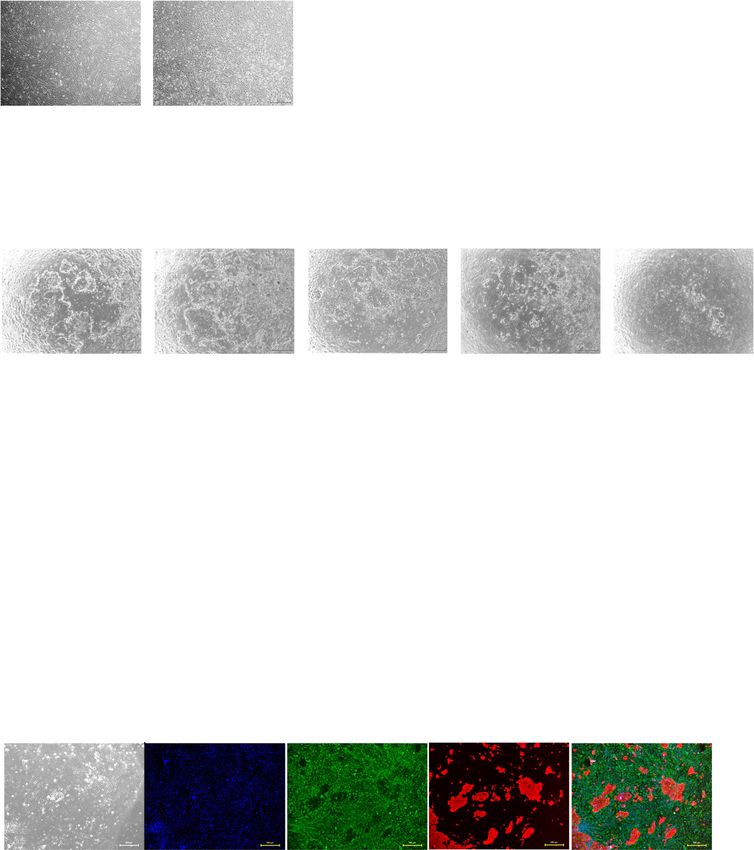
Yokomizo et al. Stem Cell Research & Therapy   (2021) 12:130                                                     Page 2 of 13

Background                                                      Methods
Infertility is defined as a failure to conceive after 12        Human tissue collection
months of regular and unprotected sexual intercourse            Endometrial specimens without any abnormalities or
and is estimated to affect 8–12% of couples of repro-           malignancies were obtained from women of reproductive
ductive age [1]. Various etiologies are associated with         age undergoing hysterectomy for benign gynecological
infertility, and endometrium is recognized as one of            diseases after written informed consent. Characteristics of
the significant factors [2]. Endometrium is structurally        donor individuals are provided in Table 1.
divided into two indistinct layers, epithelial cells and          Endometrial epithelial cells and stromal cells were
stromal cells, and undergoes remarkable histological            isolated from endometrial tissues as reported previously
and structural changes throughout the menstrual cycle           [14]. Briefly, after enzymatic digestion of minced tissue
in preparation for embryonic implantation and subse-            with 100 μg/ml collagenase type 1 in a shaking incubator
quent shedding and regeneration in non-conception               for 2 h at 37 °C, cells were separated by filtration through
cycles. Endometrial thickness and a sonographic                 100 μm and 40 μm nylon mesh. The dispersed fragments
pattern of the endometrium are widely accepted as               were collected by centrifugation, resuspended in
prognostic indicators for endometrial receptivity; lower        DMEM-high glucose (Gibco, catalog number 11965-
endometrial thickness is associated with lower                  118) containing 1% penicillin-streptomycin (Gibco, cata-
pregnancy rate and live birth rate [3]. Regardless of its       log number 15140-122) and 10% fetal bovine serum
significance, gynecologists often perform dilatation and        (Gibco, catalog number 16000-044) and seeded on bio-
curettage for spontaneous miscarriage and abortion              coat culture dishes (Corning, catalog number 354450) as
[4], and interventional radiologists perform embolization       endometrial stromal cells. The residual tissue fragments
for feeding arteries to control vaginal bleeding or reducing    and cell clumps were collected into a new 50-ml tube
size of uterine myoma in clinical practice [5]; nevertheless,   using Accumax (Innovative Cell Technologies, catalog
these invasive interventions cause endometrial damage           number AM105) and 0.25% Trypsin/EDTA (Gibco, cata-
and result in thin endometrium [6, 7].                          log number 25200-056) and then incubated for 10 min
  Thin endometrium adversely affects reproductive               at room temperature with continuous pipetting. Cells
success rates with fertility treatment [8]. Several treat-      separated by filtration through a 40-μm nylon mesh,
ment modalities are presented to patients with thin             which were regarded as endometrial epithelial cells, were
endometrium to improve endometrial thickness [9].
These approaches comprise estradiol replacement, sup-           Table 1 Patients’ information
plementation of elemental minerals and vitamins, and            Parameters                                       Mean ± SD
uncountable experimental approaches. Regenerative               Age                                              47.0 ± 2.8
medicine including application of platelet-rich plasma
                                                                BMI                                              23.4 ± 4.1
(PRP) and cell therapy utilizing menstruation-derived
stem cells is expected to be a promising therapeutic            BMI, body mass index; SD, standard deviation
strategy [10, 11]. However, these strategies are not            Parameters                                       N
broadly available and there is no reliable clinical             Gynecological disorder (including duplication)
evidence supporting their use. Autologous transplant-             Uterine myoma                                  9
ation of exogenously prepared endometrium is currently            Endometriosis                                  3
a challenge for clinical practice [12]. In addition, endo-
                                                                  Ovarian tumor                                  2
metrial organoid with endometrial cells is established
[13], but endometrial epithelial cells themselves have not        Adenomyosis                                    1
been cultured in vitro. Considering these limitations,          Operation
further study is needed to determine how to maintain              Abdominal hysterectomy                         5
endometrial epithelial cells efficiently under xeno-free          Laparoscopic hysterectomy                      4
conditions.                                                     Menstrual regularity
  We herein established a model of in vitro-cultured
                                                                  Regular                                        3
endometrium as a potential therapeutic option for
refractory thin endometrium. The three-dimensional                Irregular                                      4
culture model with endometrial epithelial and stromal             Unknown                                        2
cell orchestration will provide a new framework for             Menstrual phase
exploring the mechanisms underlying the phenomenon                Proliferative                                  5
of implantation. Additionally, modified embryo culture,           Secretory                                      2
so-called “in vitro implantation”, will be possible thera-
                                                                  Unknown                                        2
peutic approaches in fertility treatment.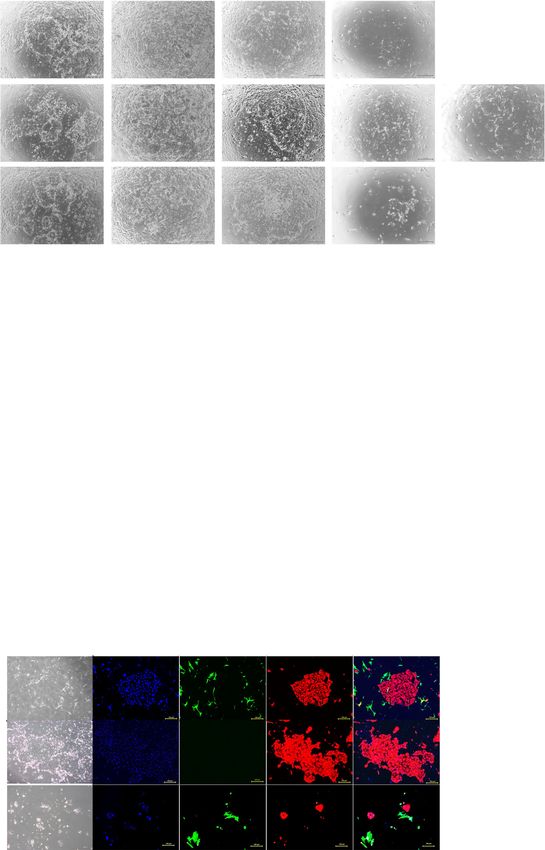
Yokomizo et al. Stem Cell Research & Therapy           (2021) 12:130                                                  Page 3 of 13

resuspended in ESTEM-HE medium (GlycoTechnica,                         Aldrich, Saint Louis, MO, USA), and 0.5 mM 8-Br-cAMP
Japan) and seeded on culture dishes. Portions of endo-                 (B5386, Sigma-Aldrich, Saint Louis, MO, USA). Detail
metrial epithelial cells were frozen with Stem Cellbanker              protocol is shown in Supplemental Figure 1.
(Nippon Zenyaku Kogyo, Japan) in − 80 °C. Endometrial
stromal cells and epithelial cells were incubated at 37 °C,
                                                                       Real-time quantitative polymerase chain reaction
95% air and 5% CO2. These cells were passaged serially
                                                                       RNA was extracted from cells using the RNeasy Mini kit
when they reached confluent by using TrypLE Express
                                                                       (Qiagen, #74104). An aliquot of total RNA was reverse-
(Gibco, catalog number 12605-010) and frozen with
                                                                       transcribed using an oligo (dT) primer (Invitrogen,
STEM CELLBANKER in − 80 °C.
                                                                       #18418-020). For the thermal cycle reactions, the cDNA
                                                                       template was amplified (Applied Biosystems Quantstu-
Immunocytochemical analysis
                                                                       dio 12 K Flex Real-Time PCR System) with gene-specific
Cells were fixed with 4% paraformaldehyde (PFA) in
                                                                       primer sets (Table 3) using the Platinum SYBR Green
PBS for 10 min at 4 °C. After washing with PBS and
                                                                       qPCR SuperMix-UDG with ROX (Invitrogen, #11733-
treatment with 0.1% Triton X-100 (Sigma-Aldrich,
                                                                       046) under the following reaction conditions: 40 cycles
#T8787-100 ML) for 10 min at 4 °C, the cells were incu-
                                                                       of PCR (95 °C for 15 s and 60 °C for 1 min) after an
bated with Protein Block Serum-Free Ready-To-Use
                                                                       initial denaturation (95 °C for 2 min). Fluorescence was
(Dako, #X 0909) for 30 min at room temperature,
                                                                       monitored during every PCR cycle at the annealing step.
followed by reaction with primary antibody in blocking
                                                                       mRNA levels were normalized using glyceraldehyde-3-
buffer for 24 h at 4 °C. After washing with PBS, the cells
                                                                       phosphate dehydrogenase as a housekeeping gene.
were incubated with fluorescently conjugated secondary
antibody. Anti-rabbit or anti-mouse immunoglobulin G
(IgG) bound to Alexa 488 or 546 (1:1000) was incubated                 Preparation of mouse embryonic fibroblasts
in blocking buffer for 30 min at room temperature. The                 Mouse embryonic fibroblasts (MEF) were prepared for
nuclei were stained with DAPI (Biotium, #40043). All                   use as nutritional support cells (feeder cells). E12.5 ICR
images were captured using confocal microscopy (con-                   mouse fetuses (Japan CLEA) were excised and the fetus
focal microscope C2+) or fluorescence microscopy (BZ-                  head, limbs, tail, and internal organs were all removed,
X700, KEYENCE). Antibody information is provided in                    minced with a blade, and seeded in culture dishes in a
Table 2.                                                               medium (DMEM containing 10% FBS, 1% Penstrep.) to
                                                                       allow cell growth. X-ray irradiation was applied (Hitachi,
Decidualization                                                        MBR-1520 R-3) to the cells in 1/100 amount of 1 M
For decidualization, endometrial stromal cells were                    HEPES Buffer Solution (Invitrogen, 15630-106). Follow-
plated in 6-well plates, then the cells were cultured for 8            ing irradiation with X-rays (dose, 30 Gy), the cells were
days in DMEM supplemented with low-serum medium                        frozen using a TC protector (DS Pharma Biomedical,
(2% FBS), 10 nM β-estradiol (E2758, Sigma-Aldrich, Saint               TCP-001) and subsequently used as feeder cells for
Louis, MO, USA), 1 μM progesterone (E8783, Sigma-                      culturing endometrial epithelial cells.

Table 2 List of antibodies for immunochemistry
Name                                                                          Class                 Company               Dilution
Primary antibodies
  Vimentin (D21H3) XP #5741                                                   Rabbit IgG            CST                   1/100
  Anti-pan Cytokeratin antibody [AE1/AE3 + 5D3] ab86734                       Mouse IgG1            abcam                 1/100
  Anti-Estrogen Receptor alpha antibody [ESR1/3557] ab259427                  Mouse IgG1            abcam                 1/200
  Anti-Progesterone Receptor antibody [SP2] ab16661                           Rabbit IgG            abcam                 1/100
  Anti-CD10 antibody [EPR22865-73] ab255609                                   Rabbit IgG            abcam                 1/50
  Anti-CD13 antibody [EPR4058] ab108310                                       Rabbit IgG            abcam                 1/100
  Anti-E Cadherin antibody [HECD-1] ab1416                                    Mouse IgG1            abcam                 1/50
Secondary antibodies
  DAPI #40043                                                                 None                  Biotium               1/1000
  Goat anti-rabbit IgG Secondary antibody, Alexa Fluor 488 A11008             None                  Invitrogen            1/500
  Goat anti-mouse IgG1 Secondary antibody, Alexa Fluor 546 A21123             None                  Invitrogen            1/500
DAPI, 4′,6-Diamidino-2-Phenylindole, dihydrochloride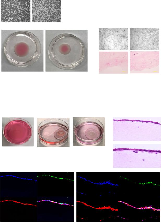
Yokomizo et al. Stem Cell Research & Therapy            (2021) 12:130                                                        Page 4 of 13

Table 3 List of primers for quantitative reverse transcription-             added tetracycline into the medium. Using an X-ray
polymerase chain reaction                                                   irradiation apparatus (Hitachi, MBR-1520 R-3), 1/100
Gene              Primer sequence                                           amount of 1 M HEPES Buffer Solution (Invitrogen,
PRL               Forward             5′ TCATCTGGTCACGGAAGTACGT 3′          15630-106) was added to the feeder cells except for
                  Reverse             5′ GCCCTCTAGAAGCCGTTTGG 3′            hESCFC-3. Following irradiation with X-rays (dose, 30
                                                                            Gy), the feeder cells were frozen using a TC protector
IGFBP1            Forward             5′ ATGGCTCGAAGGCTCTCCAT 3′
                                                                            (DS Pharma Biomedical, TCP-001). We did not use X-
                  Reverse             5′ TCCTGTGCCTTGGCTAAACTC 3′
                                                                            ray irradiation for hESCFC-3, and these cells were also
GAPDH             Forward             5′ TGTTGCCATCAATGACCCCTT 3′           frozen using a TC protector.
                  Reverse             5′ CTCCACGACGTACTCAGCG 3′
PRL prolactin, IGFBP1 insulin-like growth factor binding protein 1, GAPDH   Culture of endometrial epithelial cells
glyceraldehyde 3-phosphate dehydrogenase                                    Endometrial epithelial cells were cultured on feeder cells.
                                                                            We cultured the endometrial epithelial cells for approxi-
Preparation of human embryonic stem cell-derived feeder                     mately 2 weeks and then passaged them when they
cells                                                                       reached confluency or ceased colony enlargement. We
The human embryonic stem cell line (SEES5) was main-                        continued to observe growth of small colonies and termi-
tained on irradiated MEF feeder layers [15]. To prepare                     nated the culture when the cells did not show the signs of
human embryonic stem cell (hESC)-derived feeder cells,                      proliferation. In serial passages, the feeder cells were
we processed SEES5 cells as previously reported [16].                       detached from culture dishes 1–2 min after exposure to
Briefly, to generate embryonic bodies (EBs), SEES5 (5 ×                     TrypLE Express, but colonies of endometrial epithelial
103/well) were dissociated into single cells with 0.5 mM                    cells remained attached on the dish. MEFs and hESCFCs
EDTA (Life Technologies) after exposure to the rock                         were cultured on gelatin-coated dishes in α-MEM supple-
inhibitor (Y-27632: A11105-01, Wako, Japan) and                             mented with 10% FBS (Gibco) and 1% Pen-Strep as feeder
cultivated in 96-well plates (Thermo Fisher Scientific) in                  cells. Endometrial stromal cells were cultured in DMEM
the EB medium (76% Knockout DMEM, 20% 35 kGy                                (Gibco) supplemented with 10% FBS (Gibco) and 1% Pen-
irradiated Xeno-free Knockout Serum Replacement (XF-                        Strep. The endometrial epithelial cells were overlaid on
KSR, Life Technologies, CA, USA), 2 mM GlutaMAX-I,                          the feeder cells in ESTEM-HE medium (GlycoTechnica,
0.1 mM NEAA, Pen-Strep, and 50 μg/ml l-ascorbic acid                        Japan). The media were replaced in 2 or 3 days.
2-phosphate (Sigma-Aldrich, St. Louis, MO, USA)) for 4
days. The EBs were transferred to T25 flasks coated with                    Assessment of cell proliferation
NMP collagen PS (Nippon Meat Packers Inc.) and culti-                       Endometrial epithelial cells were plated into 24 well plates
vated in the XF32 medium (85% Knockout DMEM, 15%                            (10 × 104/well). In each well, corresponding feeder cells
35 kGy-irradiated XF-KSR, 2 mM GlutaMAX-I, 0.1 mM                           were plated in advance (5 × 10⁴/well). Cell clusters appeared
NEAA, Pen-Strep, 50 μg/ml l-ascorbic acid 2-phosphate,                      2 to 3 days after seeding, and the medium was replaced
10 ng/ml heregulin-1β (recombinant human NRG-beta                           every 2 or 3 days. Phase-contrast photomicrographs were
1/HRG-beta 1 EGF domain; Wako, Japan), 200 ng/ml                            taken at each passage when the cells reached sub-
recombinant human IGF-1 (LONGR3-IGF-1; Sigma-                               confluence or stopped growing. Numbers of cells and col-
Aldrich), and 20 ng/ml human bFGF (Kaken Pharma-                            onies were counted in central field of pictures to compare
ceutical Co. Ltd.)) for 60 to 70 days. We propagated                        feeder activities. Two investigators (R. Y. and Y.F) counted
them in α-MEM medium supplemented with 10% FBS                              the total number of endometrial epithelial cells/well and
(Gibco or HyClone) and 1% Pen-Strep.                                        the number of colonies formed, and calculated the area of
  These cells were immortalized by inoculation with                         colonies using imaging software Fiji [18]. These measure-
CSII-CMV-TERT, CSII-CMV-Tet-Off, CSII-TRE-Tight-                            ments were conducted entirely independent of all other
cyclin D1, and CSII-TRE-Tight-CDK4R24C (mutant                              variables. Each experiment was done in triplicate.
CDK4: an INK4a-resistant form of CDK4) lentiviruses
according to our previous report [17]. We further                           Growth curves
infected the immortalized cells with the lentiviruses                       Endometrial stromal cells were plated into 6-well plates
carrying a combination of the R-WNT3A, R-SPO, and                           (1 × 10⁵/well). The total number of cells/well, which was
NOGGIN genes. We designated the hESC-derived feeder                         cultured in DMEM and ESTEM-HE medium, was
cells (hESCFCs) and named as listed in Supplemental                         counted 1, 3, 5,7, and 10 days after the plating.
Table 1. We propagated these cells in a gelatin (Sigma,
G1890)-coated culture dish in α-MEM medium supple-                          Three-dimensional cell culture
mented with 10% FBS (Gibco) and 1% Pen-Strep to                             Three-dimensional cell culture was performed according
allow cell growth. For hESCFC-3 cultivation, we further                     to a previously described protocol [19]. Atelocollagen
Yokomizo et al. Stem Cell Research & Therapy   (2021) 12:130                                                        Page 5 of 13

(Koken, #IPC-50) and endometrial stromal cells were              stromal cells even after the freeze-thaw procedure and
mixed and poured into an untreated 60-mm Petri dish              subsequent passages.
and allowed to gel at 37 °C for 1 h to prepare the stromal
layer. Contraction of the collagen gel was facilitated by        Successful cultivation of endometrial epithelial cells with
pulling the gel from the surface of the Petri dish. The          feeder cells
medium (DMEM) was changed every 2 or 3 days until                We then examined various cells as feeders for endometrial
day 7. Frozen-thaw endometrial epithelial cells were             epithelium (Fig. 2a–f). We serially passaged endometrial
plated at 1 × 10⁶cells inside a glass ring (10 mm diameter)      epithelial cells on mouse embryonic fibroblasts (MEF) up
on the surface of the contracted collagen gel at Day 7.          to 6 passages for approximately 4 months (Fig. 2b). Since
Endometrial epithelial cells were grown in ESTEM-HE              the adhesive ability of MEFs was low, MEFs were easily
medium and the medium was replaced every 2 or 3 days.            separated from the endometrial epithelial cells. The endo-
Three-dimensional endometrium was obtained on day 21.            metrial epithelial cells were efficiently propagated on MEFs,
                                                                 as shown by the cumulative area of colonies (P < 0.05), area
Statistical analysis                                             of colonies (P < 0.05), and population doubling (P = 0.016)
Changes in gene expression were compared by paired t             (Fig. 2a–f). Endometrial epithelial cells on MEF showed a
tests. Unpaired Student’s t test was used to compare             cobblestone appearance and expressed pan-cytokeratin, an
differences of continuous variables in two groups and            epithelial cell-specific marker, at passage 4 (Fig. 2g, Supple-
one-way ANOVA was used in three groups. Prism 8.01               mental Figure 2A). Interestingly, endometrial epithelial cells
software (GraphPad Inc.) was used for the statistical            on MEF lost expression of vimentin, which is expressed in
analyses. P < 0.05 was considered to be statistically            both endometrial epithelial cells and stromal cells.
significant for all analyses.
                                                                 Patient-derived endometrial stromal cell is one of the
Results                                                          best human feeder cells
Endometrial epithelium and stroma                                Anticipating clinical applications, we examined human
We collected endometrial samples from healthy women              cells as feeder cells for endometrial epithelial cells. We
undergoing benign gynecological surgery and prepared             assumed that endometrial stromal cells simultaneously
primary human endometrial epithelial cells and stromal           obtained from the same patient would be good feeders
cells from the endometrial samples by enzymatic diges-           for endometrial epithelial cells. We hypothesized that
tion for in vitro experiments (Table 1, Fig. 1a–d). During       endometrial stromal cells could serve as feeder cells if
culture of stromal cells, epithelial cells were observed at      their proliferation activities were suppressed. We there-
early passages; however, they disappeared after serial           fore investigated differences in cell proliferation of endo-
passages (Fig. 1c, d). We failed to culture the endomet-         metrial stromal cells in two types of media. Endometrial
rial cells from the endometrial samples derived from             stromal cells in proliferative and non-proliferative states
older patients and patients in the secretory phase of            were spindle-shaped or flat polygonal-shaped, respect-
their cycles because the cells ceased proliferating. Endo-       ively (Fig. 3a, b). The proliferation rate of endometrial
metrial cells may have differential growth properties that       stromal cells in the conventional medium (DMEM
depend on donor age and menstrual phase.                         supplemented with 10% FBS) was significantly higher
   We then examined the thawed endometrial cells to              than that in the epithelium-specific medium (P < 0.05)
investigate whether endometrial characteristics were             (Fig. 3c). Based on these findings, we decided to utilize
preserved (Fig. 1e–g). Endometrial cells in culture exhib-       the endometrial stromal cells as feeder cells for subse-
ited a single columnar epithelium structure and spindle-         quent experiments. The endometrial epithelial cells were
shaped fibroblast-like morphology that were similar to           propagated on endometrial stromal cells up to passage 5
normal endometrial tissue. For endometrial stromal cells,        for 81 days (Fig. 3d–g). Estrogen receptor α and progester-
we further explored protein expression and hormone               one receptor were preserved in the endometrial epithelial
responsiveness (Fig. 1h–l). Endometrial stromal cells pre-       cells (Supplemental Figure 3A and B). Endometrial epithe-
sented the same protein expression pattern as endometrial        lial cells after serial passages exhibited a cobblestone
tissue. We successfully induced decidualization in endo-         appearance and expressed pan-cytokeratin, but they failed
metrial cells which exhibited aggregation of large cells         to express vimentin at early passage (passage 2) and late
with abundant cytoplasm, marked nucleolar enlargement            passage (passage 4) (Fig. 3h, Supplemental Figure 3C).
and prominent nuclei (Fig. 1i, j; Supplemental Figure 1).           We then examined human-derived cells (hESCFCs) as
Additionally, the decidualization markers PRL and IGFBP-         feeder cells for endometrial epithelial cells. We simultan-
1 were significantly upregulated in the decidualized cells       eously compared feeder activities of endometrial stromal
(P = 0.0017 and 0.0033, respectively) (Fig. 1k, l), suggesting   cells and hESCFCs by examination of the proliferation
that hormone responsiveness is preserved in endometrial          rate of endometrial epithelial cells under the same
Yokomizo et al. Stem Cell Research & Therapy (2021) 12:130 Page 6 of 13
a b c d
e f g
h
i j k l
Fig. 1 Preparation of endometrial epithelial cells and stromal cells from endometrial specimens. a, b Microscopic appearance of endometrial epithelial
cells in primary culture. Low magnification (a) and high magnification (b). Black bar is 500 μm. c, d Microscopic appearance of endometrial stromal
cells. Epithelial components were mixed in primary culture (c); however, these disappeared with serial passage (d). Black bar is 500 μm. e–g
Hematoxylin and eosin staining for endometrial tissue (e), cultured endometrial epithelial cells (f), and stromal cells (g). Black bar is 100 μm. h
Immunohistochemical staining for endometrial stromal cells in serial culture. Endometrial tissue was used as a control. Endometrial stromal cells were
positive for ERɑ, PR, CD10, and CD13 like endometrial tissue. Nuclei were stained with DAPI. Yellow bar is 500 μm. Epithelial component of endometrial
tissue was positive for ERɑ and PR, negative for CD10, and weakly positive for CD13. i–l Decidualization of endometrial stromal cells. Microscopic
appearance of endometrial stromal cells cultured in the control medium (i) and supplemented with estrogen, progesterone, and cAMP (j) (see
Experimental procedure). Black bar is 500 μm. The PRL (k) and IGFBP-1 (l) genes were significantly upregulated after decidualization (P = 0.0017 and
0.0033, respectively). Expression of the genes in the controls is designated as 1.0. Error bar indicates SEM. Each experiment was done in triplicate.
Abbreviation: ER, estrogen receptor; PR, progesterone receptor; DAPI, 4′,6-diamidino-2-phenylindole; cAMP, cyclic adenosine monophosphate; PRL,
prolactin; IGFBP-1, insulin-like growth factor binding protein-1; SEM, standard error of the mean
experimental conditions, including cell density and expression (Fig. 4f). The endometrial epithelial cells
culture media. The endometrial epithelial cells were expressed pan-cytokeratin and lost expression of vimentin
successfully cultured on hESCFCs up to passage 5 for when cultured with hESCFCs. The proliferation rate of
4 months (Fig. 4a–e). Furthermore, we examined the endometrial epithelial cells on the endometrial stromal cells
endometrial epithelial cells for morphology and protein was significantly higher than that on hESCFCs (P < 0.05).Yokomizo et al. Stem Cell Research & Therapy (2021) 12:130 Page 7 of 13
a c
b
d
f
e
g
Fig. 2 Culture of endometrial epithelial cells with mouse embryonic fibroblasts. a, b Microscopic appearance of endometrial epithelial cells without
feeder cells (a) and with MEF (b) in serial passage. Black bar is 500 μm. c–e Cumulative area of colonies (c), colony formation (number) (d), and area of
colonies (e) of endometrial epithelial cells in serial passages. Error bar indicates SEM. An asterisk means P < 0.05. ns means “not significant”. f
Population doubling levels of endometrial epithelial cells when culture with MEF (red) and without feeder cells (blue). We could propagate
endometrial epithelial cells with MEF for 111 days. Error bar indicates SEM. Dotted line indicated the observation period until the culture was
terminated. g Immunohistochemical staining for endometrial epithelial cells and MEF at passage 4. Endometrial epithelial cells kept positive for pan-
cytokeratin in serial passage. MEF expressed vimentin. Endometrial epithelial cells did not express vimentin. Nuclei were stained with DAPI. Yellow bar
is 500 μm. Each experiment was done in triplicate. Abbreviation: MEF, mouse embryonic fibroblasts; SEM, standard error of the mean
Endometrial stromal cells can therefore be used as feeder artificial endometrium network depended on the num-
cells to support proliferation of endometrial epithelial cells, ber of endometrial stromal cells. Endometrial stroma
as they were among the best human-derived cells tested. was evenly embedded in the atelocollagen gel. Endomet-
rial stromal cells (1 × 10⁶cells) embedded in atelocolla-
Three-dimensional culture of thawed endometrial cells gen formed stromal layer, and gradually shrunk during
Our successful cultivation of endometrial epithelial cells 7 days of culture (Fig. 5d). We then plated endometrial
for use in co-cultures with endometrial stromal cells epithelial cells on formed stromal layers and maintained
motivated us to investigate whether three-dimensional the three-dimensional-culture for 14 days (Fig. 5e–g).
culture can be achieved using thawed endometrial cells. Epithelial cells in three-dimensional-culture were posi-
We investigated whether variation in the numbers of tive for both epithelial markers (cytokeratins and E-
endometrial stromal cells in the atelocollagen gel affects cadherin) and mesenchymal markers (vimentin and
three-dimensional-culture (Fig. 5a–c). Construction of CD13), like intact human endometrium (Fig. 5h,Yokomizo et al. Stem Cell Research & Therapy (2021) 12:130 Page 8 of 13 a b c d e f g h Fig. 3 Culture of endometrial epithelial cells with endometrial stromal cells. a, b Microscopic appearance of endometrial stromal cells cultured in conventional medium (DMEM) (a) and epithelium-specific medium (ESTEM-HE medium) (b). Black bar is 500 μm. c Growth curves of endometrial stromal cells cultured in conventional and epithelium-specific medium. Error bar indicates SEM. An asterisk means P < 0.05. d Microscopic appearance of endometrial epithelial cells with endometrial stromal cells in serial passage. Black bar is 500 μm. e–g Cumulative area of colonies (e), colony formation (number) (f), and area of colonies (g) of endometrial epithelial cells in serial passage with endometrial stromal cells. Error bar indicates SEM. An asterisk means P < 0.05. h Immunocytochemical staining for endometrial epithelial cells and endometrial stromal cells at passage 4. Endometrial epithelial cells (surrounded with white dotted lines) continued to express pan-cytokeratin, but not vimentin, at passage 4. Endometrial stromal cells were positive for vimentin. Nuclei were stained with DAPI. Yellow bar is 500 μm. Each experiment was done in triplicate. Abbreviation: DMEM, Dulbecco’s modified Eagle’s medium; SEM, standard error of the mean
Yokomizo et al. Stem Cell Research & Therapy (2021) 12:130 Page 9 of 13
a
b
e
c
d
f
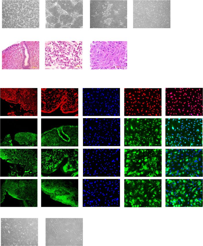
Fig. 4 Culture of endometrial epithelial cells with human embryonic stem cell-derived mesenchymal cells. a Microscopic appearance of
endometrial epithelial cells with hESCFCs (hESCFC-1, 2, 3) in serial passage. Black bar is 500 μm. b–d Cumulative area of colonies (b), colony
formation (number) (c), and area of colonies (d) of endometrial epithelial cells in serial passages with hESCFCs. Error bar indicates SEM. An asteriskYokomizo et al. Stem Cell Research & Therapy (2021) 12:130 Page 10 of 13
means P < 0.05. ns means “not significant”. e Population doubling levels of endometrial epithelial cells when cultured with endometrial stromal
cells and hESCFCs. Endometrial stromal cells showed the best feeder activities among these feeder cells (P = 0.015 when comparing with hESCFC-
1, P = 0.0177 when compared with hESCFC-2, and P = 0.0035 when comparing with hESCFC-3). Endometrial epithelial cells continued to
proliferate on endometrial stromal cells for 81 days. Error bar indicates SEM. Dotted line indicated the observation period until the culture was
terminated. f Immunocytochemical staining for endometrial epithelial cells and hESCFCs at passage 4. Endometrial epithelial cells kept positive for
pan-cytokeratin with serial passage. The endometrial epithelial cells did not express vimentin. hESCFCs expressed vimentin. Nuclei were stained
with DAPI. Yellow bar is 500 μm. Each experiment was done in triplicate. Abbreviation: EMSC, endometrial stromal cells; hESCFCs, human
embryonic stem cell-derived feeder cells; SEM, standard error of the mean
Supplemental Figure 2B). These data suggest that endo- growth of endometrial epithelial cells. We also investi-
metrial epithelium and stroma, after the freeze-thaw gated feeder activities of hESCFCs established in our
procedure and sequential culture, are able to establish laboratory. hESCFCs were easy to maintain. Further-
an endometrial three-dimensional model. The success of more, other types of human cells such as epidermal
this study may lead to the development of an in vitro cells, intestinal epithelial cells, and hepatocytes can be
implantation model. propagated on hESCFCs in our laboratory (not
published). These results motivated us to investigate
Discussion whether hESCFCs can accelerate proliferation of endo-
It is difficult to maintain endometrial epithelial cells metrial epithelial cells. As for the differences among the
in vitro and co-culture them with endometrial stromal feeder cells, various combinations of the genes have
cells. Similar three-dimensional-structures have been been transduced to investigate which genes contribute
established in the cornea, intestine, and liver [20–22]. to good feeder activities. hESCFCs contribute the small
Likewise, we hypothesized that an endometrial three- success to propagate the endometrial epithelial cells.
dimensional model can be established. In this study, we However, the difference among hESCFCs was subtle;
demonstrated that endometrial stroma is one of the best Wnt3a and Rspo1 are sufficient to confer efficient
feeder cell types for propagation of endometrial epithe- feeder activity to hESCFCs. Further studies will be
lium. We also established an endometrial three- needed to clarify which genes are necessary to support
dimensional model with frozen-thaw endometrial epi- proliferation of not only endometrial epithelial cells, but
thelial cells and endometrial stromal cells. also other types of human epithelial or parenchymal cells.
The underlying mechanisms for the successful expansion
Endometrial stromal cells as feeder cells of endometrial epithelium on homologous stroma developed
Feeder cells have the capacity to support in vitro survival in this study were not revealed; however, previous reports
and growth of orthologous epithelial or parenchymal suggest that the FGF-MAPK, WNT-R-spondin-3, BMP-
cells through a variety of soluble or membrane-bound Noggin, and TGFβ signaling pathways are involved [12, 13,
growth factors and receptors [23–25]. Functional epithelial 36]. The success of endometrial epithelial cell proliferation
and parenchymal cell types are dependent on physical con- via cell interaction is attributed to four mechanisms: First,
tact with feeder cells for survival and expansion. On the endometrial stromal cells secrete signal molecules that act
other hand, feeder-dependent cells can also be grown under as local mediators, affect cells in the immediate environ-
feeder-free conditions when coated with extracellular ment, and exert a paracrine effect on endometrial epithelial
matrix proteins such as laminin, vitronectin, or a mixture cells [37]. Second, cell membrane-bound molecules of
of the extracellular matrix components [26–29]. Feeder endometrial stromal cells influence endometrial epithelial
cells usually consist of adherent growth-arrested, but viable cells [38]. Third, endometrial stromal cells contribute to the
cells. It may be necessary to maintain feeder cells in a non- stabilization of the culture environment by producing extra-
multiplying state by irradiation or exposure to anticancer cellular matrices [39]. Fourth, the heterocellular contribution
drugs to prevent overgrowth [23]. This is observed in other of endometrial stromal cells to endometrial epithelial cells
types of feeder cells such as MEFs and immortalized feeder may be through gap junctions [40].
cells [30–35]. Irradiation of the feeder cells, i.e., MEFs,
hESCFC-1 cells, hESCFC-2 cells, and hESCFC-3 cells with Potential clinical application
an inducible Tet-ON system, indeed enabled us to estab- The culture method developed in this study has four
lish the endometrial model, implying that endometrial strengths that suggest the cells will be useful for clinical
epithelial cells are supported by non-dividing endomet- applications. First, we employed a simplified culture
rial stromal cells, but not endometrial stromal cells method which acquires more viable cells without usage of
retaining proliferation activities. This finding motivated magnetic cell sorting and/or fluorescence-activated cell
us to use endometrial stromal cells as feeder cells. In- sorting. Second, patient-derived endometrial stromal cells
deed, this study showed that endometrial stromal- can be substituted for Matrigel. Third, we show proof-of-
derived cells are able to support long-term survival and concept regarding the use of frozen cells; we successfullyYokomizo et al. Stem Cell Research & Therapy (2021) 12:130 Page 11 of 13
a
b c
d
e f
g
h
Fig. 5 Endometrial three-dimensional cell culture model. a Microscopic appearance of endometrial stromal cells embedded in atelocollagen on Day 1.
Cell numbers were 1 × 10⁶cells (left) and 2 × 10⁶cells (right), respectively. Black bar is 500 μm. b Gross appearance of endometrial stromal cells
embedded in atelocollagen on day 7. Cell numbers were 1 × 10⁶cells (left) and 2 × 10⁶cells (right), respectively. Black bar is 1 cm. c Microscopic
appearance (bright field and HE staining) of endometrial stromal cells embedded in atelocollagen on day 7. Cell numbers were 1 × 10⁶cells (left) and
2 × 10⁶cells (right), respectively. Black bar is 1 cm. d Protocol for three-dimensional cell culture. e Gross appearance of endometrial three-dimensional
cell culture. Endometrial stromal cells were embedded in atelocollagen (left), then endometrial epithelial cells were plated on formed stromal layers
using a glass ring on day 7 (middle). Endometrial three-dimensional model was developed during further 14 days of culture (right). f HE staining for
three-dimensional cultured endometrial cells. Black bar is 100 μm. g Magnification of box area in figure f. Endometrial epithelial cells (arrows) and
stromal cells (arrowheads) in three-dimensional cell culture. Black bar is 50 μm. h Immunohistochemistry of endometrial cells in three-dimensional
culture. The endometrial epithelial cells were positive for pan-cytokeratin, vimentin, E-cadherin, and CD13. This is consistent with expression of these
markers in intact endometrial tissue. Nuclei were stained with DAPI. Yellow bar is 200 μm
utilized thawed endometrial cells that were cultured for reproductive endocrinologists prepare patient-derived
more than 3 months. Indeed, cryopreservation of freshly endometrial cells for ongoing fertility treatment in a clin-
biopsied tissue was challenged [41]. These findings are ical setting. Fourth, endometrial stromal cells served the
critical for clinical application from the viewpoint that best condition for endometrial epithelial cells in vitro.Yokomizo et al. Stem Cell Research & Therapy (2021) 12:130 Page 12 of 13
Epithelial cells in vivo require close interaction with sur- Funding
rounding mesenchymal cells [23]. To solve the problem of This research was supported by The Jikei University Research Fund for
Graduate Students; by JSPS KAKENHI Grant Number JP20J14152; by the
thin endometrium with regenerative medicine, we used Grant of National Center for Child Health and Development.
endometrial somatic cells as a source of epithelial cells
and feeder cells. Alternatively, endometrium-derived Availability of data and materials
pluripotent stem cells and progenitors may also be an at- The datasets used and/or analyzed during the current study are available
from the corresponding author on reasonable request.
tractive source [42]. Although refinement of the protocol
and proof-of-concept by in vivo experimentation are Ethics approval and consent to participate
needed before applying these cells in clinical practice, The protocol for the use of human cells in the present study was approved
findings from our study can lead to development of novel by the Institutional Review Board of the National Center for Child Health and
Development of Japan (approval number: 2289) and The Jikei University
therapeutic strategies in fertility medicine. School of Medicine (approval number: 28-083(8326)) and was in full
compliance with the Ethical Guidelines for Clinical Studies (Ministry of Health,
Labor, and Welfare). The animal use protocol was approved by the Institutional
Animal Care and Use Committee of the National Center for Child Health and
Conclusions Development. All animal experiments were based on the 3R principle (refine, re-
Our study demonstrates not only the promise for duce, and replace), and all efforts were made to minimize animal suffering and
in vitro endometrial regeneration, but also advances our to reduce the number of animals used.
understanding of reproductive biology. Novel in vitro
Consent for publication
approaches, including modifying embryo culture so-
Not applicable.
called “in vitro implantation”, may be possible thera-
peutic approaches to increase success rates of fertility Competing interests
treatment and diminish unnecessary miscarriage. The authors declare that there is no conflict of interest regarding the work
described herein.
Author details
1
Supplementary Information Center for Regenerative Medicine, National Center for Child Health and
The online version contains supplementary material available at https://doi. Development Research Institute, 2-10-1 Okura, Setagaya, Tokyo 157-8535,
org/10.1186/s13287-021-02188-x. Japan. 2Center for Maternal-Fetal, Neonatal and Reproductive Medicine,
National Center for Child Health and Development, 2-10-1 Okura, Setagaya,
Tokyo 157-8535, Japan. 3Department of Obstetrics and Gynecology, The Jikei
Additional file 1: Supplemental Figure 1. Protocol of decidualization.
University School of Medicine, 3-25-8 Nishi-Shinbashi, Minato, Tokyo
Control medium was DMEM with low-serum medium (2% FBS) and Pen-
105-8461, Japan. 4Project for Prevention of HPV-related Cancer, Exploratory
strep. For decidualization, β-estradiol, progesterone and 8-Br-cAMP were
Oncology Research and Clinical Trial Center, National Cancer Center, Chiba
added in control medium as supplement. Medium replacement (MR) was
277-8577, Japan.
performed every other day.
Additional file 2: Supplemental Figure 2. Immunohistochemistry for Received: 5 December 2020 Accepted: 24 January 2021
endometrial tissues. (A) The epithelial component of endometrial tissue is
positive for pan-cytokeratin and vimentin. Nuclei were stained with DAPI.
Yellow bar is 200 μm. (B) The epithelial component of endometrial tissue
References
is positive for E-cadherin and vimentin. Nuclei were stained with DAPI.
1. Vander Borght M, Wyns C. Fertility and infertility: definition and
Yellow bar is 200 μm.
epidemiology. Clin Biochem. 2018;62:2–10.
Additional file 3: Supplemental Figure 3. Immunocytochemical 2. Abrao MS, Muzii L, Marana R. Anatomical causes of female infertility and
staining for endometrial epithelial cells cultured on endometrial stromal their management. Int J Gynaecol Obstet. 2013;123(Suppl 2):S18–24.
cells at passage 2. A, B: Endometrial epithelial cells (surrounded with 3. Zhang T, Li Z, Ren X, Huang B, Zhu G, Yang W, et al. Endometrial thickness
white dotted lines) remained positive for estrogen receptor α (A: ERα) as a predictor of the reproductive outcomes in fresh and frozen embryo
and progesterone receptor (B: PR). C: Endometrial epithelial cells transfer cycles: a retrospective cohort study of 1512 IVF cycles with
(surrounded with white dotted lines) were positive for pan-cytokeratin. morphologically good-quality blastocyst. Medicine. 2018;97 Wolters Kluwer
Endometrial stromal cells expressed vimentin, but endometrial epithelial Health. Available from: https://www.ncbi.nlm.nih.gov/pmc/articles/pmc5
cells did not. Yellow bar is 100 μm. 794374/.
Additional file 4: Supplemental Table 1. List of vectors and genes. 4. Griebel CP, Halvorsen J, Golemon TB, Day AA. Management of spontaneous
abortion. Am Fam Physician. 2005;72:1243–50.
5. Keung JJ, Spies JB, Caridi TM. Uterine artery embolization: a review of
current concepts. Best Pract Res Clin Obstet Gynaecol. 2018;46:66–73.
Acknowledgements 6. Sentilhes L, Gromez A, Clavier E, Resch B, Verspyck E, Marpeau L. Fertility
We would like to express our sincere thanks to K. Miyado and H. Akutsu for and pregnancy following pelvic arterial embolisation for postpartum
fruitful discussion, to M. Ichinose for providing expert technical assistance, to haemorrhage. BJOG. 2010;117:84–93.
C. Ketcham for English editing and proofreading, and to E. Suzuki and K. 7. Mahajan N, Sharma S. The endometrium in assisted reproductive
Saito for secretarial work. technology: how thin is thin? J Hum Reprod Sci. 2016;9:3–8.
8. Gao G, Cui X, Li S, Ding P, Zhang S, Zhang Y. Endometrial thickness and IVF
cycle outcomes: a meta-analysis. Reprod BioMed Online. 2020;40:124–33.
Authors’ contributions 9. Lebovitz O, Orvieto R. Treating patients with “thin” endometrium - an
Conceptualization: RY, AU. Data curation: RY, YF, HK. Formal analysis: RY, YF, ongoing challenge. Gynecol Endocrinol. 2014;30:409–14.
AU. Funding acquisition: RY, AU. Investigation: RY, YF, HK, SN. Methodology: 10. Maleki-Hajiagha A, Razavi M, Rouholamin S, Rezaeinejad M, Maroufizadeh S,
RY, TK, AU. Project administration: AO, HS, AU. Software: RY, YF. Supervision: Sepidarkish M. Intrauterine infusion of autologous platelet-rich plasma in
HK, AO, HS, AU. Writing–original draft: RY. Writing–review and editing: AO, women undergoing assisted reproduction: a systematic review and meta-
HS, AU. The authors read and approved the final manuscript. analysis. J Reprod Immunol. 2020;137:103078.Yokomizo et al. Stem Cell Research & Therapy (2021) 12:130 Page 13 of 13
11. Tan J, Li P, Wang Q, Li Y, Li X, Zhao D, et al. Autologous menstrual blood- 35. Paerhati P, Ito A, Yoshioka K, Iwamoto K, Fujiwara S, Horie M, et al. Neural
derived stromal cells transplantation for severe Asherman’s syndrome. Hum differentiation of mouse induced pluripotent stem cells using cadherin
Reprod. 2016;31:2723–9. gene-engineered PA6 feeder cells. J Biosci Bioeng. 2019;127:633–40.
12. Chen JC, Erikson DW, Piltonen TT, Meyer MR, Barragan F, McIntire RH, et al. 36. Gargett CE, Schwab KE, Zillwood RM, Nguyen HPT, Wu D. Isolation and
Coculturing human endometrial epithelial cells and stromal fibroblasts alters cell- culture of epithelial progenitors and mesenchymal stem cells from human
specific gene expression and cytokine production. Fertil Steril. 2013;100:1132–43. endometrium. Biol Reprod. 2009;80:1136–45.
13. Turco MY, Gardner L, Hughes J, Cindrova-Davies T, Gomez MJ, Farrell L, et al. 37. Roy A, Krzykwa E, Lemieux R, Néron S. Increased Efficiency of γ-Irradiated
Long-term, hormone-responsive organoid cultures of human endometrium in versus Mitomycin C-Treated Feeder Cells for the Expansion of Normal
a chemically defined medium. Nat Cell Biol. 2017;19:568–77. Human Cells in Long-Term Cultures. J Hematother Stem Cell Res. 2001;10:
14. Katoh N, Kuroda K, Tomikawa J, Ogata-Kawata H, Ozaki R, Ochiai A, et al. 873–80 Mary Ann Liebert, Inc., publishers.
Reciprocal changes of H3K27ac and H3K27me3 at the promoter regions of the 38. Puck TT, Marcus PI. A rapid method for viable cell titration and clone
critical genes for endometrial decidualization. Epigenomics. 2018;10:1243–57. production with HELA cells in tissue culture: the use of x-irradiated cells to
15. Akutsu H, Machida M, Kanzaki S, Sugawara T, Ohkura T, Nakamura N, et al. supply conditioning factors. Proc Natl Acad Sci U S A. 1955;41:432–7.
Xenogeneic-free defined conditions for derivation and expansion of human 39. Namba M, Fukushima F, Kimoto T. Effects of feeder layers made of human,
embryonic stem cells with mesenchymal stem cells. Regen Ther. 2015;1:18–29. mouse, hamster, and rat cells on the cloning efficiency of transformed
16. Ando Y, Saito M, Machida M, Yoshida-Noro C, Akutsu H, Takahashi M, et al. human cells. In Vitro. 1982;18:469–75.
Can human embryonic stem cell-derived stromal cells serve a starting 40. Ehmann UK, Calderwood SK, Stevenson MA. Gap-junctional communication
material for myoblasts? Stem Cells Int. 2017;2017:7541734. between feeder cells and recipient normal epithelial cells correlates with
17. Nishiwaki M, Toyoda M, Oishi Y, Ishida S, Horiuchi S-I, Makino-Itou H, et al. growth stimulation. In Vitro Cell Dev Biol Anim. 2001;37:100–10.
Immortalization of human hepatocytes from biliary atresia with CDK4R24C, 41. Bui BN, Boretto M, Kobayashi H, van Hoesel M, Steba GS, van Hoogenhuijze
cyclin D1, and TERT for cytochrome P450 induction testing. Sci Rep. 2020; N, et al. Organoids can be established reliably from cryopreserved biopsy
10:17503. catheter-derived endometrial tissue of infertile women. Reprod BioMed
18. Schindelin J, Arganda-Carreras I, Frise E, Kaynig V, Longair M, Pietzsch T, Online. 2020;41:465–73.
et al. Fiji: an open-source platform for biological-image analysis. Nat 42. Singh P, Bhartiya D. Pluripotent stem (VSELs) and progenitor (EnSCs) cells
Methods. 2012;9:676–82. exist in adult mouse uterus and show cyclic changes across estrus cycle.
19. Kajiwara K, Tanemoto T, Wada S, Karibe J, Ihara N, Ikemoto Y, et al. Fetal Reprod Sci. 2021;28:278–90.
therapy model of myelomeningocele with three-dimensional skin using
amniotic fluid cell-derived induced pluripotent stem cells. Stem Cell
Reports. 2017;8:1701–13.
Publisher’s Note
Springer Nature remains neutral with regard to jurisdictional claims in
20. Hayashi R, Ishikawa Y, Sasamoto Y, Katori R, Nomura N, Ichikawa T, et al. Co- published maps and institutional affiliations.
ordinated ocular development from human iPS cells and recovery of
corneal function. Nature. 2016;531:376–80.
21. Uchida H, Machida M, Miura T, Kawasaki T, Okazaki T, Sasaki K, et al. A
xenogeneic-free system generating functional human gut organoids from
pluripotent stem cells. JCI Insight. 2017;2:e86492.
22. Koike H, Iwasawa K, Ouchi R, Maezawa M, Giesbrecht K, Saiki N, et al.
Modelling human hepato-biliary-pancreatic organogenesis from the
foregut-midgut boundary. Nature. 2019;574:112–6.
23. Llames S, García-Pérez E, Meana Á, Larcher F, del Río M. Feeder layer cell
actions and applications. Tissue Eng Part B Rev. 2015;21:345–53.
24. Green H. Terminal differentiation of cultured human epidermal cells. Cell.
1977;11:405–16.
25. Hynds RE, Bonfanti P, Janes SM. Regenerating human epithelia with cultured stem
cells: feeder cells, organoids and beyond. EMBO Mol Med. 2018;10:139–50.
26. Miyazaki T, Futaki S, Suemori H, Taniguchi Y, Yamada M, Kawasaki M, et al.
Laminin E8 fragments support efficient adhesion and expansion of
dissociated human pluripotent stem cells. Nat Commun. 2012;3:1236.
27. Rodin S, Domogatskaya A, Ström S, Hansson EM, Chien KR, Inzunza J, et al.
Long-term self-renewal of human pluripotent stem cells on human
recombinant laminin-511. Nat Biotechnol. 2010;28:611–5.
28. Takayama K, Nagamoto Y, Mimura N, Tashiro K, Sakurai F, Tachibana M,
et al. Long-term self-renewal of human ES/iPS-derived hepatoblast-like cells
on human laminin 111-coated dishes. Stem Cell Reports. 2013;1:322–35.
29. Nishishita N, Muramatsu M, Kawamata S. An effective freezing/thawing
method for human pluripotent stem cells cultured in chemically-defined
and feeder-free conditions. Am J Stem Cells. 2015;4:38–49.
30. Ramirez RD, Morales CP, Herbert BS, Rohde JM, Passons C, Shay JW, et al.
Putative telomere-independent mechanisms of replicative aging reflect
inadequate growth conditions. Genes Dev. 2001;15:398–403.
31. Nakano T, Kodama H, Honjo T. In vitro development of primitive and
definitive erythrocytes from different precursors. Science. 1996;272:722–4.
32. Takayama N, Nishikii H, Usui J, Tsukui H, Sawaguchi A, Hiroyama T, et al.
Generation of functional platelets from human embryonic stem cells in vitro
via ES-sacs, VEGF-promoted structures that concentrate hematopoietic
progenitors. Blood. 2008;111:5298–306.
33. Vazin T, Becker KG, Chen J, Spivak CE, Lupica CR, Zhang Y, et al. A novel
combination of factors, termed SPIE, which promotes dopaminergic neuron
differentiation from human embryonic stem cells. PLoS One. 2009;4:e6606.
34. Nishimura T, Kaneko S, Kawana-Tachikawa A, Tajima Y, Goto H, Zhu D, et al.
Generation of rejuvenated antigen-specific T cells by reprogramming to
pluripotency and redifferentiation. Cell Stem Cell. 2013;12:114–26.You can also read