Inhibition of ERK 1/2 kinases prevents tendon matrix breakdown
←
→
Page content transcription
If your browser does not render page correctly, please read the page content below
www.nature.com/scientificreports
OPEN Inhibition of ERK 1/2 kinases
prevents tendon matrix breakdown
Ulrich Blache1,2,3, Stefania L. Wunderli1,2,3, Amro A. Hussien1,2, Tino Stauber1,2,
Gabriel Flückiger1,2, Maja Bollhalder1,2, Barbara Niederöst1,2, Sandro F. Fucentese1 &
Jess G. Snedeker1,2*
Tendon extracellular matrix (ECM) mechanical unloading results in tissue degradation and breakdown,
with niche-dependent cellular stress directing proteolytic degradation of tendon. Here, we show
that the extracellular-signal regulated kinase (ERK) pathway is central in tendon degradation of
load-deprived tissue explants. We show that ERK 1/2 are highly phosphorylated in mechanically
unloaded tendon fascicles in a vascular niche-dependent manner. Pharmacological inhibition of
ERK 1/2 abolishes the induction of ECM catabolic gene expression (MMPs) and fully prevents loss of
mechanical properties. Moreover, ERK 1/2 inhibition in unloaded tendon fascicles suppresses features
of pathological tissue remodeling such as collagen type 3 matrix switch and the induction of the
pro-fibrotic cytokine interleukin 11. This work demonstrates ERK signaling as a central checkpoint to
trigger tendon matrix degradation and remodeling using load-deprived tissue explants.
Tendon is a musculoskeletal tissue that transmits muscle force to bone. To accomplish its biomechanical function,
tendon tissues adopt a specialized extracellular matrix (ECM) s tructure1. The load-bearing tendon compart-
ment consists of highly aligned collagen-rich fascicles that are interspersed with tendon stromal cells. Tendon
is a mechanosensitive tissue whereby physiological mechanical loading is vital for maintaining tendon archi-
tecture and homeostasis2. Mechanical unloading of the tissue, for instance following tendon rupture or more
localized micro trauma, leads to proteolytic breakdown of the tissue with severe deterioration of both structural
and mechanical properties3–5. These mechanisms are implicated in failed healing response and longer-term
tendinopathies, with associated individual pain and socioeconomic burden that afflicts our increasingly aging
society6,7. At present, the underlying molecular mechanisms of tendinopathies are unknown and present a major
roadblock to achieving targeted and effective t herapies8–10.
In the search for mechanisms that are involved in tendon degradation, tendon explants offer a well-suited
tissue model11,12. Tendon explants retain features of native tissue including the ECM architecture/structure, allow
for highly controlled experimental conditions, and enable direct functional (mechanical) testing. In this sense,
tendon explants overcome important limitations of traditional 2D cell culture systems, engineered 3D tissues
and in vivo animal m odels13. Ex vivo, tendon explants lose their mechanical properties under load-deprived
conditions due to proteolytic matrix degradation involving different proteases, such as matrix metalloproteases
(MMP)12,14–18. We have previously used murine tendon fascicles to show that vascular like niches drive a cellular
stress response and proteolytic tendon d egradation12. However, the molecular pathways that govern ECM deg-
radation and the subsequent loss of tendon biomechanical function remain unknown. In this work, we identify
and probe the extracellular-signal regulated kinases ERK 1/2 as a central checkpoint that regulates proteolytic
tendon matrix breakdown.
Results
Standard ex vivo tissue culture of tendon explants is widely documented to provoke tissue deterioration. In con-
trast to the nutrient hyper-availability that characterizes standard tissue culture conditions, we employed more
physiological ex vivo culture conditions comprising standard culture medium but at 3% oxygen and 29 °C—con-
ditions we have identified to maintain tendon fascicle viability while preserving native biomechanical properties12
(Fig. 1A). Here, we extend these findings by exploring the potential molecular mechanisms that underpin the
niche-dependent tendon degeneration in this model system.
We started by re-examining our published transcriptome dataset of tendon fascicles (E-MTAB-7832), in
which we have identified ~ 3000 genes to be differentially expressed between standard and reduced niche condi-
tions. First, we applied the Expression2Kinase (E2K) pipeline to computationally infer key regulatory kinases
1
Department of Orthopedics, Balgrist University Hospital, University of Zurich, Zurich, Switzerland. 2Institute
for Biomechanics, ETH Zurich, Zurich, Switzerland. 3These authors contributed equally: Ulrich Blache and
Stefania L. Wunderli. *email: jess.snedeker@hest.ethz.ch
Scientific Reports | (2021) 11:6838 | https://doi.org/10.1038/s41598-021-85331-1 1
Vol.:(0123456789)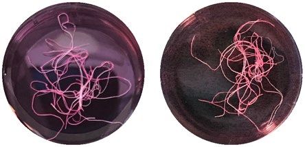
www.nature.com/scientificreports/
A B
FGFR2FGFR3
TRKC
EphB2 TRKB FGFR1
FGFR4
TRKA KDR
EphB1 FLT1 FMS
EphA5 ROR2 KIT
MUSK ROR1
Standard culture Reduced culture EphA3
EphA4
EphB3 DDR2
DDR1
MER
RET
AXL TYRO3 FLT4
PDGFRa
FLT3
IGF1R IRR PDGFRb
EphA6
ErbB2
YES INSR MET EGFR
EphB4 RON MLK3
SRC ROS MLK1
ALK TIE2
LYN EphA7 TIE1
LTK ErbB4
HCK FYN RYK MLK4
EphA8 CCK4 MLK2
Temperature
TNK1 TYK2
37°C 29°C LCK FGR ACK JAK1
TKL
JAK2
ErbB3
EphA2 JAK3
TK
BLK
ZAP70 ANKRD3 SgK288 DLK
PYK2SYK LZK
EphA1 FAK LMR1
LMR2 ALK4
ITK ZAK RAF1 TGFbR1
TEC FRK KSR1 BRAF
EphB6
SRM RIPK2 KSR2
TXK IRAK1
BTK BRK LMR3 IRAK3 ALK7
LIMK1
Oxygen
LIMK2 ARAF BMPR1B
21% 3%
BMX EphA10 TESK1 ILK BMPR1A
CTK RIPK3 TESK2 TAK1 ALK1
CSK HH498 ALK2
ABL2 ACTR2
ABL1 FES IRAK2 ACTR2B
FER RIPK1
STE
JAK3_b TGFbR2
MAP3K2
JAK2_b LRRK1 MISR2 MAP3K3
TYK2_b LRRK2 BMPR2 MAP3K5
DMEM DMEM
SuRTK106 IRAK4 MAP3K19
ANPa JAK1_b MAP3K7
ANPb KHS1 MST1 YSK1
MOS
Medium + 10% FBS + 10% FBS DYRK2
HSER SgK496 Wnk1
Wnk3
MAP3K6
HPK1 KHS2 MST2
GCK
MST3
MST4
PBK
+ 200µM AA + 200µM AA
DYRK3 CYGD MAP3K4
DYRK1A NRBP2 Wnk2 HGK
DYRK4 NRBP1 OSR1
DYRK1B CYGF MAP3K1 MINK
Wnk4 STLK3 NRK TNIK
MLKL
STLK5
PEK STLK6 MYO3A
SgK307 SLK MYO3B
PKR SgK424
Mechanical
HIPK1 HIPK3 GCN2 LOK
TAO1
yes no
SCYL3
HIPK2
SCYL2
SCYL1 NIK COT TAO3 TAO2
PRP4 PAK1
degradation
CLK4 HIPK4 HRI PAK3 PAK4
CLK1 IRE1 CLIK1 PAK2
CLK2 CLIK1L MAP2K5 PAK5
IRE2 TBCK PAK6
CLK3
MAP2K7 MAP2K1
RNAseL GCN2_b MAP2K2
CMGC MSSK1 TTK SgK071
SRPK2 KIS MAP2K3
MYT1 MAP2K4 MAP2K6
SRPK1 CK2a1 Wee1
MAK CK2a2 CDC7 SgK196 Wee1B CK1d
TTBK1
GSK3B ICK PRPK CK1e
Haspin TTBK2
GSK3A MOK CK1a
CDKL3 CK1a2
CDKL2 SgK269 PINK1 SgK493 VRK3 CK1g2
CDKL1 CDKL5 Erk7 SgK396
CK1
SgK223 CK1g1
CDKL4 Erk4 Slob
Erk3 SgK110 CK1g3
NLK PIK3R4
Erk5 SgK069 BUB1
SBK BUBR1
IKKa
Erk1 VRK1
IKKb
CDK7 IKKe PLK4 VRK2
Erk2 TBK1
p38g PITSLRE MPSK1
p38d JNK1 TLK2
JNK3JNK2 CDK10 GAK PLK3
AAK1 ULK3 TLK1
p38b CDK8 CaMKK1 PLK2
CDK11 PLK1
p38a CDK4 CCRK BIKE CaMKK2 BARK1
CDK6 ULK1 RHOK
Fused BARK2
GPRK5
GPRK7
PFTAIRE2 ULK2 ULK4 SgK494
PFTAIRE1 CDK9 NEK6 GPRK6 GPRK4
PCTAIRE2 NEK7 NEK10 RSKL1
NEK8 SgK495 PASK PDK1 RSK1
NEK9 MSK1 RSKL2
LKB1 MSK2
DYRK2 PCTAIRE1PCTAIRE3 CDK5CRK7CHED
NEK2 CHK1 p70S6K RSK3
RSK4 RSK2
AKT2
DYRK3 CDC2
NEK11 AurA p70S6Kb AKT3
AKT1
DYRK1A DYRK4
CDK3
CDK2
NEK4
Trb3 PIM1
AurB
PKN1
SGK1
SGK2SGK3
PIM2
DYRK1B NEK3
Trb2 PIM3
AurC LATS1
LATS2
PKG2
PKG1
PKN2
PKN3
NEK5 NDR1
Trb1 Obscn_b NDR2 PRKY
SPEG_b YANK1 PRKX PKCh
NEK1 MAST3
Obscn MASTL PKCz PKCe
PEK SPEG
STK33 YANK2
YANK3
PKACg
PKCi PKCd
PKACa
PKCt
PKCg
TTN
HIPK3 PKR Trio
MAST2 PKACb
HIPK1 GCN2 smMLCK Trad
TSSK4 CHK2 ROCK1
ROCK2
PKCa
HUNK PKCb
HIPK2 skMLCK SNRK
AGC
DMPK1
SCY DRAK2 SSTK
NIM1
MAST4 MAST1
CRIK
PRP4 TSSK3 PKD2
CLK4 HIPK4 HRI SgK085
DRAK1
TSSK1
DCAMKL3 DCAMKL1 DMPK2
CLK1 IRE1 CLIK1 caMLCK
TSSK2
MELK
PKD1
PKD3
DCAMKL2
CLK2
CASK
IRE2 CLIK1L DAPK3 DAPK2 VACAMKL MRCKb
TBCK DAPK1 PHKg1
MAPKAPK5 MRCKa
CLK3 PHKg2 PSKH1
AMPKa2 MNK1
MNK2 PSKH2
MAPKAPK2 CaMK2g
RNAseL
AMPKa1 CaMK2a CaMK2b
BRSK2 MAPKAPK3CaMK2d CaMK4
CMGC
BRSK1
TTK
RSK4_b
MSSK1 SgK071 NuaK2
NuaK1 RSK1_b MSK1_b
SRPK2 KIS QSK
MSK2_b CaMK1b
RSK2_b
SRPK1 CK2a1
RSK3_b
CaMK1g
CAMK
SIK
MAK CK2a2 CDC7 QIK
CaMK1a
GSK3B ICK CaMK1d
MARK4
GSK3A MOK MARK1 MARK3
CDKL3 MARK2
CDKL2 S
CDKL1 CDKL5 Erk7
CDKL4 Erk4
NLK Erk3
Erk5
Erk1
CDK7
Erk2
p38g PITSLRE
p38d JNK1
JNK3JNK2 CDK10
p38b CDK8
p38a CDK11
CDK4 CCRK
CDK6
PFTAIRE2 -log10(p-value)
PFTAIRE1 CDK9
PCTAIRE2
PCTAIRE1PCTAIRE3 CDK5CRK7CHED
CDC2
CDK3
CDK2
Min Max
C D
2.5
2.0
p-ERK / t-ERK
1.5
Tubulin
1.0
p-ERK 1/2
0.5
t-ERK 1/2
0.0
re
c e
re
re
c e
re
u
u
ltu
uc ultu
ltu
da tiss
uc ultu
da iss
cu
cu
an h t
h
ed
rd
es
ed
rd
St res
Fr
F
an
ed
ed
St
R
R
Figure 1. Niche-dependent ERK 1/2 activity and phosphorylation in load-deprived tendon explant cultures. (A)
Overview of experimental conditions and starting phenotypes. DMEM = Dulbecco’s modified Eagle’s medium, FBS =
fetal bovine serum, AA = ascorbic acid. (B) Upstream kinase enrichment prediction based on the transcriptome data
set E-MTAB-7832 (ArrayExpress), which analyses differentially expressed genes in tendon tissues cultured ex vivo
in standard and reduced niches. Kinome tree dendrogram mapping of all the predicted protein kinases generated by
Coral42. The circle color and size reflect the enrichment significance with red color encoding higher significance. (C)
Bar plots show the 20 most significantly enriched kinases. Red bars represent the ERK 1 and 2 kinases. GraphPad
Prism (version 8.4.3) was used to generate the figure. (D) Western blot validation of ERK 1/2 phosphorylation (ERK
1: Thr202/Tyr204; ERK 2: Thr185/Tyr187) in freshly isolated or cultured tendon fascicles (12 days). Full Western blot
images are provided in the supplementary material (Supplementary Fig. 3). Quantification of band pixel intensities of
phosphorylated ERK (p-ERK 1/2) compared to the total ERK (t-ERK). GraphPad Prism (version 8.4.3) was used to
perform statistical analyses and generate the figure. Bars show mean values + SD, with individual replicate values as data
points, N = 8. Repeated measure ANOVA with Tukey’s multiple comparisons test: **p < 0.01, ***p < 0.001.
Scientific Reports | (2021) 11:6838 | https://doi.org/10.1038/s41598-021-85331-1 2
Vol:.(1234567890)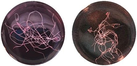
www.nature.com/scientificreports/
upstream of the differentially expressed genes (DEG). E2K algorithm predicted ~ 100 upstream protein kinases
that are likely responsible for regulating DEG. Mapping the identified kinases into Kinome phylogenetic tree
showed that the majority of these kinase hits clustered around the CMGC kinases, which is a large kinase group
including the mitogen-activated protein kinase (MAPK) family (Fig. 1B). The most significantly enriched kinases
are depicted in red color and the MAPK members ERK 1/2 were among the 5 most significantly enriched kinases
(Fig. 1B,C, Supplementary table 1). Taking this into consideration, we reasoned that ERK 1/2 may be centrally
involved in the niche-dependent degradation of tendon fascicles. Next, we experimentally validated ERK 1/2
predictions using Western Blot analysis and indeed found that the ERK kinases 1/2 are phosphorylated under
standard culture conditions but not under reduced culture conditions nor in native, uncultured fascicles (fresh
tissue) (Fig. 1D).
To further delineate the role of ERK 1/2, we pharmacologically inhibited their downstream activity using
a small molecule inhibitor (SCH772984). First, we tested whether ERK inhibition affects viability in tendon
explants (Fig. 2A,B). We observed a high cell viability (> 80%) in all conditions except for the methanol devi-
talized control tissue (Fig. 2A,B). These results show that the ERK inhibitor has no cytotoxic effects, and that
the ex vivo culture retains the viability of the tendon fascicles. In contrast to fascicles cultured under standard
conditions, ERK inhibition suppressed macroscopic contraction of fascicles after 12 days of culture similar to
fascicles cultured under reduced, non-degrading conditions (Fig. 2C). Next, we assessed the functional prop-
erties of tendon explants (Fig. 2D). Mechanical testing revealed a previously observed, drastic drop in elastic
moduli (EMod) of tissues maintained in standard culture (mean: 70 MPa) when compared to fresh tissue (mean:
1300 MPa) and reduced culture conditions (mean: 1260 MPa)12. Surprisingly, ERK inhibition fully rescues the
mechanical properties in standard conditions (mean: 1090 MPa). Additionally, we tested whether mechanical
unloading alone was responsible for ERK 1/2 phosphorylation and the loss of functional properties in standard
conditions. Hence, we cultured tendon fascicles under static load conditions using a custom-made loading
device. While static mechanical load fully prevents fascicles from losing their mechanical properties, ERK 1/2
were phosphorylated under this loaded ex vivo condition (Supplementary figure S1A, S1B).
To examine how ERK inhibition suppresses tissue breakdown in tendon fascicles, we measured the gene
expression of ECM degrading enzymes (matrix metalloproteinases Mmp3, 9, 10, 13), which we had identi-
fied previously by RNA-sequencing and mass spectrometry-based proteomics12 (Fig. 3A). The expression of all
measured MMPs was strongly increased under standard conditions compared to the fresh, uncultured control
tissue (Mmp3: 26-fold, Mmp9: 50-fold, Mmp10: 230-fold, Mmp13: 269-fold). Non-degrading, reduced culture
showed generally lower up-regulation of MMPs than standard culture. Strikingly, when ERK 1/2 activity was
inhibited in standard culture conditions the expression of Mmp3, Mmp9, and Mmp10 was not induced and fully
remained at baseline level of fresh control tissue. For Mmp13 we found that its expression was increased even in
the ERK inhibition condition (16-fold) but was much lower than in the ex vivo standard condition (269-fold).
To verify that these effects occur in other tendon types and in human tissue, we cultured human tendon tis-
sues ex vivo and measured MMP expression. Indeed, we could confirm the MMP induction and inhibition in
human tendons without and with ERK-inhibition, respectively (Fig. 3B). Collectively, these data show that ERK
inhibition prevents the induction of ECM degrading enzymes (MMPs) and the loss of mechanical properties
in tendon explants.
To investigate whether ERK influences further components of degenerative ECM remodeling we assessed the
gene expression of collagen type 1 (Col1a1), collagen type 3 (Col3a1) and interleukin 11 (Il11) (Fig. 3C). Expres-
sion of collagen type 1, the most abundant component of the healthy tendon ECM, was reduced in all ex vivo
conditions including when ERK 1/2 was inhibited. However, ERK 1/2 inhibition partially rescues the expression
of Col1a1. In contrast, collagen type 3 expression, which is a known early phenotypic marker in tendinopathic
ECM remodeling19–21, was increased in tendon fascicles. Interestingly, blocking ERK 1/2 inhibited the increased
expression of Col3a1. We have previously observed that interleukin 11 (Il11), a stromal-derived cytokine and
strong fibrosis driver in many t issues22–24, might be an important cytokine for tendon deterioration as it was
heavily up-regulated in mechanically degraded tissues compared to non-degrading conditions (Supplementary
figure S2A). Here, we show that Il11 was increased in degrading fascicles (Fig. 3C), and required ERK 1/2 activity
to be expressed and released into the supernatant (Fig. 3D). When further confirming that Il11 is only expressed
in degraded tendon tissue, we observed that mechanical loading of the tissue fully blocks the protein expression
of IL11 (Supplementary figure S2B).
Discussion
The molecular events leading to tendon degeneration are poorly understood. Mechanical unloading of tendon
sub-structures due to tissue rupture or microdamage is thought to be an ignition switch of the tissue breakdown
cascade16,20,25,26. Here, we show that the ERK pathway drives tendon matrix degradation in mechanically unloaded
tissue in a culture niche-dependent manner. We found that ERK 1/2 are highly phosphorylated in degrading
tendon fascicles, and that ERK 1/2 activity is required for tendon deterioration. However, ERK 1/2 phospho-
rylation alone is not sufficient to induce loss of tendon mechanics as mechanically loaded explants show ERK
1/2 phosphorylation but retain their mechanical properties. These findings nicely show that biomechanical and
biochemical cues act together in the maintenance of tendon mechanics and functional properties. In addition,
the strong increase of MMP gene expression under the degrading conditions of a vascular-like niche depends
on ERK 1/2 activity. We have previously shown that under standard culture conditions reactive oxygen species
(ROS) are implicated in the MMP-driven tendon breakdown in load-deprived explants12. Since ERK is known to
mediate ROS s tress27, it seems plausible that a ROS-ERK-MMP axis pathway is a central mechanism of tendon
tissue breakdown. Our data, establishing a link between ERK, MMPs and tendon matrix degradation, are in line
Scientific Reports | (2021) 11:6838 | https://doi.org/10.1038/s41598-021-85331-1 3
Vol.:(0123456789)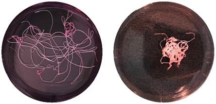
www.nature.com/scientificreports/
A Fresh tissue Standard culture B ***
*
****
*** **** *
100
Viability (% live cells)
50
Devitalized + ERK inhibition
0
e
cu Sta d c e
ar ture
re
n
liz ssu
u
tio
ltu
uc tiss
bi
ul
ER cu
ti
hi
h
ed
In
d
es
e
K
Fr
nd
ita
ed
ev
R
+
D
re
ltu
Cell nuclei
rd
Dead cells
da
an
St
C Day 0 Day 12 D
****
2000 **** ****
Reduced culture
E Modulus (MPa)
1500
1000
Standard culture 500
0
e
re
re
n
su
tio
ltu
ltu
tis
bi
Standard culture
cu
cu
hi
h
In
ed
rd
+ ERK inhibition
es
da
K
uc
Fr
ER
an
ed
St
R
+
re
ltu
cu
rd
da
an
St
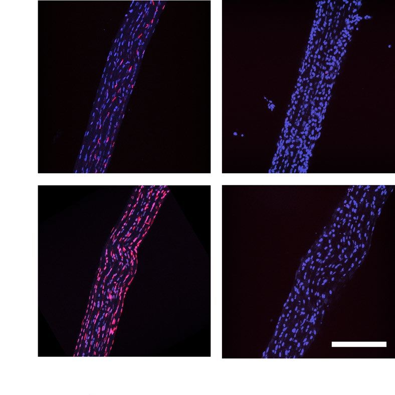
Figure 2. Inhibition of ERK 1/2 activity prevents loss of tendon biomechanical properties in tendon explants
cultured for 12 days. (A) Representative fluorescence microscopy images of tendon fascicles with nuclear
staining of all cells (blue) and dead cells (EthD-1, violet). Scale bar 200 μm. (B) Quantification of cell viability
in uncultured tendon explants (fresh tissue), methanol treated uncultured negative control tendons (devitalized
tissue) and ex vivo cultured explants in reduced and standard culture treated without and with an ERK 1/2
inhibitor, N = 6. Devitalized tissue significantly differs from all other conditions. (C) Representative macroscopic
images of tendon fascicles before treatment (uncultured tendon explants, day 0) and after 12 days of incubation
in indicated culture conditions. Scale bar 10 mm. (D) Elastic moduli of isolated tendon fascicles (freshly isolated
or cultured ex vivo in reduced conditions, standard conditions or treated with an ERK 1/2 inhibitor), N = 12
(N = 10 for standard culture, due to tissue rupture during the mounting procedure. Negative values of elastic
modulus resulting from preliminary tissue failure were set to zero). The standard culture significantly differs
from all other conditions. GraphPad Prism (version 8.4.3) was used to perform statistical analyses and generate
the figures. Box plots (25th and 75th percentiles) with whiskers (5th and 95th percentiles), median (line) and
mean (+). Bar plots represent mean values + SD. All statistical tests unless otherwise stated: Repeated measures
ANOVA with Tukey’s multiple comparisons test with *p < 0.05, ***p < 0.001, ****p < 0.0001.
with previous work reporting the control of MMP expression through ERK 1/2 in cultured fibroblasts, smooth
muscle cells and cancer c ells28–31.
Scientific Reports | (2021) 11:6838 | https://doi.org/10.1038/s41598-021-85331-1 4
Vol:.(1234567890)www.nature.com/scientificreports/
A Mouse B Human
*** ** * 1000
** ** * * ** ** **
1000
mRNA fold change
mRNA fold change 100
100
10
10
1 1
0.1 0.1
0.01 MMP3 MMP9 MMP10 MMP13
Mmp3 Mmp9 Mmp10 Mmp13
C D 6000 *
1000 *** **
IL11 (pg/ml)
* *** ** ** 4000
mRNA fold change
100
10
2000
1
0.1 0
re
re
n
tio
ltu
ltu
0.01
bi
cu
cu
hi
Col1a1 Col3a1 Il11
In
ed
rd
da
K
uc
ER
an
ed
St
R
+
re
ltu
cu
rd
da
an
St
Figure 3. Inhibition of ERK 1/2 activity prevents the induction of matrix metalloproteinases, collagen type
3 and interleukin 11 gene expression in tendon explants. (A) Gene expression of murine Mmps after 12 days
ex vivo culture. Data are normalized to 2 reference genes, N = 8. (B) Gene expression of human MMPs after
3-days ex vivo culture. Data are normalized to 2 reference genes, N = 3. (C) Gene expression of Col1a1, Col3a1
and Il11 in murine tissue after 12 days ex vivo culture. Data are normalized to 2 reference genes, N = 8. (D)
Detection of IL11 protein levels in supernatants obtained from cultured tendon fascicles by ELISA, N = 4.
Friedman test with Dunn’s multiple comparison test. Gene expression data is plotted on a logarithmic scale.
GraphPad Prism (version 8.4.3) was used to perform statistical analyses and generate the figures. Box plots (25th
and 75th percentiles) with whiskers (5th and 95th percentiles), median (line) and mean (+). Bar plots represent
mean values + SD. All statistical tests unless otherwise stated: Repeated measures ANOVA with Tukey’s multiple
comparisons test with *p < 0.05, **p < 0.01, ***p < 0.001.
Our finding showing that ERK 1/2 inhibition protects tendon tissue from degradation is in agreement with
previous studies reporting the involvement of ERK 1/2 in early tendinopathy. Using cultured human tenocytes,
ERK 1/2 have been recognized to be key signal transducers between pathological triggers and tendinopathic
phenotypes. For instance, Morita et al. showed that ERK 1/2 mediates the IL1β induced fibrotic phenotype of
diseased tendon c ells32. Furthermore, IL17A, IL33 and hypoxia were all shown to induce the expression of inflam-
matory cytokines and collagen type 3 in human tenocytes in an ERK-dependent manner19,21,33. Accordingly, we
found that ERK 1/2 inhibition abrogates collagen type 3 expression, which is induced in load-deprived tendon
explants. Similarly, we found that the stromal cytokine IL11, which is a potent fibrosis driver and therapeutic
target in many t issues22–24, is highly upregulated in degrading tendon tissue but not when ERK 1/2 was blocked.
This finding mirrors previous reports by Nishina et al. which demonstrated that the ERK pathway mediates
ROS-induced production of IL11 during early events of acute damage in hepatic s troma34.
In this study we focused on gene expression, functional read-outs and ERK 1/2 protein levels using the fascicle
ex vivo explant system as it allows for the controlled parametric investigation of the load-bearing tendon stromal
compartment. Although the relatively simple structure of tail tendon fascicles offers advantages for the study of
isolated cellular mechanisms, we acknowledge that there are limitations with using this model. In tendon fascicles
the ratio of ECM to cells is very high and measuring ECM turnover, in particular cellular collagen, at the protein
level by bulk methods is difficult due to these high ECM baseline amounts. Although we cannot rule out that the
observed decrease in collagen type I gene expression in standard culture is responsible for the measured loss of tissue
stiffness, we know from our previous proteomic quantification studies12 that the overall content of collagen type
1 protein does not significantly differ between standard and reduced culture despite clear differences in ERK 1/2
phosphorylation state. Further, recent literature has revealed that tendon is a complex tissue with heterogeneous cell
Scientific Reports | (2021) 11:6838 | https://doi.org/10.1038/s41598-021-85331-1 5
Vol.:(0123456789)www.nature.com/scientificreports/
populations, including various stromal, endothelial and immune cells35,36. Crosstalk between the cells in tendon core
and the surrounding synovial-like sheath is vital for tendon physiology and healing mechanisms20,37. The finite syno-
vial tissue layer is often damaged during fascicle extraction ex vivo which compromises the cross-compartmental
signaling12,37. Furthermore, murine tendons differ from their human counterparts in terms of function, composition
and cross-linking patterns, with the mechanical and structural properties of tail tendons significantly differing from
those of energy-storing t endons38,39. Nonetheless, there are compelling similarities between the murine tail tendon
and human load bearing tendon, both showing an ERK-dependent MMP regulation that strongly resonates with
previous studies on diseased human tendons. In this sense, the present work underscores the utility of the ex vivo
fascicle model for dissecting the early molecular events that may precede the clinical presentation of chronic tendon
pathologies. Future ongoing work is envisioned to investigate the role of ERK 1/2 pathway in degenerated human
tendons using patient-derived materials from different anatomical locations.
Experimental procedures
Kinase enrichment analysis. Upstream protein kinases associated with the DEG from dataset (Array-
Express: E-MTAB-7832)12 were identified using Expression2Kinases suite (MacOS, Version 1.6.1207, http://
www.maayanlab.net/X2K/)40. Briefly, the top 3000 DEG were fed to Expression2Kinases pipeline which applies
enrichment analysis to infer and rank potential transcription factors regulating the quired genes. Next, it identi-
fies binding partners and constructs protein–protein transcriptional regulatory subnetwork, which are loaded to
the Kinase Enrichment Analysis (KEA) module41. The identified kinases were mapped to kinome tree dendro-
gram using Coral tool and phylogenetic information derived from Manning et al.42,43 (http://phanstiel-lab.med.
unc.edu/CORAL/. Source code is available at https://github.com/dphansti/CORAL).
Murine tissue explantation and load‑deprived culture. Twelve 11-weeks-old wild-type C57BL/6
male and female mice were obtained from the ETH Phenomics Center (EPIC) and euthanized with CO2 previ-
ous to tissue harvest. Tendon fascicles were cautiously extracted from the murine tails using a surgical clamp.
For fresh tissue controls we used untreated fascicles directly after extraction. For ex vivo culture and treatments,
fascicles were immediately hydrated in PBS after extraction and randomly distributed among the test groups.
Subsequently, fascicles were maintained deprived from load in 6-well plates filled with 2 ml culture medium for
12 days in a humidified atmosphere at 5 kPa/5% CO2 and at 37 °C, 20 kPa/20% O2 (standard culture) or at 29 °C,
3 kPa/3% O2 (reduced culture). As culture medium we used high glucose Dulbecco’s Modified Eagle’s Medium
(Sigma Aldrich, D6429) containing 10% FBS (Gibco #10500-64 heat inactivated), 1% Penicillin–Streptomycin
(Sigma Aldrich, P0781) and 200 µM ascorbic acid (Wako Chemicals 013-19641). Medium was additionally sup-
plemented with an ERK 1/2 inhibitor (5 µM, SCH772984, Selleck S7101) from the beginning of the experiment.
SCH772984 blocks the ERK auto activation loop and the phosphorylation of the downstream target riboso-
mal S6 kinase. Fascicles for mechanical testing and viability were cultured individually, while fascicles for gene
expression and protein analysis were pooled (up to 15 fascicles with a maximum length per fascicle of approxi-
mately 50 mm). Medium was changed after 6 days. All experimental readouts were performed after 12 days of
ex vivo culture. The Ethics Committee of the Cantonal Veterinary office of Zurich ethically approved all animal
experiments under the permit number ZH239/17. All experiments were performed in accordance with relevant
local guidelines and regulations and in compliance with the ARRIVE g uidelines44.
Tissue culture under mechanical load. Fascicles from 5 different mice were clamped at a length of
20 mm and their mechanical properties were measured before (day 0, fresh tissue) and after (day 12, static
loading) cultivation under static mechanical load using a custom-designed uniaxial stretcher (see section “Bio-
mechanical testing”). Fresh, clamped fascicles were placed into silicon chambers (filled with 2 ml medium) and
cultured in a custom-designed bioreactor45 at crimp disappearance length (static load) and under standard cul-
ture conditions. Crimp disappearance length was visually set for each fascicle using a stereomicroscope (Nikon
SMZ 745T). As control, load-deprived fascicles (n = 5) were cultured in standard conditions as described above
and mechanical tests were performed after 12 days of culture.
Human tissue explantation and culture. Hamstring tendon pieces (Gracilis, Semitendinosus) were
collected from male and female patients undergoing anterior cruciate ligament reconstruction surgeries (mean
age: 28 years, sd: 10.7). Immediately after dissection, tendons were immersed in standard culture medium and
cut into smaller pieces (3–5 mm × 5–10 mm). Small pieces were either snap frozen as fresh control or subse-
quently cultivated in a 6-well plate with 5 ml culture medium for 3 days in standard culture or reduced culture
(see section “Murine tissue explantation and load-deprived culture”). As culture medium we used Dulbecco’s
Modified Eagle’s Medium/Nutrient Mixture F12 (Sigma Aldrich, D8437) containing 10% FBS (Gibco #10500-64
heat inactivated), 1% Penicillin–Streptomycin (Sigma Aldrich, P0781) and 200 µM ascorbic acid (Wako Chemi-
cals 013-19641). Medium was additionally supplemented with an ERK 1/2 inhibitor (5 µM, SCH772984, Selleck
S7101) from the beginning of the experiment. The Cantonal Ethics Commitee of Zurich reviewed and ethically
approved all experiments under the permit number 2020-01119. Experiments were conducted according to the
approved guidelines and tissues were collected with signed patient consent.
Biomechanical testing. Mechanical testing was performed according to previously described methods12.
Briefly, force–displacement data were recorded from 12 individually clamped fascicles (cut to a length of 20 mm
after cultivation) from different mice on a custom-designed uniaxial stretcher. Fascicles were kept hydrated
between Kapton films (DuPont, 25 µm thickness) during mechanical testing. Average diameter was determined
previous to treatment to calculate the cross-sectional area. Nominal stress was calculated based on this initial
Scientific Reports | (2021) 11:6838 | https://doi.org/10.1038/s41598-021-85331-1 6
Vol:.(1234567890)www.nature.com/scientificreports/
cross-sectional area and the pre-load length at 0.015 N corresponded to 0% strain. Tangential elastic modulus
was calculated after 5 preconditioning cycles in the linear part of the stress–strain curve (0.5–1% strain) by
fitting a linear slope (Matlab R2016a, Version 9.0.0.341360). Any spurious negative values of elastic modulus
resulting from severe tissue failure during mechanical characterization were set to zero to accord with the total
functional impairment of the tissue.
Cell viability analysis. To measure cell viability, dead cells were stained with Ethidium Homodimer-1 (EthD-
1) solution (2 mM, AS 83208, Anaspec) in PBS for 10 min. Cells in negative controls were devitalized by treatment
with 100% methanol (MeOH) for 10 min. Fascicles were thoroughly washed in PBS after EthD-1 and MeOH treat-
ment and subsequently fixed in 10% formalin for 20 min, previous to nuclear staining with NucBlue reagent (1
drop/ml, R37606, Thermo Fisher). Stained fascicles were embedded in a 50% Glycerol:PBS solution and Z-stacks
of 40 µm height (9 stacks by 5 µm) were acquired on a laser scanning confocal microscope (A1R Nikon) in trip-
licates per fascicle by using a 20 × objective (Nikon Ti2) (https://www.microscope.healthcare.nikon.com/en_EU/
products/software/nis-elements). Quantification of the viability was performed using ImageJ (Fiji version 2.0.0-rc-
59/1.51n, https://imagej.net/). We separated the channels and maximum projected all images of a stack into a single
image before subtracting background using the rolling ball method with a ball radius of 20 pixels. Next, we adjusted
image contrast/brightness and converted the image into 8-bit grayscale to then binarize it. To avoid counting cell
clusters instead of single cells, we watershed the image. Finally, we used the analyze particles tool to count the
imaged dead cells (stained with EthD-1, size: 10–200 pxn2) and all imaged cells (stained with NucBlue, size: 30–200
pxn2). Percentage cell viability was calculated by dividing the number of live cells (nuclei stained with NucBlue
only) by the total number of cells (nuclei stained with NucBlue only plus nuclei stained with EthD and NucBlue).
RNA extraction and quantitative real‑time PCR. Fascicles from three mice were pooled for each N
of gene expression analysis. RNA was extracted from 10–15 murine fascicles and 70–90 mg of human tissue
adapting a formerly established protocol12. In summary, tissues were snap frozen in liquid nitrogen, ground in
100 µl GENEzol (Labforce LSBio, 4402-GZR200) in a cryogenic mill (Spex6775 FreezerMill) for 2–4 milling
cycles at 15 cps and lysed in 1–1.5 ml GENEzol. To completely homogenize the tissue and remove insoluble
ECM components, human samples were passed ten times through a 21G syringe needle and were centrifuged at
5000 g for 5 min. Next, chloroform was added at a ratio of 1:5 from the extract volume and the lysate was centri-
fuged (12,000 g, 15 min, 4 °C). The RNA containing upper (aqueous) phase was obtained, mixed with the equal
amount of 70% EtOH and loaded onto the PureLink silica membrane column. Total RNA was isolated using
the PureLink RNA Micro Scale Kit for murine tissue (Invitrogen, 12183016) and the PureLink RNA Mini Scale
Kit for human tissue (Invitrogen, 12183018) according to the manufacturer’s instructions. 100–400 ng of RNA
was transcribed into 40 µl cDNA using the High-Capacity cDNA Reverse Transcription Kit (Applied Biosys-
tems, 4368814). qRT-PCR was performed using 1.5 µl cDNA and the KAPA PROBE FAST Master Mix (Sigma-
Aldrich, KK4707). Gene expression was carried out using the StepOnePlus Real‐Time PCR System (Applied
Biosystems) and the following TaqMan probe/primer sets: Col1a1 (Mm00801666_g1), Col3a1 (Mm01254476_
m1), Mmp3 (Mm00440295_m1), MMP3, (Hs00968305), Mmp9 (Mm00442991_m1), MMP9 Hs00957562),
Mmp10 (Mm01168399_m1), MMP10 (Hs01055413), Mmp13 (Mm00439491_m1), MMP13 (Hs00942584) and
Il11 (Mm00434162_m1). Relative gene expression was calculated by the comparative Ct method by normalizing
the data to the reference genes Anxa5 (Mm01293059_m1), ANXA5 (Hs00996186) and Eif4a2 (Mm01730183_
gH), EIF4A2 (Hs00756996) or RPL13A (Hs04194366) and to freshly isolated fascicles.
Interleukin 11 ELISA. The supernatants (2 ml) from 10–15 fascicles pooled from 3 mice were collected and
stored at − 80 °C for further use. Samples were 4 × diluted in sample buffer and interleukin 11 was detected by a
SimpleStep ELISA following the manufacturer’s instructions (Abcam, ab215084).
Western blot analysis. Fascicles from three mice were pooled for each N of protein analysis. 10–15 fasci-
cles were thoroughly washed in PBS and snap frozen in liquid nitrogen previous to lysis in RIPA buffer (Sigma-
Aldrich, R0278; supplemented with a phosphatase inhibitor, Thermo Fischer, A32957). Fascicles were homoge-
nized with surgical scissors in 50 µl ice-cold RIPA and lysed for 10 min on ice with 3 times vortexing the samples
for 10 s. Subsequently, lysates were centrifuged (20,000 g, 10 min, 4 °C) and equal protein amounts (8 μg) were
boiled in 6 × Laemmli sample buffer including 2-mercaptoethanol (10 min, 95 °C, Alfa Aesar, J61337). Next,
Mini-PROTEAN TGX stain-free protein gels (4–15%, Bio-Rad, 4568086) were used to separate proteins before
they were transferred on to polyvinylidene fluoride (PVDF) membranes using the Trans-Blot Turbo System
(Bio-Rad). Membranes were blocked in nonfat dry milk/TBS-T (5%, 30 min, RT) and primary antibodies were
incubated in BSA/TBST-T (5%) overnight at 4 °C. After washing the membranes three times in TBS-T they were
incubated with the corresponding horseradish peroxidase (HRP)-conjugated secondary antibodies in nonfat
dry milk/TBS-T (2.5%, 60 min, RT). The UltraScence Pico Ultra Western Substrate (GeneDireX, CCH345-B)
and the ChemiDoc MP imaging system (Bio-Rad) were used to visualize HRP.
Antibodies used in this study were: rabbit anti-ERK 1/2 (1:1000, Cell Signaling 9102), rabbit anti-phospho-
ERK 1/2 (1:1000, Cell Signaling 9101), mouse anti-α-tubulin (B-5-1-2, 1:3500, Santa Cruz Biotechnology sc
23948), goat-anti-mouse-HRP (1:15 000, Sigma-Aldrich, SAB3701073-2), and goat-anti-rabbit-HRP (1:15 000
Sigma- Aldrich, SAB3700878-1). For each experiment, the membrane was first probed for phospho-ERK 1/2,
then stripped by a Western blot stripping buffer (Restore Plus, Thermo Fisher 46,430) and finally probed for
total ERK 1/2. Afterwards, the membrane was developed for α-tubulin. For quantification, signal intensities were
measured using the gels tool of ImageJ (Fiji version 2.0.0-rc-59/1.51n, https://imagej.net/) and phosphorylation
levels were calculated by normalizing the values of phospho-ERK 1/2 to total ERK 1/2.
Scientific Reports | (2021) 11:6838 | https://doi.org/10.1038/s41598-021-85331-1 7
Vol.:(0123456789)www.nature.com/scientificreports/
Statistical analysis. Statistical tests were performed using GraphPad Prism (version 8.4.3, https://www.
graphpad.com/scientific-software/prism/). Between group differences of normally distributed datapoints were
evaluated by repeated measure ANOVA tests with the Tukey’s post hoc multiple comparison method. Differences
in IL11 secretion between culture conditions were evaluated using the non-parametric Friedman test and Dunn’s
post hoc multiple comparison test. In all cases, significance was defined as *p < 0.05, **p < 0.01, ***p < 0.001,
****p < 0.0001. Data were represented either as box and whisker plots with individual data points, showing 25th
and 75th percentiles (box) and 5th and 95th percentiles (whiskers) or bar charts (showing mean + SD). Negative
values of viability and elastic moduli resulting from limitations in the quantification analysis were set to 0.
Received: 10 September 2020; Accepted: 26 February 2021
References
1. Screen, H. R., Berk, D. E., Kadler, K. E., Ramirez, F. & Young, M. F. Tendon functional extracellular matrix. J. Orthop. Res. 33,
793–799. https://doi.org/10.1002/jor.22818 (2015).
2. Gracey, E. et al. Tendon and ligament mechanical loading in the pathogenesis of inflammatory arthritis. Nat. Rev. Rheumatol.
https://doi.org/10.1038/s41584-019-0364-x (2020).
3. Kinugasa, R., Hodgson, J. A., Edgerton, V. R., Shin, D. D. & Sinha, S. Reduction in tendon elasticity from unloading is unrelated
to its hypertrophy. J. Appl. Physiol. 109, 870–877. https://doi.org/10.1152/japplphysiol.00384.2010 (2010).
4. Frizziero, A. et al. Effect of training and sudden detraining on the patellar tendon and its enthesis in rats. BMC Musculoskelet.
Disord. 12, 20. https://doi.org/10.1186/1471-2474-12-20 (2011).
5. de Boer, M. D. et al. The temporal responses of protein synthesis, gene expression and cell signalling in human quadriceps muscle
and patellar tendon to disuse. J. Physiol. 585, 241–251. https://doi.org/10.1113/jphysiol.2007.142828 (2007).
6. Nourissat, G., Berenbaum, F. & Duprez, D. Tendon injury: From biology to tendon repair. Nat. Rev. Rheumatol. 11, 223–233.
https://doi.org/10.1038/nrrheum.2015.26 (2015).
7. Riley, G. Tendinopathy–from basic science to treatment. Nat. Clin. Pract. Rheumatol. 4, 82–89. https://doi.org/10.1038/ncprh
eum0700 (2008).
8. Docheva, D., Muller, S. A., Majewski, M. & Evans, C. H. Biologics for tendon repair. Adv. Drug Deliv. Rev. 84, 222–239. https://
doi.org/10.1016/j.addr.2014.11.015 (2015).
9. Millar, N. L., Murrell, G. A. & McInnes, I. B. Inflammatory mechanisms in tendinopathy—towards translation. Nat. Rev. Rheumatol.
13, 110–122. https://doi.org/10.1038/nrrheum.2016.213 (2017).
10. Andarawis-Puri, N., Flatow, E. L. & Soslowsky, L. J. Tendon basic science: Development, repair, regeneration, and healing. J. Orthop.
Res. 33, 780–784. https://doi.org/10.1002/jor.22869 (2015).
11. van Vijven, M., Wunderli, S. L., Ito, K., Snedeker, J. G. & Foolen, J. Serum deprivation limits loss and promotes recovery of tenogenic
phenotype in tendon cell culture systems. J. Orthop. Res. https://doi.org/10.1002/jor.24761 (2020).
12. Wunderli, S. L. et al. Tendon response to matrix unloading is determined by the patho-physiological niche. Matrix Biol. https://
doi.org/10.1016/j.matbio.2019.12.003 (2020).
13. Wunderli, S. L., Blache, U. & Snedeker, J. G. Tendon explant models for physiologically relevant in vitro study of tissue biology—a
perspective. Connect. Tissue Res. https://doi.org/10.1080/03008207.2019.1700962 (2020).
14. Abreu, E. L., Leigh, D. & Derwin, K. A. Effect of altered mechanical load conditions on the structure and function of cultured
tendon fascicles. J. Orthop. Res. 26, 364–373. https://doi.org/10.1002/jor.20520 (2008).
15. Leigh, D. R., Abreu, E. L. & Derwin, K. A. Changes in gene expression of individual matrix metalloproteinases differ in response
to mechanical unloading of tendon fascicles in explant culture. J. Orthop. Res. 26, 1306–1312. https://doi.org/10.1002/jor.20650
(2008).
16. Hannafin, J. A., Arnoczky, S. P., Hoonjan, A. & Torzilli, P. A. Effect of stress deprivation and cyclic tensile loading on the material
and morphologic properties of canine flexor digitorum profundus tendon: An in vitro study. J. Orthop. Res. 13, 907–914. https://
doi.org/10.1002/jor.1100130615 (1995).
17. Cousineau-Pelletier, P. & Langelier, E. Relative contributions of mechanical degradation, enzymatic degradation, and repair of the
extracellular matrix on the response of tendons when subjected to under- and over-mechanical stimulations in vitro. J. Orthop.
Res. 28, 204–210. https://doi.org/10.1002/jor.20982 (2010).
18. Arnoczky, S. P., Lavagnino, M., Egerbacher, M., Caballero, O. & Gardner, K. Matrix metalloproteinase inhibitors prevent a decrease
in the mechanical properties of stress-deprived tendons: An in vitro experimental study. Am. J. Sports Med. 35, 763–769. https://
doi.org/10.1177/0363546506296043 (2007).
19. Millar, N. L. et al. MicroRNA29a regulates IL-33-mediated tissue remodelling in tendon disease. Nat. Commun. 6, 6774. https://
doi.org/10.1038/ncomms7774 (2015).
20. Snedeker, J. G. & Foolen, J. Tendon injury and repair—A perspective on the basic mechanisms of tendon disease and future clinical
therapy. Acta Biomater. 63, 18–36. https://doi.org/10.1016/j.actbio.2017.08.032 (2017).
21. Millar, N. L. et al. Hypoxia: A critical regulator of early human tendinopathy. Ann. Rheum. Dis. 71, 302–310. https://doi.org/10.
1136/ard.2011.154229 (2012).
22. Ng, B. et al. Interleukin-11 is a therapeutic target in idiopathic pulmonary fibrosis. Sci. Transl. Med. https://doi.org/10.1126/scitr
anslmed.aaw1237 (2019).
23. Schafer, S. et al. IL-11 is a crucial determinant of cardiovascular fibrosis. Nature 552, 110–115. https://d oi.o
rg/1 0.1 038/n
ature 24676
(2017).
24. Widjaja, A. A. et al. Inhibiting interleukin 11 signaling reduces hepatocyte death and liver fibrosis, inflammation, and steatosis in
mouse models of nonalcoholic steatohepatitis. Gastroenterology 157, 777-792.e714. https://doi.org/10.1053/j.gastro.2019.05.002
(2019).
25. Arnoczky, S. P. et al. Loss of homeostatic strain alters mechanostat “set point” of tendon cells in vitro. Clin. Orthop. Relat. Res. 466,
1583–1591. https://doi.org/10.1007/s11999-008-0264-x (2008).
26. Arnoczky, S., Tian, T., Lavignano, M. & Gardner, K. Ex vivo static tensile loading inhibits MMP-1 expression in rat tail tendon
cells through a cytoskeletally based mechanotransduction mechanism. J. Orthop. Res. 22, 328–333. https://doi.org/10.1016/S0736-
0266(03)00185-2 (2004).
27. Son, Y., Kim, S., Chung, H. T. & Pae, H. O. Reactive oxygen species in the activation of MAP kinases. Methods Enzymol. 528, 27–48.
https://doi.org/10.1016/b978-0-12-405881-1.00002-1 (2013).
28. Kajanne, R. et al. EGF-R regulates MMP function in fibroblasts through MAPK and AP-1 pathways. J. Cell Physiol. 212, 489–497.
https://doi.org/10.1002/jcp.21041 (2007).
Scientific Reports | (2021) 11:6838 | https://doi.org/10.1038/s41598-021-85331-1 8
Vol:.(1234567890)www.nature.com/scientificreports/
29. Kuo, L., Chang, H. C., Leu, T. H., Maa, M. C. & Hung, W. C. Src oncogene activates MMP-2 expression via the ERK/Sp1 pathway.
J. Cell Physiol. 207, 729–734. https://doi.org/10.1002/jcp.20616 (2006).
30. Yang, C. Q. et al. MCP-1 stimulates MMP-9 expression via ERK 1/2 and p38 MAPK signaling pathways in human aortic smooth
muscle cells. Cell Physiol. Biochem. 34, 266–276. https://doi.org/10.1159/000362997 (2014).
31. Yu, T. et al. CXCR4 promotes oral squamous cell carcinoma migration and invasion through inducing expression of MMP-9 and
MMP-13 via the ERK signaling pathway. Mol. Cancer Res. 9, 161–172. https://doi.org/10.1158/1541-7786.Mcr-10-0386 (2011).
32. Morita, W. et al. ERK1/2 drives IL-1beta-induced expression of TGF-beta1 and BMP-2 in torn tendons. Sci. Rep. 9, 19005. https://
doi.org/10.1038/s41598-019-55387-1 (2019).
33. Millar, N. L. et al. IL-17A mediates inflammatory and tissue remodelling events in early human tendinopathy. Sci. Rep. 6, 27149.
https://doi.org/10.1038/srep27149 (2016).
34. Nishina, T. et al. Interleukin-11 links oxidative stress and compensatory proliferation. Sci. Signal. 5, 5. https://doi.org/10.1126/
scisignal.2002056 (2012).
35. Swanson, J. B. et al. A single-cell transcriptional atlas identifies extensive heterogeneity in the cellular composition of tendons.
bioRxiv https://doi.org/10.1101/801266 (2019).
36. Harvey, T., Flamenco, S. & Fan, C. M. A Tppp3(+)Pdgfra(+) tendon stem cell population contributes to regeneration and reveals
a shared role for PDGF signalling in regeneration and fibrosis. Nat. Cell Biol. 21, 1490–1503. https://doi.org/10.1038/s41556-019-
0417-z (2019).
37. Stauber, T., Blache, U. & Snedeker, J. G. Tendon tissue microdamage and the limits of intrinsic repair. Matrix Biol. 85–86, 68–79.
https://doi.org/10.1016/j.matbio.2019.07.008 (2020).
38. Lee, A. H. & Elliott, D. M. Comparative multi-scale hierarchical structure of the tail, plantaris, and Achilles tendons in the rat. J.
Anat. https://doi.org/10.1111/joa.12913 (2018).
39. Lee, A. H. & Elliott, D. M. Multi-scale loading and damage mechanisms of plantaris and rat tail tendons. J. Orthop. Res. https://
doi.org/10.1002/jor.24309 (2019).
40. Chen, E. Y. et al. Expression2Kinases: mRNA profiling linked to multiple upstream regulatory layers. Bioinformatics 28, 105–111.
https://doi.org/10.1093/bioinformatics/btr625 (2012).
41. Lachmann, A. & Ma’ayan, A. KEA: kinase enrichment analysis. Bioinformatics 25, 684–686. https://doi.org/10.1093/bioinforma
tics/btp026 (2009).
42. Metz, K. S. et al. Coral: Clear and customizable visualization of human kinome data. Cell Syst. 7, 347–350. https://d oi.o
rg/1 0.1 016/j.
cels.2018.07.001 (2018).
43. Manning, G., Whyte, D. B., Martinez, R., Hunter, T. & Sudarsanam, S. The protein kinase complement of the human genome.
Science 298, 1912–1934. https://doi.org/10.1126/science.1075762 (2002).
44. Kilkenny, C. et al. Animal research: Reporting in vivo experiments: The ARRIVE guidelines. Br. J. Pharmacol. 160, 1577–1579.
https://doi.org/10.1111/j.1476-5381.2010.00872.x (2010).
45. Wunderli, S. L. et al. Minimal mechanical load and tissue culture conditions preserve native cell phenotype and morphology in
tendon-a novel ex vivo mouse explant model. J. Orthop. Res. 36, 1383–1390. https://doi.org/10.1002/jor.23769 (2018).
Acknowledgements
We gratefully thank the animal facility of ETH (EPIC) for providing the mice. We also gratefully acknowledge
the Swiss Center for Musculoskeletal Biobanking, as well as Ms. Nathalie Kuehne and Mr. Dario Frustaci for
their support of the human tissue collection. This work has been funded by internal funding of the University
of Zurich and the ETH Zurich.
Author contributions
Study concept and design: U.B., S.L.W., J.G.S. Collection and/or assembly of data: U.B., S.L.W., B.N., S.F.F., M.B.,
G.F. Data analysis: U.B., S.L.W., A.H., T.S. Data interpretation and manuscript writing: U.B., S.L.W., A.H., J.G.S.
Competing interests
The authors declare no competing interests.
Additional information
Supplementary Information The online version contains supplementary material available at https://doi.org/
10.1038/s41598-021-85331-1.
Correspondence and requests for materials should be addressed to J.G.S.
Reprints and permissions information is available at www.nature.com/reprints.
Publisher’s note Springer Nature remains neutral with regard to jurisdictional claims in published maps and
institutional affiliations.
Open Access This article is licensed under a Creative Commons Attribution 4.0 International
License, which permits use, sharing, adaptation, distribution and reproduction in any medium or
format, as long as you give appropriate credit to the original author(s) and the source, provide a link to the
Creative Commons licence, and indicate if changes were made. The images or other third party material in this
article are included in the article’s Creative Commons licence, unless indicated otherwise in a credit line to the
material. If material is not included in the article’s Creative Commons licence and your intended use is not
permitted by statutory regulation or exceeds the permitted use, you will need to obtain permission directly from
the copyright holder. To view a copy of this licence, visit http://creativecommons.org/licenses/by/4.0/.
© The Author(s) 2021
Scientific Reports | (2021) 11:6838 | https://doi.org/10.1038/s41598-021-85331-1 9
Vol.:(0123456789)You can also read