Effect of metal concentration on growth and luminescence of luminous bacteria strains isolated from golfo de Nicoya, Costa Rica Efecto de la ...
←
→
Page content transcription
If your browser does not render page correctly, please read the page content below
Effect of metal concentration on growth and
luminescence of luminous bacteria strains isolated from
              golfo de Nicoya, Costa Rica
    Efecto de la concentración de metales en el crecimiento
    y la luminiscencia de cepas de bacterias luminiscentes
            aisladas del golfo de Nicoya, Costa Rica
                            Luis Vega-Corrales1* & Carolina Marín-Vindas1,2

ABSTRACT
Luminescence in bacteria is catalyzed by luciferase. When these microorganisms are exposed to
toxic substances, the bioluminescent enzyme system can be inhibited. The objective of this study
was to analyze the potential that these microorganisms offer as native bioindicators of coastal
marine pollution. The dynamics of luminescence intensity by visual classification and the effect
of metal concentration on the growth and luminescence of 25 strains of luminescent bacteria,
isolated during 2016 from seawater samples from the gulf of Nicoya, Costa Rica, was evaluated
by the disk diffusion method. The sensitivity of each strain to different concentrations (0.1, 0.5
and 1 mg mL-1) of Cd, Cu, Cr, Pb and Zn was determined by its bioluminescent phenotype. In
susceptible strains, a range of metal concentrations less than the growth inhibitory concentration
affected the expression of luminescence. Strains with intense luminescence and defined zones
of luminescence inhibition were considered to have greater potential as native bioindicators
for monitoring environmental toxicity. More studies are required to determine the minimum
concentrations that inhibit growth and luminescence with respect to the tested metals and other
potentially toxic substances for the coastal marine ecosystems of Costa Rica.

Keywords: ecotoxicology, indicator species, luminous organism, marine biotechnology,
luminous organism

RESUMEN
La luminiscencia en bacterias es catalizada por la luciferasa. Cuando estos microorganismos
se exponen a sustancias tóxicas, el sistema enzimático bioluminiscente puede ser inhibido.
El objetivo de este estudio fue analizar el potencial que tienen estos microorganismos, como

1    Laboratorio de Microbiología Marina (LaMMar), Estación de Biología Marina Juan Bertoglia Richards, Es-
     cuela de Ciencias Biológicas, Universidad Nacional, Puntarenas, Costa Rica. luis.vega.corrales@una.ac.cr*
     ORCID: https://orcid.org/0000-0003-3389-4373
2    Departament de Biologia Marina i Oceanografia, Institut de Ciències del Mar, CSIC, Barcelona, Catalunya, Spain.
     carolina.marin.vindas@una.ac.cr ORCID: https://orcid.org/0000-0002-9013-2378

Recibido: 15 setiembre 2020 • Corregido: 16 febrero 2020 • Aceptado: 16 febrero 2020
DOI: http://dx.doi.org/10.15359/revmar.13-1.2                                                                               27
Rev. Mar. Cost. Vol. 12 (1): 27-38, enero-junio 2021                                         Licencia Creative Commons
                                                                                               Atribución-No-Comercial
ISSN: 1659-455X • e-ISSN: 1659-407X                                                        Compartir Igual 4.0 Costa Rica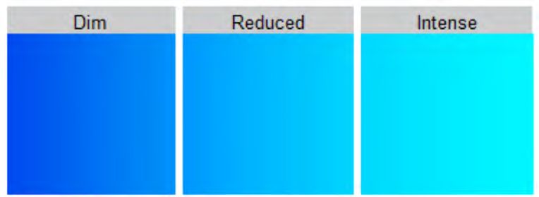
Luis Vega-Corrales & Carolina Marín-Vindas

bioindicadores nativos de contaminación marino costera. La dinámica de la intensidad de la
luminiscencia por clasificación visual y el efecto de la concentración de metales en el crecimiento
y la luminiscencia de 25 cepas de bacterias luminiscentes, aisladas durante el 2016, a partir de
muestras de agua marina del golfo de Nicoya, Costa Rica, fue evaluada por el método de difusión
en disco. La sensibilidad de cada cepa a diferentes concentraciones (0.1, 0.5 y 1 mg mL-1) de Cd, Cu,
Cr, Pb y Zn fue determinada por su fenotipo bioluminiscente. En las cepas sensibles, un rango de
concentraciones del metal menor a la concentración inhibitoria del crecimiento afectó la expresión.
Se consideró que las cepas con luminiscencia intensa y zonas de inhibición de luminiscencia
esta, definidas, tienen un mayor potencial como bioindicadores nativos para la vigilancia de la
toxicidad ambiental. Se requieren más estudios para determinar las concentraciones mínimas que
inhiben el crecimiento y la luminiscencia con respecto a los metales analizados y demás sustancias,
potencialmente tóxicas, para los ambientes marino costeros de Costa Rica.

Palabras claves: biotecnología marina, contaminación marina, ecotoxicología, especies
indicadoras, organismos luminiscentes

INTRODUCTION                                        substances, and the intensity of light
                                                    decreases quickly. Measuring the light
      Most luminous bacteria, which                 intensity of bacteria exposed to dif-
are widely distributed in marine en-                ferent concentrations of a substance is
vironments, are taxonomically classi-               used to evaluate its toxicity (Ma et al.
fied as belonging to Aliivibrio, Vibrio,            2014). Sensitive, repeatable, and easy
and Photobacterium genera from the                  to transport luminous bacteria-based
Vibrionaceae family. Several of these               assays have been developed to detect
bacteria emit high levels of light eas-             environmental contaminants (Camanzi
ily visible in laboratory cultures. Lu-             et al. 2011; Bolelli et al. 2016).
minescence in these microorganisms                         Therefore, luminous bacteria
is produced by the expression of lux                have been used as bioindicators in as-
genes, and the light emission reaction              sessments of environmental pollution
is catalyzed by luciferase (Dunlap &                (Burga et al. 2012; Ma et al. 2014),
Urbanczyk, 2013).                                   toxicity evaluation of pharmaceuti-
      Luciferase synthesis and lumi-                cal wastewater (Yu et al. 2014) and
nescence in these bacteria is regulated             toxicological tests to assess pollution
by population density through a system              caused by metals, pesticides, and an-
called quorum sensing (Dunlap & Ur-                 tibiotics in aquatic systems (Ranjan et
banczyk, 2013). Hence, there is a rela-             al. 2012; Shanware et al. 2013). More-
tionship between the emission of lumi-              over, Bagordo et al. (2012) have used
nescence with cellular metabolism. The              them to assess anthropogenic impacts
bioluminescent enzyme system can be                 on estuarine ecosystems.
inhibited by exposing bacteria to toxic

28                                                      Rev. Mar. Cost. Vol. 12 (1): 27-38, enero-junio 2021.
                                                                    ISSN: 1659-455X • e-ISSN: 1659-407X
                                                              DOI: http://dx.doi.org/10.15359/revmar.13-1.2
Effect of metal concentration on growth and luminescence of luminous bacteria
strains isolated from golfo de Nicoya, Costa Rica
Luminous bacteria represent a the methodology proposed by Kumar
powerful tool for the initial assessment et al. (2015), with some variations. Ev-
of environmental samples or substanc- ery 2 h during a 14 h period, a record
es with unknown ecotoxicological or was made of growth and luminescence
toxicological characteristics (Menz intensity of each strain inoculated by
et al. 2013). In Costa Rica, there are streaking swabs on MA plates (Dif-
no references of bioassays to assess co®) from a cell suspension equivalent
the effect that toxic compounds might to a 0.5 McFarland standard in distilled
be causing in coastal marine ecosys- water with 2% NaCl prepared from an
tems and, according to Diepens et al. overnight MA plate. At the end of the
(2014), promotion of ecotoxicological incubation period and according to the
assessments in the country using native final luminescence intensity, the strains
species is required. This study aimed were classified as intense, reduced, or
to analyze the effect of metal concen- dim, based on the luminescence inten-
tration on growth and luminescence sity scale defined in Fig. 1.
of luminous bacteria strains isolated
from the golfo de Nicoya, Costa Rica,
to generate base information about the
application that these microorganisms
have as native bioindicators of coastal
marine pollution in the country.
Fig. 1. Luminescence intensity scale
MATERIALS AND METHODS defined for the classification of luminous
bacteria strains isolated from the golfo de
Nicoya, Costa Rica
In 2016, twenty-five (25) lumi- Fig. 1. Escala de la intensidad de
nous bacteria strains isolated from sur- luminiscencia definida por la clasificación
face seawater samples collected at the de las cepas de bacterias bioluminiscentes
golfo de Nicoya, Costa Rica, were used. aisladas del golfo de Nicoya, Costa Rica
These strains were isolated using Ma-
rine Agar (MA) (Difco®) and stored in The effect of metal concentra-
Brain Heart Infusion (OXOID®) with tion on growth and luminescence of
20% glycerol at -80°C until they were these strains was assessed using the
tested. These strains were numerically disk diffusion method. Each lumi-
identified in a consecutive manner us- nous strain was inoculated by streak-
ing the CL prefix. ing swabs in MA plates plates from
Strains were characterized ac- a cell suspension equivalent to a 0.5
cording to growth and luminescence in- McFarland standard in distilled water
tensity by visual classification following with 2% NaCl. Thereafter that, 6 mm
Rev. Mar. Cost. Vol. 12 (1): 27-38, enero-junio 2021. 29
ISSN: 1659-455X • e-ISSN: 1659-407X
DOI: http://dx.doi.org/10.15359/revmar.13-1.2Luis Vega-Corrales & Carolina Marín-Vindas
diameter Whatman #3 filter paper ster- were analyzed and plotted using R (R
ile disks were separately impregnat- Core Team, 2018).
ed each with 10 µL of 0.1, 0.5, and 1
mg mL-1 metal ion solutions; namely, RESULTS
cadmium (Cd), copper (Cu), chromi-
um (Cr), lead (Pb), and zinc (Zn), then The luminescence intensity scale
they were aseptically dried at room is shown in Fig. 1. All strains grew 4 h
temperature and placed on the agar after the incubation time started, and
surface. Disks impregnated with 10 showed luminescence from 6 to 8 h af-
µL of sterile distilled water were used ter the start of the incubation (Fig. 2).
as negative controls. Tests were per- The luminous strains assessed did not
formed duplicate. Plates were incubat- show growth and luminescence inhi-
ed in the dark at 30°C for 24 h. bition zones for the tested Cr concen-
After the incubation period, re- trations. CL6, CL11, CL12, and CL19
cords were made concerning the di- strains showed dim luminescence (Fig.
ameter in mm of the growth inhibition 2), and no inhibition zones (growth o
zones (in the light) and the diameter luminescent) were observed (Fig. 5).
in mm of the luminescence inhibition CL1, CL4, CL5, CL21, CL23,
zones (in the dark). Every 2 h during CL24, and CL25 strains showed re-
14 h of incubation, a photographic re- duced luminescence intensities (Fig.
cord was also made of the assay with 2) and undefined luminescence inhi-
Cd for CL4 and CL22 strains to illus- bition zones. Only a degradation in
trate the results. Photographs were tak- luminescence (Fig. 3) was observed
en in the dark with a 30 s exposure and mainly around disks impregnated with
under the same conditions (ISO500, Cd and with 1 mg mL-1 of Cu, Pb, and
F8) for comparison. Zn (Fig. 4). CL2 showed intense lu-
Mean difference and 95% confi- minescence (Fig. 1.), and undefined
dence intervals were calculated between luminescence inhibition zones for Cd,
the diameters for inhibition zones of Pb, and Zn (Fig. 4.). Growth inhibition
growth or luminescence by metals and zones were registered for CL1, CL2,
metal ion concentration. Samples mean CL4, CL5, CL21, and CL25 strains
pair-wise comparisons were evaluated only in the 1 mg mL-1 concentration of
using a t-test (P < 0.05). In a case were Cd; the mean of the diameter of the in-
the statistical assumption (t-test) was hibition zones was 8, 10, 10, 11.5, 10,
not meet the non-parametric Wilcox- and 10.5 mm, respectively.
on test was applied (P < 0.05). Results
30 Rev. Mar. Cost. Vol. 12 (1): 27-38, enero-junio 2021.
ISSN: 1659-455X • e-ISSN: 1659-407X
DOI: http://dx.doi.org/10.15359/revmar.13-1.2Effect of metal concentration on growth and luminescence of luminous bacteria
strains isolated from golfo de Nicoya, Costa Rica
Fig. 2. Dynamic of luminescence intensity by incubation time (h) of luminous bacteria
strains isolated from the golfo de Nicoya, Costa Rica. Strains are classified, according
to the final luminescence intensity, as intense, reduced, or dim
Fig. 2. Dinámica en la intensidad de luminiscencia por tiempo de incubación (h) de
las cepas de bacterias bioluminiscentes aisladas del golfo de Nicoya, Costa Rica. Las
cepas se clasificaron de acuerdo con la intensidad de luminiscencia final como intensa,
reducida o tenue
After the incubation period, CL3, Cd, 1 mg mL-1 of Zn, and minimum
CL7, CL8, CL9, CL10, CL13, CL14, diameters of 7 mm for 1 mg mL-1 con-
CL15, CL16, CL17, CL18, CL20, and centration of Cu and Pb (Fig. 5).
CL22 strains showed intense lumines- Those as mentioned above
cence (Fig. 2) and defined lumines- showed luminescence inhibition zones
cence inhibition zones (Fig. 3). concerning Pb at all assayed concen-
The diameters of the inhibition trations. These strains showed defined
zones (growth and luminescence) luminescence inhibition zones for 0.5
were very similar, and the averages and 1 mg mL-1 Cd and Zn, respectively.
were used (Fig. 5). Most of the strains Only CL15 and CL17 strains showed
showed growth inhibition zones for the same condition for 0.1 mg mL-1
0.5 and 1 mg mL-1 concentrations of Cd. CL9, CL10, and CL17 strains also
Rev. Mar. Cost. Vol. 12 (1): 27-38, enero-junio 2021. 31
ISSN: 1659-455X • e-ISSN: 1659-407X
DOI: http://dx.doi.org/10.15359/revmar.13-1.2Luis Vega-Corrales & Carolina Marín-Vindas
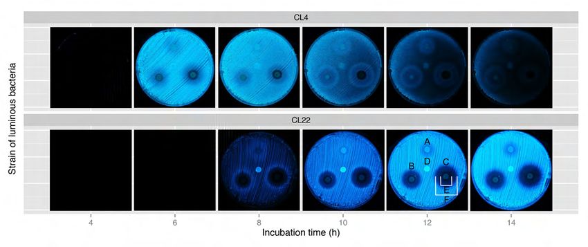
Fig. 3. Photographical record of the effect of 0.1 (A), 0.5 (B), and 1 (C) mg mL-1
cadmium on the variation of luminescence intensity for CL4 and CL22 luminous
strains at different incubation times. D = control. Strain CL4 exhibits a degradation of
luminescence around disks impregnated with Cd; strain CL22 shows defined growth
inhibition zones (E) and luminescence inhibition zones (F) for 0.5 and 1 mg mL-1 Cd
Fig. 3. Registro fotográfico del efecto de 0.1 (A), 0.5 (B) y 1 (C) mg mL-1 de cadmio en la
variación de la intensidad de la luminiscencia para las cepas luminiscentes CL4 y CL22
a diferentes tiempos de incubación. D = control. La cepa CL4 muestra degradación de
la luminiscencia alrededor de los discos impregnados con Cd, y la cepa CL22 muestra
zonas de inhibición del crecimiento (E) y zonas de inhibición de la luminiscencia (F)
definidas para concentraciones de Cd de 0.5 y 1 mg mL-1
Fig. 4. Luminescence reduction (●)
by different concentrations (mg mL-
1
) of cadmium, copper, lead, and zinc
ions on bacterial strains with reduced
luminescence intensity isolated from
the golfo de Nicoya, Costa Rica. Data
for chromium ions not shown due to the
absence of growth and luminescence
inhibition zones
Fig. 4. Reducción de luminiscencia (●)
por concentración (mg mL-1) de cadmio,
cobre, plomo y zinc de cepas bacterianas
con intensidad de luminiscencia reducida
aislada del golfo de Nicoya, Costa Rica. No
se muestran los ensayos con cromo debido
a que no se observaron halos de inhibición
de luminiscencia ni de crecimiento
32 Rev. Mar. Cost. Vol. 12 (1): 27-38, enero-junio 2021.
ISSN: 1659-455X • e-ISSN: 1659-407X
DOI: http://dx.doi.org/10.15359/revmar.13-1.2Effect of metal concentration on growth and luminescence of luminous bacteria
strains isolated from golfo de Nicoya, Costa Rica
Fig. 5. Diameter (average of duplicates) of the growth and luminescence inhibition
zone (mm) for different concentration (mg mL-1) of cadmium, copper, lead, and zinc
ions of bacterial strains with intense luminescence isolated from the golfo de Nicoya,
Costa Rica. The diameters of the growth inhibition zones (mm) for Cu and Pb are
the same. Strains with dim luminescent (CL6, CL11, CL12, and CL19) did no exhibit
growth or luminescent inhibition zones
Fig. 5. Diámetro (promedio de las réplicas) de las zonas de inhibición de crecimiento y
de luminiscencia (mm) por concentración (mg mL-1) de cadmio, cobre, plomo y zinc de
cepas bacterianas con luminiscencia intensa aisladas del golfo de Nicoya, Costa Rica.
El diámetro de las zonas de inhibición del crecimiento (mm) para Cu y Pb es el mismo.
Las cepas con luminiscencia tenue (CL6, CL11, CL12 and CL19) no mostraron zonas
de inhibición de crecimiento o luminiscencia
showed luminescence inhibition zones the luminescence inhibition zones vary
for 0.5 and 1 mg mL-1 Cu, while CL13 in the order of Cd > Zn > Cu (Fig. 5).
strain only for 1 mg mL-1 Cu (Fig. 5). The mean difference between
Strains affected by two or more inhibition zones of growth or lumines-
concentrations of the same metal cence by metal and metal concentra-
showed a directly proportional rela- tion is shown in Table 1. According to
tionship between the diameter of the the statistical tests, growth inhibition
growth and luminescence inhibition zones by Cd and luminesce inhibition
zones and the corresponding metal zones by Zn and Cd were significant-
concentration. CL9, CL10, CL13, and ly higher at 1 mg mL-1 than 0.5 mg
CL17 strains showed that diameters of mL-1 for each metal. Luminescence
Rev. Mar. Cost. Vol. 12 (1): 27-38, enero-junio 2021. 33
ISSN: 1659-455X • e-ISSN: 1659-407X
DOI: http://dx.doi.org/10.15359/revmar.13-1.2Luis Vega-Corrales & Carolina Marín-Vindas
Table 1. Pair-wise comparison between the diameter of the growth inhibition
zone or luminescence inhibition zone caused by different metals at different
concentration (mg mL-1) levels upon luminous bacteria strains isolated from
golfo de Nicoya, Costa Rica
Cuadro 1. Comparación por pares entre el diámetro de las zonas de inhibición
del crecimiento o zonas de inhibición de luminiscencia causada por distintos
metales a diferentes concentraciones (mg mL-1) sobre las cepas de bacterias
luminiscentes aisladas del golfo de Nicoya, Costa Rica
Comparison Mean difference Significance
Inhibition Metal mg mL-1 mm (± 95% CI) (P < 0.05)**
Growth:Growth Cd:Cd 1:0.5 1.86 (1.49, 2.23) 1
Cd:Zn 1:1 1.75 (0.85, 2.65) 0
Luminescence:Luminescence Cd:Cd 1:0.5 4.38 (3.47, 5.30) 1
Zn:Zn 1:0.5 4.88 (3.87, 5.90) 1
Cd:Zn 1:1 7.88 (6.75, 9.02) 1
Cd:Zn 0.5:0.5 8.38 (7.46, 9.31) 1
Luminescence:Growth Cd:Cd 1:1 15.15 (14.22, 16.09) 1
Cd:Cd 0.5:0.5 12.86 (11.43, 14.30) 1
Zn:Zn 1:1 8.75 (7.41, 10.09) 1
Notes: *CI = confidence interval; **1 and 0 denote significant and insignificant, respectively, at the 0.05 significance
level.
inhibition zones for 1 and 0.5 mg mL-1 DISCUSSION
Cd and 1 mg mL-1 Zn were statistical-
ly significantly higher than the cor- This study’s culture medium
responding growth inhibition zones. promoted the growth and lumines-
These strains showed higher lumines- cence of the tested strains; therefore,
cent inhibition zones (P < 0.05) by it was appropriate for the assays. Most
Cd than Zn at 0.5 and 1 mg mL-1. The research on luminous bacteria use the
difference for mean pair-wise compar- SWC (Seawater Complete) culture
ison for growth inhibition zones by Cd medium (Bagordo et al. 2012; Martini
and Zn at 1 mg mL-1 did not show a et al. 2013; Jabalameli et al. 2015) or
statistical significance. culture media with a similar composi-
tion (Efremenko et al. 2014; Drozdov
et al. 2015; Urbanczyk et al. 2015). In
this study, strains grew over (4 h) in
34 Rev. Mar. Cost. Vol. 12 (1): 27-38, enero-junio 2021.
ISSN: 1659-455X • e-ISSN: 1659-407X
DOI: http://dx.doi.org/10.15359/revmar.13-1.2Effect of metal concentration on growth and luminescence of luminous bacteria
strains isolated from golfo de Nicoya, Costa Rica
the Marine Agar (MA) and lumines- without requiring specialized equip-
cence occurred 2 or 4 h later. ment. It could also be considered a
The assessment luminescence useful technique for the screening of
intensity dynamics of the assayed isolated strains and to define the opti-
strains confirms that bacterial lumi- mal incubation period of each strain in
nescence is an indicator of the activity view of evaluating the effects of toxic
of intracellular metabolic processes. substances in solid-phase biolumines-
The maximum luminescence intensi- cence assays.
ty in a bacterial culture occurs during The luminescence inhibition rate
the exponential growth phase. Then of some bacterial strains has been used
luminescence dynamics is affected in toxicological and ecotoxicological
because of the reduction in metabol- assessments (Menz et al. 2013; Ma et
ic processes kinetics (Drozdov et al. al. 2014). Our results show that some
2015). Efremenko et al. (2014) report metals at different concentration levels
that the luminescence intensity in free affect the growth and luminescence of
cells of Photobacterium phosphoreum luminous bacterial strains to different
decreases with time. degrees. Also, we observed that the
Differences between assessed luminescence of some bacteria was
strains in terms of luminescence du- inhibited by a metal ion concentration
ration and intensity coincide with the lower than the minimum growth inhib-
findings reported by Efremenko et al. itory concentration.
(2014). The authors indicate that du- The sensitivity of each strain to
ration and intensity significantly dif- different concentrations of metal ions
fer between species and strains of the is determined by its bioluminescent
Photobacterium genus. In this way, the phenotype and for practical bioassay
amount of light produced by luminous applications the higher intensity lumi-
bacteria, even belonging to the same nescent strains with luminescence in-
genus, makes these microorganisms hibition zones afford greater potential
show different bioluminescent phe- as pollution indicators. For example,
notypes even though bioluminescence strain CL17 that is potentially sen-
is regulated by a similar basic genetic sitive to cadmium at concentrations
system (Jabalameli et al. 2015). lower than 0.1 mg mL-1.
Although the classification of The intensity of the lumines-
the bioluminescence intensity used in cence (in free and immobilized cells)
this study is not a quantitative meth- decreases with increasing metal con-
od, it is adequate for the determina- centration. Kumar et al. (2015) report-
tion of bacterial luminescence dynam- ed the effect of copper and zinc at 1 mg
ics (mainly in solid culture media) mL-1 on luminescence sensitivity of 20
Rev. Mar. Cost. Vol. 12 (1): 27-38, enero-junio 2021. 35
ISSN: 1659-455X • e-ISSN: 1659-407X
DOI: http://dx.doi.org/10.15359/revmar.13-1.2Luis Vega-Corrales & Carolina Marín-Vindas
strains isolated from the Bay of Bengal For sensible strains, a range of con-
While Efremenko et al. (2014) showed centrations of the metal ion lower than
that luminescence intensity in cells the growth inhibition concentration
of P. phosphoreum, immobilized in a affected the expression of lumines-
polyvinyl alcohol cryogel, decreases cence. Strains showing intense lumi-
with increasing metal ion concentra- nescence and defined luminescence
tion, and further concluded that met- inhibition zones were assigned great-
al toxicity for these immobilized cells er application potential as bioindica-
varies in the order Cu+2 > Co+2 > Hg+2 tors for environmental toxicity mon-
> Zn+2. Our study reveals the order Cd itoring. However, more studies are
> Zn > Cu, as well as the absence of required to determine the minimum
luminescence inhibition by Cr+3 and growth and luminescence inhibition
Pb+2 at all the tested concentrations. concentrations for the assayed metals
Many application tests employ- ions and other substances potentially
ing luminous bacteria are known, and toxic to coastal marine ecosystems in
their distinctive features depend on the Costa Rica.
specific characteristics of the bacteri-
al strain used. The isolation of native ACKNOWLEDGEMENTS
luminous bacteria allows bioprospect-
ing for strains useful for monitoring This research was funded by the
environmental toxicity. Moreover, the Ley de Pesca y Acuicultura (Fishery
isolation of strains of microorganisms and Aquaculture Act) from the Gov-
resistant to metals is significant. The ernment of Costa Rica as part of the
expectation is that microorganisms project entitled “Distribución y abun-
designated for bioremediation are tol- dancia de bacterias luminiscentes y su
erant of metals (Kumar et al. 2015). potencial como indicadoras de con-
taminación en el Golfo de Nicoya,
CONCLUSION Costa Rica” (Distribution and abun-
dance of luminous bacteria and their
Differences in luminescence in- potential as pollution indicators in the
tensity dynamics between strains, the Golfo de Nicoya, Costa Rica), UNA-
definition of luminescence inhibition SIA-0338-13. Special thanks to Oscar
zones, and the determination of growth Pacheco Prieto for taking the photo-
inhibition zone diameters and lumi- graphs and two anonymous reviewers
nescence inhibition zone diameters for improving the manuscript.
for different metals ions at different
concentration levels were determined.
36 Rev. Mar. Cost. Vol. 12 (1): 27-38, enero-junio 2021.
ISSN: 1659-455X • e-ISSN: 1659-407X
DOI: http://dx.doi.org/10.15359/revmar.13-1.2Effect of metal concentration on growth and luminescence of luminous bacteria
strains isolated from golfo de Nicoya, Costa Rica
REFERENCES Stackebrandt & F. Thompson (Eds),
The Prokaryotes. (4th, pp. 495-
528). Springer, Germany. https://doi.
Bagordo, F., Serio, F., Lugoli, F., Idolo, A.,
org/10.1007/978-3-642-30141-4_75
Gabutti, G. & De Donno, A. (2012).
Efremenko, E. N., Senko, O. V., Aleskerova,
Phenotypic characterization of cultur-
L. E., Alenina, K. A., Mazhul, M. M.
able marine luminous bacteria isolat-
& Ismailov, A. D. (2014). Biosensors
ed from coastal waters of the southern
based in luminous bacteria Photobacte-
Adriatic Sea (Otranto, Italy). Cienc.
rium phosphoreum imobilized in poly-
Mar., 38(4), 599-608. https://doi.
vinil alcohol cryogel for the monitor-
org/10.7773/cm.v38i4.2119
ing of ecotoxicants. Applied Biochem.
Bolelli, L., Ferri, E. N.& Girotti, S. (2016). The
Microbiol., 50(5), 477-482. https://doi.
management and exploitation of natu-
org/10.1134/S0003683814050032
rally light-emitting bacteria as a flexi-
Jabalameli, L., Razavi, M. R., Hosseinkhani,
ble analytical tool: A tutorial. Analytica
S. & Akhavan Sepahi, A. (2015). Isola-
Chimica Acta., 934, 22-35. https://doi.
tion, identification and characterization
org/10.1016/j.aca.2016.05.038
of new luminous bacteria from Chah
Burga, K. F., Charlatchka, R., Sahli, L. &
Bahar Port, southern marine habitat
Férard, J. (2012). New methodolog-
of Iran. Iranian J. Fish. Sci., 14(3),
ical improvements in the Microtox®
555-566.
solid phase assay. Chemosphere., 86,
Kumar, A. R., Jayaprakashvel, M., FeuK-La-
105-110. https://doi.org/10.1016/j.
gerstedt, E. & Hussain, A. J. (2015).
chemosphere.2011.08.042
Factors affecting bioluminescence in
Camanzi, L., Bolelli, M., Girotti, S. & Mat-
free living Photobacterium spp. Isolat-
teuzzi, D. (2011). Optimal conditions
ed from Bay of Bengal, India. J. Mar.
for stability of photoemission and
Biosci., 1(1), 33-49.
freeze drying of two luminescent bac-
Ma, X. Y., Wang, X. C., Hao, H., Guo, W.,
teria use in a biosensor. Environ. Toxi-
Wu, M. N. & Wang, N. (2014). Bio-
col. Chem., 30(4), 801-805. https://doi.
assay based luminescent bacteria:
org/10.1002/etc.452
interferences, improvements, and ap-
Diepens, N. J., Pfenning, S., Van den Brink,
plications. Sci. Total Environ., 468-
P. J., Gunnarsson, J. S., Ruepert, C. &
469, 1-11. https://doi.org/10.1016/j.
Castillo, L. E. (2014). Effect of pes-
scitotenv.2013.08.028
ticides used in banana and pineapple
Martini, S., Al Ali, B., Garel, M., Nerini, D.,
plantations on aquatic ecosystems in
Grossi, V., Pacton, M., & Tamburini, C.
Costa Rica. J. Environ. Biol., 35(1),
(2013). Effects of hydrostatic pressure
73-84.
on growth and luminescence of a mod-
Drozdov, A. V., Gromozova, E. N. & Gretsky,
erately-piezophilic luminous bacteria
I. A. (2015). An analysis of the biolu-
Photobacterium phosphoreum ANT-
minescence intensity dynamics of the
2200. PLoS One, 8(6), e66580. https://
luminous bacteria. Photobact phosph.
doi.org/10.1371/journal.pone.0066580
Biophys., 60(2), 251-255. https://doi.
Menz, J., Schneider, M. & Kümmerer, K.
org/10.1134/S0006350915020050
(2013). Toxicity testing with lu-
Dunlap, P. V. & Urbanczyk, H. (2013).
minescent bacteria- Characteriza-
Luminous Bacteria. In E., Rosen-
tion of an automated method for the
berg E.F., DeLong S., Lory E.
Rev. Mar. Cost. Vol. 12 (1): 27-38, enero-junio 2021. 37
ISSN: 1659-455X • e-ISSN: 1659-407X
DOI: http://dx.doi.org/10.15359/revmar.13-1.2Luis Vega-Corrales & Carolina Marín-Vindas
combined assessment of acute and Urbanczyk, Y., Ogura, Y., Hayashi, T. &
chronic effects. Chemosphere., 93(6), Urbanczyk, H. (2015). Description
990-996. https://doi.org/10.1016/j. of a novel marine bacterium, Vib-
chemosphere.2013.05.067 rio hyugaensis sp. nov., based on ge-
R Core Team. (2018). R: a language and en- nomic and phenotypic characteriza-
vironment for statistical computing. tion. System. Applied Microbiol., 38,
Austria: R Foundation for Statistical 300-304. https://doi.org/10.1016/j.
Computing. https://www.R-project. syapm.2015.04.001
org/ Yu, X., Zuo, J., Tang, X., Li, R., Li, Z. &
Ranjan, R., Rastogi, N. K. & Thakur, M. S. Zhang, F. (2014). Toxicity evaluation
(2012). Development of immobilized of pharmaceutical wastewaters using
biophotonic beads consisting of Photo- the alga Scenedesmus obliquus and
bacterium leiognathi for the detection the bacterium Vibrio fischeri. J. Haz-
of heavy metals and pesticide. J. Haz- arduos Mat., 266, 68-74. https://doi.
ardous Mat., 225-226, 114-123. https:// org/10.1016/j.jhazmat.2013.12.012
doi.org/10.1016/j.jhazmat.2012.04.076
Shanware, A., Thakre, N. & Pande, S. (2013).
Isolation and characterization of nov-
el marine luminescent bacteria from
Diu beach, India. J. Pharm. Res., 7(6),
529-533. https://doi.org/10.1016/j.
jopr.2013.05.019
38 Rev. Mar. Cost. Vol. 12 (1): 27-38, enero-junio 2021.
ISSN: 1659-455X • e-ISSN: 1659-407X
DOI: http://dx.doi.org/10.15359/revmar.13-1.2You can also read