Digital Dermatopathology and Its Application to Mohs Micrographic Surgery
←
→
Page content transcription
If your browser does not render page correctly, please read the page content below
Perspective
Yonsei Med J 2022 Jan;63 Suppl:S112-114
https://doi.org/10.3349/ymj.2022.63.S112 pISSN: 0513-5796 · eISSN: 1976-2437
Digital Dermatopathology and Its Application
to Mohs Micrographic Surgery
Yeongjoo Oh1*, Hye Min Kim2*, Soon Won Hong2, Eunah Shin2, Jihee Kim1, and Yoon Jung Choi2
Departments of 1Dermatology and 2Pathology, Yongin Severance Hospital, Yonsei University College of Medicine, Yongin, Korea.
Digital pathology is being gradually adopted in hospitals due to technological advances. We propose that digital pathology can be
used in Mohs micrographic surgery (Mohs surgery) to precisely check residual tumor cells in frozen tumor margin tissues. This
would aid surgeons and pathologists in accurately recording tumor margins and give patients the benefit of shorter operation time.
Key Words: Mohs micrographic surgery, dermatology, clinical pathology, margin of excision, neoplasm
Digital pathology, which includes automated digital scanning gy during residency and in most training institutions; therefore,
of tissue slides, file storage, and displaying of files using high- additional glass slides are utilized in the dermatology depart-
resolution monitor, has existed for several decades. However, ment. This creates conflict between the pathology and derma-
it has only been recently introduced and adopted in the clini- tology departments in several institutes due to the amount of
cal field, as a result of technological development with faster resources used for sectioning and supplying additional slides.
scanning time and higher graphic resolution.1,2 While using digital pathology, however, there would be no wast-
In Yongin Severance hospital, the first hospital with a fully ing of resources for sharing the slides, as the data would be digi-
digitalized pathology system, all pathology slides are scanned tally stored and can be shared via a computer and visualized on
and shared with clinicians, and used to actively communicate a high-resolution monitor.
especially with dermatologists. Mohs micrographic surgery, a surgical procedure for skin
During clinical practice, dermatologists perform numerous cancer, which is performed mainly by dermatologists, can assess
skin biopsies as well as surgical procedures. Due to the pleth- the complete tumor margin by examining the frozen section re-
ora of dermatological conditions and subtle differences based sults of the outermost shell of margin tissue during surgery.5
on the natural course of disease, reviewing the biopsy speci- When the frozen section result is found to be positive for tumor
men is essential for clinical and academic training of derma- cells, surgeons visit the frozen section reading room to check
tologists.3,4 Accordingly, dermatologists study dermatopatholo- precisely where the tumor cells remain among the margin areas
using conventional pathology systems. With a digital pathology
Received: September 8, 2021 Revised: November 4 2021 system, frozen margin tissues can also be scanned and shared.
Accepted: November 12, 2021 The pathologists can annotate the exact area where tumor cells
Co-corresponding authors: Jihee Kim, MD, PhD, Department of Dermatology,
Yongin Severance Hospital, Cutaneous Biology Research Institute, Yonsei University exist, and surgeons can check the shared digital slides in the
College of Medicine, 363 Dongbaekjukjeon-daero, Giheung-gu, Yongin 16995, Korea. operation room (Fig. 1).
Tel: 82-2-2228-2080, Fax: 82-2-393-9157, E-mail: MYGIRLJIHEE@yuhs.ac and Compared to a conventional system, digital pathology system
Yoon Jung Choi, MD, PhD, Department of Pathology, Yongin Severance Hospital,
Yonsei University College of Medicine, 363 Dongbaekjukjeon-daero, Giheung-gu, takes one more step of scanning the slide, which takes about 10–
Yongin 16995, Korea. 30 minutes; therefore, some time delay may occur in reporting
Tel: 82-31-5189-8447, Fax: 82-31-5189-8247, E-mail: CHRIS316@yuhs.ac pathology results (Fig. 2). However, the digital pathology sys-
*Yeongjoo Oh and Hye Min Kim contributed equally to this work. tem still saves time for both surgeons and pathologists, as they
•The authors have no potential conflicts of interest to disclose. do not have to move from the operating room to the pathology
© Copyright: Yonsei University College of Medicine 2022 room.
This is an Open Access article distributed under the terms of the Creative Com-
One of the advantages of a digital pathology system is its re-
mons Attribution Non-Commercial License (https://creativecommons.org/licenses/
by-nc/4.0) which permits unrestricted non-commercial use, distribution, and repro- producibility. In a conventional system, pathologists directly de-
duction in any medium, provided the original work is properly cited. scribe the microscopic findings of frozen section of the tumor
S112 www.eymj.org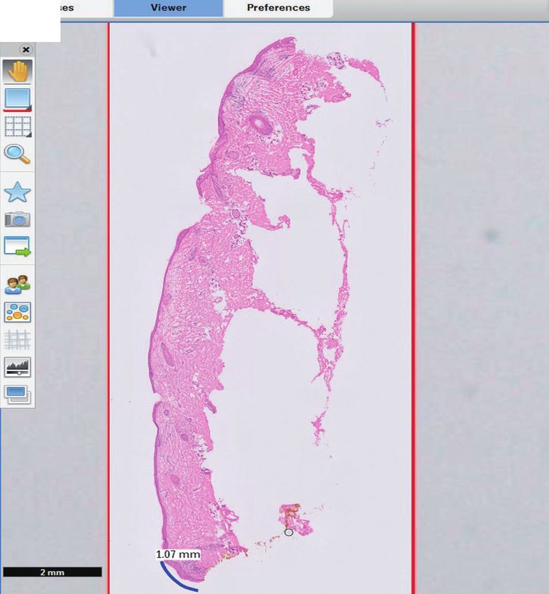
Yeongjoo Oh, et al.
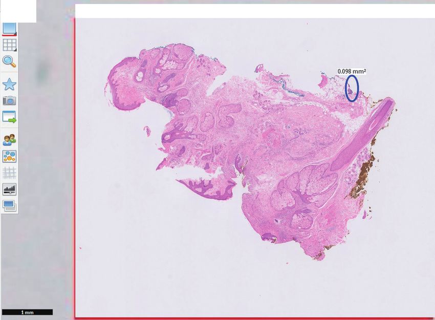
A B
Fig. 1. Shared digital pathology of frozen margin during Mohs surgery. (A) Blue mark at the lateral tumor positive area. (B) Blue mark at the base focal tu-
mor cell cluster.
Tissue sample Clinician Report Tissue arrival Report time
time
Fig. 2. Frozen pathology report during Mohs surgery with digital pathology. The upper four frozen tissues are tissues from the first Mohs stage. It took
about 30 minutes for the tissues to arrive at the pathology department to be made into slides and scanned, and for the pathologist to report the results
with scanned digital pathology. The last tissue is from the second Mohs stage, and it took only 10 minutes to arrive and be reported.
margin to the surgeon, which limits the exact pictorial descrip- Despite various advantages, there are still some technological
tion of positive margins. In a digital system, pathologists can challenges that must be overcome.2,3 Digitally scanned pathol-
freely mark or attach notes on the slide file. Even if the clinician ogy specimens require much less physical storage area. How-
is not as well-trained as the pathologist, they can quickly check ever, each file could be of a massive size, requiring huge storage
the cancer cells and the area by checking the marked area only.6 and server systems. Despite rapid advances in the scanning
Therefore, using a digital pathology system is convenient and technology, thick specimens may present blurred focus due to
precise when retrospectively reviewing the tumor margin dur- resolution differences within the tissue. Additionally, when the
ing and after the surgery. slide is sectioned with some irregularity, it may be difficult to
The advantages of digital pathology described above are also focus during scanning, and the area of interest could become
applicable to other procedures and surgeries, which require blurred on the final scanned file. Nevertheless, these techno-
confirmation using frozen tissue results. In surgeries other logical limitations will undoubtedly be resolved within few
than Mohs surgery, surgeons do not directly check the patholo- years. After overcoming these few limitations, additional bene-
gy slides, but only check whether the result for tumor cell is fits are expected in the era of digital pathology. Currently, addi-
positive. However, with digital pathology, surgeons in the oper- tional pathology slides must be sectioned when transferring the
ating room can easily and accurately check tumor cell charac- patient to other hospitals. This sectioning of additional tissues
teristics and locations via a computer screen. requires several human and material resources and a greater
https://doi.org/10.3349/ymj.2022.63.S112 S113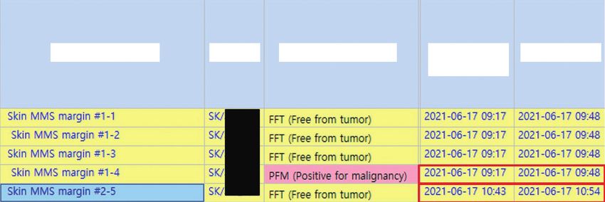
Digital Dermatopathology for Mohs Surgery

number of tissue samples. If most hospitals are equipped with a           Visualization: Yeongjoo Oh and Hye Min Kim. Writing—original
digital pathology system and such file exchange is legally rec-           draft: Yeongjoo Oh, Hye Min Kim, and Jihee Kim. Writing—review &
                                                                          editing: Yeongjoo Oh, Hye Min Kim, Jihee Kim, and Yoon Jung Choi.
ognized, hospitals can share patient pathology slides through
                                                                          Approval of final manuscript: all authors.
simple file transfer without using additional resources.7
   Digital pathology aids surgeons and pathologists in accu-
rately recording tumor margins, which indirectly gives patients           ORCID iDs
the benefit of reduced surgery time in Mohs surgery. With the
                                                                          Yeongjoo Oh          https://orcid.org/0000-0003-4973-9335
reproducibility of digital pathology, the patient can get an ex-          Hye Min Kim          https://orcid.org/0000-0002-2899-9480
planation of the frozen slides even after the surgery. If the pa-         Soon Won Hong        https://orcid.org/0000-0002-0324-2414
tient is referred to another hospital after the surgery, the patient      Eunah Shin           https://orcid.org/0000-0001-5961-3563
can digitally carry pathology test results, including frozen slides,      Jihee Kim            https://orcid.org/0000-0002-0047-5941
                                                                          Yoon Jung Choi       https://orcid.org/0000-0002-5701-8864
thereby minimizing physical and economic costs and efforts.
   In addition, by applying artificial intelligence (AI) to the
digital pathology system, faster and more accurate diagnosis              REFERENCES
may be possible. AI may not be able to confirm a diagnosis or
                                                                           1. Evans AJ, Salama ME, Henricks WH, Pantanowitz L. Implementa-
describe the disease characteristics. However, it may aid pa-                 tion of whole slide imaging for clinical purposes: issues to consider
thologists to make a diagnosis and provide simple information                 from the perspective of early adopters. Arch Pathol Lab Med 2017;
to clinicians before pathologic confirmation.2,8 Moreover, AI                 141:944-59.
can quickly identify tumor cells of frozen margins during sur-             2. Nam S, Chong Y, Jung CK, Kwak TY, Lee JY, Park J, et al. Introduc-
gery and shorten the operation time.8                                         tion to digital pathology and computer-aided pathology. J Pathol
                                                                              Transl Med 2020;54:125-34.
   Despite the technical and legal limitations at every hospital,          3. Blum AE, Murphy GF, Lee JJ. Digital dermatopathology: the time
digital pathology is expected to advance rapidly in a few years.              is now. J Cutan Pathol 2021;48:469-71.
Therefore, we propose that digital pathology be used in Mohs               4. Glines KR, Haidari W, Ramani L, Akkurt ZM, Feldman SR. Digital
surgery, as it would be advantageous for both surgeons and                    future of dermatology. Dermatol Online J 2020;26:2.
pathologists, and give the benefit of shortened operation time             5. Mariwalla K, Aasi SZ, Glusac EJ, Leffell DJ. Mohs micrographic sur-
                                                                              gery histopathology concordance. J Am Acad Dermatol 2009;60:
to the patient.
                                                                              94-8.
                                                                           6. Marsch AF, Espiritu B, Groth J, Hutchens KA. The effectiveness of
AUTHOR CONTRIBUTIONS                                                          annotated (vs. non-annotated) digital pathology slides as a teach-
                                                                              ing tool during dermatology and pathology residencies 2014;41:
Conceptualization: Yeongjoo Oh, Hye Min Kim, Jihee Kim, and Yoon              513-8.
Jung Choi. Data curation: all authors. Formal analysis: Yeongjoo Oh,       7. García-Rojo M. International clinical guidelines for the adoption
Hye Min Kim, Jihee Kim, and Yoon Jung Choi. Investigation: Yeong-             of digital pathology: a review of technical aspects. Pathobiology
joo Oh, Hye Min Kim, Jihee Kim, and Yoon Jung Choi. Methodology:              2016;83:99-109.
Yeongjoo Oh, Hye Min Kim, Jihee Kim, and Yoon Jung Choi. Project           8. Cui M, Zhang DY. Artificial intelligence and computational pathol-
administration: all authors. Resources: all authors. Supervision: Jihee       ogy. Lab Invest 2021;101:412-22.
Kim and Yoon Jung Choi. Validation: Yeongjoo Oh and Hye Min Kim.

S114                                                                                                         https://doi.org/10.3349/ymj.2022.63.S112
You can also read


