Description of a New Scaled Species of Ptychostomella (Gastrotricha: Macrodasyida) from the Brazilian Coast and a Cladistics Analysis of the Genus
←
→
Page content transcription
If your browser does not render page correctly, please read the page content below
Article
Description of a New Scaled Species of Ptychostomella
(Gastrotricha: Macrodasyida) from the Brazilian Coast and a
Cladistics Analysis of the Genus
Thiago Q. Araújo *              and André R. S. Garraffoni

                                          Department of Animal Biology, Institute of Biology, University of Campinas, Campinas 13083-970, SP, Brazil;
                                          arsg@unicamp.br
                                          * Correspondence: araujotq@gmail.com

                                          Abstract: A new species of marine Gastrotricha from the north coast of São Paulo state, Brazil,
                                          is described. Adults of Ptychostomella sebastiana sp. nov. are unique in that they possess a pair
                                          of dorsolateral “rod-like” cephalic sensory organs and subrectangular scales covering the lateral
                                          body surfaces. A cladistic analysis was performed to investigate the internal relationship of the
                                          representatives of the taxa based on morphological data. Our analysis supported the monophyly of
                                          the taxon Ptychostomella, but its internal phylogenetic relationships are not well established due to
                                          the low phylogenetic signal of morphological characters used in the present study.

                                          Keywords: Gastrotricha; morphology; phylogeny; macrodasyida


                                   1. Introduction
Citation: Araújo, T.Q.; Garraffoni,             The family Thaumastodermatidae is the most specious and diverse group of Macro-
A.R.S. Description of a New Scaled        dasyida and comprises eight genera and up to 140 described species [1,2]. Specimens of
Species of Ptychostomella                 this taxon are usually recognized by the diversity of cuticular ornamentations of the dorsal
(Gastrotricha: Macrodasyida) from         and ventral surfaces (e.g., scales, spines, spined scales, ancres).
the Brazilian Coast and a Cladistics            Belonging to this taxon, specimens of the genus Ptychostomella Remane, 1926 were
Analysis of the Genus. Taxonomy 2021,     originally recognized by their smooth cuticle, large oral hood, and lateral club-shape
1, 278–289. https://doi.org/10.3390/      or knob-like tentacle. Some authors have hypothesized that the lack of ornamentation
taxonomy1040022                           is a secondary reduction [1,3,4]. This panorama changed when the first specimen of
                                          Ptychostomella with cuticular ornamentation was described, P. lepidota Clausen, 2000. Since
Received: 4 August 2021                   then, another four ornamented species have been described: P. orientalis Lee & Chang, 2003,
Accepted: 1 October 2021
                                          P. brachycephala (Levi, 1954), P. papillata Lee and Chang 2003, and P. lamelliphora Todaro 2013.
Published: 8 October 2021
                                                In this study, we describe a new species of Ptychostomella with cuticular ornamentation
                                          collected on the Brazilian Coast and present the first phylogenetic hypothesis using external
Publisher’s Note: MDPI stays neutral
                                          morphological data to investigate the internal relationship of the taxon Ptychostomella.
with regard to jurisdictional claims in
published maps and institutional affil-   2. Materials and Methods
iations.
                                          2.1. Sampling
                                                Samples were obtained (22/02/2018) at São Sebastião Island (where the municipality
                                          of Ilhabela is housed) at Fome Beach (23◦ 440 2700 S; 45◦ 160 0100 W). The top 10–15 cm of sedi-
                                          ment at a depth of 8–10 m (total of 8 L of sediments) was taken by scuba diving. Sediments
Copyright: © 2021 by the authors.
                                          were placed in plastic buckets and gastrotrichs were extracted at the Universidade Estadual
Licensee MDPI, Basel, Switzerland.
                                          de Campinas (UNICAMP). The specimens were extracted from the sediment with isotonic
This article is an open access article
                                          7% MgCl2 using the anesthetization-decantation technique [5] in 1 L plastic Erlenmeyer
distributed under the terms and
                                          flasks. Animals were decanted onto 35 µm sieves and washed with ambient seawater into
conditions of the Creative Commons
Attribution (CC BY) license (https://
                                          Petri dishes. Gastrotrichs were observed alive with a Leica EZ4 stereomicroscope and then
creativecommons.org/licenses/by/
                                          transferred to glass slides for specific identification using a Zeiss Axio Imager M2 light
4.0/).                                    microscope equipped with differential interference contrast optics (DIC) connected to a

Taxonomy 2021, 1, 278–289. https://doi.org/10.3390/taxonomy1040022                                         https://www.mdpi.com/journal/taxonomy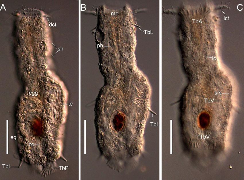
Taxonomy 2021, 1                                                                                                                    279

                                      camera. Photomicrographs and measurements were taken using the software ZEN-blue
                                      edition. The position of anatomical characters is provided in percent units (U) of total body
                                      length (anterior tip of body (excluding cilia) = U00, posterior tip of body = U100) [6].

                                      2.2. Phylogenetic Analysis
                                      2.2.1. Ingroup Taxa
                                           All 13 formally described species within the genus Ptychostomella were used in the
                                      present analysis (Table 1).

                                      2.2.2. Outgroup Taxa
                                           The species Tetranchyroderma esarabdophorum Tongiorgi & Balsamo, 1984 and T. quadri-
                                      tentaculatum Todaro, Balsamo & Tongiorgi, 1992 were used as outgroups. Previous phy-
                                      logenetic reconstructions placed Tetranchyroderma species as a sister group to the genus
                                      Ptychostomella [1,7].

                                      2.2.3. Character Coding and Analysis
                                           We coded 12 morphological characters (Table 2). Most characters were coded as
                                      multistate or binary, but some were coded as contingent. Contingent coding [8], also called
                                      C coding [9] or conditional coding [10], first codes a binary character in one column of
                                      the matrix, and an additional code for the presence and absence of the feature in the next
                                      column. Non-applicable states were coded as ‘–’ and unknown stated as ‘?’ (Table 1).

     Table 1. Matrix of morphological characters considered in the phylogenetic analysis of Ptychostomella. “?”: unknown data;
     “-”: inapplicable character state.

                                                                         0   1   2   3   4   5   6   7   8    9   10   References
 Tetranchyroderma esarabdophorum Tongiorgi & Balsamo, 1984               2   0   1   1   0   1   1   3   1    0   0    [11]
 Tetranchyroderma quadritentaculatum Todaro, Balsamo & Tongiorgi, 1992   2   0   1   1   0   1   1   3   1    0   0    [12]
 Ptychostomella bergensis Clausen, 1996                                  2   0   0   1   1   0   1   1   0    0   0    [13]
 Ptychostomella brachycephala (Levi, 1954)                               1   0   0   1   0   0   1   1   0    0   0    [14]
 Ptychostomella helana Roszczak, 1939                                    1   0   0   0   1   0   0   -   0    0   0    [15]
 Ptychostomella jejuensis Lee, Hwang & Chang, 2009                       2   1   0   1   0   0   1   3   0    0   0    [16]
 Ptychostomella higginsi Clausen, 2004                                   2   1   0   1   1   0   1   3   0    0   0    [14]
 Ptychostomella lamelliphora Todaro, 2013                                0   1   0   1   0   0   1   2   0    0   1    [3]
 Ptychostomella lepidota Clausen, 2000                                   2   0   0   1   1   0   1   1   0    1   0    [17]
 Ptychostomella mediterranea Remane, 1927                                2   1   0   1   0   0   0   -   0    0   0    [18]
 Ptychostomella ommatophora Remane, 1927                                 2   0   0   ?   ?   0   0   -   0    0   0    [18]
 Ptychostomella orientalis Lee & Chang, 2003                             2   1   0   1   0   0   1   1   0    1   0    [19]
 Ptychostomella papillata Lee & Chang, 2003                              2   0   0   1   0   0   1   3   0    0   0    [19]
 Ptychostomella pectinata Remane, 1926                                   2   0   0   1   0   0   0   -   0    0   0    [20]
 Ptychostomella tyrrhenica Hummon, Todaro & Tongiorgi, 1993              2   1   0   1   0   0   1   3   0    0   0    [21]
                                                                                                                       [present
 Ptychostomella sebastiana sp. nov.                                      1   0   1   1   0   0   1   1   0    0   1
                                                                                                                       study]

                                           Parsimony analysis was carried out using the computer program TNT under equal and
                                      implied weights [22,23] to perform a ‘sensitivity analysis’ of the data in distinct situations.
                                      We used an exhaustive search for equal weights. For implied weight, the values of K
                                      were assigned to a ‘not perfectly hierarchical’ character fit of 85%, 86.11%, 87.22%, 88.33%,
                                      89.44%, 90.55%, 91.66%, 92.77%, 93.88%, and 95% (script commands aaa 3 10 85 95 7). The
                                      authors in [24] proposed regular intervals fit/distortion values, which can be obtained
                                      under different K-values (Appendix S3 of [24]–file aaa.run). The software WINCLADA
                                      1.00.08 [25] was used for character optimization and tree editing.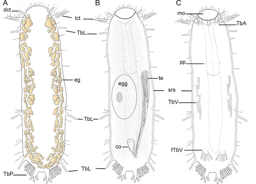
Taxonomy 2021, 1                                                                                        280

                   Table 2. Morphological characters of Ptychostomella coded in the present analysis.

                                            0. Cephalic sensorial organs–Lateral tentacle type
                                                                • 0 Short
                                                                • 1 Long
                                                               • 2 absent

                                              1. Cephalic sensorial organs–Knob-like organ
                                                               • 0 absent
                                                              • 1 present

                                           2. Cephalic sensorial organs–Dorsolateral tentacle
                                                              • 0 absent
                                                              • 1 present

                                      3. Anterior adhesive tubes–Inserted along the mouth margin
                                                              • 0 absent
                                                              • 1 present

                                     4. Anterior adhesive tubes–Inserted in a ventrolateral column
                                                              • 0 absent
                                                             • 1 present

                                                        5. Dorsal adhesive tubes
                                                              • 0 absent
                                                              • 1 present

                                                       6. Ventral adhesives at U85
                                                               • 0 absent
                                                              • 1 present

                                                   7. Ventral adhesives at U85–Type
                                                              • 0 Paired
                                                             • 1 Foot-like
                                                 • 2 Bulked with many adhesive tube
                                                               • 3 Single

                                                    8. Dorsal ornamentation–Ancres
                                                               • 0 absent
                                                              • 1 present

                                         9. Dorsal ornamentation–Scale-like cuticular elevation
                                                             • 0 absent
                                                            • 1 present

                                                    10. Lateral ornamentation–Plates
                                                               • 0 absent
                                                               • 1 present

                   2.3. Morphological Description of the Characters
                       In this section, we present brief comments on the different characters. Schematic
                   drawings of some structures were provided to better understand the assessment of homol-
                   ogy hypotheses (Figure 1). Character numbers and transformation series in the following
                   descriptions correspond to the character summary in Table 2 and data matrix in Table 1.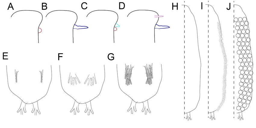
2.3. Morphological Description of the Characters
In this section, we present brief comments on the different characters. Schematic
drawings of some structures were provided to better understand the assessment of
Taxonomy 2021, 1 homology hypotheses (Figure 1). Character numbers and transformation series in the281
following descriptions correspond to the character summary in Table 2 and data matrix
in Table 1.
Figure 1. Schematic drawing of the morphological features. (A–D) Types and arrangements of cephalic sensorial organs:
Figure 1. Schematic drawing of the morphological features. (A–D) Types and arrangements of cephalic sensorial organs:
(A) Lateral knob (red); (B) Lateral large cephalic tentacle (dark blue); (C) Lateral knob (red) + lateral short cephalic tentacle
(A) Lateral knob (red); (B) Lateral large cephalic tentacle (dark blue); (C) Lateral knob (red) + lateral short cephalic tentacle
(light
(lightblue);
blue);(D)
(D)Dorsolateral
Dorsolateralrod-like
rod-like(pink)
(pink) ++ lateral
lateral long (dark blue)
long (dark blue)tentacle.
tentacle.(E–G)
(E–G)Arrangement
ArrangementofofTbV
TbVatat U80
U80 region:
region:
(E)(E)Single adhesive tubes;
Single adhesive tubes; (F) “Foot-like” tubes; (G) Bulkiness cluster of adhesive tubes. (H–J) Dorsal ornamentation:
“Foot-like” tubes; (G) Bulkiness cluster of adhesive tubes. (H–J) Dorsal ornamentation: (H)
(H)Smooth
Smooth body
bodysurfaces (I) (I)
surfaces Subrectangular
Subrectangularscales; (J) Scale-like
scales; cuticular
(J) Scale-like elevations.
cuticular elevations.
Cephalic
Cephalic sensorial
sensorial organs.
organs. TwoTwotypestypesofofbilateral
bilateralcephalic
cephalictentacles
tentaclesare present
are present Pty-
in in
chostomella
Ptychostomellaspecies:
species:long andand
long short tentacles.
short TheThe
tentacles. long tentacles
long are are
tentacles 3–43–4times larger
times than
larger
TbA,
than and
TbA,theand short
the tentacles possess
short tentacles the same
possess size assize
the same TbA as(Figure 1B,D),1B,D),
TbA (Figure whichwhichwe believe
we
to
believe to be equivalent to the same transformation series with two different states0).
be equivalent to the same transformation series with two different states (character
There is also
(character 0).one
There type of rod-like
is also one typedorsolateral cephalic tentacle
of rod-like dorsolateral (character
cephalic tentacle 2) (Figure 2)
(character 1D)
and one 1D)
(Figure typeandof bilateral
one typeknob-like
of bilateralorgan (character
knob-like organ1)(character
(Figure 1A,C).
1) (Figure 1A,C).
Ventral
Ventral adhesive tubesatatU85
adhesive tubes U85region.
region.TheThe ventral
ventral adhesive
adhesive tubestubes
at theatU85the region
U85 region
can
can be arranged
be arranged in three
in three different
different forms: forms:
pairedpaired
tubes tubes (character
(character 7, state7,0)state 0) (Figure
(Figure 1E), foot-1E),
foot-like
like withwith
few few adhesive
adhesive tubestubes (character
(character 7, state
7, state 3) (Figure
3) (Figure 1F), and
1F), and a bulkiness
a bulkiness cluster
cluster of
of adhesive
adhesive tubes
tubes (character
(character 7, state
7, state 2) (Figure
2) (Figure 1G).1G).
Coverage
Coverage of dorsal and dorsolateral
dorsolateral bodybody wall.
wall.The
Theornamentation
ornamentationcan canbebepresent
presentalong
along
all
alldorsal
dorsal surfaces
surfaces as scale-like cuticular elevations
scale-like cuticular elevations(Figure
(Figure1J)1J)and
andsubrectangular
subrectangularscales scales
(Figure1J)
(Figure 1J) covering
covering the lateral surfaces.
surfaces.
3. Results
3.1. Taxonomic Account
Order Macrodasyida Remane, 1925 [Rao & Clausen, 1970]
Family Taumastodermatidae Remane, 1927
Subfamily Taumastodermatinae Remane, 1927
Genus Ptychostomella Remane, 1926
Ptychostomella sebastiana sp. nov.
Examined Material. Holotype. Adult, collected from Praia da Fome in São Sebastião,
State of São Paulo, Brazil (23◦ 450 2400 S; 45◦ 210 1.500 W), at 10 m depth, deposited at the Museu
de Zoologia, Universidade Estadual de Campinas, Brazil, under accession number ZUEC-
PIC: 432. Paratype. Subadult, collected from Praia da Fome in São Sebastião, State of São
Paulo, Brazil (23◦ 450 2400 S; 45◦ 210 1.500 W), at 10 m depth, deposited at the Museu de Zoologia,
Universidade Estadual de Campinas, Brazil, under accession number ZUEC-PIC: 433.Taxonomy 2021, 1 282
Etymology. The specific epithet sebastiana refers to the city of São Sebastião, where the
specimens were sampled.
Repository: urn:lsid:zoobank.org:act:3918251C-3391-43B6-9E5F-FBE32B10EE20.
Diagnosis. A Ptychostomella with an adult length up to 250 µm; pharynx length up to
78 µm, with pharyngeal pores at base. PhIJ at U38; body with almost parallel sides and
short, bilobed caudum. Head bearing paired club-shape lateral tentacles and a paired small
“rod-like” tentacle at dorsolateral surface; few sensory hairs forming columns along lateral
side of the body, scattered sensory hairs around the oral opening, few sensory hairs along
the lateral tentacle surface and tufts at the tip of the dorsolateral tentacle; a great number
of epidermal glands along the length of the body. Dorsal and ventral surfaces are generally
smooth, except ventrolateral surfaces that are covered by subrectangular scales arranged in
a column on each side of the body. Ten TbA arranged in groups of four on each side, and
one of two at middle line on ventral surface (4 + 2 + 4). Three TbL on each side of the body
at U12, U60, and U90. TbV noticeable in two distinct groups: four to five adhesive tubes
spaced along the ventral surface from U45–U65 and paired “feet” with five adhesive tubes
each at U83. Eleven TbP per side arranged as eight inserted laterally, two at the tip of the
lobes, and one on the inner part of the lobe. Locomotory cirri covering the entire ventral
surface of the body. Testis on the right side of the body, pyriform caudal organ, frontal
organ above the caudal organ filled with spermatozoa, and presence of a dorsally placed
egg in intestine the region.
Description. Adult holotype with 250 µm in total length. Pharynx 78 µm in length.
Pharyngeal pores near the base, at U31; pharyngeo-intestinal junction at U38. Head bearing
two paired tentacles: dorsolateral “rod-like” small tentacle with 12 µm in length at U03
(Figures 2A, 3A and 4A), and a lateral club-shape tentacle with 25 µm in total length at
U8. Widths of the head/neck/trunk/caudal base, 40/50/53/30 µm at U07/U25/U46/U90,
respectively. Oral opening with scattered sensory cilia along the entire margin. Sparse
sensory cilia along the dorsal surface of the lateral tentacle and tufts of sensory cilia on the
tip of the dorsolateral tentacle. Few sensory hairs forming columns along lateral sides of
the body.
Cuticular armature. Dorsal and ventral surfaces apparently smooth. However, each
ventrolateral side is covered by subrectangular scales with similar size (3–4 µm) overlapping
each other partially, forming a column running from U08 to U90 (Figures 2B,C and 3C).
Adhesive tubes. Ten TbA arranged in 4 + 2 + 4 at U09, 7 µm in length laterally and medially
(Figures 2C, 3C and 4C). Three TbL per side (12 µm in length) inserting at U12, U60, and
U90, respectively (Figures 2, 3A,B and 4C). TbD, absent. TbV noticeable in two distinct
groups: four to five spaced along the ventral surface from U45–U65 (8–13 µm in length)
(Figures 2C and 3C) and paired “feet” with five adhesive tubes each at U83 (12 µm in length)
(Figures 2C, 3C and 4D). Eleven TbP per side arranged as eight inserted laterally (11 µm in
length), two at tip of the lobes (6 µm in length), and one flanking medially on the inner part
of the caudal pedicle (7 µm in length) (Figures 2, 4B and 5A).Taxonomy 2021, 1 283
Taxonomy 2021, 1, FOR PEER REVIEW 6
Figure 2. Schematic drawing of Ptychostomella sebastiana sp. nov. A–Dorsal body. B–Internal body. C–Ventral body.
Figure 2. Schematic drawing of Ptychostomella sebastiana sp. nov. (A)—Dorsal body. (B)—Internal body. (C)—Ventral body.
Abbreviations: co, caudal organ; dct, dorsal cephalic tentacle; eg, epidermal glands; fTbV, foot-like ventral adhesive tubes;
Abbreviations: co, caudal
lct, lateral cephalic organ;
tentacle; mo,dct, dorsalpp,
mouth; cephalic tentacle;
pharyngeal eg, epidermal
pores; glands; fTbV,
srs, subrectangular foot-like
scales; ventral adhesive
TbA, anterior adhesivetubes;
tubes;
lct, lateral cephalic tentacle; mo, mouth; pp, pharyngeal pores; srs, subrectangular scales; TbA,
TbL, lateral adhesive tubes; TbP, posterior adhesive tubes; TbV, ventral adhesive tubes; te, testis.anterior adhesive tubes; TbL,
lateral adhesive tubes; TbP, posterior adhesive tubes; TbV, ventral adhesive tubes; te, testis.Taxonomy 2021, 1 284
Taxonomy 2021, 1, FOR PEER REVIEW 7
Figure 3. DIC photomicrographies of Ptychostomella sebastiana sp. nov. (A) Dorsal body. (B) Internal body. (C) Ventral
Figure 3. DICbody.
photomicrographies
Abbreviations: co, caudal of Ptychostomella
organ; dct, dorsalsebastiana sp. nov.
cephalic tentacle; (A) Dorsal
eg, epidermal body.
glands; fTbV,(B) Internal
foot-like body.
ventral (C) Ventral body.
adhesive
Abbreviations:tubes;
co, lc, locomotory
caudal organ; cilia; lct,dorsal
dct, lateral cephalic
cephalictentacle; mo, mouth;
tentacle; ph, pharynx;
eg, epidermal sh, sensory
glands; fTbV,hairs; srs, subrectangular
foot-like ventral adhesive tubes;
scales; TbA, anterior adhesive tubes; TbL, lateral adhesive tubes; TbP, posterior adhesive tubes; TbV, ventral adhesive
lc, locomotorytubes;
cilia;te,lct, lateral
testis. cephalic
Scale bars: 50 µmtentacle; mo, mouth; ph, pharynx; sh, sensory hairs; srs, subrectangular scales; TbA,
anterior adhesive tubes; TbL, lateral
Taxonomy 2021, 1, FOR PEER REVIEW
adhesive tubes; TbP, posterior adhesive tubes; TbV, ventral adhesive tubes; 8
te, testis.
Scale bars: 50 µm.
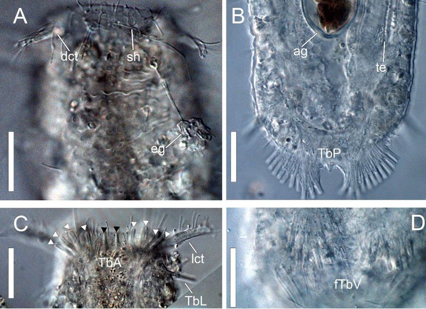
Figure 4. DIC photomicrographies of Ptychostomella sebastiana sp. nov. (A) Dorsal head surface. (B) Posterior end of body.
Figure 4. DIC(C) photomicrographies of Ptychostomella
Close-up of TbA arrangement. (D) Close-up ofsebastiana sp. nov.
foot-like ventral (A) tubes.
adhesive Dorsal head surface.
Abbreviations: (B) dct,
ag, algae; Posterior
dorsal end of body.
cephalic tentacle; eg, epidermal glands; fTbV, foot-like ventral adhesive tubes; lc, locomotory cilia;
(C) Close-up of TbA arrangement. (D) Close-up of foot-like ventral adhesive tubes. Abbreviations: ag, algae; dct, dorsal lct, lateral cephalic
tentacle; sh, sensory hair; TbA, anterior adhesive tubes; TbL, lateral adhesive tubes; TbP, posterior adhesive tubes; te,
cephalic tentacle; eg, epidermal
testis. Black glands;
triangles indicate centralfTbV, foot-like
TbA; White ventral
triangles adhesive
indicate lateral TbA.tubes; lc, locomotory
Scale bars: 50 µm. cilia; lct, lateral cephalic
tentacle; sh, sensory hair; TbA, anterior adhesive tubes; TbL, lateral adhesive tubes; TbP, posterior adhesive tubes; te, testis.
Cuticular armature. Dorsal and ventral surfaces apparently smooth. However, each
Black triangles indicate central TbA; White triangles indicate lateral TbA. Scale bars: 50 µm.
ventrolateral side is covered by subrectangular scales with similar size (3–4 µm)
overlapping each other partially, forming a column running from U08 to U90 (Figures
2B,C and 3C).
Adhesive tubes. Ten TbA arranged in 4 + 2 + 4 at U09, 7 µm in length laterally and
medially (Figures 2C, 3C, and 4C). Three TbL per side (12 µm in length) inserting at U12,
U60, and U90, respectively (Figures 2, 3A,B, and 4C). TbD, absent. TbV noticeable in two
distinct groups: four to five spaced along the ventral surface from U45–U65 (8–13 µm in
length) (Figures 2C and 3C) and paired “feet” with five adhesive tubes each at U83 (12 µm+ lateral club-shape tentacles in the new species and lateral pairs knob-like + short cephalic
tentacles in P. lamelliophora).
3.2. Phylogenetic Analysis
Taxonomy 2021, 1
A total of 221 most parsimonious trees with 22 steps, Ci = 59 and Ri = 68, were285
obtained under equal weight analysis. Six MPTs (Figures 5 and 6) were recovered using
implied weight on all K-values obtained from the used script and besides that, the present
topologies were recovered from the unweighted MPTs.
Figure
Figure 5. Most
5. Most parsimonious
parsimonious treestrees obtained
obtained by implied
by implied weight analysis
weight analysis of the
of the genus genus Ptychostomella.
Ptychostomella. Black circlesBlack circles =
= synapomorphy;
synapomorphy;
white white circles
circles = homoplasy. Numbers= homoplasy. Numbers
above branches defineabove branches
characters; define
numbers characters;
below numbers
branches belowstates.
are character branches are
character states.
Ventral ciliation. A continuous, dense field of locomotory cilia that extend from the
ventral border of the oral opening to the base of the caudal pedicles (Figure 3C).
Reproductive system. Testis on the right body side, pyriform caudal organ at U82; Eggs
dorsal to the mid intestine (Figures 2B and 3A).
Epidermal Glands. Numerous epidermal glands forming two columns along the lateral
length of the body from U01, joining at caudal base U89 (Figures 2A and 4A).
Taxonomic Remarks
Ptychostomella sebastiana sp. nov. falls within a clade that includes P. lamelliophora,
sharing the apomorphic feature of subrectangular scales on each lateral side of the body.
However, the new species can be distinguished by the presence of adhesive tubes inserted
laterally of the pedicles, TbV as foot-like instead of a cluster of high number of adhesive
tubes at U85, and cephalic tentacle shape and arrangement (dorsolateral pairs of rod-like +
lateral club-shape tentacles in the new species and lateral pairs knob-like + short cephalic
tentacles in P. lamelliophora).
3.2. Phylogenetic Analysis
A total of 221 most parsimonious trees with 22 steps, Ci = 59 and Ri = 68, were
obtained under equal weight analysis. Six MPTs (Figures 5 and 6) were recovered using
implied weight on all K-values obtained from the used script and besides that, the present
topologies were recovered from the unweighted MPTs.Taxonomy 2021, 1 286
Taxonomy 2021, 1, FOR PEER REVIEW 10
Figure
Figure 6. Most
6. Most parsimonioustrees
parsimonious treesobtained
obtained by
by implied
impliedweight
weightanalysis of the
analysis of genus Ptychostomella.
the genus Black circles
Ptychostomella. Black =circles
synapomorphy;
=
synapomorphy; white circles = homoplasy. Numbers above branches define characters; numbers below branches
white circles = homoplasy. Numbers above branches define characters; numbers below branches are character states. are
character states.
These six topologies presented five monophyletic groups that were recovered from the
six MPTs and presented in the consensus tree (Figure 7). The first group, (P. higginsi (P. bergen-
These
sis, six topologies
(P. lepidota, presented
P. orientallis), five monophyletic
(P. helana (P. brachycephala groups that were recovered
(P. lamelliphora, from shares
P. sp. nov.)))))
the six MPTs and presented in the consensus tree (Figure 7). The first
the synapomorphic presence of anterior adhesive tubes, inserted in a ventrolateral column group, (P. higginsi
(P. bergensis,
(character(P.4, lepidota,
state 1). P. orientallis),
The second group(P. helana (P. brachycephala
is defined by the foot-like(P. lamelliphora,
arrangement P. of
sp.ventral
nov.))))
adhesive tubes at U85 (Figure 1F) of (P. bergensis (P. lepidota, P. orientallis) (P. helana (P.a brachy-
shares the synapomorphic presence of anterior adhesive tubes, inserted in
ventrolateral
cephala (P.column (character
lamelliphora, P. sp. 4, state 1).
nov.)))), butThe
the second groupofisP.defined
real position bergensisbyis the
not foot-like
clear. The third
arrangement
group, (P.oflepidota,
ventralP.adhesive tubes
orientallis) at U85by
is defined (Figure 1F) of (P.
the presence bergensis elevations
of cuticular (P. lepidota,(Figure
P. 1J)
orientallis)
covering(P. helana (P. brachycephala
the dorsal body surface (P.(character
lamelliphora, P. sp.1).
9, state nov.)))), but the real position
The monophyletic group (P. of helana
P. bergensis is not clear.
(P. brachycephala The thirdP.group,
(P. lamelliphora, (P. islepidota,
sp. nov.))) definedP. byorientallis) is defined bythethe
unique synapomorphy, presence
presence of cuticular elevations (Figure 1J) covering the dorsal body surface
of bilateral long cephalic tentacles (character 0, state 1) (Figure 1B–D). The last monophyletic(character 9, ,
stategroup
1). Theis monophyletic group (P. helana (P. brachycephala (P. lamelliphora, P.
defined by the presence of lateral plates (subrectangular scales) (character 10, state 1)sp. nov.))))
is defined
covering byeach
unique synapomorphy,
lateral body surface of the(P.presence
lamelliphora,of bilateral
P. sp. nov.)long cephalic
(Figure 1I). tentacles
(character 0, state 1) (Figure 1B–D). The last monophyletic group is defined by the
presence of lateral plates (subrectangular scales) (character 10, state 1) covering each
lateral body surface of (P. lamelliphora, P. sp. nov.) (Figure 1I).Taxonomy 2021, 1 287
Taxonomy 2021, 1, FOR PEER REVIEW 11
Figure 7. Consensus tree of the six most parsimonious trees obtained by implied weight analysis of the genus
Figure 7. Consensus tree of the six most parsimonious trees obtained by implied weight analysis of the genus Ptychostomella.
Ptychostomella.
4. Discussion
4. Discussion
Thaumastodermatidae was originally described as a group of marine animals with
Thaumastodermatidae was originally described as a group of marine animals with
an extraordinary ornamented
an extraordinary ornamented cuticle cuticle[18].
[18].However,
However, it it
is is possible
possible to to observe
observe at least
at least twotwo
exceptions this group: Oregodasys
exceptions within this group: Oregodasys Hummon, 2008, and Ptychostomella, which waswas
within Hummon, 2008, and Ptychostomella, which
previously
previously recognized
recognized by by aa group
groupof ofsmooth
smoothcuticle
cuticlespecies
species [3,17].
[3,17]. Specimens
Specimens belonging
belonging
totothese two genera present some papillae covering the dorsal
these two genera present some papillae covering the dorsal body surfaces when body surfaces when
anyany
kind of scale-like structures are not observed. However, since the
kind of scale-like structures are not observed. However, since the first description of an first description of an
ornamented Ptychostomella
ornamented speciesconducted
Ptychostomella species conductedbyby[16], [16], currently
currently half
half of of
thethe species
species of this
of this
genus
genus were
were described
described with withornamentation
ornamentation onon thethe dorsal
dorsal bodybody surface.
surface. It is important
It is important to
tohighlight
highlightthatthat despite
despite thesethese species
species presenting
presenting somesomekind of kind of cuticular
cuticular ornamentation,
ornamentation, it is
itnecessary
is necessaryto betoaware
be aware
whenwhen a comparison
a comparison is made is between
made between the structures,
the structures, since they since
they
cannotcannot be properly
be properly homologous,
homologous, for example,
for example, scales and scales and are
papillae papillae are not
not formed byformed
the
bysamethe biological
same biological
process process
and cannotandbe cannot be considered
considered a state of athe state
same ofcharacter.
the same Due character.
to
Due
this,towethis, we decided
decided not tonot useto papilla
use papilla as cuticular
as cuticular ornamentations,
ornamentations, sincesince
amongamong the the
thaumastodematids
thaumastodematids it it is
is possible
possibletotoobserve
observethis thisfeature
feature onon other
other smooth
smooth bodied
bodied species,
species,
whereititis is
where notnot treated
treated as ornamentation
as ornamentation (e.g.,(e.g., Oregodasys).
Oregodasys). ThereThere
werewere two types
two types of
of cuticular
cuticular ornamentations
ornamentations that were considered:
that were considered: a rounded ascale-like
roundedornamentation
scale-like ornamentation
embossed on
embossed
the dorsal onandthe dorsalsurfaces
lateral and lateral surfaces
[17,19], and [17,19], and subrectangular
subrectangular scales covering scales each
covering
lateral
each lateral
surface of thesurface of the
body [3]. bodyanalysis,
In our [3]. In our
the analysis,
smooth dorsalthe smooth
bodydorsal
surfaces body surfacesas a
appeared
appeared as astate,
plesiomorphic plesiomorphic
and some kind state,ofand some cover
cuticular kind of cuticular cover
is homoplastic, is homoplastic,
arising in two distinct
arising inin
moments twothedistinct
evolutionmoments
of theingroup.
the evolution of the group.
Another important feature is the presence of a foot-like TbV among the Ptychostomella
specimens. As pointed out by [3], this feature is not a novelty among the thaumastoder-Taxonomy 2021, 1 288
matids, but it was important to define a monophyletic group in our analysis. This feature
appears to evolve from a plesiomorphic condition of a single paired adhesive tube to a
synapomorphic condition foot-like structure with four or five tubes. The autapomorphy
bulkiness clusters of TBV in P. lamelliophora are derived from the foot-like TbV. The presence
of these TbV arrangements could be related to the type of sandy beach that they were
present in (high, medium, low) or to a reproductive strategy [3].
A phylogenetic analysis based on morphological data was previously performed for
two genera of Thaumastodermatidae, Thaumastoderma [26], and Pseudostomella [27]. These
analyses were carried out using 37 and 33 morphological characters, respectively, and the
monophyly of these groups was recovered in the same way as that proposed by phylo-
genetic analysis using molecular data [1], and the internal relationship of both taxa were
well resolved. Despite that, we were able to ascertain that Ptychostomella is monophyletic,
and we listed only 11 characters, but they did not provide the same phylogenetic signals
or the internal support as observed in Thaumastoderma and Pseudostomella [26,27]. This
low phylogenetic signal does not allow us to infer hypotheses about the biogeographic
distribution of the Ptychostomella species as proposed by [27] and regarding the distribu-
tional pattern since, for the moment, we only have the sampling locations of all species
(Table S1, Figure S1). Acquisition of information on morphological structures of Ptychos-
tomella specimens is mainly done by optical microscopy and this technique addresses a low
number of external morphological characters. However, as pointed out by [28], to obtain
detailed morphological information on the external and internal morphology it is required
to use other techniques such as scanning electron microscopy (SEM), transmission electron
microscopy (TEM), confocal laser scanning microscopy (cLSM) combined with immuno-
histochemistry, and X-ray microtomography (Micro-CT). Beyond this, it is necessary to
increase the number of the outgroup with non-scaled and scaled thaumastodermatids in
future phylogenetic analyses to further test the monophyly of this taxon.
Supplementary Materials: The following are available online at https://www.mdpi.com/article/10
.3390/taxonomy1040022/s1, Figure S1: Global distribution of Ptychostomella species, Table S1: List of
Ptychostomella species with sampling biogeographic records.
Author Contributions: Conceptualization, T.Q.A. and A.R.S.G.; Methodology, T.Q.A. and A.R.S.G.;
Software, T.Q.A. and A.R.S.G.; Formal analysis, T.Q.A. and A.R.S.G.; Investigation, T.Q.A.; Resources,
A.R.S.G.; Data curation, T.Q.A. and A.R.S.G.; Writing—original draft preparation, T.Q.A. and A.R.S.G.;
Writing—review and editing, T.Q.A. and A.R.S.G.; Visualization, T.Q.A. and A.R.S.G.; Supervision,
A.R.S.G.; Project administration, A.R.S.G.; Funding acquisition, A.R.S.G. Both authors have read and
agreed to the published version of the manuscript.
Funding: This research was funded by the São Paulo Research Foundation—FAPESP, grant numbers
2014/23856-0 and 2018/11166-0.
Data Availability Statement: The data of geographic location of all Ptychostomella species presented
in this study are openly available in the supplementary material of this study.
Acknowledgments: We are grateful to the two anonymous reviewers for their constructive criticism
that improved the first version of the manuscript and Yasmina Shah Esmaeil for editing the English text.
Conflicts of Interest: The authors declare no conflict of interest.
References
1. Todaro, M.A.; Kånneby, T.; Dal Zotto, M.; Jondelius, U. Phylogeny of Thaumastodermatidae (Gastrotricha: Macrodasyida)
inferred from nuclear and mitochondrial sequence data. PLoS ONE 2011, 6, e0017892. [CrossRef]
2. Todaro, M.A. Marine and Freshwater Gastrotricha. Available online: http://www.gastrotricha.unimore.it (accessed on 30 September 2021).
3. Todaro, M. A new non-naked species of Ptychostomella (Gastrotricha) from Brazil. Zookeys 2013, 289, 13–24. [CrossRef] [PubMed]
4. Ruppert, E.E. Gastrotricha. In Introduction to the Study of Meiofauna; Higgins, R.P., Thiel, H., Eds.; Smithsonian Institution Press:
Washington, DC, USA, 1988; pp. 302–321.
5. Pfannkuche, O.; Thiel, H. Sampling processing. In Introduction to the Study Meiofauna; Higgins, R.P., Thiel, H., Eds.; Smithsonian
Institution Press: Washington, DC, USA, 1988; Volume 1, pp. 134–145.Taxonomy 2021, 1 289
6. Hummon, W.D.; Balsamo, M.; Todaro, M.A. Italian marine Gastrotricha: I. Six new and one redescribed species of Chaetonotida.
Boll. Zool. 1992, 59, 499–516. [CrossRef]
7. Bosco, I.; Lourenço, A.P.; Guidi, L.; Balsamo, M.; Hochberg, R.; Garraffoni, A.R.S. Integrative description of a new species
of Acanthodasys Remane, 1927 (Gastrotricha, Macrodasyida, Thaumastodermatidae) based on four distinct morphological
techniques and molecular data. Zool. Anz. 2020, 286, 31–42. [CrossRef]
8. Forey, P.L.; Kitching, I.J. Experiments in coding multistate characters. In Homology and Systematics: Coding Characters for Phylogenetic
Analysis; Scotland, R., Pennington, T., Eds.; Taylor & Francis: London, UK, 2000; pp. 54–80.
9. Pleijel, F. On character coding for phylogeny reconstruction. Cladistics 1995, 11, 309–315. [CrossRef]
10. Hawkins, J.A.; Hughes, C.E.; Scotland, R.W. Primary Homology Assessment, Characters and Character States. Cladistics 1997,
13, 275–283. [CrossRef]
11. Tongiorgi, P.; Balsamo, M. A new Tetranchyroderma species (Gastrotricha, Macrodasyoidea) from the Adriatic coast. Boll. Zool.
1984, 51, 335–338. [CrossRef]
12. Balsamo, M.; Todaro, M.A.; Tongiorgi, P. Marine gastrotrichs from the Tuscan achipelago (Tyrrhenian Sea): II. Chaetonotida, with
description of three new species. Boll. Zool. 1992, 59, 487–498. [CrossRef]
13. Clausen, C. Three new species of Gastrotricha Macrodasyida from the Bergenarea, Western Norway. Sarsia 1996, 81, 119–219.
[CrossRef]
14. Clausen, C. Gastrotricha from the Faroe Bank. Sarsia 2004, 89, 423–458. [CrossRef]
15. Roszczak, R. Die Psammitgastrotricha des polnischen Ostseestrandes. Zool. Pol. 1939, 4, 1–24.
16. Lee, J.M.; WookHwang, U.; Chang, C.Y. A new gastrotrich species of the genus Ptychostomella (macrodasyida, thaumastodermati-
dae) from south korea. Anim. Cells Syst. 2009, 13, 25–30. [CrossRef]
17. Clausen, C.; Båmstedt, U. Gastrotricha Macrodasyida from the Tromsø region, northern Norway. Sarsia 2000, 85, 357–384.
[CrossRef]
18. Remane, A. Neue Gastrotricha Macrodasyoidea. Zool. Jahrb. Abt. Syst. Ökologie Geogr. Tiere 1927, 54, 230–242.
19. Lee, J.M.; Chang, C.Y. Two New Marine Gastrotrichs of the Genus Ptychostomella (Macrodasyida, Thaumastodermatidae) from
South Korea. Zool. Sci. 2003, 20, 481–489. [CrossRef] [PubMed]
20. Remane, A. Morphologie und verwandtschaftsbeziehungen der aberranten gastrotrichen I. Z. Morphol. Ökologie Tiere 1926,
5, 625–754. [CrossRef]
21. Hummon, W.D.; Tod Aro, M.A.; Tongiorgi, P. Italian marine gastrotricha: II. one new genus and ten new species of macrodasyida.
Bolletino Zool. 1993, 60, 109–127. [CrossRef]
22. Goloboff, P.A.; Farris, J.S.; Nixon, K.C. TNT: Tree Analysis Using New Technology. Syst. Biol. 2003, 54, 176–178.
23. Goloboff, P.A.; Farris, J.S.; Nixon, K.C. TNT, a free program for phylogenetic analysis. Cladistics 2008, 24, 774–786. [CrossRef]
24. Marcos Mirande, J. Weighted parsimony phylogeny of the family Characidae (Teleostei: Characiformes). Cladistics 2009, 25, 574–
613. [CrossRef]
25. Nixon, K.C. WinClada Version. 1.00.08. Published by the Author. Ithaca, N.Y. 2002. Available online: http://www.cladistics.com/
(accessed on 16 February 2007).
26. Kieneke, A. A new species of Thaumastoderma (Gastrotricha: Macrodasyida) from the Antarctic deep sea with a phylogenetic
analysis of the whole genus. J. Mar. Biol. Assoc. UK 2010, 90, 575–584. [CrossRef]
27. Garraffoni, A.R.S.; Araújo, T.Q. Phylogeny of Pseudostomella Swedmark, 1956 (Gastrotricha: Macrodasyida) based on morphologi-
cal data and first insights on the historical biogeography of Thaumastodermatidae. Proc. Biol. Soc. Washingt. 2017, 130, 222–238.
[CrossRef]
28. Fonseca, G.; Fontaneto, D.; Di Domenico, M. Addressing biodiversity shortfalls in meiofauna. J. Exp. Mar. Biol. Ecol. 2018,
502, 26–38. [CrossRef]You can also read