Deep Learning of the Retina Enables Phenome- and Genome-Wide Analyses of the Microvasculature - Spiral
←
→
Page content transcription
If your browser does not render page correctly, please read the page content below
Circulation

                                                                ORIGINAL RESEARCH ARTICLE

                                                                Deep Learning of the Retina Enables
                                                                Phenome- and Genome-Wide Analyses of the
                                                                Microvasculature
                                                                Seyedeh Maryam Zekavat , BS*; Vineet K. Raghu , PhD*; Mark Trinder, MSc; Yixuan Ye , BS;
                                                                Satoshi Koyama, MD, PhD; Michael C. Honigberg , MD, MPP; Zhi Yu , PhD; Akhil Pampana , MS;
                                                                Sarah Urbut , MD, PhD; Sara Haidermota, BS; Declan P. O’Regan, MBBS, PhD; Hongyu Zhao , PhD;
                                                                Patrick T. Ellinor , MD, PhD; Ayellet V. Segrè, PhD; Tobias Elze , PhD; Janey L. Wiggs , MD, PhD;
                                                                James Martone, MD, MPH; Ron A. Adelman, MD, MPH, MBA; Nazlee Zebardast, MD, MSc; Lucian Del Priore, MD, PhD;
                                                                Jay C. Wang, MD; Pradeep Natarajan , MD, MMSc

                                                                BACKGROUND: The microvasculature, the smallest blood vessels in the body, has key roles in maintenance of organ health and
                                                                tumorigenesis. The retinal fundus is a window for human in vivo noninvasive assessment of the microvasculature. Large-
                                                                scale complementary machine learning-based assessment of the retinal vasculature with phenome-wide and genome-wide
                                                                analyses may yield new insights into human health and disease.

                                                                METHODS: We used 97 895 retinal fundus images from 54 813 UK Biobank participants. Using convolutional neural
                                                                networks to segment the retinal microvasculature, we calculated vascular density and fractal dimension as a measure
                                                                of vascular branching complexity. We associated these indices with 1866 incident International Classification of
                                                                Diseases–based conditions (median 10-year follow-up) and 88 quantitative traits, adjusting for age, sex, smoking
Downloaded from http://ahajournals.org by on January 21, 2022

                                                                status, and ethnicity.

                                                                RESULTS: Low retinal vascular fractal dimension and density were significantly associated with higher risks for incident
                                                                mortality, hypertension, congestive heart failure, renal failure, type 2 diabetes, sleep apnea, anemia, and multiple ocular
                                                                conditions, as well as corresponding quantitative traits. Genome-wide association of vascular fractal dimension and density
                                                                identified 7 and 13 novel loci, respectively, that were enriched for pathways linked to angiogenesis (eg, vascular endothelial
                                                                growth factor, platelet-derived growth factor receptor, angiopoietin, and WNT signaling pathways) and inflammation (eg,
                                                                interleukin, cytokine signaling).

                                                                CONCLUSIONS: Our results indicate that the retinal vasculature may serve as a biomarker for future cardiometabolic and
                                                                ocular disease and provide insights into genes and biological pathways influencing microvascular indices. Moreover, such a
                                                                framework highlights how deep learning of images can quantify an interpretable phenotype for integration with electronic
                                                                health record, biomarker, and genetic data to inform risk prediction and risk modification.

                                                                                  Key Words: deep learning ◼ epidemiology ◼ genomics ◼ mendelian randomization analysis ◼ microvessels ◼ retina

                                                                Correspondence to: Pradeep Natarajan, MD, MMSc, Massachusetts General Hospital, 185 Cambridge St, CPZN 3.184, Boston, MA 02114. Email pnatarajan@mgh.
                                                                harvard.edu
                                                                *S.M. Zekavat and V.K. Raghu contributed equally.
                                                                This work was presented AHA Scientific Sessions 2021, November 13–15, 2021, Boston, MA.
                                                                Supplemental Material, the podcast, and transcript are available with this article at https://www.ahajournals.org/doi/suppl/10.1161/circulationaha.121.057709.
                                                                For Sources of Funding and Disclosures, see page 147–148.
                                                                © 2021 The Authors. Circulation is published on behalf of the American Heart Association, Inc., by Wolters Kluwer Health, Inc. This is an open access article under the
                                                                terms of the Creative Commons Attribution License, which permits use, distribution, and reproduction in any medium, provided that the original work is properly cited.
                                                                Circulation is available at www.ahajournals.org/journal/circ

                                                                134      January 11, 2022                                                Circulation. 2022;145:134–150. DOI: 10.1161/CIRCULATIONAHA.121.057709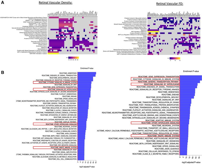
Zekavat et al Deep Learning to Analyze Retinal Microvasculature
T
he microvasculature influences health and disease
Clinical Perspective throughout all organ systems. Dysregulation of the
ORIGINAL RESEARCH
microvasculature contributes to many ocular and sys-
What Is New? temic conditions.1–5 The formation of blood vessels is con-
ARTICLE
• We leveraged deep learning to quantify geometric trolled by 2 processes: vasculogenesis and angiogenesis.
microvasculature indices across >100 000 retinal Vasculogenesis involves the differentiation of endothelial
fundus photographs and used these indices toward cells from mesodermal precursors and is under control of
characterizing phenome-wide clinical associations many signaling cues, including the WNT/B-catenin6 and
and genomic risk factors. Notch7 signaling pathways. Angiogenesis then follows a
• Epidemiologically, low microvascular density and series of sequential steps for vascular branching and is
fractal dimension (a measure of vascular branch-
mediated by growth factors and cytokines, including vas-
ing complexity) were associated with higher risk
cular endothelial growth factor (VEGF), fibroblast growth
of future mortality and cardiometabolic and ocular
disease; genetically, these microvascular indices factor, tumor necrosis factor-α, transforming growth
were enriched in pathways related to angiogen- factor-β, platelet-derived growth factor, and angiopoietins,
esis and inflammation. as well as intracellular signaling pathways incorporating
• Genetically higher risk for hypertension and diabe- Rho GTPases, protein kinase C, and Notch signaling.8 The
tes was associated with lower microvascular den- microvasculature is also of significance in cancer, wherein
sity; in turn, genetically lower microvascular density angiogenesis is necessary for tumor growth and enables
was independently associated with higher risk of metastasis.9 Antiangiogenic agents such as anti-VEGF
retinal detachments and skin cancer. antibodies are key aspects of cancer therapy, whereby the
actions of VEGF, which is induced by the tumor microen-
What Are the Clinical Implications? vironment (eg, hypoxia) and stimulates abnormal neovas-
• We illustrate the potential for deep learning of the cularization, are inhibited.9 The same anti-VEGF therapies
retina to understand the microvasculature among that treat cancers are also a mainstay of treatment for
humans in vivo, with wide applications across
neovascularization and macular edema secondary to neo-
diseases.
• Retinal microvascular indices may be clinically vascular age-related macular degeneration and prolifera-
useful as biomarkers of cardiometabolic disease tive diabetic retinopathy, respectively.10,11
severity and for risk prediction of ocular condi- Given the anatomic and physiological similarities
between the retinal microvasculature and that of other
Downloaded from http://ahajournals.org by on January 21, 2022
tions; however, more research is required to
assess clinical efficacy. organs, fundus photographs of the retina allow noninva-
• Genetic contributors to microvascular indices, sive in vivo assessment of the microvasculature. Current
including those influencing angiogenesis and computerized approaches of assessing the microvascula-
inflammation, may provide insight into therapeutic ture include retinal vessel caliber estimation of arterioles
targets for microvascular disease in the eye, can- and venules and their ratio.12 Smaller studies have shown
cer, and diseases in other tissues. that smaller retinal vascular caliber and smaller arte-
riolar/venular ratio are linked to antecedent and future
hypertension.13–17 Furthermore, retinal vascular caliber is
associated with incident stroke.18–20 However, associa-
Nonstandard Abbreviations and Acronyms tions with coronary artery disease and renal disease are
CHASE Child Heart and Health Study in less consistent.21–23 Current approaches are now able to
England extract geometric and branching patterns of the retinal
DBP diastolic blood pressure microvasculature such as retinal vascular tortuosity,24
fractal dimension (FD),25–31 and vascular density.32–36
FD fractal dimension
Such indices are more consistently linked to stroke, cor-
GWAS genome-wide association study
onary artery disease, and renal disease in small cross-
HbA1c hemoglobin A1c
sectional analyses of ≈1000 individuals.37-41 The extent
HR hazard ratio to which retinal microvasculature geometric alterations
MITF melanocyte-inducing transcription factor may be linked to incident phenome-wide consequences
OR odds ratio is poorly understood. In addition, complementary genetic
PheWAS phenome-wide association study discovery analyses of the retinal microvasculature offer
PoPS Polygenic Priority Score the prospects of identifying new therapeutic targets for
PRS polygenic risk score both ocular and nonocular conditions.
SBP systolic blood pressure Here, we leveraged deep learning for automated
VEGF vascular endothelial growth factor image quality control and segmentation of the micro-
vasculature across >100 000 retinal fundus photo-
graphs. We subsequently quantified 2 vascular features:
Circulation. 2022;145:134–150. DOI: 10.1161/CIRCULATIONAHA.121.057709 January 11, 2022 135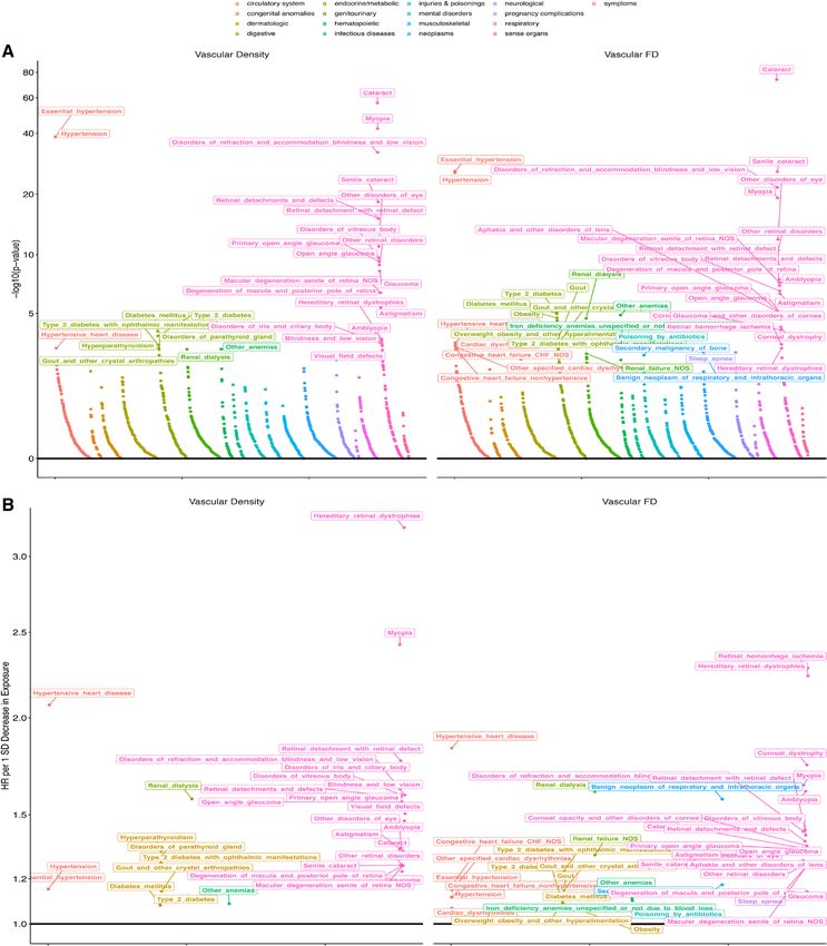
Zekavat et al                                                                                   Deep Learning to Analyze Retinal Microvasculature

                                                                       branching complexity as measured with FD42 and vas-                         centers across the United Kingdom with sample collections,
                                                                       cular density, defined as the total number of segmented                     including blood-derived DNA. Retinal fundus imaging was per-
ORIGINAL RESEARCH

                                                                       pixels given a consistent field of view and fixed pixel                     formed at enrollment with the TOPCON 3D OCT 1000 Mk2
                                                                       dimensions across all individuals. We then performed a                      instrument, which takes a 3-dimensional scan and photograph
     ARTICLE

                                                                                                                                                   of the retina with a magnified photograph of the fundus. Of the
                                                                       phenome-wide association study (PheWAS) for FD and
                                                                                                                                                   67 339 genotyped individuals with retinal imaging available at
                                                                       vascular density across 1866 phenotypes and 88 quan-
                                                                                                                                                   enrollment, we analyzed 97 895 images across 54 813 partici-
                                                                       titative clinical traits and biomarkers. Secondarily, we                    pants after applying the quality control filters as indicated below.
                                                                       performed genome-wide association studies (GWASs)                           Use of the data was approved by the Massachusetts General
                                                                       for FD and vascular density across common variants in                       Hospital Institutional Review Board (protocol 2013P001840)
                                                                       the genome, as well as a rare variant association study                     and facilitated through UK Biobank Application 7089. Details
                                                                       across rare predicted disruptive variants in the genome.                    on UK Biobank array genotyping,43 whole-exome sequenc-
                                                                       We leveraged these results to investigate causal rela-                      ing,44,45 genomic quality control,46–48 sample exclusion criteria,
                                                                       tionships through mendelian randomization. Overall, our                     and poor-quality fundus image removal49,50 are provided in the
                                                                       study highlights how deep learning enables a large-                         Supplemental Methods.
                                                                       scale, unbiased connection between the retinal micro-
                                                                       vasculature and clinical outcomes (Figure 1A).                              Deep Learning for Vessel Segmentation
                                                                                                                                                   An overview of previous methods for retinal vessel segmenta-
                                                                                                                                                   tion42,51–56 is provided in the Supplemental Methods. Here, we
                                                                       METHODS                                                                     developed a deep learning ensemble model of U-Nets57 on the
                                                                                                                                                   Google Cloud’s artificial intelligence platform to automatically
                                                                       UK Biobank Cohort, Retinal Fundus                                           segment vasculature from retinal fundus photographs. The model
                                                                       Imaging, and Quality Control                                                was developed from 90 photographs and associated hand-drawn
                                                                       The UK Biobank is a population-based cohort of ≈500 000 par-                segmentations from 3 publicly available data sets: (1) Digital
                                                                       ticipants recruited from 2006 to 2010 with existing genomic                 Retinal Images for Vessel Extraction, photographs from a diabetic
                                                                       and longitudinal phenotypic data and median 10-year follow-                 retinopathy screening program in the Netherlands of subjects 25
                                                                       up.43 Baseline assessments were conducted at 22 assessment                  to 90 years of age; (2) fundus photographs with hand-labeled
       Downloaded from http://ahajournals.org by on January 21, 2022

                                                                       Figure 1. Study schematic and vascular features.
                                                                       A, Here, we first used deep learning toward large-scale automated removal of low-quality images, followed by vessel segmentation. Next,
                                                                       using the vascular segmentations, we quantified 2 vascular indices: branching complexity as measured by fractal dimension (FD) and vascular
                                                                       density. Last, phenome- and genome-wide association analyses of retinal FD and vascular density were performed to discover phenotypes
                                                                       associated with the microvasculature and genotypes influencing these vascular indices. B, Significant Spearman correlations were observed
                                                                       between FD and vascular density of the right vs left eyes, with right eyes having significantly higher FD and vascular density compared with
                                                                       left eyes. Blue line reflects best-fit line; dotted purple line reflects the unity line (x=y). C, Relationship of FD and vascular density (averaged
                                                                       across right and left eyes) with age.

                                                                       136     January 11, 2022                                        Circulation. 2022;145:134–150. DOI: 10.1161/CIRCULATIONAHA.121.057709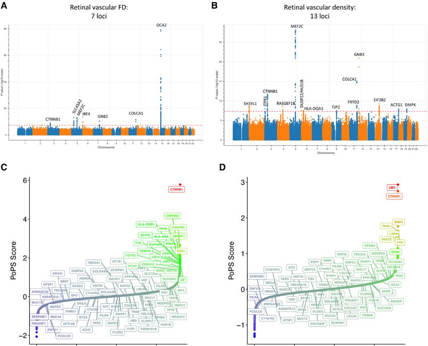
Zekavat et al                                                                         Deep Learning to Analyze Retinal Microvasculature

                                                                vessel segmentations from the Structured Analysis of the Retina       sample of 794 retinal fundus photographs from the UK
                                                                database58 extracted from clinical visits to the Shiley Eye Center    Biobank. This model achieved a sensitivity of 97.4% and

                                                                                                                                                                                                          ORIGINAL RESEARCH
                                                                at the University of California, San Diego; and (3) CHASE (Child      a specificity of 100.0% for detecting poor-quality im-
                                                                Heart and Health Study in England),59 retinal fundus photographs      ages in the independent testing set of 206 fundus pho-

                                                                                                                                                                                                               ARTICLE
                                                                of 9- and 10-year-old children of different ethnic origin from
                                                                                                                                      tographs. We then applied this model across 134 653
                                                                England. Seventy-five images were used for training and hyper-
                                                                                                                                      photographs acquired at the UK Biobank enrollment
                                                                parameter tuning of the model, and the remaining 15 images
                                                                were used as an independent validation data set (with the 3 data      visit across 67 339 individuals. This resulted in removal
                                                                sets proportionally represented). A second external validation        of 26% of the original images with 99 736 images from
                                                                data set consisted of 143 images from the Automated Retinal           55 603 participants remaining, similar to filtering rates
                                                                Image Analysis database. These were retinal fundus photos from        from other studies.49,50 Further details on sensitivity anal-
                                                                adults with age-related macular degeneration, adults with diabetic    yses for poor-quality image removal are provided in the
                                                                retinopathy, and healthy control subjects collected between 2004      Supplemental Methods (Table S1).
                                                                and 2006 from the St. Paul’s Eye Unit in Liverpool, UK.60 Details         To implement large-scale vessel segmentation, we
                                                                on model training and FD42,61 and vascular density calculation are    developed an ensemble of deep convolutional neu-
                                                                provided in the Supplemental Methods.                                 ral networks as detailed in the Methods section. On
                                                                                                                                      the 15-image testing data set, this ensemble model
                                                                PheWAS Analyses                                                       achieved an 82.1% Dice similarity coefficient (a mea-
                                                                Four sets of PheWAS analyses were performed, corresponding            sure of spatial overlap accuracy), 97.4% pixel-wise accu-
                                                                to association of retinal vascular FD and vascular density with       racy, 99.1% area under the curve, and correlation of
                                                                (1) prevalent phenotypes at enrollment,62,63 (2) incident pheno-      0.92 for FD and 0.88 for vascular density with the true
                                                                types developed after enrollment,62,63 (3) quantitative systemic      hand-labeled vessel segmentations on the independent
                                                                biomarkers, and (4) quantitative ocular traits,46–48 as detailed in   testing data set (Figure S1). These results were compa-
                                                                the Supplemental Methods including the statistical analysis. All
                                                                                                                                      rable or superior to results of other deep learning-based
                                                                models were adjusted for age, age squared, sex, smoking sta-
                                                                                                                                      approaches.52 On the Automated Retinal Image Analy-
                                                                tus (current/previous/never smoker), and ethnicity (data field
                                                                21000). For each analysis, statistical significance was defined       sis external validation data set, the model achieved an
                                                                with false discovery rate–corrected P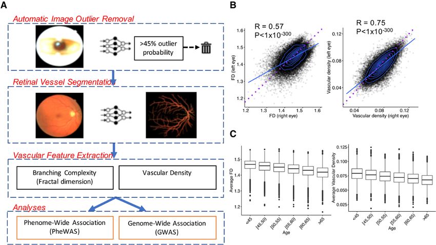
Zekavat et al                                                                   Deep Learning to Analyze Retinal Microvasculature

                                                                       (Figure S3). Retinal vascular density and FD across both        heart disease (HR Density, 2.07; HR FD, 1.84). Accordingly,
                                                                       eyes were strongly correlated (right eye: RSpearman=0.77,       systolic blood pressure (SBP) and diastolic blood pres-
ORIGINAL RESEARCH

                                                                       P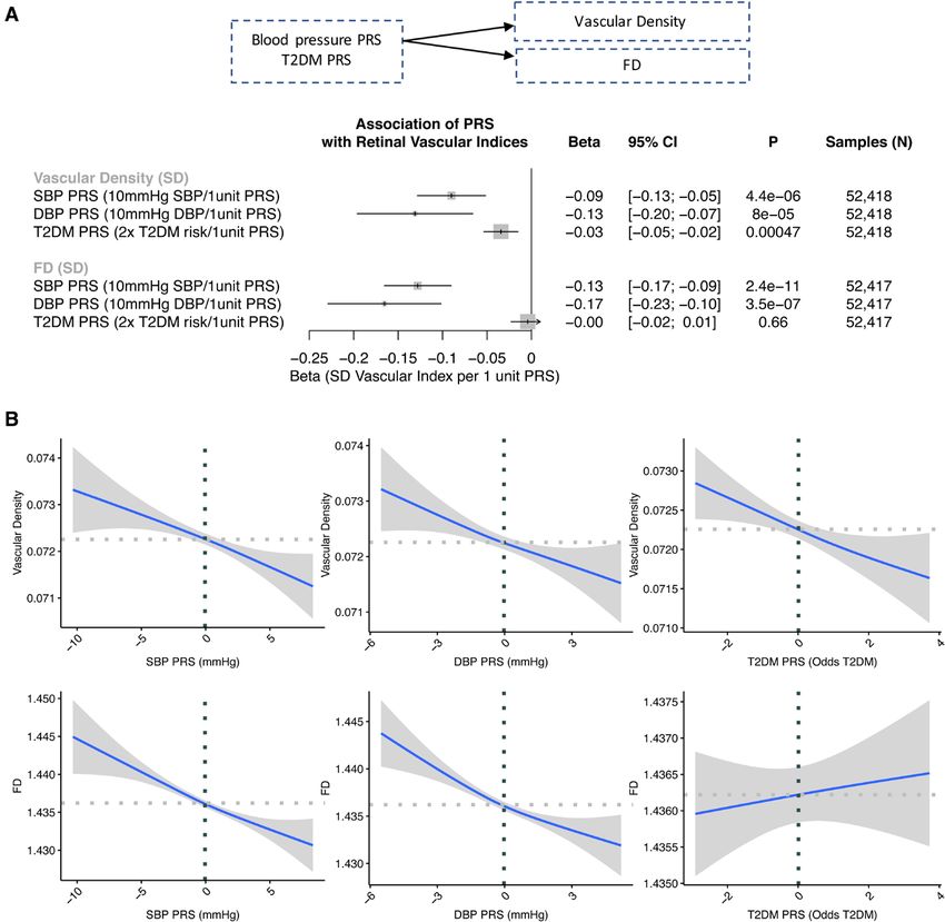
Zekavat et al                                                                                  Deep Learning to Analyze Retinal Microvasculature

                                                                                                                                                                                                                        ORIGINAL RESEARCH
                                                                                                                                                                                                                             ARTICLE
                                                                Figure 2. Association of retinal vascular FD and density with incident mortality.
                                                                A, Association of low (≥2 SD below the mean) fractal dimension (FD) and density with incident mortality, stratified by whether the person has
                                                                prevalent type 2 diabetes (T2D) or hypertension (HTN) at time of image acquisition. Analyses are adjusted for age, age squared, sex, smoking
Downloaded from http://ahajournals.org by on January 21, 2022

                                                                status, and ethnicity. B, Cumulative incidence of mortality across individuals with low FD and density who have a diagnosis of prevalent T2D or
                                                                hypertension compared with those who do not. HR indicates hazard ratio.

                                                                and mean spherical equivalent (Figure S11 and Table                         vascular density in chromosome 12 (β, −0.13 SD;
                                                                S9) are discussed in the Supplemental Results.                              P=1.11×10⁻²¹) and is predicted to be deleterious by
                                                                                                                                            several in silico prediction tools, including PolyPhen,74
                                                                                                                                            SIFT,75 and PrimateAI.76 It was also suggestively asso-
                                                                Common Variant GWAS and In Silico Analyses                                  ciated with vascular FD (β, −0.069 SD; P=1.06×10⁻⁷).
                                                                GWASs for retinal vascular density and FD were carried                      Further sensitivity analyses and comparison with variants
                                                                out across 38 932 unrelated individuals and 15 580 782                      identified in a previous GWAS of vascular tortuosity24 are
                                                                variants with minor allele frequency >0.001 in the UK                       provided in the Supplemental Results (Figure S13 and
                                                                Biobank, identifying 13 and 7 genome-wide–significant                       S14 and Table S12).
                                                                (P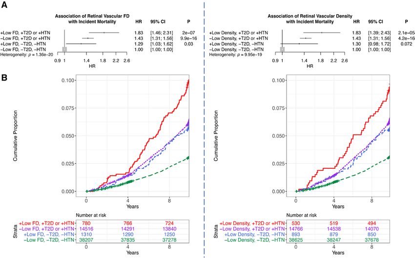
Zekavat et al                                                                                 Deep Learning to Analyze Retinal Microvasculature
ORIGINAL RESEARCH
     ARTICLE
       Downloaded from http://ahajournals.org by on January 21, 2022

                                                                       Figure 3. Phenome-wide associations with incident disease.
                                                                       A, –Log10(P value) of associations of retinal vascular density and fractal dimension (FD) with incident disease plotted as grouped by phenotypic
                                                                       category. Associations were performed with Cox proportional hazards models adjusted for age, age squared, sex, smoking status, and ethnicity. B,
                                                                       Hazard ratio (HR) per 1-SD decrease in either vascular density (left) or FD (right). Labeled phenotypes across both plots have false discovery
                                                                       rate–corrected P
Zekavat et al Deep Learning to Analyze Retinal Microvasculature
ORIGINAL RESEARCH
ARTICLE
Downloaded from http://ahajournals.org by on January 21, 2022
Figure 4. Genome-wide association studies and gene prioritization.
A and B, Manhattan plots visualizing the genome-wide association results for retinal vascular fractal dimension (FD; A) and density (B), which
identify 7 and 13 loci, respectively. Gene prioritization with the Polygenic Priority Score (PoPS) method.67 C and D, Prioritized genes at each locus
(see locus-specific prioritizations in Figure S15). The locus-specific genes prioritized by PoPS at each locus are labeled in A and B. The x axes in
C and D are arbitrary and reflect the ordered genes by PoPS score.
GNB3, EIF2B2, ACTG1, DMPK, SH3YL1, ITPR1, RAS- detachment, SBP, and DBP (Figure S16a and S16b
GEF1B, HLA-DA1, and TJP2. Outside of these loci, sev- and Table S14). Further fine mapping of genome-wide–
eral other genes were also suggestively prioritized by significant loci was performed to prioritize potentially
PoPS, including HLA-DRA, HLA-DRB1, ESR1, FGFR2, causal variants66 (Table S15). Retinal vascular density–
PDGFRA, and TYR for FD and UBC for vascular density and FD-lowering alleles at the fine-mapped variants
(Figure S15). had heterogeneous effects on the previous phenotypes
Second, we identified other traits associated with the assessed in genetic correlation analyses (Figure S16c
top variants, assessing traits with significant associa- and S16d and Table S16). In particular, retinal density–
tions from the PheWAS (such as systemic traits includ- and FD-lowering alleles at several PoPS-prioritized
ing blood pressure and diabetes, as well as ocular traits genes showed associations with higher risk of skin neo-
such as retinal detachment, myopia, glaucoma, diabetic plasms, malignant melanoma, and eye cancer (IRF4/
retinopathy, and macular degeneration), in addition to DUSP22, SLC45A2); a separate set of loci showed
traits showing strong associations on PhenoScanner for associations with lighter skin color (GNB2, ACTG1). One
top variants (including melanoma, eye cancer, and skin locus showing consistency with the phenotypic associa-
color). Genetic correlation analyses across the genome tions previously described was the retinal vascular den-
identified significant inverse correlations between retinal sity–lowering variant rs8070929-T (βDensity, −0.04 SD;
vascular FD and density with previous published loci for PDensity=1.27×10⁻⁸) at the PoPS-prioritized gene ACTG1
myopia, age-related cataracts, lighter skin color, retinal encoding the actin gamma 1 protein, for which each reti-
Circulation. 2022;145:134–150. DOI: 10.1161/CIRCULATIONAHA.121.057709 January 11, 2022 141Zekavat et al Deep Learning to Analyze Retinal Microvasculature
nal vascular density-–lowering allele was genome-wide vascular density (P=8.0×10⁻⁵) and 0.17-SD decrease in
significantly associated with higher risk of advanced age- FD (P=3.5×10⁻⁷; Figure 6). Each 2-fold higher genetic
ORIGINAL RESEARCH
related macular degeneration (OR, 1.13; P=1.65×10⁻¹¹), risk for type 2 diabetes was associated with a 0.03-SD
reduced refractive error (β=−0.03; P=2.68×10⁻³⁰), and decrease in retinal vascular density (P=4.7×10⁻⁴); no
ARTICLE
lighter skin color (β=0.01; P=8.17×10⁻²⁷) and sugges- significant association was observed between the type 2
tively associated with higher risk of retinal detachment, diabetes PRS and FD.
age-related cataracts, skin neoplasms, and higher DBP Second, to assess the phenome-wide influence of
(PZekavat et al Deep Learning to Analyze Retinal Microvasculature
ORIGINAL RESEARCH
ARTICLE
Downloaded from http://ahajournals.org by on January 21, 2022
Figure 5. Pathway enrichment analysis.
Pathway enrichment analyses of the retinal vascular fractal dimension (FD) and density genome-wide association study results were performed
using the prioritized genes with Polygenic Priority Score z score >1 across the (A) Elsevier pathways and (B) Reactome pathways. Top
Bonferroni-significant results are listed (A), along with the enrichment odds ratios (ORs) and the enrichment P values (B). Further details on
genes included in each pathway and enrichment statistics are given in Tables S17 and S18.
those without either condition. This suggests that lower induced sheer stress and anemia-induced deficits in gas
microvascular density and branching complexity are and nutrient exchange may similarly impair the systemic
associated with higher severity of disease among those microvasculature. Moreover, pulmonary conditions such
with existing cardiometabolic disease. We also observed as sleep apnea are a well-described clinical risk factor
significant associations between lower retinal vascular for ocular conditions.81,82 Of note, previous studies in
density and FD and higher prevalence of, and separately small cohorts have been inconclusive or contradictory in
higher risk for, cardiometabolic (hypertension, hyper- the reported relationships between vascular FD and dia-
tensive heart disease, diabetes, renal failure, elevated betes and hypertension.25–28
HbA1c, and body mass index), pulmonary (sleep apnea, Second, we identify associations between lower reti-
abnormal pulmonary function tests), and hematopoietic nal vascular density and FD and higher risk of future ocu-
(anemia) conditions. Retinal microvascular dysfunction lar conditions. Although previous studies in small cohorts
may signify more widespread microvascular alterations. have identified associations between retinal vascular
For example, hyperglycemia-induced cellular dysfunc- indices and prevalent diabetic retinopathy,25,26,37 this is,
tion and death are linked to widespread insufficient to the best of our knowledge, one of the first large-scale
renewal of vascular endothelial and smooth muscle cells studies identifying associations with diverse future ocu-
through sorbitol accumulation, glycosylation, and reac- lar conditions. Specifically, we identify multiple associa-
tive oxygen formation.26,79,80 Furthermore, hypertension- tions between lower retinal vascular FD and density and
Circulation. 2022;145:134–150. DOI: 10.1161/CIRCULATIONAHA.121.057709 January 11, 2022 143Zekavat et al Deep Learning to Analyze Retinal Microvasculature
ORIGINAL RESEARCH
ARTICLE
Downloaded from http://ahajournals.org by on January 21, 2022
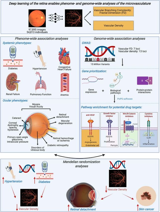
Figure 6. One-sample mendelian randomization for DBP PRS, SBP PRS, and T2D PRS on retinal vascular density and FD.
A, Association of the diastolic blood pressure (DBP), systolic blood pressure (SBP), and type 2 diabetes (T2D) polygenic risk score (PRS) with
normalized retinal vascular density and fractal dimension (FD) in a linear regression model adjusted for age, age squared, sex, smoking status,
and the first 10 principal components of genetic ancestry. B, Relationship of the SBP, DBP, and T2D PRSs with retinal vascular FD and density.
Horizontal and vertical dotted lines reflect the average value for the respective axis. Shaded gray region reflects the 95% CI using a restricted
maximum likelihood generalized additive model with integrated smoothness from the gam() function in R.
higher risk of future incident conditions influencing the tion of individuals with lower retinal vascular density may
posterior segment of the eye (retinal detachment, dia- enable improved monitoring and blindness risk reduction
betic retinopathy, macular degeneration, vitreous hemor- in this high-risk population. Moreover, we also identify
rhage). In particular, retinal detachment is a potentially associations with the anterior segment of the eye (glau-
blinding ocular condition for which limited risk factors coma, cataracts), suggesting that the retinal vasculature
have been described, including ocular trauma, myopia, may have physiological significance beyond the retina
and family history. Here, we find evidence of significant and vitreous fluid. Our observations are aligned with pre-
association between lower retinal vascular density and vious studies hypothesizing a link between the retinal
FD and higher incidence of retinal detachment, indepen- vasculature and normal-tension glaucoma.83,84 Hypoth-
dently of myopia. These findings suggest that identifica- esized contributors to normal-tension glaucoma include
144 January 11, 2022 Circulation. 2022;145:134–150. DOI: 10.1161/CIRCULATIONAHA.121.057709Zekavat et al Deep Learning to Analyze Retinal Microvasculature
ORIGINAL RESEARCH
ARTICLE
Downloaded from http://ahajournals.org by on January 21, 2022
Figure 7. One-sample mendelian randomization for vascular FD PRS and density PRS on retinal detachment.
A, Association of the vascular density PRS and fractal dimension (FD) polygenic risk score (PRS) with combined prevalent and incident retinal
detachment. Original model includes the following covariates: age, age squared, sex, smoking status, and the first 10 principal components of
genetic ancestry. +Myopia adjustment reflects additional adjustment for prevalent myopia at the time of image acquisition. B, Relationship of
vascular density PRS and vascular FD PRS with fraction of individuals developing retinal detachments and defects during their lifetime. Horizontal
and vertical dotted lines reflect the average value for the respective axis. Shaded gray region reflects the 95% CI using a restricted maximum
likelihood binomial generalized additive model with integrated smoothness from the gam() function in R. OR indicates odds ratio.
vascular abnormalities impeding nutrient delivery to the mapped top variants are strongly associated with skin
inner retina, thereby resulting in ganglion cell degen- neoplasms and lighter skin color (ie, at the PoPS-prior-
eration.83,84 Together, these findings linking the retinal itized genes IRF4, SLC45A2, OCA2, DUSP22, ACTG1).
vasculature with ocular pathophysiology highlight the Indeed, OCA2 encodes the oculocutaneous albinism 2
importance of the retinal microvasculature in ophthalmic protein that is known to result in lighter skin color and pre-
health and help us understand the diverse mechanisms disposes to skin cancers.85 Additionally identified in both
linking the retinal microvascular to impaired visual acuity the rare variant association study and GWASs were pre-
and blindness. dicted deleterious variants in MITF, a transcription factor
Third, genome-wide association analyses identified necessary for normal melanocyte differentiation. MITF has
genetic links between genes involved in angiogenesis, been associated with Waardenburg syndrome, character-
cancer, pigmentation, and inflammation and microvascula- ized by pigmentation anomalies of eyes, hair, and skin.86–88
ture architecture. We observed a significant enrichment in Previous work has also identified that MITF (melanocyte-
pathways related to angiogenesis (VEGF, platelet-derived inducing transcription factor) protein labeling in human
growth factor, angiopoietin), which are currently thera- tumor samples is strong around the vessels.89 Moreover,
peutically targeted to inhibit neovascularization in diabetic 2 of the GWAS loci identified across the FD and vascular
retinopathy, advanced age-related macular degeneration, density GWASs, namely GNB3 and GNB2, are G proteins
and many cancers.9–11 A significant genetic correlation and are known to be key moderators of chemokine sig-
with skin color was also observed, and several of the fine- nal transduction pathways. Notably, the missense variant
Circulation. 2022;145:134–150. DOI: 10.1161/CIRCULATIONAHA.121.057709 January 11, 2022 145Zekavat et al Deep Learning to Analyze Retinal Microvasculature
ORIGINAL RESEARCH
ARTICLE
Downloaded from http://ahajournals.org by on January 21, 2022
Figure 8. Summary of key findings.
Here, we successfully implemented deep learning toward image quality control and vessel segmentation to extract 2 features of the retina:
vascular density and fractal dimension (FD). Through phenome-wide analyses, we identified significant associations between low vascular
density and FD and higher risk of multiple systemic and ocular phenotypes, including ocular conditions influencing both the anterior and posterior
segments. Genome-wide association analyses of these microvascular indices discovered multiple loci enriched among pathways related to
vascular biology, inflammation, and neovascularization in cancer and may hypothesize potential drug targets for risk modification. Mendelian
randomization analyses identified that higher genetic risk for hypertension and type 2 diabetes is associated with lower microvascular density
and that higher genetic risk for lower microvascular density is associated with retinal detachment (independently of myopia) and with skin cancer
(independently of genetic ancestry principal components, self-reported skin color, and self-reported sun exposure and sun sensitivity). More
broadly, our results illustrate the potential for using deep learning on retinal imaging to understand the microvasculature, with wide applications
across diseases. This image was made with Biorender. GWAS indicates genome-wide association study.
146 January 11, 2022 Circulation. 2022;145:134–150. DOI: 10.1161/CIRCULATIONAHA.121.057709Zekavat et al Deep Learning to Analyze Retinal Microvasculature
rs5442-A in GNB3 variant has previously been associ- neous in magnification, a range in image magnification
ated with retinal microvascular diameter,90 hypertension,91 exists that is correlated with an individual’s spherical
ORIGINAL RESEARCH
refractive error,92,93 and advanced age-related macular equivalent. Sensitivity analyses adjusted for spherical
degeneration.94 Inflammatory and chemokine pathways equivalence and myopia indicated consistent associa-
ARTICLE
were significantly enriched in both GWAS studies, with tions. Third, given the paucity of accessible data sets
contributing genes prioritized by PoPS including IL2RA, with fundus images and genomic data and designation
IL23A, IL1R2, IL2, IL7R, IL6, MEF2A, PDGFRA, HLA mark- of retinal images as protected health information by the
ers, and others (Tables S16 and S17). Interleukins are Health Insurance Portability and Accountability Act, we
known modulators of angiogenesis and antiangiogenesis were unable to systematically replicate our GWAS results
in tumors,95 and inhibition of interleukins has been found in adequately powered data sets. Previous GWASs of
to suppress VEGF expression in tumors. Previous work on retinal vascular caliber performed in smaller studies90,97,98
retinal vascular tortuosity in the UK Biobank also identified identified overlapping loci with our present results at the
genetic loci linked to cardiometabolic diseases and can- MEF2C, OCA2, and GNB3 loci. Fourth, although hyper-
cer.24 Genetic contributors to microvascular indices may tension and diabetes may be the causal pathway from
provide insights for therapeutic targets with pleiotropic microvascular dysfunction to cardiovascular disease, it
effects for retinopathy, cancer, and microvascular disease is possible that hypertension and diabetes are true con-
in other tissues. founders in associations with cardiovascular disease.
Fourth, mendelian randomization analysis allowed Fifth, the present analysis was done with the UK Bio-
assessment of directionality of the links observed in the bank, which is composed predominantly of Europeans,
epidemiological analysis. Mendelian randomization uses and had only fundus images acquired from TOPCON
human genetics for causal inference by leveraging the OCT scanner with limited retinal views. Further analyses
random assortment of genetic variants during meiosis in diverse ethnic cohorts and with other imaging modali-
at conception, which diminishes susceptibility to con- ties are necessary.
founding or reverse causality.96 Here, we identified that Overall, these findings support retinal microvascular
individuals with genetically elevated blood pressure have indices as biomarkers for risk prediction and disease
lower retinal vascular density and FD. Similarly, individu- monitoring of systemic and ocular conditions. Further-
als with genetically elevated risk for type 2 diabetes also more, genome-wide association provided an unbiased
had lower retinal vascular density, although no significant assessment of the genes and biological pathways linked
association was detected with FD, suggesting that the to the microvasculature. More research is needed to
Downloaded from http://ahajournals.org by on January 21, 2022
relationship with vascular density may be through vessel evaluate added benefit beyond existing clinical risk pre-
diameter as opposed to branching complexity, as sup- dictors and protocols and feasibility for incorporation into
ported by previous work on retinal vascular caliber.14,16 a clinical screening workflow. More broadly, our results
In addition, individuals predisposed to genetically lower illustrate the potential for using deep learning on retinal
vascular density have higher risk of myopia and higher imaging to understand the microvasculature, with wide
risk of retinal detachments (independently of myopia applications across diseases.
and spherical equivalent) and higher risk of skin can-
cer (independently of principal components of genetic
ARTICLE INFORMATION
ancestry, self-reported skin color, and self-reported sun
Received September 27, 2021; accepted November 3, 2021.
exposure and sun sensitivity). In particular, this genetic
link between vascular density and retinal detachment, in Affiliations
addition to the phenotypic association of retinal vascular Department of Ophthalmology and Visual Science, Yale School of Medicine, New
density with incident retinal detachment, highlights the Haven, CT (S.M.Z., J.M., R.A.A., L.D.P., J.C.W.). Computational Biology & Bioinfor-
matics Program (S.M.Z., Y.Y., H.Z.) and School of Public Health (H.Z.), Yale Uni-
likely causal link between these 2 phenotypes, thereby versity, New Haven, CT. Program in Medical and Population Genetics and Car-
potentially identifying a new causal risk factor for retinal diovascular Disease Initiative, Broad Institute of MIT and Harvard, Cambridge,
detachment that may be used for monitoring and thera- MA (S.M.Z., V.K.R., M.T., S.K., M.C.H., Z.Y., A.P., S.U., P.T.E., P.N.). Cardiovascular
Research Center (S.M.Z., V.K.R., M.C.H., S.U., S.H., P.T.E., P.N.) and Cardiovascu-
peutic modulation. lar Imaging Research Center (V.K.R.), Massachusetts General Hospital, Harvard
Although our study has several strengths, there are Medical School, Boston. Centre for Heart Lung Innovation, University of British
important limitations to consider. First, it is possible that Columbia, Vancouver, Canada (M.T.). MRC London Institute of Medical Sciences,
Imperial College London, UK (D.P.O.). Department of Ophthalmology, Massachu-
contributors to image quality, including the turbidity of setts Eye and Ear, Harvard Medical School, Boston (A.V.S., T.E., J.L.W., N.Z.).
the optical media, cataracts, and fundus pigmentation,
may influence and confound the phenotypic and geno- Acknowledgments
UK Biobank analyses were conducted with Application 7089. The authors thank
typic associations. However, we performed analyses all study participants and staff for contributing to the UK Biobank Cohort.
conditioning on retinal conditions such as cataracts, reti-
nal detachments, and myopia, as well as skin color, with Sources of Funding
Dr Natarajan is supported by a Hassenfeld Scholar Award from the Massachu-
largely unchanged associations with systemic traits. Sec- setts General Hospital and grants from the National Heart, Lung, and Blood
ond, although the TOPCON images are largely homoge- Institute (R01HL1427, R01HL148565, and R01HL148050). S.M. Zekavat
Circulation. 2022;145:134–150. DOI: 10.1161/CIRCULATIONAHA.121.057709 January 11, 2022 147Zekavat et al Deep Learning to Analyze Retinal Microvasculature
is supported by the National Health Institute National Heart, Lung, and Blood 12. Cheung CY, Ikram MK, Sabanayagam C, Wong TY. Retinal microvascula-
Institute (1F30HL149180-01) and the National Health Institute Medical Sci- ture as a model to study the manifestations of hypertension. Hypertension.
ORIGINAL RESEARCH
entist Training Program Training Grant (T32GM136651). Dr O’Regan is sup- 2012;60:1094–1103. doi: 10.1161/HYPERTENSIONAHA.111.189142
ported by the Medical Research Council (MC-A658-5QEB0), National Institute 13. Sharrett AR, Hubbard LD, Cooper LS, Sorlie PD, Brothers RJ, Nieto FJ,
for Health Research Imperial College Biomedical Research Centre, and British Pinsky JL, Klein R. Retinal arteriolar diameters and elevated blood pres-
ARTICLE
Heart Foundation (RG/19/6/34387, RE/18/4/34215). Dr Zebardast is sup- sure: the Atherosclerosis Risk in Communities study. Am J Epidemiol.
ported by the National Eye Institute 1K23EY032634. Dr Wiggs is supported in 1999;150:263–270. doi: 10.1093/oxfordjournals.aje.a009997
part by National Eye Institute (R01EY020928, R01EY022305, R01EY031820, 14. Sun C, Wang JJ, Mackey DA, Wong TY. Retinal vascular caliber: systemic,
R01EY032559). The opinions expressed by the authors are their own and this environmental, and genetic associations. Surv Ophthalmol. 2009;54:74–95.
material should not be interpreted as representing the official viewpoint of the doi: 10.1016/j.survophthal.2008.10.003
National Institutes of Health. 15. Wong TY, Hubbard LD, Klein R, Marino EK, Kronmal R, Sharrett AR,
Siscovick DS, Burke G, Tielsch JM. Retinal microvascular abnormalities and
Disclosures blood pressure in older people: the Cardiovascular Health Study. Br J Oph-
Dr Natarajan reports grants from Amgen during the conduct of the study and thalmol. 2002;86:1007–1013. doi: 10.1136/bjo.86.9.1007
grants from Boston Scientific, grants and personal fees from Apple, personal 16. Wong TY, Islam FM, Klein R, Klein BE, Cotch MF, Castro C, Sharrett AR,
fees from Novartis and Blackstone Life Sciences, and other support from Vertex Shahar E. Retinal vascular caliber, cardiovascular risk factors, and inflamma-
outside the submitted work. Dr Ellinor has received grant support from Bayer AG tion: the Multi-Ethnic Study of Atherosclerosis (MESA). Invest Ophthalmol
and has served on advisory boards or consulted for Bayer AG, Quest Diagnostics, Vis Sci. 2006;47:2341–2350. doi: 10.1167/iovs.05-1539
MyoKardia, and Novartis outside of the present work. Dr Wiggs has received 17. Wong TY, Wong T, Mitchell P. The eye in hypertension. Lancet.
grant support from Aerpio and served as a consultant for Allergan, Avellino, Edi- 2007;369:425–435. doi: 10.1016/S0140-6736(07)60198-6
tas, Maze, and Regenxbio outside of the present work. The other authors report 18. Ikram MK, de Jong FJ, Bos MJ, Vingerling JR, Hofman A, Koudstaal PJ,
no conflicts. de Jong PT, Breteler MM. Retinal vessel diameters and risk of stroke: the
Rotterdam Study. Neurology. 2006;66:1339–1343. doi: 10.1212/01.
Supplemental Material wnl.0000210533.24338.ea
Supplemental Methods 19. McGeechan K, Liew G, Macaskill P, Irwig L, Klein R, Klein BE, Wang JJ,
Supplemental Results Mitchell P, Vingerling JR, de Jong PT, et al. Prediction of incident stroke
Figures S1–S9 events based on retinal vessel caliber: a systematic review and individual-
Excel Files S1–S25 participant meta-analysis. Am J Epidemiol. 2009;170:1323–1332. doi:
Reference 99 10.1093/aje/kwp306
20. Mitchell P, Wang JJ, Wong TY, Smith W, Klein R, Leeder SR. Retinal mi-
crovascular signs and risk of stroke and stroke mortality. Neurology.
2005;65:1005–1009. doi: 10.1212/01.wnl.0000179177.15900.ca
REFERENCES
21. McGeechan K, Liew G, Macaskill P, Irwig L, Klein R, Klein BE, Wang JJ,
1. Berry C, Sidik N, Pereira AC, Ford TJ, Touyz RM, Kaski JC, Hainsworth Mitchell P, Vingerling JR, Dejong PT, et al. Meta-analysis: retinal vessel cali-
AH. Small-vessel disease in the heart and brain: current knowledge, ber and risk for coronary heart disease. Ann Intern Med. 2009;151:404–
unmet therapeutic need, and future directions. J Am Heart Assoc. 413. doi: 10.7326/0003-4819-151-6-200909150-00005
2019;8:e011104. doi: 10.1161/JAHA.118.011104 22. McGeechan K, Liew G, Macaskill P, Irwig L, Klein R, Sharrett AR, Klein
2. Patel H, Aggarwal NT, Rao A, Bryant E, Sanghani RM, Byrnes M, Kalra D, BE, Wang JJ, Chambless LE, Wong TY. Risk prediction of coronary heart
Downloaded from http://ahajournals.org by on January 21, 2022
Dairaghi L, Braun L, Gabriel S, et al. Microvascular disease and small-ves- disease based on retinal vascular caliber (from the Atherosclerosis Risk
sel disease: the nexus of multiple diseases of women. J Womens Health In Communities [ARIC] Study). Am J Cardiol. 2008;102:58–63. doi:
(Larchmt). 2020;29:770–779. doi: 10.1089/jwh.2019.7826 10.1016/j.amjcard.2008.02.094
3. Taqueti VR, Di Carli MF. coronary microvascular disease pathogenic mecha- 23. Michelson EL, Morganroth J, Nichols CW, MacVaugh H 3rd. Retinal arte-
nisms and therapeutic options: JACC state-of-the-art review. J Am Coll Car- riolar changes as an indicator of coronary artery disease. Arch Intern Med.
diol. 2018;72:2625–2641. doi: 10.1016/j.jacc.2018.09.042 1979;139:1139–1141.
4. Lesnik Oberstein SA, Jukema JW, Van Duinen SG, Macfarlane PW, 24. Tomasoni M, Beyeler MJ, Mounier N, Porcu E, Vela SO, Button AL,
van Houwelingen HC, Breuning MH, Ferrari MD, Haan J. Myocardial infarc- Corre T, Abouzeid H, Bochud M, Krefl D, et al. Genome-wide associa-
tion in cerebral autosomal dominant arteriopathy with subcortical infarcts tion studies of retinal vessel tortuosity identify 173 novel loci, captur-
and leukoencephalopathy (CADASIL). Medicine (Baltimore). 2003;82:251– ing genes and pathways associated with disease and vascular tissue
256. doi: 10.1097/01.md.0000085054.63483.40 pathomechanics. medRxiv. Preprint posted online February 7, 2021.
5. Riverol M, Becker JT, López OL, Raji CA, Thompson PM, Carmichael OT, doi:10.1101/2020.06.25.20139725
Gach HM, Longstreth WT Jr, Fried L, Tracy RP, et al. Relationship be- 25. Avakian A, Kalina RE, Sage EH, Rambhia AH, Elliott KE, Chuang EL, Clark
tween systemic and cerebral vascular disease and brain structure integ- JI, Hwang JN, Parsons-Wingerter P. Fractal analysis of region-based vascu-
rity in normal elderly individuals. J Alzheimers Dis. 2015;44:319–328. doi: lar change in the normal and non-proliferative diabetic retina. Curr Eye Res.
10.3233/JAD-141077 2002;24:274–280. doi: 10.1076/ceyr.24.4.274.8411
6. Foulquier S, Daskalopoulos EP, Lluri G, Hermans KCM, Deb A, Blankesteijn 26. Gardiner TA, Archer DB, Curtis TM, Stitt AW. Arteriolar involvement
WM. WNT signaling in cardiac and vascular disease. Pharmacol Rev. in the microvascular lesions of diabetic retinopathy: implications
2018;70:68–141. doi: 10.1124/pr.117.013896 for pathogenesis. Microcirculation. 2007;14:25–38. doi: 10.1080/
7. Siekmann AF, Lawson ND. Notch signalling and the regulation of angiogen- 10739680601072123
esis. Cell Adh Migr. 2007;1:104–106. doi: 10.4161/cam.1.2.4488 27. Grauslund J, Green A, Kawasaki R, Hodgson L, Sjølie AK, Wong TY.
8. Ucuzian AA, Gassman AA, East AT, Greisler HP. Molecular me- Retinal vascular fractals and microvascular and macrovascular compli-
diators of angiogenesis. J Burn Care Res. 2010;31:158–175. doi: cations in type 1 diabetes. Ophthalmology. 2010;117:1400–1405. doi:
10.1097/BCR.0b013e3181c7ed82 10.1016/j.ophtha.2009.10.047
9. Katayama Y, Uchino J, Chihara Y, Tamiya N, Kaneko Y, Yamada T, Takayama 28. Liew G, Wang JJ, Cheung N, Zhang YP, Hsu W, Lee ML, Mitchell P, Tikellis
K. Tumor neovascularization and developments in therapeutics. Cancers G, Taylor B, Wong TY. The retinal vasculature as a fractal: methodology, reli-
(Basel). 2019;11:E316. doi: 10.3390/cancers11030316 ability, and relationship to blood pressure. Ophthalmology. 2008;115:1951–
10. Martin DF, Maguire MG, Ying GS, Grunwald JE, Fine SL, Jaffe GJ; CATT 1956. doi: 10.1016/j.ophtha.2008.05.029
Research Group. Ranibizumab and bevacizumab for neovascular age-re- 29. Cheung CY, Thomas GN, Tay W, Ikram MK, Hsu W, Lee ML, Lau QP, Wong
lated macular degeneration. N Engl J Med. 2011;364:1897–1908. doi: TY. Retinal vascular fractal dimension and its relationship with cardiovascu-
10.1056/NEJMoa1102673 lar and ocular risk factors. Am J Ophthalmol. 2012;154:663–674.e1. doi:
11. Wells JA, Glassman AR, Ayala AR, Jampol LM, Aiello LP, Antoszyk AN, 10.1016/j.ajo.2012.04.016
Arnold-Bush B, Baker CW, Bressler NM, Browning DJ, et al; Diabetic Reti- 30. Dinesen S, Jensen PS, Bloksgaard M, Blindbæk SL, De Mey J, Rasmussen
nopathy Clinical Research Network. Aflibercept, bevacizumab, or ranibizum- LM, Lindholt JS, Grauslund J. Retinal vascular fractal dimensions and
ab for diabetic macular edema. N Engl J Med. 2015;372:1193–1203. doi: their association with macrovascular cardiac disease. Ophthalmic Res.
10.1056/NEJMoa1414264 2021;64:561–566. doi: 10.1159/000514442
148 January 11, 2022 Circulation. 2022;145:134–150. DOI: 10.1161/CIRCULATIONAHA.121.057709Zekavat et al Deep Learning to Analyze Retinal Microvasculature
31. Liew G, Gopinath B, White AJ, Burlutsky G, Yin Wong T, Mitchell P. Retinal 49. MacGillivray TJ, Cameron JR, Zhang Q, El-Medany A, Mulholland C, Sheng
vasculature fractal and stroke mortality. Stroke. 2021;52:1276–1282. doi: Z, Dhillon B, Doubal FN, Foster PJ, Trucco E, et al; UK Biobank Eye and
ORIGINAL RESEARCH
10.1161/STROKEAHA.120.031886 Vision Consortium. Suitability of UK Biobank retinal images for auto-
32. Cao D, Yang D, Yu H, Xie J, Zeng Y, Wang J, Zhang L. Optic nerve head matic analysis of morphometric properties of the vasculature. PLoS One.
perfusion changes preceding peripapillary retinal nerve fibre layer thinning 2015;10:e0127914. doi: 10.1371/journal.pone.0127914
ARTICLE
in preclinical diabetic retinopathy. Clin Exp Ophthalmol. 2019;47:219–225. 50. Welikala RA, Fraz MM, Foster PJ, Whincup PH, Rudnicka AR, Owen CG,
doi: 10.1111/ceo.13390 Strachan DP, Barman SA; UK Biobank Eye and Vision Consortium. Au-
33. Shin YI, Nam KY, Lee WH, Ryu CK, Lim HB, Jo YJ, Kim JY. Peripapillary tomated retinal image quality assessment on the UK Biobank data-
microvascular changes in patients with systemic hypertension: an optical set for epidemiological studies. Comput Biol Med. 2016;71:67–76. doi:
coherence tomography angiography study. Sci Rep. 2020;10:6541. doi: 10.1016/j.compbiomed.2016.01.027
10.1038/s41598-020-63603-6 51. Imran A, Li J, Pei Y, Yang J-J, Wang Q. Comparative analysis of vessel
34. Vujosevic S, Muraca A, Gatti V, Masoero L, Brambilla M, Cannillo B, Villani segmentation techniques in retinal images. IEEE Access. 2019;7:114862–
E, Nucci P, De Cillà S. Peripapillary microvascular and neural changes in 114887.
diabetes mellitus: an OCT-angiography study. Invest Ophthalmol Vis Sci. 52. Chen C, Chuah JH, Raza A, Wang Y. Retinal vessel segmentation using
2018;59:5074–5081. doi: 10.1167/iovs.18-24891 deep learning: a review. IEEE Access. 2021;9:111985–112004.
35. Alan G, Guenancia C, Arnould L, Azemar A, Pitois S, Maza M, Bichat F, 53. Azzopardi G, Strisciuglio N, Vento M, Petkov N. Trainable COSFIRE filters
Zeller M, Gabrielle PH, Bron AM, et al. Retinal vascular density as a novel for vessel delineation with application to retinal images. Med Image Anal.
biomarker of acute renal injury after acute coronary syndrome. Sci Rep. 2015;19:46–57. doi: 10.1016/j.media.2014.08.002
2019;9:8060. doi: 10.1038/s41598-019-44647-9 54. Bankhead P, Scholfield CN, McGeown JG, Curtis TM. Fast retinal vessel
36. Hannappe MA, Arnould L, Méloux A, Mouhat B, Bichat F, Zeller M, Cottin detection and measurement using wavelets and edge location refinement.
Y, Binquet C, Vergely C, Creuzot-Garcher C, et al. Vascular density with PLoS One. 2012;7:e32435. doi: 10.1371/journal.pone.0032435
optical coherence tomography angiography and systemic biomarkers in 55. Soares JV, Leandro JJ, Cesar Júnior RM, Jelinek HF, Cree MJ. Retinal
low and high cardiovascular risk patients. Sci Rep. 2020;10:16718. doi: vessel segmentation using the 2-D Gabor wavelet and supervised clas-
10.1038/s41598-020-73861-z sification. IEEE Trans Med Imaging. 2006;25:1214–1222. doi: 10.1109/
37. Cheung N, Donaghue KC, Liew G, Rogers SL, Wang JJ, Lim SW, Jenkins tmi.2006.879967
AJ, Hsu W, Li Lee M, Wong TY. Quantitative assessment of early diabetic 56. Sofka M, Stewart CV. Retinal vessel centerline extraction using multiscale
retinopathy using fractal analysis. Diabetes Care. 2009;32:106–110. doi: matched filters, confidence and edge measures. IEEE Trans Med Imaging.
10.2337/dc08-1233 2006;25:1531–1546. doi: 10.1109/tmi.2006.884190
38. Doubal FN, MacGillivray TJ, Patton N, Dhillon B, Dennis MS, Wardlaw JM. 57. Ronneberger O, Fischer P, Brox T. U-net: convolutional networks for bio-
Fractal analysis of retinal vessels suggests that a distinct vasculopathy medical image segmentation. Paper presented at: International Confer-
causes lacunar stroke. Neurology. 2010;74:1102–1107. doi: 10.1212/ ence on Medical Image Computing and Computer-Assisted Intervention.
WNL.0b013e3181d7d8b4 2015:234–241.
39. Kawasaki R, Che Azemin MZ, Kumar DK, Tan AG, Liew G, Wong TY, 58. Hoover A, Kouznetsova V, Goldbaum M. Locating blood vessels in retinal
Mitchell P, Wang JJ. Fractal dimension of the retinal vasculature and risk of images by piecewise threshold probing of a matched filter response. IEEE
stroke: a nested case-control study. Neurology. 2011;76:1766–1767. doi: Trans Med Imaging. 2000;19:203–210. doi: 10.1109/42.845178
10.1212/WNL.0b013e31821a7d7d 59. Fraz MM, Remagnino P, Hoppe A, Uyyanonvara B, Rudnicka AR, Owen CG,
40. Liew G, Mitchell P, Rochtchina E, Wong TY, Hsu W, Lee ML, Wainwright A, Wang Barman SA. An ensemble classification-based approach applied to retinal
JJ. Fractal analysis of retinal microvasculature and coronary heart disease blood vessel segmentation. IEEE Trans Biomed Eng. 2012;59:2538–2548.
Downloaded from http://ahajournals.org by on January 21, 2022
mortality. Eur Heart J. 2011;32:422–429. doi: 10.1093/eurheartj/ehq431 doi: 10.1109/TBME.2012.2205687
41. Sasongko MB, Wong TY, Donaghue KC, Cheung N, Jenkins AJ, 60. Farnell DJ, Hatfield F, Knox P, Reakes M, Spencer S, Parry D, Harding SP.
Benitez-Aguirre P, Wang JJ. Retinal arteriolar tortuosity is associated with Enhancement of blood vessels in digital fundus photographs via the appli-
retinopathy and early kidney dysfunction in type 1 diabetes. Am J Ophthal- cation of multiscale line operators. J Franklin Inst. 2008;345:748–765.
mol. 2012;153:176–183.e1. doi: 10.1016/j.ajo.2011.06.005 61. Meyer HV, Dawes TJW, Serrani M, Bai W, Tokarczuk P, Cai J, de Marvao
42. Huang F, Dashtbozorg B, Zhang J, Bekkers E, Abbasi-Sureshjani S, A, Henry A, Lumbers RT, Gierten J, et al. Genetic and functional insights
Berendschot TT, Ter Haar Romeny BM. Reliability of using retinal vascular into the fractal structure of the heart. Nature. 2020;584:589–594. doi:
fractal dimension as a biomarker in the diabetic retinopathy detection. J 10.1038/s41586-020-2635-8
Ophthalmol. 2016;2016:6259047. doi: 10.1155/2016/6259047 62. Wu P, Gifford A, Meng X, Li X, Campbell H, Varley T, Zhao J, Carroll R,
43. Bycroft C, Freeman C, Petkova D, Band G, Elliott LT, Sharp K, Motyer A, Bastarache L, Denny JC, et al. Mapping ICD-10 and ICD-10-CM codes to
Vukcevic D, Delaneau O, O’Connell J, et al. The UK Biobank resource with phecodes: workflow development and initial evaluation. JMIR Med Inform.
deep phenotyping and genomic data. Nature. 2018;562:203–209. doi: 2019;7:e14325. doi: 10.2196/14325
10.1038/s41586-018-0579-z 63. Denny JC, Ritchie MD, Basford MA, Pulley JM, Bastarache L, Brown-
44. Van Hout CV, Tachmazidou I, Backman JD, Hoffman JD, Liu D, Pandey Gentry K, Wang D, Masys DR, Roden DM, Crawford DC. PheWAS:
AK, Gonzaga-Jauregui C, Khalid S, Ye B, Banerjee N, et al; Geisinger- demonstrating the feasibility of a phenome-wide scan to discover gene-
Regeneron DiscovEHR Collaboration; Regeneron Genetics Center. Exome disease associations. Bioinformatics. 2010;26:1205–1210. doi: 10.1093/
sequencing and characterization of 49,960 individuals in the UK Biobank. bioinformatics/btq126
Nature. 2020;586:749–756. doi: 10.1038/s41586-020-2853-0 64. Currant H, Hysi P, Fitzgerald TW, Gharahkhani P, Bonnemaijer PWM,
45. Jurgens SJ, Choi SH, Morrill VN, Chaffin M, Pirruccello JP, Halford JL, Senabouth A, Hewitt AW, Atan D, Aung T, Charng J, et al; UK Biobank
Weng L-C, Nauffal V, Roselli C, Hall AW, et al. Rare genetic variation un- Eye and Vision Consortium; International Glaucoma Genetics Consor-
derlying human diseases and traits: results from 200,000 individuals in tium. Genetic variation affects morphological retinal phenotypes extracted
the UK Biobank. bioRxiv. Preprint posted online November 29, 2020. from UK Biobank optical coherence tomography images. PLoS Genet.
doi:10.1101/2020.11.29.402495 2021;17:e1009497. doi: 10.1371/journal.pgen.1009497
46. Zekavat SM, Aragam K, Emdin C, Khera AV, Klarin D, Zhao H, 65. Finucane HK, Bulik-Sullivan B, Gusev A, Trynka G, Reshef Y, Loh PR,
Natarajan P. Genetic association of finger photoplethysmography- Anttila V, Xu H, Zang C, Farh K, et al; ReproGen Consortium; Schizophrenia
derived arterial stiffness index with blood pressure and coronary ar- Working Group of the Psychiatric Genomics Consortium; RACI Consor-
tery disease. Arterioscler Thromb Vasc Biol. 2019;39:1253–1261. doi: tium. Partitioning heritability by functional annotation using genome-wide
10.1161/ATVBAHA.119.312626 association summary statistics. Nat Genet. 2015;47:1228–1235. doi:
47. Zekavat SM, Honigberg M, Pirruccello JP, Kohli P, Karlson EW, 10.1038/ng.3404
Newton-Cheh C, Zhao H, Natarajan P. Elevated blood pressure in- 66. Benner C, Spencer CC, Havulinna AS, Salomaa V, Ripatti S, Pirinen M.
creases pneumonia risk: epidemiological association and mendelian FINEMAP: efficient variable selection using summary data from genome-
randomization in the UK Biobank. Med (N Y). 2021;2:137–148.e4. doi: wide association studies. Bioinformatics. 2016;32:1493–1501. doi:
10.1016/j.medj.2020.11.001 10.1093/bioinformatics/btw018
48. Zekavat SM, Roselli C, Hindy G, Lubitz SA, Ellinor PT, Zhao H, Natarajan 67. Weeks EM, Ulirsch JC, Cheng NY, Trippe BL, Fine RS, Miao J, Patwardhan
P. Genetic link between arterial stiffness and atrial fibrillation. Circ Genom TA, Kanai M, Nasser J, Fulco CP, et al. Leveraging polygenic enrichments
Precis Med. 2019;12:e002453. doi: 10.1161/CIRCGEN.118.002453 of gene features to predict genes underlying complex traits and diseases.
Circulation. 2022;145:134–150. DOI: 10.1161/CIRCULATIONAHA.121.057709 January 11, 2022 149You can also read