Cutaneous mucormycosis or black fungus of right shoulder regionrare case report
←
→
Page content transcription
If your browser does not render page correctly, please read the page content below
International Surgery Journal
Gupta M et al. Int Surg J. 2021 Aug;8(8):2512-2514
http://www.ijsurgery.com                                                                   pISSN 2349-3305 | eISSN 2349-2902

                                                                 DOI: https://dx.doi.org/10.18203/2349-2902.isj20213160
Case Report

        Cutaneous mucormycosis or black fungus of right shoulder region-
                              rare case report
                                     Madhusoodan Gupta1*, Deepti Varshney2

  1
      Department of Plastic Surgery, Sri Sai Superspeciality Hospital, Moradabad, Uttar Pradesh, India
  2
      Department of Pathology, Pathkind labs Moradabad, Uttar Pradesh, India

  Received: 25 May 2021
  Revised: 30 June 2021
  Accepted: 01 June 2021

  *Correspondence:
  Dr. Madhusoodan Gupta,
  E-mail: drmsgupta12@gmail.com

  Copyright: © the author(s), publisher and licensee Medip Academy. This is an open-access article distributed under
  the terms of the Creative Commons Attribution Non-Commercial License, which permits unrestricted non-commercial
  use, distribution, and reproduction in any medium, provided the original work is properly cited.

   ABSTRACT

   Cutaneous mucormycosis or black fungus is a fungal infection which is caused by fungi of class zygomycetes and
   having high morbidity and mortality. It is an opportunistic infection, which is prevalent in immunocompromised
   patients. Risk factors associated with mucormycosis are diabetes mellitus, organ transplant, trauma, burn and long-
   term steroid use. Here author reports a case of mucormycosis of right shoulder complicated with necrotizing fasciitis
   in a diabetic patient. Patient underwent multiple sittings of radical debridement along with empirical therapy of
   liposomal Amphotericin B followed by skin grafting.

   Keywords: Mucormycosis, Black fungus, Zygomycetes, Opportunistic infection, Amphotericin B, Skin grafting

INTRODUCTION                                                        thrombosis leads to widespread necrotizing soft tissue
                                                                    infection. It requires high suspicion for making early
Cutaneous mucormycosis or black fungus is a fungal                  diagnosis and timely appropriate treatment in the form of
infection which is caused by fungi of class zygomycetes             radical debridement and use of liposomal amphotericin B.
and having high morbidity and mortality.1,2                         Overall mortality is up to 40% in mucormycosis
Mucormycosis infection is the third most common                     infection.6 Here author reports a case of mucormycosis of
invasive fungal infection after candidiasis and                     right shoulder complicated with necrotizing fasciitis in a
aspergilosis.3 Mucormycosis most commonly involves                  diabetic patient. Patient underwent multiple sittings of
rhino cerebral, pulmonary followed by cutaneous                     radical debridement along with liposomal amphotericin B
infections.4 It is an opportunistic infection, which is             followed by skin grafting.
prevalent in immunocompromised patients. Risk factors
associated with mucormycosis are diabetes mellitus,                 CASE REPORT
organ transplant, trauma, burn and long-term steroid use.5
Mode of transmission in cutaneous mucormycosis is                   A 33 years old male patient, smoker with history of type
direct inoculation of spores from the contaminated soil             2 diabetes mellitus came to department of plastic surgery.
into the wound. Spores of mucormycosis are                          Patient presented with chief complaints of pain, swelling
phagocytosed by the polymorphonuclear cells inside the              and skin discoloration of right shoulder, neck and upper
body. Iron metabolisms play a critical role in the                  chest region. There was history of road traffic accident 11
pathogenesis of mucormycosis infection. Iron overload               days back and sustained soft tissue injury of right
can facilitate the spread of infection in the body.                 shoulder, neck and upper chest region without any other
Mucormycosis invasion causes cutaneous arteriole                    significant injury. Initially patient was treated with broad

                                                            International Surgery Journal | August 2021 | Vol 8 | Issue 8   Page 2512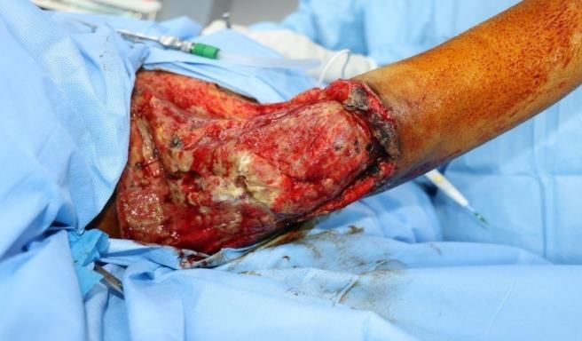
Gupta M et al. Int Surg J. 2021 Aug;8(8):2512-2514
spectrum antibiotics at primary health care centre, patient
did not respond to those antibiotics, and referred to us for
further management. There was history of very fast
spreading of necrotic patch in surrounding normal tissues.
At the time of presentation patient was conscious,
oriented to time, place and person. His vitals were stable
except mild tachycardia (pulse rate 112/minute) and had
history of multiple spikes of fever in the past. At local
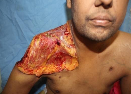
examination patient had necrotic patch with eschar
formation size 12x10 cm with oozing pus from
underneath the eschar (Figure1). Between the margins of
necrotic patch and normal skin there was suspicious
fungal areas seen. KOH smear was sent which came
positive for fungal growth. Intravenous liposomal
amphotericin B started and patient was taken up for Figure 3: Excised tissue specimen for HPE.
surgery with emergency OT clearance. Extensive
debridement of necrotic patches with 2 cm normal skin
margins and few fibres of pectoralis major muscle were
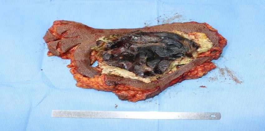
excised (Figure 2). Tissue sent for HPE Figure 3).
Meanwhile patient’s dressing was done with gauge piece
soaked in diluted Amphotericin B solution. After few
days wound margins, discoloration was again noticed
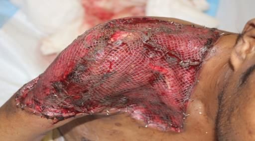
(Figure 4). Patient was again taken up for surgery and
extensive debridement of wound was done up to the
healthy bleeding edges (Figure 5). HPE reports shown
non-septate branching hyphae accompanied with
angioinvasion and tissue necrosis. Patient responded well
to antifungal treatment and few days later once wound
was well granulated, skin grafting was done over the right
shoulder raw areas (Figure 6). Patient was discharged
Figure 4: Post debridement recurrence of fungal
from the hospital in stable condition later on.
growth and skin margins necrosis.
Figure 1: Post RTA mucormycosis or black fungus of Figure 5: Post second debridement of right shoulder
right shoulder. region.
Figure 2: Post first radical debridement of right Figure 6: Post meshed split thickness skin graft cover
shoulder region. for right shoulder region.
International Surgery Journal | August 2021 | Vol 8 | Issue 8 Page 2513
Gupta M et al. Int Surg J. 2021 Aug;8(8):2512-2514

DISCUSSION                                                         REFERENCES

Primary cutaneous mucormycosis infection is necrotizing            1.  Wall S, Lee KH, Alvarez JD, Bigelow DC. Quiz
fungal infection and having angioinvasion and vascular                 case, Cutaneous mucormycosis of the external ear.
thrombosis formation leads to widespread soft tissue                   Arch Otolaryngol Head Neck Surg. 2000;126:236-9.
necrosis.7 Initial presentation of primary cutaneous               2. Elgart ML. Zygomycosis. Dermatol Clin.
mucormycosis can confuse with necrotizing fasciitis. It                1996;14:141-6.
requires high degree of suspicion to make a diagnosis of           3. Prabhu RM, Patel R. Mucormycosis and
mucormycosis. Most commonly involved region is upper                   entomophthoramycosis: a review of the clinical
extremity followed by head and neck region, chest, flanks              manifestations, diagnosis and treatment. Clin
and genitalia.8,9 In this case affected site is upper                  Microbiol Infect. 2004;10(1):31-47.
extremity and shoulder region. In our case report patient          4. Roden MM, Zaoutis TE, Buchanan WL, Knudsen
had multiple major risk factors -first history of road                 TA, Sarkisova TA, Schaufele RL et al. Epidemiology
traffic accident second patient had diabetes mellitus; third           and outcome of zygomycosis: a review of 929
patient was not responding to broad spectrum antibiotics.              reported cases. Clin Infect Dis. 2005;41:634-53.
All these arises the high suspicion of necrotizing fungal          5. Carvalhal GF, Machado MG, Pompeo A, Saldaña L,
infection. Initially screening with KOH smear and then                 Sabbaga E, Arap S. Mucormycosis presenting as a
histopathological examination confirmed the diagnosis of               renal mass in a patient with the human
mucormycosis, which is the gold standard method for                    immunodeficiency virus. J Urol. 1997;158:2230-1.
diagnosing mucormycosis.10 Early diagnosis, appropriate            6. Pérez-Uribe A, Molina de Soschin D, Arenas R,
antifungal treatment along with radical surgical                       Reyes M. Mucormicosis cutánea primaria en un
procedures are the key to manage the primary cutaneous                 paciente con virus de la inmunodeficiencia humana.
mucormycosis.                                                          Revista Iberoam Micol. 2005;22:118-21.
                                                                   7. Kontoyiannis DP, Lewis RE. Invasive zygomycosis:
CONCLUSION                                                             update on pathogenesis, clinical manifestations, and
                                                                       management. Infect Dis Clin North Am.
Our case demonstrates the usefulness of making early                   2006;20:581-607.
diagnosis and emergent surgical treatment along with               8. Tehmeena W, Hussain W, Zargar HR, Sheikh AR,
amphotericin B. It can reduce morbidity and mortality in               Iqbal S. Primary cutaneous mucormycosis in an
cutaneous mucormycosis, although early presentation of                 immunocompetent
cutaneous mucormycosis can be confused with bacterial                  host. Mycopathologia. 2007;164:197-99.
necrotizing fasciitis. The multidisciplinary team is               9. Simbli M, Hakim F, Koudieh M, Tleyjeh IM.
required for the management of complicated cases of                    Nosocomial           post-traumatic        cutaneous
primary cutaneous mucormycosis or black fungus.                        mucormycosis: A systematic review. Scand J Infect
                                                                       Dis. 2008;40:577-82.
The most affected areas of the skin are the arms and legs.         10. Hata TR, Johnson RA, Barnhill R, Dover JS.
Other locations include the scalp, face, thorax, back,                 Ecthyma-like lesions on the leg of an
abdomen, perineum, breast, neck and gluteal area.                      immunocompromised patient. Primary cutaneous
                                                                       mucormycosis. Arch Dermatol. 1995;131:833-4.
Funding: No funding sources
Conflict of interest: None declared                                     Cite this article as: Gupta M, Varshney D.
Ethical approval: Not required                                          Cutaneous mucormycosis or black fungus of right
                                                                        shoulder region-rare case report. Int Surg J
                                                                        2021;8:2512-4.

                                                           International Surgery Journal | August 2021 | Vol 8 | Issue 8   Page 2514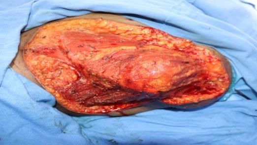
You can also read