Characterization of a Dengue Virus Serotype 1 Isolated from a Patient in Ciudad Juarez, Mexico - MDPI
←
→
Page content transcription
If your browser does not render page correctly, please read the page content below
pathogens
Article
Characterization of a Dengue Virus Serotype 1 Isolated from a
Patient in Ciudad Juarez, Mexico
Pedro M. Palermo 1, *, Antonio de la Mora-Covarrubias 2 , Jeanette Orbegozo 1 , Jessica A. Plante 3 ,
Kenneth S. Plante 3 , Florinda Jimenez-Vega 2 and Douglas M. Watts 1

                                            1   Department of Biological Sciences and Border Biomedical Research Center, University of Texas at El Paso,
                                                El Paso, TX 79968, USA; jporbegozor@miners.utep.edu (J.O.); dwatts2@utep.edu (D.M.W.)
                                            2   Instituto de Ciencias Biomedicas, Universidad Autonoma de Ciudad Juarez, Juarez City 32100, Mexico;
                                                adelamor@uacj.mx (A.d.l.M.-C.); fjimenez@uacj.mx (F.J.-V.)
                                            3   Department of Microbiology and Immunology, University of Texas Medical Branch,
                                                Galveston, TX 77555, USA; japlante@utmb.edu (J.A.P.); ksplante@utmb.edu (K.S.P.)
                                            *   Correspondence: ppalermo@utep.edu; Tel.: +1-915-747-8528

                                            Abstract: Dengue (DEN) is the most important human arboviral disease worldwide. Sporadic
                                            outbreaks of DEN have been reported since 1980 in urban communities located along the border in
                                            southeast Texas and northern Mexico. Other than the Rio Grande Valley region of TX, autochthonous
                                            transmission of DENV has not been reported from any other US border communities. As part of
                                            a surveillance program for arthropod-borne viruses in Ciudad Juarez, Mexico, during November
                                  2015, a blood sample was obtained from a female patient who experienced an undifferentiated fever

                                            and arthralgia. The plasma of the sample was tested for virus in Vero-76 and C6/36 cells. DENV
Citation: Palermo, P.M.;
                                            serotype 1 (DENV-1) was isolated in the C6/36 cells, and nucleotide sequencing of the envelope
de la Mora-Covarrubias, A.;
                                            gene and full genome grouped the DENV-1 isolate in the Central America clade. The patient had
Orbegozo, J.; Plante, J.A.; Plante, K.S.;
                                            not traveled outside of Ciudad Juarez, Mexico, thus suggesting DENV-1 infection was acquired in
Jimenez-Vega, F.; Watts, D.M.
                                            this community.
Characterization of a Dengue Virus
Serotype 1 Isolated from a Patient in
Ciudad Juarez, Mexico. Pathogens
                                            Keywords: dengue virus; Mexico; Central America clade
2021, 10, 872. https://doi.org/
10.3390/pathogens10070872

Academic Editor:                            1. Introduction
Andrew Taylor-Robinson                            Dengue is the most important mosquito-borne viral human disease in the tropical
                                            and subtropical regions of the world. The dengue disease is caused by any of the four
Received: 4 June 2021
                                            (1–4) dengue virus (DENV) serotypes, which can range from a mild febrile illness to severe
Accepted: 8 July 2021
                                            hemorrhagic fever and/or fatal shock syndrome. DENV is transmitted by the Aedes aegypti
Published: 10 July 2021
                                            and Ae. albopictus mosquitoes, and the distribution of both species has increased in the last
                                            30 years, mainly in the temperate regions of Europe and North America [1,2].
Publisher’s Note: MDPI stays neutral
                                                  Dengue cases in the Americas have increased over time from 1980 to 2007, with Mexico
with regard to jurisdictional claims in
                                            being the country with the third highest number of dengue cases [3]. Ae. aegypti is widely
published maps and institutional affil-
                                            distributed in Mexico, including the urban communities in the southern, western, and eastern
iations.
                                            regions, extending along the Gulf Coast as far north as Matamoros, Mexico, as well as
                                            the southern half of the United States (US) [2,4]. Along the US–Mexico border, DENV is
                                            endemic only in the southern region of Texas, even though Ae. aegypti is found in almost
                                            all urban border communities. More recently, data accumulated suggested that DENV is
Copyright: © 2021 by the authors.
                                            endemic in Ciudad Juarez, Mexico, an urban community in the state of Chihuahua located in
Licensee MDPI, Basel, Switzerland.
                                            Northwestern Mexico, which is the second-largest community located along the US–Mexico
This article is an open access article
                                            border. The first reported evidence was the detection of DENV ribonucleic acid (RNA) in Ae.
distributed under the terms and
                                            aegypti mosquitoes in Ciudad Juarez [5]. Subsequently, serological evidence of DENV infection
conditions of the Creative Commons
                                            in humans was also reported in the Anapra neighborhood of Ciudad Juarez, including
Attribution (CC BY) license (https://
creativecommons.org/licenses/by/
                                            seroconversions to DENV infection in 10.4% (n = 5) of 48 individuals who were negative for
4.0/).
                                            DENV antibody during the baseline survey [6]. The individuals who seroconverted did not

Pathogens 2021, 10, 872. https://doi.org/10.3390/pathogens10070872                                           https://www.mdpi.com/journal/pathogens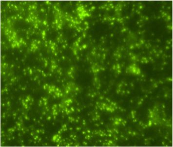
Pathogens 2021, 10, x FOR PEER REVIEW 2 of 12
Pathogens 2021, 10, 872 2 of 11
Subsequently, serological evidence of DENV infection in humans was also reported in the
Anapra neighborhood of Ciudad Juarez, including seroconversions to DENV infection in
10.4% (n = 5) of 48 individuals who were negative for DENV antibody during the baseline
report [6].
survey anyThetravel outside of
individuals theseroconverted
who community, thus suggesting
did not report anythat infection
travel was
outside of acquired
the as a
result of local
community, DENV
thus transmission.
suggesting Furtherwas
that infection evidence suggesting
acquired that of
as a result DENV
local isDENV
endemic in the
Juarez community
transmission. wasevidence
Further supported by the observation
suggesting that DENV reported in this study
is endemic in theofJuarez
the isolation of
DENV-1 in was
community November 2015
supported byfrom a febrile patient
the observation in the
reported in Felipe Angeles
this study of theneighborhood
isolation of located
DENV-1
adjacentintoNovember
the Anapra 2015 from a febrile
community patientJuarez,
of Ciudad in the Mexico.
Felipe Angeles neighborhood
located adjacent to the Anapra community of Ciudad Juarez, Mexico.
2. Results
2. Results
A Flavivirus was isolated from the patient’s plasma sample in the C6/36 cells accord-
ing A Flavivirus
to the was of
detection isolated from the
viral antigen by patient’s plasma sample
immunofluorescence in the
assay C6/36
(IFA) usingcells
polyclonal
according to the detection of viral antigen by immunofluorescence assay (IFA) using
Saint Louis encephalitis virus (SLEV) antibodies, which are cross-reactive to Flavivirus
polyclonal Saint Louis encephalitis virus (SLEV) antibodies, which are cross-reactive to
(Figure 1C). The Flavivirus isolate was further identified as DENV-1 using DENV-1 mon-
Flavivirus (Figure 1C). The Flavivirus isolate was further identified as DENV-1 using
oclonal antibody (Figure 1D) and by the sequencing of the generic Flavivirus RT-PCR
DENV-1 monoclonal antibody (Figure 1D) and by the sequencing of the generic Flavivirus
amplicon
RT-PCR (data not
amplicon shown).
(data Positive
not shown). controls
Positive (DENV-1-infected
controls (DENV-1-infectedC6/36
C6/36cells)
cells)were
were also reac-
tive by IFA to SLEV polyclonal (Figure 1A) and DENV-1 monoclonal antibodies
also reactive by IFA to SLEV polyclonal (Figure 1A) and DENV-1 monoclonal antibodies (Figure 1B).
The infectivity titer of the DENV-1 in the plasma of the patient was 5 × 10 2 PFU/mL at the
(Figure 1B). The infectivity titer of the DENV-1 in the plasma of the patient was 5 × 10²
time theatblood
PFU/mL sample
the time wassample
the blood drawn.was drawn.
A C E
B D F
Indirect
Figure1.1.Indirect
Figure immunofluorescent
immunofluorescent antibody
antibody assayassay in C6/36
in C6/36 cells
cells (200× (200× magnification).
magnification). Cells were Cells were
inoculated
inoculated with DENV-1
with DENV-1(A,B), human
(A,B), plasma
human (C,D),(C,D),
plasma and cell culture
and mediummedium
cell culture (E,F). Viral antigen
(E,F). Viral antigen
was detected by using polyclonal antibodies to Saint Louis encephalitis virus (A,C,E)
was detected by using polyclonal antibodies to Saint Louis encephalitis virus (A,C,E) and and
monoclonal
monoclonal antibody (15F3) to DENV-1 (B,D,F).
antibody (15F3) to DENV-1 (B,D,F).
Viral replication of the second passage of DENV-1 was compared in Vero and C6/36
Viral replication of the second passage of DENV-1 was compared in Vero and
cells up to 10 days post infection (dpi). DENV-1 replication in C6/36 cells was
C6/36 cells up
approximately to 10
3 logs days
higher in post
C6/36infection
cells than (dpi). DENV-1
in Vero-76 replication
cells from 2 to 10 dpiin(pC6/36
< 0.05),cells was
approximately 3 logs higher in C6/36 cells than in Vero-76
reaching the highest titer (2 × 10 PFU/mL) after 7 dpi (Figure 2).
6 cells from 2 to 10 dpi (p < 0.05),
Pathogens 2021, 10, x FOR PEER REVIEW 3 of 12
reaching the highest titer (2 × 10⁶ PFU/mL) after 7 dpi (Figure 2).
Figure 2. Replication curves of the Ciudad Juarez DENV-1 isolate in Vero-76 and C6/36 cells. DENV-1
isolate replicated at significant high titers (** p < 0.05) in C636 cells in comparison to Vero-76 cells.
Figure 2. Replication curves of the Ciudad Juarez DENV-1 isolate in Vero-76 and C6/36 cells. DENV-
1 isolate replicated at significant high titers (** p < 0.05) in C636 cells in comparison to Vero-76 cells.
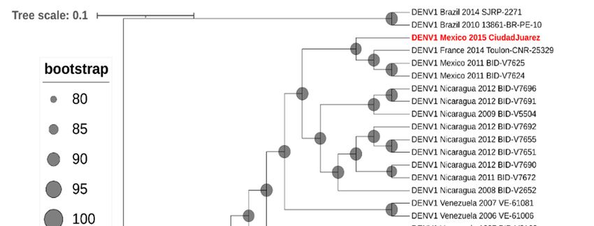
Phylogenetic analysis of the envelope gene indicated that the DENV-1 isolate
clustered in genotype V and grouped with other Central American strains of DENV-1
(Figure 3) and with other DENV-1 strains isolated in Mexico (Tamaulipas, Yucatan, and
Morelos) and Texas (Brownsville). Furthermore, a full genome analysis showed that the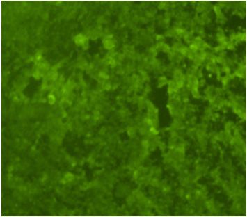
Pathogens 2021, 10, 872                                                                                                                     3 of 11

                                             Phylogenetic analysis of the envelope gene indicated that the DENV-1 isolate clustered
                                        in genotype V and grouped with other Central American strains of DENV-1 (Figure 3) and
                                        with other DENV-1 strains isolated in Mexico (Tamaulipas, Yucatan, and Morelos) and
                                        Texas (Brownsville). Furthermore, a full genome analysis showed that the
      Pathogens 2021, 10, x FOR PEER REVIEW                                                                   4 of DENV-1
                                                                                                                   12        isolate
                                        clustered with Central American DENV-1 strains (Figure 4).

                                 Figure 3. Molecular phylogenetic analysis using maximum-likelihood method derived from 100
                                 Figure 3. Molecular phylogenetic analysis using maximum-likelihood method derived from
                                  DENV-1 envelope glycoprotein gene sequences. DENV-1 genotypes (I–V) and isolates are shown
                                 100  DENV-1
                                  using brackets.envelope    glycoprotein
                                                   The tree was rooted with gene sequences.
                                                                            prototype  strains ofDENV-1
                                                                                                  DENV-3 genotypes    (I–V) and
                                                                                                          (H87) and DENV-2        isolates are shown
                                                                                                                              (NGC).
                                  DENV-1,
                                 using      strain Ciudad
                                         brackets.          Juarez,
                                                      The tree  wasisrooted
                                                                      highlighted
                                                                             withinprototype
                                                                                      red. The tree   withof
                                                                                                  strains  theDENV-3
                                                                                                               highest log likelihood
                                                                                                                        (H87)  and DENV-2 (NGC).
                                  (−12,372.1590) is shown.
                                 DENV-1, strain Ciudad Juarez, is highlighted in red. The tree with the highest log likelihood
                                 (−12,372.1590) is shown.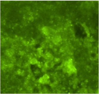
Pathogens 2021, 10, 872                                                                                                                   4 of 11
2021, 10, x FOR PEER REVIEW                                                                                                          5 of 12

                            Figure 4. Phylogenetic
                 Figure 4. Phylogenetic                 analysis Maximum-likelihood
                                          analysis of DENV-1.    of DENV-1. Maximum-likelihood              treefull
                                                                                            tree based on the     based  on the
                                                                                                                     genome              (minus the 50
                                                                                                                                 full genome
                                                                                                                              sequences
                            sequences
                 and 30 UTRs)           (minus
                                of 68 strains     the 5′ and
                                              of DENV-1      3′ UTRs)
                                                           from         of 68along
                                                                 GenBank,      strains
                                                                                    withof the
                                                                                           DENV-1     from GenBank,
                                                                                                2015 Ciudad              along(highlighted
                                                                                                               Juarez isolate   with the 2015
                                                                                                                                           in red and
                            Ciudad   Juarez  isolate  (highlighted  in red  and  bold   text). DENV-2     strain New   Guinea   was  included
                 bold text). DENV-2 strain New Guinea was included to root the tree. In the cladogram, branch lengths are ignored, but
                            to root
                 bootstrapping       the≥tree.
                                 values         Inrepresented
                                           80 are  the cladogram,    branch lengths
                                                               as proportionately        aregray
                                                                                     sized    ignored,
                                                                                                  circlesbut  bootstrapping
                                                                                                          at the               values ≥80 are
                                                                                                                 relevant nodes.
                            represented as proportionately sized gray circles at the relevant nodes.

                                The amino-acid diversity of the DENV-1 isolate was compared with other Mexican
                             DENV-1 strains. The DENV-1 isolate was unique amongst the other Mexican strains in
Pathogens 2021, 10, 872                                                                                                      5 of 11

                                       The amino-acid diversity of the DENV-1 isolate was compared with other Mexican
                                  DENV-1 strains. The DENV-1 isolate was unique amongst the other Mexican strains in
                                  the amino-acid alignment due to five amino-acid changes in the NS1 (D139E), NS2 (L41F,
                                  K218R), and NS3 (V332A, R338K) proteins (Table 1).

           Table 1. Amino-acid changes in the Mexican DENV-1 strains in comparison to the DENV-1 Ciudad Juarez strain.

                                                                       GenBank
            Tree Name             Collection Year   Strain Name                       NS1           NS2A               NS3
                                                                     Accession No.
                                                                                      139      41          218   332         338
 DENV1_Mexico_2006_BID_V3658           2006          BID_V3658        GU131958.1       D        L          K     V           R
  DENV1_Mexico_2007_BID-V3673          2007          BID-V3673        GU131964.1       D        L          K     V           R
  DENV1_Mexico_2007_BID-V3679          2007          BID-V3679        GU131966.1       D        L          K     V           R
  DENV1_Mexico_2007_BID-V3709          2007          BID-V3709        GQ868513.1       D        L          K     V           R
  DENV1_Mexico_2007_BID-V3727          2007          BID-V3727        GQ868521.1       D        L          K     V           R
  DENV1_Mexico_2007_BID-V3739          2007          BID-V3739        GQ868527.1       D        L          K     V           R
  DENV1_Mexico_2007_BID-V7572          2007          BID-V7572        KJ189317.1       D        L          K     V           R
  DENV1_Mexico_2007_BID-V7589          2007          BID-V7589        KJ189323.1       D        L          K     V           R
  DENV1_Mexico_2007_BID-V7590          2007          BID-V7590        KJ189324.1       D        L          K     V           R
  DENV1_Mexico_2008_BID-V3758          2008          BID-V3758        GQ868537.1       D        L          K     V           R
  DENV1_Mexico_2011_BID-V7298          2011          BID-V7298        KJ189306.1       D        L          K     V           R
  DENV1_Mexico_2011_BID-V7624          2011          BID-V7624        KJ189348.1       D        L          K     V           R
  DENV1_Mexico_2011_BID-V7625          2011          BID-V7625        KJ189349.1       D        L          K     V           R
  DENV1_Mexico_2012_BID-V8195          2012          BID-V8195        KJ189368.1       D        L          K     V           R
 DENV1_Mexico_2015_CiudadJuarez        2015         CiudadJuarez      MZ343259         E        F          R     A           K

                                  3. Discussion
                                        The findings herein represent the first reported isolation of a DENV from a patient
                                  in Ciudad Juarez, Mexico, where previous serological evidence of DENV infection was
                                  reported among a human cohort [6]. The patient’s clinical data were consistent with a
                                  febrile illness with a DENV-1 viremia level of 5 × 10² PFU/mL in the plasma and the
                                  consequent isolation of DENV-1 in C6/36 cells, in addition to being consistent with clinical
                                  criteria routinely used for DENV isolation from clinical samples [7].
                                        Viral replication of the DENV-1 isolate was higher in C6/36 cells than in Vero cells
                                  (Figure 2); even though in vitro testing was performed, the findings suggested the ability
                                  of the DENV-1 isolate to replicate in its mosquito vector in nature. Furthermore, a limited
                                  number of passages were performed, restricting the adaptation of the DENV-1 isolate in
                                  the mosquito cell line.
                                        Mexico has experienced several large outbreaks of dengue in the last two decades
                                  (2000–2019) causing more than 520,000 human cases [8]. Furthermore, most of the dengue
                                  cases in the last 10 years were reported from the northern states of Mexico, where DENV-1
                                  and DENV-2 are the predominant serotypes. The DENV-1 isolate from this study clustered
                                  with other Mexican and Central American strains of DENV-1 belonging to genotype V.
                                  DENV-1 genotype V has been circulating in Mexico since the late 1970s and has experi-
                                  enced several lineage replacements since its introduction, causing a high degree of genetic
                                  variability [9]. Moreover, DENV-1 genotype V is widely distributed in the Americas, and
                                  its clades are associated with distinct regions (South America, Caribbean, and Central
                                  America) [10]. Furthermore, the presence of various country-specific DENV-1 founder
                                  effects may have supported the geographical structure of DENV-1 genotype V in the
                                  Americas [11].
                                        Variation in the virus genome has been reported to be associated with virulence.
                                  For example, amino-acid substitutions on the DENV E and NS3 proteins have resulted
                                  in enhanced virulence in animal models and neurotoxicity, respectively [12,13]. Other
                                  amino-acid substitutions on the DENV NS1, NS4B, and NS5 proteins have increased the
                                  viral fitness in endemic regions [14]. Our study identified five amino-acid changes in
                                  the DENV-1 isolate that were unique in comparison with other Mexican DENV isolates
                                  (Table S1). Most of the changes involved amino acids with similar properties (e.g., NS1
                                  (D139E), NS2 (K218R), and NS3(V332A, R338K)), which may not of have resulted in a viral
                                  protein dysfunction. Furthermore, four amino-acid changes in the Ciudad Juarez strain
                                  were also found in other DENV-1 strains, mainly from Asia. Briefly, NS1 139D was changed
Pathogens 2021, 10, 872 6 of 11
to N in strains from China (GIZ-11), French Polynesia (BID-V2939), Hawaii (Haw03663),
Japan (Mochizuki), Malaysia (P72-1244), and Thailand (606147 and ThD1-0081-82). NS2A
41L was changed to F in Brazil (SJRP-2271), China (GIZ-11), and India (715393) strains. NS3
332V was changed to A in Brazil (SJRP-2271), Gabon (Gabon2012), and Hawaii (Haw03663),
and changed to I in the Thailand (606147) strain. Lastly, NS3 338R was changed to K in the
French Polynesia (BID-V2939) strain [15].
The results suggested that the patient acquired DENV-1 infection in Ciudad Juarez
because no travel history was reported before the onset of illness. Moreover, that DENVs
are endemic in this urban community is supported by the observation that three individuals
seroconverted to DENV-1 infection during 2015 in Ciudad Juarez, and two of the three
were positive for dengue IgM antibody [6]. Furthermore, a total of 10 human DENV-1
infections were reported in Ciudad Juarez in 2015 [16], thus documenting that the DENV-1
is endemic in this Mexican border urban community, representing a possible source for
locally acquired DENV infections. A total of 12 dengue cases have been reported in Ciudad
Juarez during the last 5 years, with DENV-1 being the most frequent cause of cases (n = 11),
whereas a single case was caused by DENV-2 in 2019 [17–19].
An alternative explanation for our findings regarding the human cases of dengue
in Ciudad Juarez is the possibility of imported dengue cases resulting in secondary focal
and transient autochthonous transmission to cause one or more cases. The reported
occurrence of sporadic outbreaks of dengue attributed to autochthonous transmission from
1980 to 2013 in Brownsville, Texas, and surrounding communities was associated with
DENV-infected travelers returning from visits to the bordering Mexican city of Matamoros
during dengue epidemics in this community [20–22]. Thus, a low number of secondary
autochthonous cases may have been acquired from returning viremic travelers as a source of
infection, before going undetected, especially in areas without active surveillance programs;
furthermore, asymptomatic or silent DENV infections are more difficult to detect than
symptomatic cases [23,24].
Further longitudinal cohort studies in humans and mosquito surveillance are needed
to obtain a better understanding of the dynamics of DENV transmission in this Mexican
border community. Lastly, the vectorial capacity of Ae. aegypti mosquitoes from this
Mexican border region for DENV would help to provide information for predicting the
risk of DENV transmission.
4. Materials and Methods
4.1. Patient and Sample Collection and Processing
A 64 year old female housewife who resided in Felipe Angeles, Ciudad Juarez, Chi-
huahua (Mexico) experienced a febrile illness on November 2015, which was characterized
by an acute onset of chills, severe headache, fever, diarrhea, joint pain, bad taste in the
mouth, and decreased appetite. She sought medical attention, and medication was admin-
istered for symptomatic treatment. A venous blood sample was collected during the acute
phase of illness from the patient with a 6 mL vacutainer tube containing ethylenediaminete-
traacetic acid (EDTA) from the arm of the patient at her residence in the neighborhood
of Felipe Angeles, Ciudad Juarez, Chihuahua, Mexico (Figure 5). The blood sample was
centrifuged at 3000× g for 10 min, and the plasma was stored in aliquots of 0.5 mL at
−80 ◦ C until tested for arboviruses in C6/36 and Vero-76 cells. A convalescent blood
sample was not available for antibody testing. In addition, the female patient did not report
any travel history prior to the onset of symptoms. The deidentified medical history and
deidentified blood sample were provided by one of the authors (A.M.C.) to another author
(D.M.W.) at the University of Texas at El Paso (UTEP), Texas, to test for arboviruses. The
patient was enrolled in accordance with the Ethics Committee at the Institute of Biomedical
Sciences of the Universidad Autonoma de Ciudad Juarez, Mexico, using a written informed
consent form and a questionnaire to obtain demographic and clinical information.1, 10, x FOR PEER REVIEW 8 of 12
Institute of Biomedical Sciences of the Universidad Autonoma de Ciudad Juarez, Mexico,
Pathogens 2021, 10, 872 7 of 11
using a written informed consent form and a questionnaire to obtain demographic and
clinical information.
Figure 5.Figure
Map of5.Ciudad
Map of Ciudad
Juarez, Juarez,
Mexico, Mexico,the
highlighting highlighting the adjoining
adjoining neighborhood neighborhood
of Felipe of Felipe
Angeles, Ciudad Juarez,
Angeles,
Chihuahua 32100. Ciudad Juarez, Chihuahua 32100.
4.2. Viral Isolation
4.2. Viral Isolation
The plasma sample was diluted
The plasma sample1:10wasin cell culture
diluted 1:10 in maintenance medium (Minimum
cell culture maintenance medium (Minimum
Essential Medium Essential
(MEM) Medium (MEM) supplemented
supplemented with 2% fetalwith 2% fetal
bovine serumbovine serum
(FBS), 1% (FBS), 1% penicillin–
penicillin–
streptomycin, and 1% non-essential amino acids). Two hundred
streptomycin, and 1% non-essential amino acids). Two hundred microliters of the diluted microliters of the diluted
plasma sample was inoculated
plasma sample was inoculated onto confluent onto confluent monolayers of Vero-76
monolayers of Vero-76 and C6/36 cells and C6/36 cells
propagated in a T-25 cm 2 flask and incubated for 1 h at 37 ◦ C and 28 ◦ C in the presence
propagated in a T-25 cm2 flask and incubated for 1 h at 37 °C and 28 °C in the presence of
of 5% CO2 , respectively. Then, 5 mL of the maintenance medium was added to each
5% CO2, respectively.
culture,Then, 5 mL
and the ofand
cells the inoculum
maintenance weremedium
incubated was
foradded
7 days to each
at 37 ◦ Cculture,
and 28 ◦ C in 5%
and the cells andCO2 . Confluent monolayers of Vero-76 and C6/36 cells were inoculated withCO
inoculum were incubated for 7 days at 37 °C and 28 °C in 5% 2.
maintenance
Confluent monolayers of Vero-76 and C6/36 cells were inoculated with maintenance
medium to serve as controls. The cells were observed for evidence of viral cytopathic
medium to serve as controls.
effect (CPE) onceThe dailycells werean
through observed
inverted for evidenceThen,
microscope. of viral cytopathic
7 days post inoculation,
effect (CPE) oncethedaily
Vero through
and C6/36 ancells were scraped
inverted off theThen,
microscope. flasks and the post
7 days suspensions were clarified
inoculation,
by centrifugation at 3000 × g for 10 min at 4 ◦ C. The clarified cells and supernatants were
the Vero and C6/36 cells were scraped off the flasks and the suspensions were clarified by
80 ◦10
3000×at g−for
centrifugation atstored C and
minlabeled
at 4 °C. as The
passage 1 (p-1).cells
clarified Theand
cell supernatants
pellets were resuspended
were in
phosphate-buffered saline (PBS) 1×, and aliquots of 20 µL were spotted onto the ringed
stored at −80 °C and labeled as passage 1 (p-1). The cell pellets were resuspended in
areas of slides and dried and fixed in cold acetone at −20 ◦ C for 10 min, before storing
phosphate-buffered saline (PBS) 1×, and aliquots of 20 µL were spotted onto the ringed
at −20 ◦ C for subsequent testing of the cell pellets for selected arboviruses using an
areas of slides and dried and fixed assay.
immunofluorescence in cold acetone at −20 °C for 10 min, before storing at
−20 °C for subsequent testing of the cell pellets for selected arboviruses using an
4.3. Immunofluorescence
immunofluorescence assay. Assay (IFA)
The IFA was performed on the cell pellet fixed to the ringed area of the slides using
polyclonal
4.3. Immunofluorescence antibodies
Assay (IFA) for West Nile, Saint Louis encephalitis, DENV serotypes 1 and 2
(DENV-1, DENV-2), Chikungunya, Western equine encephalitis, and La Crosse. Further-
The IFA was performed on the cell pellet fixed to the ringed area of the slides using
more, DENV-1 monoclonal antibody (15F3) ascites were included in the test. Briefly, 10 µL
polyclonal antibodies for West Nile, Saint Louis encephalitis, DENV serotypes 1 and 2
(DENV-1, DENV-2), Chikungunya, Western equine encephalitis, and La Crosse.Pathogens 2021, 10, 872 8 of 11
of polyclonal or monoclonal antibodies were added to the cell pellets of the slide and
incubated at 37 ◦ C for 1 h. Then, the slides were washed twice in PBS 1× and dried,
before adding 10 µL of goat anti-mouse IgG antibody conjugated with fluorescein isothio-
cyanate (1:100 diluted in PBS 1×) and incubating at 37 ◦ C for 1 h. DAPI (40 ,6-diamidino-2-
phenylindole) was used as a counterstain (1:1000 dilution). The slides were washed twice
in PBS 1× and dried, before adding a drop of a glycerol solution (nine parts glycerol + one
part PBS) onto the ringed area of the slides. Slides were examined for fluorescence using a
20× objective on a fluorescent microscope (Nikon, Ti-S). Slide controls containing known
infected cells with each of the test viruses and/or uninfected cells were subjected to the
same procedure. A Flavivirus was detected by IFA using Flavivirus polyclonal antibodies
and shown to be DENV-1 using DENV-1 monoclonal antibody.
4.4. Viremia Levels
Tenfold dilutions of the patient’s plasma sample were prepared in maintenance
medium, and 50 µL of each dilution was inoculated into a suspension of baby hamster
kidney cells (BHK-21 clone 15) propagated in 24-well plates. A plaque assay was then
performed to determine the titer of the DENV-1 isolate as described previously [25].
4.5. Molecular Identification
RNA was extracted from the Vero-76 and C6/36 cells infected with DENV-1 (p-1)
using the QIAamp viral RNA kit (Qiagen, Valencia, CA, USA), following the manufac-
turer’s protocol. A generic reverse-transcription polymerase chain reaction (RT-PCR)
assay was performed to detect nucleic acid from Flaviviruses, as previously described [26].
The amplicons obtained were purified and sequenced using the BigDye® Terminator 3.1
Cycle Sequencing Kit (Applied Biosystems, Foster City, CA, USA), with the sequencer
3730xl DNA analyzer (Applied Biosystems). Sequences were edited with the Sequencher
4.6 software (Gene Codes Corporation, Ann Arbor, MI, USA) and compared to other viral
sequences deposited in the GenBank database using the BLASTn analysis.
4.6. Envelope Gene Sequencing
The pre-membrane (preM) and envelope (E) genes of the DENV-1 isolate (p1) were am-
plified by RT-PCR [27,28]. Phylogenetic analysis of the E gene of DENV-1 was performed
using neighbor joining and maximum probability analysis implemented in MEGA7 [29].
The best-fit model (GTR+ I + G) was determined by the Mr. Model test software. Clades
were evaluated by bootstrap analyses with 1000 replicates for maximum probability anal-
yses. Initial trees for the heuristic search were obtained automatically by applying the
Neighbor-Join and BioNJ algorithms to a matrix of pairwise distances estimated using the
maximum composite likelihood (MCL) approach and then selecting the topology with the
superior log-likelihood value.
4.7. Viral Whole-Genome Sequencing
DENV-1 RNA was prepared using the NEBNext Ultra II RNA Library Prep Kit (New
England BioLabs, Ipswich, MA). Finished libraries were quality-checked on an Agilent
Bioanalyzer (Agilent, Santa Clara, CA, USA) and quantified by real-time PCR. Libraries
were pooled and sequenced on a NextSeq 550 (Illumina, San Diego, CA, USA) using the
High-output kit (Illumina, San Diego, CA, USA) and a paired-end 75 base read protocol.
Raw reads were filtered to remove low-quality reads and adapter sequences using the
Trimmomatic package [30]. The read assembly program ABySS was used to assemble the
filtered reads into longer de novo contigs to assemble the genome. The genome sequence
of DENV-1 was submitted to GenBank with the accession number MZ3432597.
Sixty-eight DENV-1 sequences, as well as DENV-2 New Guinea C to root the tree, were
downloaded from GenBank (Table S1). The resulting set of 69 strains from GenBank plus the
2015 isolate from Ciudad Juarez, Mexico, were aligned using Clustal Omega in MegAlign
Pro version 17.0.0 (DNASTAR Inc., Madison, WI, USA) and trimmed to remove the 50 andPathogens 2021, 10, 872 9 of 11
30 UTRs. Maximum-likelihood analysis was performed using the phangorn package [31]
in R version 3.5.3 (R Core Team 2017). The GTR + G + I model of nucleotide substitution
was selected from the 24 options analyzed as part of the model test analysis in phangorn
on the basis of minimizing the AIC score. A maximum likelihood tree was generated
using a rooted UPGMA tree as a starting point, and bootstrapping was performed with
1000 iterations. The resulting tree was visualized using iTOL version 5.7 [32].
4.8. Replication Curve
The infectivity replication curve of the DENV-1 isolate was determined in Vero-76 and
C6/36 cells by plaque assay in BHK-21 clone 15 cells [6]. The C6/36 cells were grown in a
T-75 cm2 flask and inoculated with the DENV-1 isolate (p-1 from C6/36 cells supernatant)
at a multiplicity of infection (MOI) of 0.01. Cells were harvested once CPE became evident
at 7 days post infection (dpi), and aliquots of the clarified supernatant (p-2) were stored
at −80 ◦ C until used to determine the virus infectivity replication curves. Briefly, Vero-76
and C6/36 cells were inoculated in triplicate with the DENV-1 suspension (p-2) at an MOI
of 0.01 and incubated at 37 ◦ C and 28 ◦ C, respectively, for 1 h. Then, the inoculum was
removed, and the cells were washed three times with sterile PBS 1× pH 7.4. Five milliliters
of maintenance medium was added to cells of each flask, and a 0.5 mL aliquot was collected
immediately (time point 0) and replaced with 0.5 mL of new medium. Cells were incubated
as mentioned above, and aliquots were collected and replaced with new medium every
day until 10 dpi. Timepoint aliquots were tested to determine virus infectivity titers by
plaque assays on BHK-21 clone 15 cells. Statistical differences in the viral replication titers
were determined using Student’s t-test.
Supplementary Materials: The following are available online at https://www.mdpi.com/article/
10.3390/pathogens10070872/s1: Table S1. DENV strains for phylogenetic analysis. Strain names,
accession numbers, and country and year of collection are included.
Author Contributions: Conceptualization, P.M.P., A.d.l.M.-C. and D.M.W.; methodology, P.M.P., J.O.,
K.S.P., J.A.P. and F.J.-V.; software analysis, J.O., K.S.P., J.A.P. and A.d.l.M.-C.; writing—original draft
preparation and writing—review and editing, P.M.P. and D.M.W. All authors have read and agreed
to the published version of the manuscript.
Funding: This research was funded by the Office Research and Sponsored Projects and by a Grant
2U54MD007592 from the National Institutes on Minority Health and Health Disparities (NIMHD), a
component of the National Institutes of Health (NIH).
Institutional Review Board Statement: The study was approved by the University Autonoma de
Ciudad Juarez Institutional Review Board for the protection of human subjects.
Informed Consent Statement: Informed consent was obtained from the human subject involved in
the study.
Data Availability Statement: Data is provided in the supplementary table of the article.
Acknowledgments: The authors are grateful to the technicians from the Universidad Autonoma de
Ciudad Juarez for collection of the patient sample.
Conflicts of Interest: The authors declare no conflict of interest. The funders had no role in the design
of the study; in the collection, analyses, or interpretation of data; in the writing of the manuscript, or
in the decision to publish the results.
References
1. Kraemer, M.U.; Sinka, M.E.; Duda, K.A.; Mylne, A.; Shearer, F.M.; Brady, O.J.; Messina, J.P.; Barker, C.M.; Moore, C.G.; Carvalho,
R.G.; et al. The global compendium of Aedes aegypti and Ae. albopictus occurrence. Sci. Data 2015, 2, 150035. [CrossRef]
[PubMed]
2. Hahn, M.B.; Eisen, L.; McAllister, J.; Savage, H.M.; Mutebi, J.P.; Eisen, R.J. Updated Reported Distribution of Aedes (Stegomyia)
aegypti and Aedes (Stegomyia) albopictus (Diptera: Culicidae) in the United States, 1995–2016. J. Med. Entomol. 2017, 5, 1420–1424.
[CrossRef] [PubMed]Pathogens 2021, 10, 872 10 of 11
3. San Martín, J.L.; Brathwaite, O.; Zambrano, B.; Solórzano, J.O.; Bouckenooghe, A.; Dayan, G.H.; Guzmán, M.G. The epidemiology
of dengue in the Americas over the last three decades: A worrisome reality. Am. J. Trop. Med. Hyg. 2010, 1, 128–135. [CrossRef]
4. De Lourdes Muñoz, M.; Mercado-Curiel, R.F.; Diaz-Badillo, A.; Pérez Ramirez, G.; Black, W.C. Gene flow pattern among Aedes
aegypti populations in Mexico. J. Am. Mosq Control. Assoc. 2013, 29, 1–18. [CrossRef] [PubMed]
5. De la Mora-Covarrubias, A.; Jiménez-Vega, F.; Treviño-Aguilar, S.M. Geospatial distribution and detection of dengue virus in
Aedes (Stegomyia) aegypti mosquitos in Ciudad Juárez, Chihuahua, Mexico. Salud Publica Mex. 2010, 52, 127–133. [PubMed]
6. Palermo, P.M.; De la Mora-Covarrubias, A.; Jimenez-Vega, F.; Watts, D.M. Serological evidence of Dengue and West Nile Virus
human infection in Juarez City, Mexico. Vector Borne Zoonotic Dis. 2019, 2, 134–141. [CrossRef] [PubMed]
7. Jarman, R.G.; Nisalak, A.; Anderson, K.B.; Klungthong, C.; Thaisomboonsuk, B.; Kaneechit, W.; Kalayanarooj, S.; Gibbons, R.V.
Factors influencing dengue virus isolation by C6/36 cell culture and mosquito inoculation of nested PCR-positive clinical samples.
Am. J. Trop. Med. Hyg. 2011, 2, 218–223. [CrossRef] [PubMed]
8. Arredondo-García, J.L.; Aguilar-López, E.G.; Aguilar Lugo-Gerez, J.; Osnaya-Romero, N.; Pérez-Guillé, G.; Medina-Cortina, H.
Epidemiological panorama of dengue in Mexico 2000–2019. Rev. Latin Infect. Pediatr. 2020, 33, 78–83.
9. González-Durán, E.; Vázquez-Pichardo, M.; Torres-Flores, J.M.; Garcés-Ayala, F.; Méndez-Tenorio, A.; Curiel-Quesada, E.;
Ortiz-Alcántara, J.M.; Castelán-Sánchez, H.G.; Salas-Benito, J.S.; Torres-Longoria, B.; et al. Genotypic variability analysis of
DENV-1 in Mexico reveals the presence of a novel Mexican lineage. Arch. Virol. 2018, 163, 1643–1647. [CrossRef]
10. Villabona-Arenas, C.J.; Zanotto, P.M. Worldwide spread of Dengue virus type 1. PLoS ONE 2013, 8, e62649. [CrossRef]
11. Carvalho, S.E.; Martin, D.P.; Oliveira, L.M.; Ribeiro, B.M.; Nagata, T. Comparative analysis of American Dengue virus type 1
full-genome sequences. Virus Genes 2010, 40, 60–66. [CrossRef]
12. Prestwood, T.R.; Prigozhin, D.M.; Sharar, K.L.; Zellweger, R.M.; Shresta, S. A mouse-passaged dengue virus strain with reduced
affinity for heparan sulfate causes severe disease in mice by establishing increased systemic viral loads. J. Virol. 2010, 82,
8411–8421. [CrossRef]
13. Duarte dos Santos, C.N.; Frenkiel, M.P.; Courageot, M.P.; Rocha, C.F.; Vazeille-Falcoz, M.C.; Wien, M.W.; Rey, F.A.; Deubel, V.;
Desprès, P. Determinants in the envelope E protein and viral RNA helicase NS3 that influence the induction of apoptosis in
response to infection with dengue type 1 virus. Virology 2000, 274, 292–308. [CrossRef] [PubMed]
14. OhAinle, M.; Balmaseda, A.; Macalalad, A.R.; Tellez, Y.; Zody, M.C.; Saborío, S.; Nuñez, A.; Lennon, N.J.; Birren, B.W.; Gordon, A.;
et al. Dynamics of dengue disease severity determined by the interplay between viral genetics and serotype-specific immunity.
Sci. Transl. Med. 2011, 3, 114ra128. [CrossRef]
15. Tang, Y.; Rodpradit, P.; Chinnawirotpisan, P.; Mammen, M.P.; Li, T., Jr.; Lynch, J.A.; Putnak, R.; Zhang, C. Comparative analysis
of full-length genomic sequences of 10 dengue serotype 1 viruses associated with different genotypes, epidemics, and disease
severity isolated in Thailand over 22 years. Am. J. Trop Med. Hyg. 2010, 83, 1156–1165. [CrossRef] [PubMed]
16. Direccion General de Epidemiologia. Mexico. Panorama Epidemiologico de Dengue 2015. Available online: https://www.gob.
mx/cms/uploads/attachment/file/45495/Pano_dengue_sem_52_2015.pdf (accessed on 21 May 2021).
17. Direccion General de Epidemiologia. Mexico. Panorama Epidemiologico de Dengue 2016. Available online: https://www.gob.
mx/cms/uploads/attachment/file/178952/Pano_dengue_sem_52_2016.pdf (accessed on 21 May 2021).
18. Direccion General de Epidemiologia. Mexico. Panorama Epidemiologico de Dengue 2017. Available online: https://www.gob.
mx/cms/uploads/attachment/file/285237/Pano_dengue_sem_52_2017.pdf (accessed on 21 May 2021).
19. Direccion General de Epidemiologia. Mexico. Panorama Epidemiologico de Dengue 2019. Available online: https://www.gob.
mx/cms/uploads/attachment/file/524262/Pano_dengue_52_2019.pdf (accessed on 21 May 2021).
20. Rawlings, J.A.; Hendricks, K.A.; Burgess, C.R.; Campman, R.M.; Clark, G.G.; Tabony, L.J.; Patterson, M.A. Dengue surveillance in
Texas, 1995. Am. J. Trop. Med. Hyg. 1998, 59, 95–99. [CrossRef]
21. Ramos, M.M.; Mohammed, H.; Zielinski-Gutierrez, E.; Hayden, M.H.; Lopez, J.L.; Fournier, M.; Trujillo, A.R.; Burton, R.;
Brunkard, J.M.; Anaya-Lopez, L.; et al. Epidemic dengue and dengue hemorrhagic fever at the Texas-Mexico border: Results of a
household-based seroepidemiologic survey, December 2005. Am. J. Trop. Med. Hyg. 2008, 78, 364–369. [CrossRef]
22. Thomas, D.L.; Santiago, G.A.; Abeyta, R.; Hinojosa, S.; Torres-Velasquez, B.; Adam, J.K.; Evert, N.; Caraballo, E.; Hunsperger, E.;
Muñoz-Jordán, J.L.; et al. Reemergence of dengue in southern Texas, 2013. Emerg. Infect. Dis. 2016, 22, 1002–1007. [CrossRef]
[PubMed]
23. Chen, W.J.; Chen, S.L.; Chien, L.J.; Chen, C.C.; King, C.C.; Harn, M.R.; Hwang, K.P.; Fang, J.H. Silent transmission of the dengue
virus in southern Taiwan. Am. J. Trop. Med. Hyg. 1996, 1, 12–16. [CrossRef]
24. Fredericks, A.C.; Fernandez-Sesma, A. The burden of dengue and chikungunya worldwide: Implications for the Southern United
States and California. Ann. Glob. Health 2014, 80, 466–475. [CrossRef] [PubMed]
25. Morens, D.M.; Halstead, S.B.; Repik, P.M.; Putvatana, R.; Raybourne, N. Simplified plaque reduction neutralization assay for
dengue viruses by semimicro methods in BHK-21 cells: Comparison of the BHK suspension test with standard plaque reduction
neutralization. J. Clin. Microbiol. 1985, 22, 250–254. [CrossRef]
26. Kuno, G.; Chang, G.J.; Tsuchiya, K.R.; Karabatsos, N.; Cropp, C.B. Phylogeny of the genus Flavivirus. J. Virol. 1998, 72, 73–83.
[CrossRef]
27. Díaz, F.J.; Black, W.C.; Farfán-Ale, J.A.; Loroño-Pino, M.A.; Olson, K.E.; Beaty, B.J. Dengue virus circulation and evolution in
Mexico: A phylogenetic perspective. Arch. Med. Res. 2006, 37, 760–773. [CrossRef]Pathogens 2021, 10, 872 11 of 11
28. Warrilow, D.; Northill, J.A.; Pyke, A.T. Sources of dengue viruses imported into Queensland, australia, 2002–2010. Emerg. Infect.
Dis. 2012, 18, 1850–1857. [CrossRef]
29. Kumar, S.; Stecher, G.; Tamura, K. MEGA7: Molecular Evolutionary Genetics Analysis version 7.0 for bigger datasets. Mol. Biol.
Evol. 2016, 33, 1870–1874. [CrossRef] [PubMed]
30. Bolger, A.M.; Lohse, M.; Usadel, B. Trimmomatic: A flexible trimmer for Illumina sequence data. Bioinformatics 2014, 30, 2114–2120.
[CrossRef] [PubMed]
31. Schliep, K.P. phangorn: Phylogenetic analysis in R. Bioinformatics 2011, 27, 592–593. [CrossRef] [PubMed]
32. Letunic, I.; Bork, P. Interactive Tree of Life (iTOL) v4: Recent updates and new developments. Nucleic Acids Res. 2019, 47, 256–259.
[CrossRef]You can also read


