Case Report Rhabdomyolysis plus Hypocalcemia and Diabetic Ketoacidosis as Concurrent Rare COVID-19 Manifestations
←
→
Page content transcription
If your browser does not render page correctly, please read the page content below
Hindawi
Case Reports in Medicine
Volume 2021, Article ID 6625086, 6 pages
https://doi.org/10.1155/2021/6625086

Case Report
Rhabdomyolysis plus Hypocalcemia and Diabetic Ketoacidosis as
Concurrent Rare COVID-19 Manifestations

          Maryam Heidarpour ,1 Mehrbod Vakhshoori ,2 Mohammad Ali Haghighatpanah ,3
          Leila Ashrafi ,1 Farzin Khorvash ,4 and Bijan Iraj 1
          1
            Isfahan Endocrine and Metabolism Research Center, Isfahan University of Medical Sciences, Isfahan, Iran
          2
            Heart Failure Research Center, Cardiovascular Research Institute, Isfahan University of Medical Sciences, Isfahan, Iran
          3
            Isfahan University of Medical Sciences, Isfahan, Iran
          4
            Acquired Immunodeficiency Research Center, Isfahan University of Medical Sciences, Isfahan, Iran

          Correspondence should be addressed to Bijan Iraj; bijaniraj@gmail.com

          Received 28 December 2020; Revised 23 February 2021; Accepted 27 February 2021; Published 10 March 2021

          Academic Editor: Mamede de Carvalho

          Copyright © 2021 Maryam Heidarpour et al. This is an open access article distributed under the Creative Commons Attribution
          License, which permits unrestricted use, distribution, and reproduction in any medium, provided the original work is
          properly cited.
          Background. Common manifestations of coronavirus disease 2019 (COVID-19) from its initial official introduction are mostly
          related to the respiratory system. However, other rarer presentations are reported nowadays. Case Presentations. We reported
          three cases of COVID-19-infected patients with rhabdomyolysis as well as two other rarer simultaneous signs, including hy-
          pocalcemia (Case 1) and diabetic ketoacidosis (DKA) (Case 2). Conclusion. Despite the fact that rhabdomyolysis is an infrequent
          manifestation of COVID-19, high clinical suspicion is required for proper diagnosis and management of this disease as well as
          other concurrent rarer presentations, including hypocalcemia and DKA for the prevention of further complications.

1. Introduction                                                        some viruses, including severe acute respiratory syndrome
                                                                       (SARS), Middle East respiratory syndrome (MERS), and
Coronavirus disease 2019 (COVID-19) emerges as a novel                 Ebolavirus [3, 4]. For instance, a study done on patients
infectious agent with several body organs’ involvement. This           suffering from SARS reported that hypocalcemia was
disease’s most common presentations are fever, dry cough,              prevalent in 70% of individuals during the hospitalization
fatigue, or myalgia [1]. However, some patients develop                duration [4]. Another study investigated the prevalence of
severe symptoms, including shortness of breath, requiring              hypocalcemia among severely ill COVID-19 patients and
further therapeutic support. On the other hand, rarer                  figured out that almost two-thirds of them had lower cal-
manifestations of COVID-19 have been declared.                         cium levels and had subsequent worse outcomes [5].
    Rhabdomyolysis is defined as muscle cell destruction                    Diabetic ketoacidosis (DKA) is another rare manifes-
resulting in subsequent leakage of cellular components to the          tation during the COVID-19 pandemic. Goldman et al.
bloodstream. This disease is mostly manifested with weak-              reported that this disorder is prevalent among 1.8% of
ness, myalgia, electrolyte imbalance, myoglobinuria, or even           admitted patients [6]. Chan et al. performed a case series on
acute kidney injury [2]. However, the exact etiology of                six patients with COVID-19 admitted with DKA and/or
rhabdomyolysis among COVID-19 patients needs to be                     hyperosmolar hyperglycemic state (HHS) and found that
investigated in future studies.                                        these two glucose imbalance disorders were the primary
    Moreover, hypocalcemia is a common phenomenon                      manifestations in most cases [7]. However, the data are
among critically ill patients, and it has been reported that           limited, and several future studies in terms of predisposing
calcium plays a pivotal role in the replication mechanisms of          factors are required.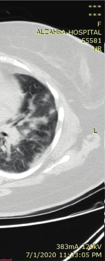
2 Case Reports in Medicine
Herein, we reported three cases of COVID-19 patients psychosis, and he consumed olanzapine, lorazepam, pro-
with rhabdomyolysis as well as other concurrent rarer pranolol, and sertraline for the last ten years. He was opium-
symptoms, including hypocalcemia and DKA. addicted and used 20 cc of daily methadone. At admission,
he was confused and lethargic with normal vital signs except
for high temperature (T � 38.5°C). Besides dehydration,
2. Case Presentation
other physical examinations did not reveal any positive
2.1. Case 1. A 22-year-old man with no previous past findings. His blood sample was taken and showed a blood
medical history was referred to the emergency department sugar of 500 mg/dl, pH of 7, and HCO3 of 2.5 mEq/l. With
with severe hypocalcemia (Ca: 5 mg/dl) as well as spastic the assumed diagnosis of DKA, insulin, tazocin, and serum
muscles in both upper and lower extremities. Also, he was therapy was initiated. The patient’s general condition and
opium-addicted and used 15 cc of daily methadone. He had blood sugar levels were improved during the next day, and
severe bloodless diarrhea from 20 days before admission the HCO3 concentration was raised to 9.5 mEq/l. On the
managed with serum therapy. His vital signs were in normal third day of admission, his HCO3 was 11 mEq/l. However,
ranges. He was conscious, but he appeared to be pale. He did insulin infusion was held due to a lower potassium level. The
not have any positive history in terms of nausea, vomiting, patient suffered from dyspnea and shortness of breath on the
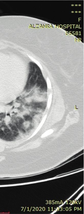
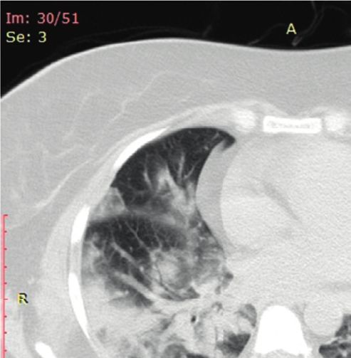
cough, or dyspnea. However, he suffered from generalized next day and was connected to the ventilator. HRCT was
muscle spasms and pain. There was not any positive finding requested, and the results were highly suggestive of COVID-
during heart and lung examinations. His abdomen was 19 infection (Figure 2). Additional laboratory data were as
nontender, but pitting edema (2+) was observed in the lower follows: potassium of 7 mEq/l, sodium of 138 mEq/l, LDH of
extremities. Chvostek’s and Trousseau’s signs were positive, 1296 U/L, CPK of 5130 U/L, phosphorus of 2 mg/dl, and Cr
and his electrocardiogram (ECG) revealed a prolonged QT of 5.3 mg/dl.
interval (QT � 550 msec). Due to the high prevalence of Prompt therapeutic measurements were done, and be-
COVID-19, lung high-resolution computed tomography cause of decreased urine output, a temporary dialysis
(HRCT) was performed and the results were highly sug- catheter was inserted, and he underwent hemodialysis.
gestive of COVID-19 infection (Figure 1). During the hospital admission, he experienced an attack of
In the first hour of admission, intravenous infusion of hypotension managed with norepinephrine as well as hy-
10% calcium gluconate was initiated promptly, and muscle drocortisone. An abdominal computed tomography (CT)
spasms disappeared within four hours. Intravenous calcium scan was performed due to increased amylase (142 U/L) and
administration was then continued based on plasma-cal- lipase (351 U/L) levels. The findings of imaging studies and
cium and ECG monitoring. The patient had a myoclonic-like the analysis of ascites fluid favored pancreatitis (Figure 3(a)).
seizure with an upward gaze on the first night of hospi- Moreover, this CT was highly suggestive of concurrent
talization, while the serum level of calcium was 8.9 mg/dl. splenic vein thrombosis (Figure 3(b)). All appropriate
The result of the requested brain CT was negative. Unfor- therapies were implemented, and most of the laboratory data
tunately, the seizure recurred, and intravenous diazepam were improved consequently. The hemodialysis course was
was prescribed. On the third day of admission, the calcium discontinued, and he was discharged from the hospital after
concentration reached 9.1 mg/dl, and the QT interval was the second abdominal CT examination indicated resolved
corrected (QT � 405 msec). Other laboratory data collected signs of pancreatitis. On the 45th day, his general condition
during the admission time were as follows: lactic acid de- was good. He received insulin twice a day and daily orally
hydrogenase (LDH) of 1740 U/L, creatine phosphokinase administered warfarin with a dosage of 5 mg adjusted to
(CPK) of 1840 U/L, potassium of 6.9 mEq/l, sodium of keep the patient’s international normalized ratio (INR) value
142 mEq/l, blood sugar of 234 mg/dl, albumin of 3.2 g/dl, of over 2.
aspartate aminotransferase (AST) of 261 IU/L, alanine
aminotransferase (ALT) of 110 IU/L, alkaline phosphatase
(ALP) of 379 IU/L, parathyroid hormone (PTH) of 145 pg/ 2.3. Case 3. A 47-year-old woman came to the hospital with
ml, 25-hydroxy vitamin D of 32 ng/mL, blood urea nitrogen respiratory symptoms, including fever, cough, and dyspnea,
(BUN) of 19 mg/dl, and creatinine (Cr) of 1.2 mg/dl. for two weeks. She had a long history of diabetes mellitus,
His laboratory findings favored rhabdomyolysis, and hypertension, hypothyroidism, and asthma, which had been
fluid therapy was started promptly with concurrent seizure medically controlled. Other vital signs were in normal
as well as hypocalcemia management to avoid acute kidney ranges. She did not complain of muscle pain. Due to high
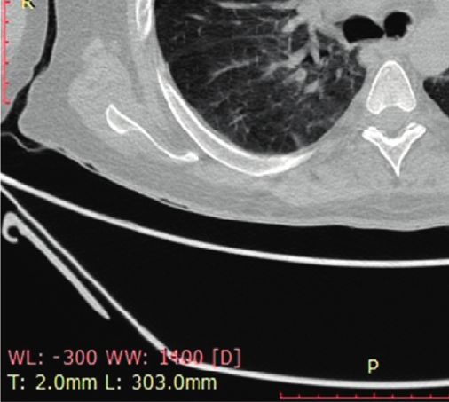
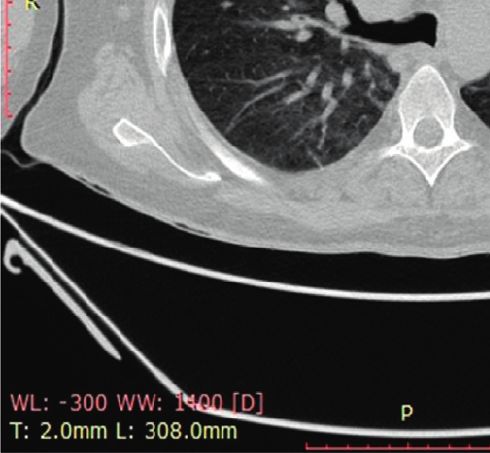
injury. Unfortunately, the patient had a cardiac and respi- suspicion for COVID-19 infection, she was tested, and the
ratory arrest, and cardiopulmonary resuscitation (CPR) was results of reverse transcriptase-polymerase chain reaction
implemented, and he was connected to a ventilator. How- (RT-PCR) and HRCT were suggestive of this viral infection
ever, on the fourth day of hospitalization, the patient could (Figure 4). She was transferred to the infectious ward. Her
not be resuscitated after the second cardiac arrest attack. laboratory findings showed LDH of 5580 U/L, CPK of
16480 U/L, Cr of 1.7 mg/dl, AST of 951 IU/L, ALT of 874 IU/
L, and ALP of 394 IU/L. With the assumed diagnosis of
2.2. Case 2. A 36-year-old man was referred to the emer- rhabdomyolysis, serum therapy was prescribed. The short-
gency department with two days of polyuria, polydipsia, ness of breath worsened on the fifth day and resulted in the
nausea, and vomiting. His medical history was positive for New York Heart Association (NYHA) class of IV worsening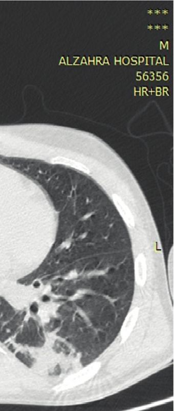
Case Reports in Medicine                                                                                                                  3

                                          (a)                                                (b)

                          Figure 1: Multifocal peribronchial consolidation and diffuse ground-glass opacities.

                                           (a)                                               (b)

                                  Figure 2: Bilateral and peripheral ground-glass pulmonary opacities.

                                           (a)                                              (b)

Figure 3: Heterogeneity and edema of the pancreatic tail with low enhancement (a) and splenic vein thrombosis with multiple collaterals (b).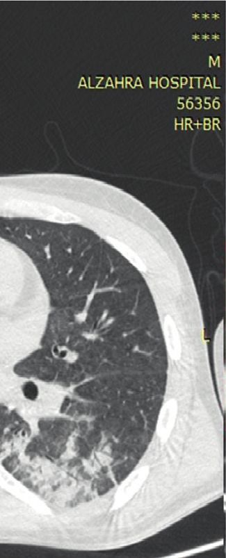
4                                                                                                          Case Reports in Medicine

                                         (a)                                             (b)

                             Figure 4: Multifocal multilobular consolidation and ground-glass opacities.

cardiac function with severe hemoglobin oxygen desatura-            genomic similarity between SARS and COVID-19 and the
tion due to bronchial secretions. Despite receiving 100%            ability of SARS to directly infect muscle cells, it probably
oxygen via a face mask, oxygen saturation only reached 80%.         seems that this new infective agent can infect myocytes. The
Therefore, the decision was made in order to intubate the           formation of immune complexes and their deposition because
patient. She was transferred to the intensive care unit (ICU),      of cross-reactivity between muscle cells and viral antigens
and the proper treatments were continued. Her laboratory            might be responsible for muscular damage. Massive cytokine
findings were improved during the next week (LDH: 1204 U/            production and cytotoxic T-cell activation could be catego-
L, CPK: 560 U/L, Cr: 0.7 mg/dl, AST: 135 IU/L, ALT: 234 IU/         rized as other possible causes in this regard [10, 11]. On the
L, and ALP: 562 IU/L). The patient remained stable he-              other hand, other rare comanifestations of COVID-19-in-
modynamically; however, she became unable to wean from a            duced rhabdomyolysis must be considered. Case 1 was ad-
ventilator secondary to probable acute respiratory distress         mitted with severe hypocalcemia as the initial presentation.
syndrome (ARDS). Unfortunately, in the third week, she had          Although the patient had myalgia and experienced seizure,
a sudden cardiac arrest. She underwent cardiorespiratory            which might be due to rhabdomyolysis complications, the
resuscitation but could not be resuscitated from her pulseless      direct virus effect for causing these presentations should be
electrical activity arrest and died.                                considered. Liu et al. performed a retrospective study to assess
                                                                    the predictive effect of hypocalcemia among patients with
3. Discussion                                                       severe COVID-19. They figured out that 62.6% of patients had
                                                                    low calcium levels, which was associated with poorer out-
Since the emergence of COVID-19, several other manifes-             comes [5]. Hypocalcemia is quite common in viral infections
tations rather than pulmonary symptoms have been reported.          and among critically ill patients. The possible factors include
Here, we presented three cases of confirmed COVID-19                 malnutrition, decreased intestinal absorption of calcium, cell
infection with concomitant rhabdomyolysis as well as hy-            membrane damage due to hypoxia and calcium influx, vi-
pocalcemia and DKA. Rhabdomyolysis is a life-threatening            tamin D deficiency, or medication interaction. Moreover,
syndrome characterized by skeletal muscle cell breakdown            serum calcium is mainly bound to albumin, and hypocal-
and leakage of cellular components, including potassium,            cemia could occur in favor of reduced albumin levels.
phosphorus, CPK, and myoglobin, to the bloodstream. The             However, cytokine production during COVID-19 infection is
typical triad of this disease consisted of myalgia, weakness,       reported to inhibit PTH secretion, and impaired response to
and dark-colored urine. However, less than 10% of patients          PTH might be responsible for the induction of hypocalcemia
experienced the classic triad [8]. Multiple etiologies have been    [12]. Several studies are required to evaluate the exact
suggested, including drug side effects, trauma, autoimmune           pathophysiological mechanism responsible for hypocalcemia
status, exertion, ischemia, and infection [9]. Common viral         among COVID-19-infected individuals. Overall, in our case,
causes include influenza A and B, herpes simplex virus,              it seems that impaired response to PTH, malnutrition, and
Epstein–Barr virus, cytomegalovirus, human immunodefi-               calcium influx were possible causes of hypocalcemia.
ciency virus, and enteroviruses [10]. Even though our patients           In terms of DKA that occurred in Case 2, several points
were not tested for other viral diseases, clinical status and       should be considered. The infection itself might be re-
laboratory data confirmed COVID-19 infection. While the              sponsible for the induction of DKA [7]. Also, abnormal
exact pathophysiological mechanism attributed to rhabdo-            blood glucose has been reported to be associated with worse
myolysis occurrence in COVID-19 infection has yet to be             outcomes in the COVID-19 pandemic. Although the data
investigated, several theories have been declared. Due to the       are still limited, further investigations for the diagnosis of the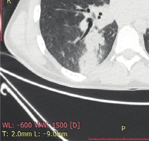
Case Reports in Medicine                                                                                                             5

exact mechanism are required. The Coronaviridae family has       are available from the corresponding author on reasonable
the tropism for both exocrine and endocrine pancreatic cells.    request.
Like SARS, angiotensin-converting enzyme 2 (ACE2) has
been suggested to be a receptor for COVID-19 S-protein           Consent
[13]. This receptor has been expressed in pancreatic beta-
cells in addition to pulmonary cells [14]. The primary role of   Written informed consent was obtained from the patient or
ACE2 is the degradation of angiotensin 2 to angiotensin1–7.      patient’s next of kin (when the death happened) for pub-
Therefore, downregulation of the ACE2 receptor during            lication of this case report and any accompanying images.
COVID-19 leads to increased angiotensin 2 concentration
resulting in reduced blood supply to pancreatic cells and
                                                                 Disclosure
subsequently delayed insulin secretion, which ultimately
results in hyperglycemia and even DKA occurrence [14, 15].       Maryam Heidarpour and Mehrbod Vakhshoori are con-
However, whether the sudden onset alterations of glucose         sidered co-first authors.
metabolism in severe COVID-19 will be persisted or re-
mitted is still unclear. The presence of this receptor on the
exocrine pancreas might explain the incidence of pancrea-        Conflicts of Interest
titis in Case 2. It seems that pancreatic involvement during     None of the authors had any personal or financial conflicts of
COVID-19 should be further addressed in clinical settings.       interest.
     In conclusion, our findings from this case report indicate
that rhabdomyolysis could be categorized as one of the
complications of COVID-19, and further suspicion for             Authors’ Contributions
proper diagnosis and therapeutic interventions should be         M. H., B. I., and F. K. contributed to study concept and
performed. Furthermore, other simultaneous rare mani-            design and provided administrative, technical, and material
festations, including hypocalcemia or DKA, must be com-          support. M. H., MA. H., and L. A. were responsible for the
prehensively addressed in future studies.                        acquisition of data. M. V., MA. H., and L. A. drafted the
                                                                 manuscript. M. V., B. I., M. H., and F. K. critically revised the
Abbreviations                                                    manuscript for valuable intellectual content. B. I., M. H., and
                                                                 F. K supervised the study. M. H. and M. V. contributed
COVID-       Coronavirus disease 2019                            equally to this manuscript.
19:
SARS:        Severe acute respiratory syndrome                   References
MERS:        Middle East respiratory syndrome
DKA:         Diabetic ketoacidosis                                [1] L. Di Filippo, A. M. Formenti, P. Rovere-Querini et al.,
HHS:         Hyperosmolar hyperglycemic state                         “Hypocalcemia is highly prevalent and predicts hospitaliza-
ECG:         Electrocardiogram                                        tion in patients with COVID-19,” Endocrine, vol. 68, no. 3,
HRCT:        High-resolution computed tomography                      pp. 1–4, 2020.
                                                                  [2] Z. Al-Ismaili, M. Piccioni, and M. Zappitelli, “Rhabdomyol-
LDH:         Lactic acid dehydrogenase
                                                                      ysis: pathogenesis of renal injury and management,” Pediatric
CPK:         Creatine phosphokinase                                   Nephrology, vol. 26, no. 10, pp. 1781–1788, 2011.
AST:         Aspartate aminotransferase                           [3] J. K. Millet and G. R. Whittaker, “Physiological and molecular
ALT:         Alanine aminotransferase                                 triggers for SARS-CoV membrane fusion and entry into host
ALP:         Alkaline phosphatase                                     cells,” Virology, vol. 517, pp. 3–8, 2018.
PTH:         Parathyroid hormone                                  [4] L. Nathan, A. L. Lai, J. K. Millet et al., “Calcium ions directly
BUN:         Blood urea nitrogen                                      interact with the ebola virus fusion peptide to promote
Cr:          Creatinine                                               structure-function changes that enhance infection,” ACS In-
CPR:         Cardiopulmonary resuscitation                            fectious Diseases, vol. 6, no. 2, pp. 250–260, 2019.
CT:          Computed tomography                                  [5] J. Liu, P. Han, J. Wu, J. Gong, and D. Tian, “Prevalence and
INR:         International normalized ratio                           predictive value of hypocalcemia in severe COVID-19 pa-
                                                                      tients,” Journal of Infection and Public Health, vol. 13, no. 9,
RT-PCR:      Reverse transcriptase-polymerase chain
                                                                      pp. 1224–1228, 2020.
             reaction                                             [6] N. Goldman, D. Fink, J. Cai, Y.-N. Lee, and Z. Davies, “High
NYHA:        New York Heart Association                               prevalence of COVID-19-associated diabetic ketoacidosis in
ICU:         Intensive care unit                                      UK secondary care,” Diabetes Research and Clinical Practice,
ARDS:        Acute respiratory distress syndrome                      vol. 166, Article ID 108291, 2020.
ACE2:        Angiotensin-converting enzyme 2.                     [7] K. H. Chan, D. Thimmareddygari, A. Ramahi, L. Atallah,
                                                                      N. G. Baranetsky, and J. Slim, “Clinical characteristics and
                                                                      outcome in patients with combined diabetic ketoacidosis and
Data Availability                                                     hyperosmolar hyperglycemic state associated with COVID-
                                                                      19: a retrospective, hospital-based observational case series,”
The datasets generated and/or analyzed during the current             Diabetes Research and Clinical Practice, vol. 166, Article ID
study are not publicly available due to confidential issues but        108279, 2020.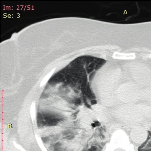
6                                                                        Case Reports in Medicine

 [8] R. Zutt, A. J. Van Der Kooi, G. E. Linthorst, R. J. A. Wanders,
     and M. De Visser, “Rhabdomyolysis: review of the literature,”
     Neuromuscular Disorders, vol. 24, no. 8, pp. 651–659, 2014.
 [9] W. H. Bagley, H. Yang, and K. H. Shah, “Rhabdomyolysis,”
     Internal and Emergency Medicine, vol. 2, no. 3, pp. 210–218,
     2007.
[10] N. F. Crum-Cianflone, “Bacterial, fungal, parasitic, and viral
     myositis,” Clinical Microbiology Reviews, vol. 21, no. 3,
     pp. 473–494, 2008.
[11] Y. Liang, M.-L. Wang, C.-S. Chien et al., “Highlight of im-
     mune pathogenic response and hematopathologic effect in
     SARS-CoV, MERS-CoV, and SARS-cov-2 infection,” Fron-
     tiers in Immunology, vol. 11, p. 1022, 2020.
[12] J. Fong and A. Khan, “Hypocalcemia: updates in diagnosis and
     management for primary care,” Canadian family physician
     Medecin de famille canadien, vol. 58, no. 2, pp. 158–162, 2012.
[13] M. Hoffmann, H. Kleine-Weber, S. Schroeder et al., “SARS-
     CoV-2 cell entry depends on ACE2 and TMPRSS2 and is
     blocked by a clinically proven protease inhibitor,” Cell,
     vol. 181, no. 2, 2020.
[14] J.-K. Yang, S.-S. Lin, X.-J. Ji, and L.-M. Guo, “Binding of SARS
     coronavirus to its receptor damages islets and causes acute
     diabetes,” Acta Diabetologica, vol. 47, no. 3, pp. 193–199, 2010.
[15] P.-O. Carlsson, C. Berne, and L. Jansson, “Angiotensin II and
     the endocrine pancreas: effects on islet blood flow and insulin
     secretion in rats,” Diabetologia, vol. 41, no. 2, pp. 127–133,
     1998.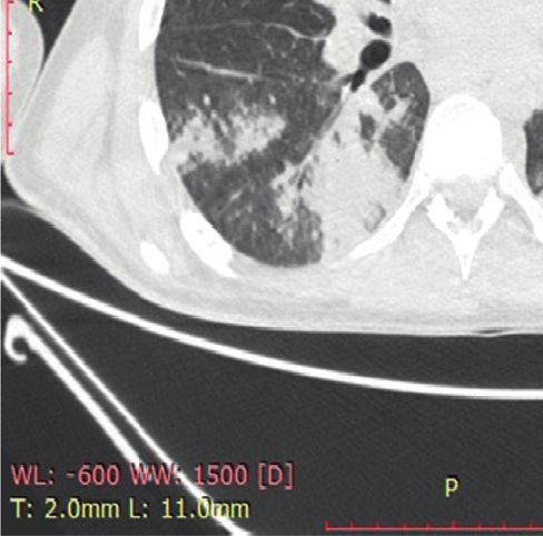
You can also read