Case Report Lung Abscess with a Refractory Bronchopleural Fistula Saved from Potentially Fatal Sepsis by Omentoplasty and Extracorporeal Membrane ...
←
→
Page content transcription
If your browser does not render page correctly, please read the page content below
Hindawi
Case Reports in Critical Care
Volume 2021, Article ID 9025990, 6 pages
https://doi.org/10.1155/2021/9025990

Case Report
Lung Abscess with a Refractory Bronchopleural Fistula
Saved from Potentially Fatal Sepsis by Omentoplasty and
Extracorporeal Membrane Oxygenation

          Jumpei Takamatsu , Jinkoo Kang, Aya Fukuhara, Yuichi Yasue, and Sae Kawata
          Department of Emergency Medicine, Kansai Rosai Hospital, Amagasaki, Japan

          Correspondence should be addressed to Jumpei Takamatsu; jtakamatsu@gmail.com

          Received 20 July 2021; Accepted 12 October 2021; Published 21 October 2021

          Academic Editor: Mehmet Doganay

          Copyright © 2021 Jumpei Takamatsu et al. This is an open access article distributed under the Creative Commons Attribution
          License, which permits unrestricted use, distribution, and reproduction in any medium, provided the original work is
          properly cited.

          Controlling air leaks during thoracic drainage in patients with lung abscesses caused by bronchopleural fistulas is challenging. To
          reduce the occurrence of air leaks, positive pressure ventilation should be avoided whenever possible. A 69-year-old man presented
          with a 10-day history of gradually worsening chest pain. He had lost consciousness and was brought to the emergency room. His
          SpO2 was approximately 70%, and his systolic blood pressure was approximately 60 mmHg. Chest radiography and computed
          tomography revealed findings suggestive of a right pyothorax. Therefore, thoracic drainage was performed. However, the
          patient’s respiratory status did not improve, and his circulatory status could not be maintained. Therefore, extracorporeal
          membrane oxygenation was introduced after the improvement in circulation by noradrenaline and fluid resuscitation, resulting
          in adequate oxygenation and ventilation without the use of high-pressure ventilator settings. Subsequently, omentoplasty for a
          refractory bronchopleural fistula was successfully performed, and the air leak was cured without recurrence of the lung abscess.

1. Introduction                                                         2. Case Presentation
Bronchopleural fistula is one of the most common causes of               A 69-year-old man was transported to the emergency room
lung abscesses. When a lung abscess is complicated by fatal             after losing consciousness; he had a 10-day history of gradu-
sepsis, drainage should be performed to control the infec-              ally worsening chest pain. The patient had no history of rel-
tion. However, air leakage may occur during drainage when               evant medical conditions, though it was reported that he
a bronchopleural fistula is present. To reduce air leakage,              smoked cigarettes. After admission to the hospital, the
positive thoracic pressure should be avoided as much as pos-            patient’s level of consciousness improved to a Glasgow
sible. However, in sepsis-associated lung abscesses, respira-           Coma Scale score of E3V4M6; however, his vital signs were
tory insufficiency necessitates the use of high-pressure                  unstable (SpO2, approximately 70% and systolic blood pres-
ventilator settings to achieve adequate oxygenation and ven-            sure, approximately 60 mmHg). The patient underwent
tilation. In this report, we present the case of a patient in           endotracheal intubation and fluid resuscitation with contin-
whom a lung abscess and refractory bronchopleural fistula                uous injection of noradrenaline, which stabilised his circula-
developed into potentially fatal sepsis.                                tory system. Chest radiography and computed tomography
2 Case Reports in Critical Care
Figure 1: Chest radiograph obtained at admission. The chest radiograph shows pleural effusion and diffuse infiltrative shadows.
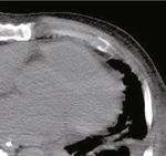
Figure 2: Computed tomography at admission. Computed tomography scans show fluid collection in the right thoracic cavity.Case Reports in Critical Care 3
Table 1: Laboratory values at admission. anterosternal route by creating a subcutaneous tunnel. The
flap was transferred to the bronchopleural fistula and
Item Result crimped with towel gauze. The chest was then temporarily
White blood cells (×103/μl) 9.6 closed to allow for necessary washing. The air leak was
Red blood cells (×106/μl) 3.61 resolved after surgery, and the omental flap became adherent
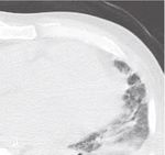
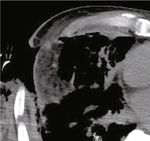
Hematocrit (%) 38.2 (Figure 4). CT performed on day 61 revealed the disappear-
Platelet (×103/μl) 335 ance of the abscess and viability of the omental flap
(Figure 5). We set the positive end-expiratory pressure
International normalized ratio of prothrombin time 1.24
(PEEP) of the ventilator to 0 cm H2O. On day 40, the patient
D-dimer (μg/ml) 23.02 was weaned from ECMO and treated via ventilatory man-
Total bilirubin (mg/dL) 1.5 agement. On day 95, the patient was weaned from the venti-
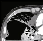
Aspartate aminotransferase (U/L) 282 lator, and CT revealed a left pneumothorax that did not
Alanine aminotransferase (U/L) 129 affect his respiratory status (Figure 6). On day 142, the
Lactate dehydrogenase (U/L) 450 patient was transferred to another hospital to continue reha-
Sodium (mmol/L) 142 bilitation for advanced disuse syndrome.
Chloride (mmol/L) 103
Potassium ((mmol/L)) 3.7 3. Discussion
Blood urea nitrogen (mg/dL) 21.4 Lung abscesses associated with refractory bronchopleural
Creatinine (mg/dL) 0.84 fistulas require a three-pronged treatment strategy: control-
Albumin (U/L) 1.5 ling the infection, treating the respiratory dysfunction, and
Lactate (mmol/L) 11.8 eliminating the air leak. Antimicrobial therapy is the first
C-reactive protein (mg/dL) 11.1 choice of treatment for this disease. However, approximately
10% of patients require drainage, and surgery is indicated
PCT (ng/mL) 0.64
when drainage fails [1]. The typical operation performed
for these patients is partial pneumonectomy or lobectomy.
(CT) were performed, which led to a diagnosis of right Oxygen is administered to treat respiratory dysfunction;
pyothorax (Figures 1 and 2) in addition to septic shock. however, mechanical ventilation is required if oxygenation
The laboratory values obtained on arrival are shown in and ventilation cannot be maintained. If the patient’s respi-
Table 1. Approximately 400 mL of pus was drained from ratory dysfunction is reversible, ECMO may be performed
the right thoracic cavity; however, the patient’s respiratory [2]. Thoracic drainage is required if air leaks develop [3].
status did not improve. Oxygenation and ventilation could However, if the patient’s condition worsens to respiratory
not be maintained, although the patient’s circulation was failure, mechanical ventilation with positive pressure venti-
stabilised by fluid resuscitation and noradrenaline. After 12 lation is required and closure of the fistula is difficult in these
hours of hospitalisation, extracorporeal membrane oxygena- patients.
tion (ECMO) was introduced via cannulation of the right EWS-based treatment is considered for bronchopleural
femoral artery, right femoral vein, and right common jugu- fistulas that do not improve with drainage alone [3]. How-
lar vein. The patient was started on veno-venous ECMO as ever, if the fistula does not respond to an EWS through the
his circulatory system stabilised. We believed that poor oxy- bronchus, surgical closure of the thoracic cavity should be
genation, in addition to sepsis, was causing worsening of considered. If occlusion using an EWS is technically difficult,
circulation-related problems. Finally, ECMO was performed a pneumonectomy that includes the bronchopleural fistula
at 3,000 rpm and a blood flow rate of 2.0 L/min. The and lung abscess can be performed. However, lung resection
patient’s clinical course during ECMO is shown in is impossible in patients with chronic inflammation and
Figure 3. Daily intrathoracic lavage was performed initially, thickened, adherent pleura. In these patients, the bronchial
but the air leak from the bronchopleural fistula persisted. fistula may be closed using a muscle or omental flap [4, 5].
On day 10 of hospitalisation, bronchial embolisation with In our patient, an omental flap was used to close the fistula.
an endobronchial Watanabe Spigot (EWS®; HARADA Cor- Omentoplasty is believed to aid in controlling the infection
poration, Osaka, Japan) was performed without success. On in the bronchopleural fistula due to the anti-inflammatory
day 15, the patient underwent a thoracotomy for thorough effects of the omentum [6]. The patient in this case report
cleaning of the thoracic cavity and abscess. The pleura on did not receive PEEP ventilation because ECMO was suc-
the visceral side appeared thickened with unclear borders, cessfully implemented. Thus, the omental flap was viable,
suggesting that the abscess was chronic. The mediastinal and the bronchopleural fistula was ultimately treated.
border was adherent and could not be detached. In addition, The management of respiratory failure due to a refrac-
an air leak was evident, though the leaking bronchus could tory bronchopleural fistula with ECMO has been previously
not be identified. On day 20, an omentoplasty was per- reported, and ECMO may be a future management option
formed for the bronchopleural fistula using a pedicled [7–9]. Although bronchial occlusion with an EWS is effec-
omental flap nourished by the right gastroepiploic artery. tive, it is often ineffective when the bronchi with fistula is
The flap was made long enough to reach the thoracic cavity difficult to locate [10]. However, if the intrathoracic air leak
by laparotomy and continued into the thoracic cavity via the is covered with an omental flap, precise identification of the4 Case Reports in Critical Care
ECMO ECMO removal
EWS-based treatment Omentum patch
Tracheotomy
Open chest hemostasis
CLDM
F-FLCZ
MEPM CFPM CFPM
VCM VCM
160 300
MAP
140 HR
RR 250
120 P/F ratio
MAP-HR-RR
200
100
P/F ratio
80 150
60
100
40
50
20
0 0
30.0 38.5
WBC 38.0
25..0 CRP
BT 37.5
WBC•CRP
20.0
37.0
BT
15.0
36.5
10.0
36.0
5.0 35.5
0.0 35.0
0 1 2 3 4 5 6 7 8 9 10 11 12 13 14 15 16 17 18 19 20 21 22 23 24 25 26 27 28 29 30 31 32 33 34 35 36 37 38 39 40
ECMO; Extracorporeal membrane oxygenation MAP; mean arterial pressure (mmHg)
EWS; Endobronchial Watanabe Spigot HR; heart rate (/minute)
MEPM; meropenem RR; respiratory rate (/minute)
VCM; vancomycin P/F ratio; PaO2/FiO2 ratio
F-FLCZ; fosfluconazole WBC; white blood cell (×103/uL)
CLDM; clindamycin CRP; C- reactive protein (mg/dL)
CFPM; cefepime BT; body temperature (°C)
Figure 3: Patient’s clinical course. The patient’s clinical course during extracorporeal membrane oxygenation (ECMO) is shown. His
PaO2/FiO2 ratio was not favourable throughout ECMO, but his level of consciousness and circulatory dynamics were maintained.Case Reports in Critical Care 5
Figure 4: Adherent omental flap. The patient’s omental flap is shown to be adherent on day 34 of hospitalisation.
Figure 5: Postoperative computed tomography on day 61 of hospitalisation. Computed tomography scans obtained on day 61 show that the
omental flap was viable, and that the abscess had disappeared.
Figure 6: Computed tomography on day 95 of hospitalisation. Improved air flow is observed in the right lung. However, a left
pneumothorax is also observed.6 Case Reports in Critical Care
location of the leak is not needed. In our patient, ECMO was [7] A. A. Grant, E. B. Lineen, A. Klima, R. Vianna, M. Loebe, and
used to support oxygenation and ventilation, and reduced A. Ghodsizad, “Refractory traumatic bronchopleural fistula: is
positive pressure ventilation with a respirator decreased the extracorporeal membrane oxygenation the new gold stan-
pressure in the airway, allowing for healing. Daily observa- dard?,” Journal of Cardiac Surgery, vol. 35, no. 1, pp. 242–
tion of the patient is required after omentoplasty, as the 245, 2020.
infection may not respond to the anti-inflammatory proper- [8] M. F. Odish, J. Yang, G. Cheng et al., “Treatment of broncho-
ties of the omentum [11]. As our patient was managed with pleural and alveolopleural fistulas in acute respiratory distress
an open chest, the infection status was noted daily. syndrome with extracorporeal membrane oxygenation, a case
series and literature review,” Crit Care Explor., vol. 3, no. 5,
In our patient, who had a lung abscess caused by a
article e0393, 2021.
bronchopleural fistula and an air leak that was difficult to
control during thoracic drainage, ECMO was used to achieve [9] J. C. Grotberg, R. C. Hyzy, J. De Cardenas, and I. N. Co,
“Bronchopleural fistula in the mechanically ventilated patient:
adequate oxygenation and ventilation without applying high
a concise review,” Critical Care Medicine, vol. 49, no. 2,
pressure. An omentoplasty over the refractory broncho- pp. 292–301, 2021.
pleural fistula was successfully engrafted, and the air leak
[10] K. Shin, T. Hifumi, R. Tsugitomi et al., “Empyema with fistula
was cured without the recurrence of any lung abscesses.
successfully treated with a comprehensive approach including
bronchial blocker and embolization receiving veno-venous
Data Availability extracorporeal membrane oxygenation,” Acute Med Surg.,
vol. 8, no. 1, article e621, 2021.
The data used to support the findings of this study are avail-
[11] H. Yokomise, T. Fukuse, O. lke et al., “Unsuccessful omento-
able from the corresponding author upon request.
pexy in thoracic surgery,” The Thoracic and Cardiovascular
Surgeon, vol. 45, no. 3, pp. 145–148, 1997.
Consent
Written consent was not obtained from the patient, as all
identifiable data were removed from this case report.
Conflicts of Interest
The authors declare that there are no conflicts of interest
regarding the publication of this paper.
Acknowledgments
We would like to thank Editage (http://www.editage.jp/) for
English language editing.
References
[1] H. C. Mwandumba and N. J. Beeching, “Pyogenic lung infec-
tions: factors for predicting clinical outcome of lung abscess
and thoracic empyema,” Current Opinion in Pulmonary Med-
icine, vol. 6, no. 3, pp. 234–239, 2000.
[2] ELSO, Extracorporeal Life Support Organization (ELSO)
Guidelines for Adult Respiratory Failure; August, 2017.
[3] S. Morikawa, T. Okamura, T. Minezawa et al., “A simple
method of bronchial occlusion with silicone spigots (endo-
bronchial Watanabe Spigot; EWS®) using a curette,” Thera-
peutic Advances in Respiratory Disease, vol. 10, no. 6,
pp. 518–524, 2016.
[4] L. De Weerd, P. C. Endresen, A. T. Numan, and S. Weum,
“Intrathoracic breast transposition: a new method in the treat-
ment of bronchopleural fistula and empyema,” Plastic and
Reconstructive Surgery. Global Open, vol. 7, no. 12, article
e2531, 2019.
[5] Y. Okumura, S. Takeda, H. Asada et al., “Surgical Results for
Chronic Empyema Using Omental Pedicled Flap: Long-
Term Follow-Up Study,” The Annals of Thoracic Surgery,
vol. 79, no. 6, pp. 1857–1861, 2005.
[6] H. Endoh, R. Yamamoto, N. Nishizawa, and Y. Satoh, “Thora-
coscopic surgery using omental flap for bronchopleural fis-
tula,” Surg Case Rep., vol. 5, no. 1, p. 5, 2019.You can also read


