Case Report Hypertrophic Osteoarthropathy: A Secondary Manifestation of Malignant Melanoma
←
→
Page content transcription
If your browser does not render page correctly, please read the page content below
Hindawi
Case Reports in Rheumatology
Volume 2021, Article ID 6691320, 3 pages
https://doi.org/10.1155/2021/6691320

Case Report
Hypertrophic Osteoarthropathy: A Secondary Manifestation of
Malignant Melanoma

          Shiva Malaty             and Aditya Gupta
          HonorHealth Internal Medicine Residency, Scottsdale, AZ, USA

          Correspondence should be addressed to Shiva Malaty; shivamalaty@gmail.com

          Received 13 October 2020; Revised 1 January 2021; Accepted 11 January 2021; Published 18 January 2021

          Academic Editor: Franco Schiavon

          Copyright © 2021 Shiva Malaty and Aditya Gupta. This is an open access article distributed under the Creative Commons
          Attribution License, which permits unrestricted use, distribution, and reproduction in any medium, provided the original work is
          properly cited.
          Background. Hypertrophic osteoarthropathy (HOA) is a rare finding in the setting of metastatic melanoma. A majority of cases of
          secondary HOA involve lung malignancies. Evaluation of presenting symptoms such as polyarthralgia and clubbing followed by
          review of imaging studies are diagnostic steps for HOA. Case Presentation. We present a 60-year-old female with a history of
          metastatic melanoma who presented with bilateral and symmetric polyarthralgia and clubbing. A plain film radiograph
          demonstrated periosteal thickening involving the metacarpals and proximal phalanges as well as the distal radius and ulna,
          consistent with HOA. The patient was treated with nonsteroidal anti-inflammatory agents for supported care. Conclusion. HOA
          may be a secondary manifestation of metastatic melanoma. Recognition and supportive care of this condition may lead to
          improved quality of life for patients.

1. Introduction                                                        metastasis to the liver and lung, presenting with swelling
                                                                       over the fingers and toes. The patient reported the presence
Hypertrophic osteoarthropathy (HOA) is characterized by                of edema for years and was told it was related to an auto-
digital clubbing, periostosis, and arthralgia of extremities.          immune condition given a previously positive ANA result.
This condition is secondary to abnormal proliferation of               The patient stated that the swelling was also associated with
osseous and soft tissue. There are two forms of HOA: a                 symmetric polyarthralgia of the upper and lower extremities.
primary genetic form, which accounts for approximately 3%              She denied any alleviating or aggravating factors. On
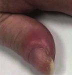
of cases, and a secondary form, mainly attributed to para-             physical exam, clubbing was present on bilateral distal
neoplastic syndrome from non-small cell lung cancer and to             phalanges (Figure 1). Additionally, when placing the pads of
a lesser degree, from pulmonary diseases such as sarcoidosis,          bilateral terminal phalanges in dorsal opposition, disap-
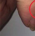
pulmonary tuberculosis, and cystic fibrosis [1, 2]. The                 pearance of the normal diamond-shaped window was found,
proposed pathophysiology involves the release of platelet-             indicating a positive Schamroth sign (Figure 2). The patient
derived growth factor and vascular endothelium-derived                 underwent lab work, which revealed an alkaline phosphatase
growth factor [3]. These growth factors lead to increased              level of 146.
bone production and vascular hyperplasia, which leads to                   Plain film radiography of the upper extremities dem-
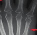
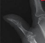
clinical signs of digital clubbing.                                    onstrated periosteal thickening involving the metacarpals
                                                                       and proximal phalanges as well as the distal radius and ulna,
                                                                       consistent with hypertrophic osteoarthropathy (Figure 3).
2. Case Presentation
                                                                       Given patient’s terminal malignancy, no further oncological
We present the case of a 60-year-old female with a medical             or surgical treatments were available. The patient was started
history of B-RAF wild-type metastatic melanoma with                    on a nonsteroidal anti-inflammatory agent, ibuprofen, for
2 Case Reports in Rheumatology
Figure 1: Clubbing of distal phalanges.
Figure 3: Periosteal thickening involving the metacarpals, prox-
imal phalanges, distal radius, and ulna.
There are no specific or reliable lab tests available for the
diagnosis of HOA. A plain radiograph is the most telling
diagnostic modality for HOA. The presence of periosteal
thickening is the most specific radiographic finding asso-
ciated with HOA. This finding may be generalized to include
more than one specific region of the bone or localized to a
specific site of the bone [4, 5].
Treatment of the underlying disease process is key for
secondary HOA. Supportive care with nonsteroidal anti-
inflammatory agents as well as bisphosphonates may pro-
vide relief. VEGF inhibitors have also been proposed as
Figure 2: Schamroth sign, disappearance of normally appearing therapeutic options for alleviating pain symptoms in sec-
diamond-shaped window between dorsal pads of opposite terminal ondary HOA [6].
phalanges. This case exhibits the importance of both, a thorough
clinical history and physical examination. This diagnosis
symptomatic relief. She demonstrated significant improve- should be considered in patients presenting with poly-
ment of arthralgias during her three-week follow-up. The arthralgia and appropriate imaging findings. Early diagnosis
patient went on to hospice care and shortly passed. of this condition combined with appropriate management
and therapy may result in improved quality of life for af-
fected patients.
3. Discussion
The proposed pathophysiology of HOA involves the release Data Availability
of platelet-derived and vascular endothelium-derived
growth factors. The exact mechanism of disease in HOA is All data in this case report are taken from the clinical
uncertain. This diagnosis should be considered in the setting records.
of clubbing with imaging findings supportive of periosteal
thickening. In the setting of HOA with no underlying cause, Conflicts of Interest
a plain film radiograph of the chest should be indicated to
rule out pulmonary etiology [3]. The authors declare no conflicts of interest.Case Reports in Rheumatology 3
References
[1] P. Cathébras, J.-B. Gaultier, S. Charmion, and I. Guichard,
“Hypertrophic osteoarthropathy of the lower limbs,” The
Journal of Rheumatology, vol. 38, no. 4, pp. 775-776, 2011.
[2] T. Ito, K. Goto, K. Yoh et al., “Hypertrophic pulmonary
osteoarthropathy as a paraneoplastic manifestation of lung
cancer,” Journal of Thoracic Oncology, vol. 5, no. 7, pp. 976–980,
2010.
[3] M. Martı́nez-Lavı́n, M. Matucci-Cerinic, I. Jajic, and C. Pineda,
“Hypertrophic osteoarthropathy: consensus on its definition,
classification, assessment and diagnostic criteria,” The Journal
of Rheumatology, vol. 20, no. 8, pp. 1386-1387, 1993.
[4] S. Nguyen and M. Hojjati, “Review of current therapies for
secondary hypertrophic pulmonary osteoarthropathy,” Clinical
Rheumatology, vol. 30, no. 1, pp. 7–13, 2011.
[5] F. Y. Yap, M. R. Skalski, D. B. Patel et al., “Hypertrophic
osteoarthropathy: clinical and imaging features,” Radio-
graphics, vol. 37, no. 1, pp. 157–195, 2017.
[6] M. A. Thompson, N. B. Warner, and W.-J. Hwu, “Hypertrophic
osteoarthropathy associated with metastatic melanoma,”
Melanoma Research, vol. 15, no. 6, pp. 559–561, 2005.You can also read


