Case Report Development of Two Types of Skin Cancer in a Patient with Systemic Sclerosis: a Case Report and Overview of the Literature
←
→
Page content transcription
If your browser does not render page correctly, please read the page content below
Hindawi
Case Reports in Oncological Medicine
Volume 2021, Article ID 6628671, 5 pages
https://doi.org/10.1155/2021/6628671

Case Report
Development of Two Types of Skin Cancer in a Patient with
Systemic Sclerosis: a Case Report and Overview of the Literature

          Firdevs Ulutaş ,1 Erdem Çomut ,2 and Veli Çobankara                                  1

          1
              Division of Rheumatology, Department of Internal Medicine, Pamukkale University Faculty of Medicine, Denizli, Turkey
          2
              Department of Pathology, Pamukkale University Faculty of Medicine, Denizli, Turkey

          Correspondence should be addressed to Firdevs Ulutaş; firdevsulutas1014@gmail.com

          Received 1 January 2021; Revised 5 February 2021; Accepted 18 February 2021; Published 26 February 2021

          Academic Editor: Raffaele Palmirotta

          Copyright © 2021 Firdevs Ulutaş et al. This is an open access article distributed under the Creative Commons Attribution License,
          which permits unrestricted use, distribution, and reproduction in any medium, provided the original work is properly cited.

          Systemic sclerosis (SSc) is an uncommon rheumatic disease in which the underlying main histopathologic feature is a thickening of
          the skin due to excessive accumulation of collagen in the extracellular tissue. Fibrogenesis, chronic inflammation, and ulceration
          may eventually promote skin neoplasms. Although nonmelanoma skin cancer (NMSC) is the most frequent type, there have
          been restricted case reports and case series with skin cancers in SSc patients in the literature. Herein, we describe a 78-year-old
          woman diagnosed with diffuse cutaneous systemic sclerosis thirteen years ago and associated nonspecific interstitial pneumonia
          that was successfully treated with high cumulative doses of cyclophosphamide. She developed basal cell carcinoma and
          squamous cell carcinoma of the skin in the follow-up. She is still on rituximab treatment with stable interstitial lung disease as
          indicated by pulmonary function tests and high-resolution chest computed tomography. To our knowledge and a literature
          search, this is the first reported patient with SSc with two types of skin cancer. In this review, we also aimed to emphasize the
          relationship between SSc and skin cancer, and possible risk factors for SSc-related skin cancer.

1. Introduction                                                          case series with skin cancers in SSc patients. Although under-
                                                                         lying pathogenetic mechanisms are not yet clear, a shred of
Systemic sclerosis (SSc), also called scleroderma, is an                 evidence is present related to few immunosuppressive drugs.
uncommon rheumatic disease. It is a slowly progressive dis-              Cyclophosphamide (CyP) is one of them and nonselectively
ease that is symbolized by vasculopathy, fibrosis of the skin             inhibits the whole immune system, and malignancy may
and visceral organs, immune rearrangement, and B-cell acti-              develop by suppressing immune surveillance [6]. Today, the
vation with characteristic autoantibodies [1]. Although evi-             use of CyP for the treatment of ILD in connective tissue dis-
dence for high risk of having malignancy is suggestive, the              orders has growing evidence for optimization of lung func-
types of malignancies and the importance of the total risk               tions [7]. Immunosuppression with methotrexate use alone
are quite variable in each patient with SSc [2]. The lung and            and/or combined with antitumor necrosis factor agents
skin are the frequently seen specific tumor sites in patients             (anti-TNF) is also related to high risk of second nonmela-
with SSc, which are commonly affected by both fibrosis and                 noma skin cancers (NMSC) in rheumatoid arthritis (RA)
underlying immune dysfunction [3]. Interstitial lung disease             patients [8].
(ILD) is also a serious complication of SSc, which leads to sig-             Herein, we report an older SSc patient who was treated
nificant morbidity and lung cancer [4]. Autoantibodies to                 with highly toxic cumulative doses of CyP due to progressive
topoisomerase I (also referred to as anti-Scl70), smoking,               ILD and who subsequently developed two types of skin can-
and some immunosuppressive drugs may also lead to a                      cer simultaneously. Managing treatment of individuals with
higher frequency of lung cancer among SSc patients [5]. On               SSc-ILD is difficult due to balancing the need for therapy in
the other hand, skin cancers are also seen in SSc patients.              severely progressive patients against the potential for adverse
Although nonmelanoma skin cancer (NMSC) is the most fre-                 effects. Clinicians should identify patients who will develop
quent subtype, there have been restricted case reports and               the progressive disease with the lowest level of unexpected
2 Case Reports in Oncological Medicine
side effects before planning treatment. In addition to treat-
ment modalities, underlying inflammatory diseases, pro-
longed life span and family histories, occupations, and sun
exposure of patients should be examined. Written informed
consent was obtained from our patient for publication.
2. Methods
A literature search was performed in PubMed using these
terms: “scleroderma,” “systemic sclerosis,” “skin cancer,”
“squamous cell carcinoma,” and “basal cell carcinoma.” An
English language filter was activated. All case reports pub-
lished before November 2020 were examined.
3. Case Report
A 78-year-old woman diagnosed with diffuse cutaneous sys-
temic sclerosis (dcSSc) thirteen years ago is on regular follow-
up in the tertiary health center, in Denizli. She is of Turkish
origin and works as a farmer for a long time. She reports
no use of tobacco and has no known allergic diseases. Her
past medical history included idiopathic venous thromboem-
bolism in 2016 without an identifiable cause. She was diag-
nosed with dcSSc based on the presence of Raynaud’s
phenomenon, digital pitting scars, sclerodactyly, and diffuse
skin sclerosis extending proximal to the metacarpophalan-
geal joints on both hands. An indirect immunofluorescence
test for antinuclear antibodies (ANA) was positive
(titer ≥ 1 : 1280), and anti-Scl-70 was also positive. In the
first year of diagnosis, she worsened and complained of dys-
pnea occurring with minimal exertion, along with chronic,
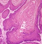
persistent cough. Transthoracic echocardiogram revealed Figure 1: Squamous cell carcinoma. Blunt-type intradermal
invasion of well-differentiated tumor nests (H&E, ×40).
no findings suggestive of pulmonary arterial hypertension.
After spirometry tests (forced vital capacity of 1.79 L (40%
of the predicted)), high-resolution chest computed tomogra- Although a malignant turn of localized scleroderma is rarely
phy (nonspecific interstitial pneumonia (NSIP)), and a 6- seen, Durcanska et al. concluded that a young 26-year-old
minute walk test (developing desaturation at 424 m), she woman who was diagnosed with localized scleroderma (mor-
was accepted as having SSc-related NSIP. CyP was adminis- phea) ultimately developed SCC camouflaged by osteomyeli-
tered at a dose of 1000 mg/m2 of body surface area per month tis on the lower extremities after a long course of the disease
for six months in an outpatient clinic, followed by [9]. An 18-year-old woman with progressive SSc developed
500 mg/m2 of body surface area every two months for one SCC of the skin. Despite resection, the tumor recurred and
year. After two years of clinical stability, the same treatment was resistant to local radiotherapy [10]. Song et al. reported
regimen was repeated due to signs of lung exacerbation (total a 46-year-old man with systemic sclerosis and BCC on his
cumulative dose has exceeded 15 g). After three years of com- face that was successfully removed by surgical excision [11].
pleted treatment, a 1-2 cm nonhealing ulcer on her right fore- Sargın et al. reported variable cancers in 7 cases among 153
arm and a pigmented mass at the tip of the nose appeared systemic sclerosis patients. Two of all the cancers were malig-
and were excised surgically. Histopathological examinations nant melanoma in the eyes and skin, in Aydın, neighboring
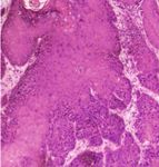
revealed squamous cell carcinoma (SCC) and basal cell carci- to Denizli [12]. Koksal et al. investigated the distribution of
noma (BCC), respectively (Figures 1 and 2). Surgical borders cancer cases in Denizli between 2000 and 2004. 10.9% of
were intact without tumor cells. Additional scanning 2185 cancer cases were skin cancers. They emphasized a sig-
methods showed no local or distant metastases. She is still nificant increase in skin cancers over the years [13]. Ceylan
on rituximab treatment with stable interstitial lung disease et al. investigated features of NMSCs in Izmir where high
as indicated by pulmonary function tests and high- ultraviolet light exposure was present such as Denizli.
resolution chest computed tomography. Tumors were commonly seen in elderly men and were also
related to sun exposure and older age. Among 3,186 NMSC
4. Discussion lesions, 71 patients had both BCC and SCC, and BCC was
the most common type among the whole group, and mostly
Skin cancers, covering SCC, BCC, and malignant melanoma located on the face [14]. Nose and lip were the most common
were found as SSc-related tumors in a literature search. locations on the face with high recurrence rates for NMSCCase Reports in Oncological Medicine 3
involving melanoma, SCC, and BCC, was significantly higher
in patients with morphea compared with the healthy subjects
[22]. Besides, a study in which malignancies were analyzed in
SSc patients and were compared with the general population
revealed 11 new cases of variable malignancies in 10 SSc
patients (4.6%) more commonly seen than the general popu-
lation. Only one of them had skin cancer [23]. Although
there is no comprehensive analysis of the prevalence of skin
cancers and other malignancies among SSc in our health cen-
ter, we can report that she is the first case with two concur-
rent types of skin cancer.
SSc-specific autoantibodies may identify patients at high
risk and play a role in the prognosis and triaging of patients
who may require further cancer screening [24]. However,
the best-known autoantibodies such as antitopoisomerase I
and anticentromere are inconsistent in predicting risk for
developing malignancy. In a comprehensive review,
increased age, diffuse SSc, and female gender were well-
known risk factors for the development of malignancy in
patients with SSc [25]. In another large cohort including
2177 patients with SSc, the presence of anti-RNA polymerase
III (anti-RNAPIII) was the most associated antibody for the
development of malignancies [26]. Female gender, older
age, and having antitopoisomerase I antibody were personal,
unchangeable risk factors in our patient.
Also, immunosuppressants used in major organ involve-
ments of scleroderma may encourage tumor formation. CyP
is a very efficient immunosuppressant drug in autoimmune
and inflammatory illnesses with possible major side effects
[27]. Hematological adverse events, bone marrow suppres-
sion, hematuria, bladder cancer, and gonadal damage have
Figure 2: Basal cell carcinoma. Nests of basaloid cells project from been demonstrated and are the most feared side effects
the overlying epidermis (H&E, ×40). [28]. Increased risk of hematological and solid organ malig-
nancies in patients treated with CyP suggests carcinogenic
activity and should be kept in mind [29]. However, in addi-
[15]. Our patient also had SCC and BCC on the tip of her tion to immunosuppressive drugs, some immunosuppressive
nose and on her right forearm which may be related to sun conditions such as HIV, chronic lymphocytic leukemia,
exposure. and/or transplant recipients have also a higher risk for devel-
The main histopathologic feature of scleroderma is a oping any skin cancer than the general population [30].
thickening of the skin due to excessive accumulation of colla- Other important points are sun exposure and advanced
gen in the extracellular tissue. Fibrogenesis, chronic inflam- age in the pathogenesis of NMSC. The most common cancer
mation, and ulceration may eventually promote skin in the world, NMSC, was developed more commonly in out-
neoplasms [16]. SCC has been the most frequently reported door workers including different job groups than indoor
skin cancer in association with chronic tissue inflammation workers due to sun exposure in a recently published paper
[17]. Also, Magro et al. stated that not only malignancies [31]. Although it is difficult to clearly distinguish the causa-
but also atypical lymphoid aggregates are seen in cutaneous tive etiopathogenic mechanism, our patient had apparent
specimens of connective tissue disease (CTD) related to B- multiple risk factors for developing skin cancer, including
cell or T-cell phenotypes [18]. Numerous theories and path- high sun exposure due to her occupation, exposure to high
ophysiological mechanisms have been emphasized to explain cumulative toxic doses of CyP, long-term immunosuppres-
this clinical association. One of the highlights is a signifi- sion with CyP and rituximab, the underlying chronic inflam-
cantly reduced percentage of CD4(+) Foxp3(+) T(reg) in matory disease, systemic sclerosis, and the advanced age. We
the skin of patients with SSc or limited scleroderma [19]. think the unfortunate outcome in this patient is the cumula-
Dysregulation of the endocannabinoid system (ECS) is also tive result of all these risk factors.
related to skin cancer in scleroderma [20]. Recent extensive
research points to the renin-angiotensin-aldosterone system- 5. Conclusion
(RAAS-) modulating drugs in the impaired regulatory func-
tion of local RAAS to be related to cancer development and We reported a rare case of BCC and SCC in a SSc patient
scleroderma-like skin changes [21]. Recent literature along with a review of the literature. To our knowledge and
revealed that the prevalence of all epithelial skin neoplasms, the literature search, this is the first reported SSc patient with4 Case Reports in Oncological Medicine
two types of skin cancer. With this review, we aimed to demographic and clinicopathological characteristics,” The
improve our knowledge of SSc-related skin cancers. Clini- Journal of Dermatology, vol. 30, no. 2, pp. 123–131, 2003.
cians should always keep in mind the patient’s risk of devel- [15] G. Eskiizmir, E. Ozgur, P. Temiz, G. Gencoglan, and A. T.
oping toxicity based on a risk-benefit analysis. These patients Ermertcan, “The evaluation of tumor histopathology, location,
are candidates for cancer development due to underlying characteristic, size, and thickness of nonmelanoma skin can-
immune dysregulation and immunosuppressive drugs, in cers of the head and neck,” Kulak Burun Boğaz Ihtisas Dergisi,
addition to traditional risk factors. vol. 22, no. 2, pp. 91–98, 2012.
[16] Y. Asano, “Systemic sclerosis,” The Journal of Dermatology,
vol. 45, no. 2, pp. 128–138, 2018.
Conflicts of Interest [17] A. T. Jacques Maria, L. Partouche, R. Goulabchand, S. Riviere,
P. Rozier, C. Bourgier et al., “Intriguing relationships between
All authors declare that they have no conflicts of interest. cancer and systemic sclerosis: role of the immune system and
other contributors,” Frontiers in Immunology, vol. 9, article
3112, 2019.
References [18] C. M. Magro, A. N. Crowson, and T. J. Harrist, “Atypical lym-
[1] C. P. Denton and D. Khanna, “Systemic sclerosis,” The Lancet, phoid infiltrates arising in cutaneous lesions of connective tis-
vol. 390, no. 10103, pp. 1685–1699, 2017. sue disease,” The American Journal of Dermatopathology,
vol. 19, no. 5, pp. 446–455, 1997.
[2] J. Barnes and M. D. Mayes, “Epidemiology of systemic sclero-
sis: incidence, prevalence, survival, risk factors, malignancy, [19] S. Klein, C. C. Kretz, V. Ruland et al., “Reduction of regulatory
and environmental triggers,” Current Opinion in Rheumatol- T cells in skin lesions but not in peripheral blood of patients
ogy, vol. 24, no. 2, pp. 165–170, 2012. with systemic scleroderma,” Annals of the Rheumatic Diseases,
[3] A. K. Rosenthal, J. K. McLaughlin, G. Gridley, and O. Nyren, vol. 70, no. 8, pp. 1475–1481, 2011.
“Incidence of cancer among patients with systemic sclerosis,” [20] Río C, E. Millán, V. García, G. Appendino, J. DeMesa, and
Cancer, vol. 76, no. 5, pp. 910–914, 1995. E. Muñoz, “The endocannabinoid system of the skin. A poten-
[4] V. Artinian and P. A. Kvale, “Cancer and interstitial lung dis- tial approach for the treatment of skin disorders,” Biochemical
ease,” Current Opinion in Pulmonary Medicine, vol. 10, no. 5, Pharmacology, vol. 157, pp. 122–133, 2018.
pp. 425–434, 2004. [21] M. Aleksiejczuk, A. Gromotowicz-Poplawska, N. Marcinczyk,
[5] M. Colaci, D. Giuggioli, M. Sebastiani et al., “Lung cancer in A. Przylipiak, and E. Chabielska, “The expression of the renin-
scleroderma: results from an Italian rheumatologic center angiotensin-aldosterone system in the skin and its effects on
and review of the literature,” Autoimmunity Reviews, vol. 12, skin physiology and pathophysiology,” Journal of Physiology
no. 3, pp. 374–379, 2013. and Pharmacology, vol. 70, no. 3, 2019.
[6] T. Mimori, “Immunosuppressants,” Nihon Rinsho, vol. 67, [22] E. Boozalis, A. A. Shah, F. Wigley, S. Kang, and S. G. Kwatra,
no. 3, pp. 582–587, 2009. “Morphea and systemic sclerosis are associated with an
[7] D. Carmier, E. Diot, L. Guilleminault, and S. Marchand-Adam, increased risk for melanoma and nonmelanoma skin cancer,”
“Interstitial lung disease in connective tissue disorders,” La Journal of the American Academy of Dermatology, vol. 80,
Revue du Praticien, vol. 64, no. 7, pp. 941–945, 2014. no. 5, pp. 1449–1451, 2019.
[8] F. Scott, R. Mamtani, C. M. Brensinger et al., “Risk of nonme- [23] S. Szamosi, A. Horvath, A. Nemeth, B. Juhasz, G. Szucs, and
lanoma skin cancer associated with the use of immunosup- Z. Szekanecz, “Malignancies associated with systemic sclero-
pressant and biologic agents in patients with a history of sis,” Autoimmunity Reviews, vol. 11, no. 12, pp. 852–855,
autoimmune disease and nonmelanoma skin cancer,” JAMA 2012.
Dermatology, vol. 152, no. 2, pp. 164–172, 2016. [24] K. Morrisroe and M. Nikpour, “Cancer, and scleroderma:
[9] V. Ďurčanská, H. Jedličková, O. Sláma, L. Velecký, recent insights,” Current Opinion in Rheumatology, vol. 32,
E. Březinová, and V. Vašků, “Squamous cell carcinoma in no. 6, pp. 479–487, 2020.
localized scleroderma,” Klinická Onkologie, vol. 27, no. 6, [25] M. Wooten, “Systemic sclerosis and malignancy: a review of
pp. 434–437, 2014. the literature,” Southern Medical Journal, vol. 101, no. 1,
[10] M. A. Öztürk, M. Benekli, M. K. Altundaĝ, and N. Güler, pp. 59–62, 2008.
“Squamous cell carcinoma of the skin associated with sys- [26] P. Moinzadeh, C. Fonseca, M. Hellmich et al., “Association of
temic sclerosis,” Dermatologic Surgery, vol. 24, no. 7, anti-RNA polymerase III autoantibodies and cancer in sclero-
pp. 777–779, 1998. derma,” Arthritis Research & Therapy, vol. 16, no. 1, p. R53,
[11] J. S. Song, S. K. Bae, S. Y. Park, and W. Park, “A case of basal 2014.
cell carcinoma of the skin in a patient with systemic sclerosis,” [27] H. Barnes, A. E. Holland, G. P. Westall, N. S. Goh, and
Rheumatology International, vol. 20, no. 1, pp. 39-40, 2000. I. N. Glaspole, “Cyclophosphamide for connective tissue
[12] G. Sargin, T. Senturk, and S. Cildag, “Systemic sclerosis and disease-associated interstitial lung disease,” Cochrane Data-
malignancy,” International Journal of Rheumatic Diseases, base of Systematic Reviews, vol. 1, no. 1, article CD010908,
vol. 21, no. 5, pp. 1093–1097, 2018. 2018.
[13] A. Koksal, H. C. Sorkin, H. Demirhan, A. G. Tomatir, T. Alan, [28] C. Bruni and D. E. Furst, “The burning question: to use or not
and F. Ozerdem, “Evaluation of cancer records from 2000- to use cyclophosphamide in systemic sclerosis,” European
2004 in Denizli, Turkey,” Genetics and Molecular Research, Journal of Rheumatology, vol. 7, pp. 237–241, 2020.
vol. 8, no. 1, pp. 64–75, 2009. [29] T. Kaşifoğlu, Ş. Yaşar Bilge, F. Yıldız et al., “Risk factors for
[14] C. Ceylan, G. Ozturk, and S. Alper, “Non-melanoma skin can- malignancy in systemic sclerosis patients,” Clinical Rheuma-
cers between the years of 1990 and 1999 in Izmir, Turkey: tology, vol. 35, no. 6, pp. 1529–1533, 2016.Case Reports in Oncological Medicine 5
[30] L. Collins, A. Quinn, and T. Stasko, “Skin cancer and immuno-
suppression,” Dermatologic Clinics, vol. 37, no. 1, pp. 83–94,
2019.
[31] A. Zink, L. Tizek, M. Schielein, A. Bohner, T. Biedermann, and
M. Wildner, “Different outdoor professions have different
risks- a cross-sectional study comparing non-melanoma skin
cancer risk among farmers, gardeners and mountain guides,”
Journal of the European Academy of Dermatology and Vener-
eology, vol. 32, no. 10, pp. 1695–1701, 2018.You can also read


