Bico Relieves Irritable Bowel Syndrome By Regulating Gut Microbiota Dysbiosis and Inammatory Cytokines in an Animal Model
←
→
Page content transcription
If your browser does not render page correctly, please read the page content below
Bifico Relieves Irritable Bowel Syndrome By
Regulating Gut Microbiota Dysbiosis and
Inflammatory Cytokines in an Animal Model
Yanlin Zhou
The First Clinical College of Zhejiang Chinese Medical University
Fan Zhang
The First Clinical College of Zhejiang Chinese Medical University
Liqi Mao
The First People's Hospital of Huzhou
Tongfei Feng
The First Clinical College of Zhejiang Chinese Medical University
Kaijie Wang
The First Clinical College of Zhejiang Chinese Medical University
Xi Wang
The First Affiliated Hospital of Zhejiang Chinese Medical University
Maosheng Xu
The First Affiliated Hospital of Zhejiang Chinese Medical University
Bin Lv ( lvbin@medmail.com.cn )
The First Affiliated Hospital of zhejiang chinese medical university https://orcid.org/0000-0002-6247-
571X
Research
Keywords: Bifico, Gut microbiota, Fecal metabolites, Inflammatory cytokines
Posted Date: July 26th, 2021
DOI: https://doi.org/10.21203/rs.3.rs-717837/v1
License: This work is licensed under a Creative Commons Attribution 4.0 International License.
Read Full License
Page 1/24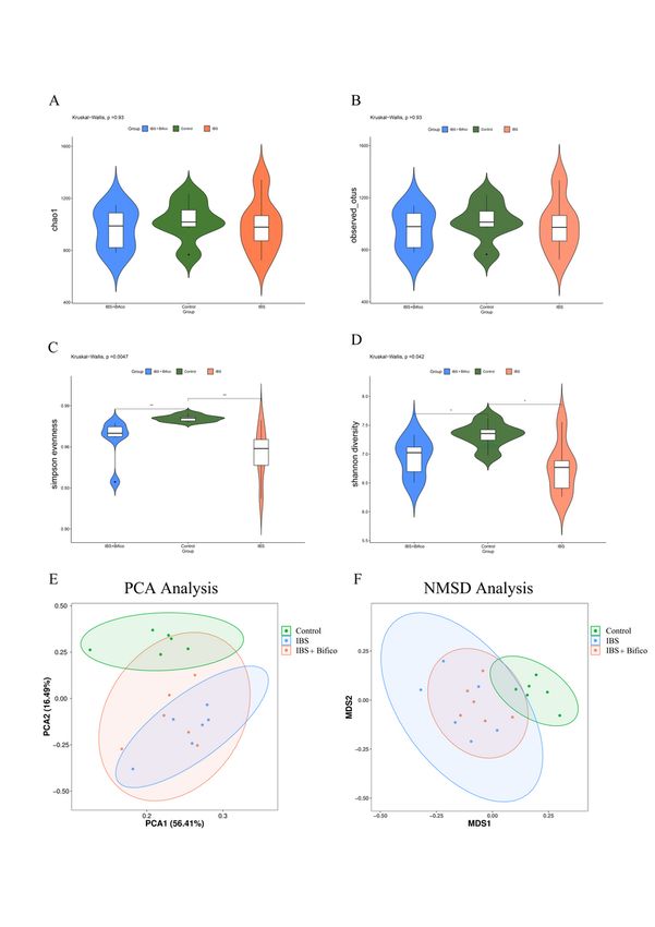
Abstract
Gut microbiota dysbiosis, a core pathophysiology of irritable bowel syndrome (IBS), is closely related to
immunological and metabolic functions. Gut microbiota-based therapeutics have been recently explored
in several studies. Bifico is a probiotic cocktail widely used in gastrointestinal disorders which relate to
the imbalance of gut microbiota. However, the efficacy and potential mechanisms of Bifico treatment in
IBS remains incompletely understood. In this animal experiment, IBS mice received Bifico by intragastric
administration. Subsequently, abdominal withdrawal reflex (AWR) scores showed a protective effect of
Bifico in IBS mice. Then 16S rDNA, 1H nuclear magnetic resonance (1H-NMR) and western blot assays
were performed to analyze alterations of gut microbiota, microbiome metabolites and inflammatory
cytokines, respectively. Results suggested that while Bifico did not increase gut microbial diversity, it
could change the composition of gut microbiota which were characterized by an increase of
Proteobacteria phylum and Actinobacteria phylum, Muribaculum genera, Bifidobacterium genera and a
decrease of Parabacteroides genera, Sutterella genera and Lactobacillus genera. Moreover, Bifico
elevated the concentration of short-chain fatty acids (SCFAs) and reduced protein levels of interleukin-6
(IL-6) and tumor necrosis factor-α (TNF-α). From further Spearman's correlation analysis, Bifidobacterium
genera were positively correlated with SCFAs including propionate, butyrate, valerate and negatively
correlated with IL-6 and TNF-α. In conclusion, this study demonstrated that Bifico could alleviate
symptoms of IBS mice through regulation of the gut microbiota, elevating production of SCFAs and
reducing the colonic inflammatory response. Therefore, Bifico may have utility in clinical practice.

1. Introduction
IBS is a chronic functional gastrointestinal disorder characterized by recurrent abdominal discomfort and
disturbed defecation such as a change in stool frequency or form [1, 2]. With a high risk of depression
and anxiety, between 5% and 10% of the general population suffers from IBS [3]. However, the underlying
etiology and pathogenesis of IBS are incompletely understood. Visceral hypersensitivity, alteration of gut
microbiota, chronic inflammation, psychological factors and genetics have been proposed as possible
mechanisms in the pathogenesis of IBS [4]. In recent years, increasing evidence suggests that gut
microbiota dysbiosis may a core pathophysiology of IBS [5, 6].

The gut microbiome has been dominated mainly by bacteria, as over 1000 species and 7000 strains have
now been characterized [7]. Further, the gut microbiome is closely related to immunological and
metabolic functions by producing a common bacterial metabolite SCFAs as mediators [8]. Studies have
proven that the gut microbiota dysbiosis could trigger host immune response, damage the intestinal
motility and barrier function [9–11]. Furthermore, the richness of the gut microbiota is negatively
associated with the symptom severity of IBS [12].

Considering the pivotal role of the microbiota in IBS, recent research in IBS treatments has been focused
on gut microbiome-based therapeutics. Generally well tolerated, probiotics in IBS have become a
relatively successful treatment option [13]. Ford AC et al. made a meta-analysis of 35 randomized

                                                   Page 2/24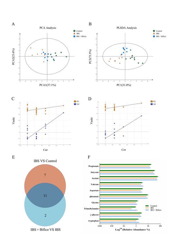
controlled trials of probiotics including Lactobacillus, Bifidobacterium, Streptococcus and combination
probiotics, involving 3452 patients suffering from IBS. They found that probiotics were effective for the
treatment IBS [14]. However, it should be noted that not all probiotic formulations are of benefit in IBS
patients [15]. S. boulardii and Probiotic mixtures containing Lactobacillus paracasei ssp paracasei F19,
Lactobacillus acidophilus La5 and Bifidobacterium Bb12 both failed to alleviate symptoms of IBS in
randomized clinical trials [16, 17]. Therefore, treatment strategies of probiotics should be further defined.

In 2002, Bifico was approved as an over-the-counter (OTC) drug by the Chinese regulatory authority, the
State Food and Drug Administration (SFDA), which contains Bifidobacterium, Lactobacillus acidophilus
and Enterococcus faecalis [18, 19]. As a mixture of viable bacteria, its regulatory functions on the gut
microbiota and anti-inflammatory effects on gastrointestinal disorders have been repeatedly confirmed.
We previously reported a prospective, randomized, controlled study of treatment of Bifico in antibiotic-
induced gut dysbiosis (AIGD) and found that Bifico could not only stabilize microbiota disorders but also
ameliorated colon inflammatory reactions [20]. Prophylactic therapy with Bifico could also reduce the
occurrence of neonatal nosocomial enteric infection (NNEI) and decrease the relapse of ulcerative colitis
(UC) [19, 21]. Using experimental colitis mice, Bifico was found to ameliorate gut inflammation by
decreasing the TNF-α level [22]. In a study on chronic functional diarrhea, Bifico was able to reduce drug
withdrawal in patients compared to the control group [23]. However, there was no relevant research to
illustrate the efficacy of Bifico in IBS and its potential mechanisms.

To solve these issues, we adopted a wrap restraint stress (WRS) -induced IBS mice model and AWR
scores to examine the treatment effect of Bifico, following by 16S rDNA gene sequencing to assess the
alterations of the gut microbiome, 1H-NMR to evaluate differential metabolites of fecal samples and
western blot assays to detect changes of inflammatory cytokines. Finally, we performed Spearman's
correlation analysis to find relationships among the gut microbiome of fecal samples, metabolites and
inflammatory cytokines.

2. Materials And Methods
2.1. Animals
Seven-week-old male C57BL/6 mice were purchased from the Laboratory Animal Center of Zhejiang
Chinese Medical University, Hangzhou, China. All mice were housed in metal barred cages (5 mice/cage)
and under controlled conditions (22 ± 1°C, 55 ± 10% humidity, low noise) with a 12-hour light/dark cycle.
Water and food were provided ad libitum. After adaptive feeding for 7 days, the mice were randomly
divided into 3 groups (n = 10/group): control group, IBS group and IBS + Bifico group. The experimental
workflow was shown in Fig. 1, the control group were given an intragastric administration of phosphate
buffer saline (PBS) (10ml/kg) once a day. The IBS group and the IBS + Bifico group were induced by WRS
procedures between 8 am and 10 am for 14 days. During forcing immobilization, they were placed in 50
mL tubes with a small hole for air and cotton ball were used to fill the extra space as described previously
[24]. Subsequently, the IBS group was given an intragastric administration of PBS (10ml/kg) once a day
                                                  Page 3/24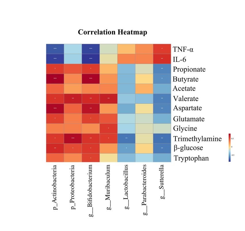
and the IBS + Bifico group was given an intragastric administration of Bifico (0.78g/kg) Shanghai
Sinepharm, Shanghai, China) once a day. Body weights of mice were recorded daily. Fecal samples were
collected from mice on the last two days and stored at − 80°C for further analyses. Experimental
protocols conformed to the requirements of the Experimental Animal Ethical Committee of the Zhejiang
Chinese Medical University (No. ZSLL-2018-014).

2.2. AWR test: visceral hypersensitivity evaluation
Visceral sensitivity was evaluated at the end of each experiment as follows [25, 26]. A disposable silicon
balloon-urethral catheter for pediatric use (6 Fr, Terumo, Tokyo, Japan) was inserted into the rectum to
apply colorectal distension (CRD). The balloon was placed 2 cm distal from the anus. After insertion, CRD
stimulation was maintained at three different levels of distention (0.25, 0.35, 0.50 mL, respectively) via
water injection. Each distention was repeated 3 times, with an interval of 4 min. Average values of AWR
scores were calculated as the final score for each mouse. The scoring of the AWR was quantified as
previously described [27]. 0 = no behavioral response to distension; 1 = brief head movements followed by
immobility; 2 = contraction of abdominal muscles without lifting of the abdomen; 3 = lifting of the
abdomen; 4 = body arching and lifting of the pelvic structure.

2.3. 16S rDNA sequencing and data analysis
DNA from different samples (at least 200 mg for each sample) was extracted using the E.Z.N.A. ®Stool
DNA Kit (D4015, Omega, Inc., USA) according to the manufacturer’s instructions. The V3-V4 region of the
bacterial 16S rRNA gene was amplified with primers 341F (5'-CCTACGGGNGGCWGCAG-3') and 805R (5'-
GACTACHVGGGTATCTAATCC-3') [28]. The PCR products were purified by AMPure XT beads (Beckman
Coulter Genomics, Danvers, MA, USA) and quantified by Qubit (Invitrogen, USA). The 5' ends of the
primers were tagged with specific sequencing universal primers. PCR amplification was performed in a
total volume of 25 µL reaction mixture containing 25 ng of template DNA, 12.5 µL PCR Premix, 2.5 µL of
each primer, and PCR-grade water to adjust the volume. The PCR conditions to amplify the prokaryotic
16S fragments consisted of an initial denaturation at 98 ℃ for 30 seconds; 32 cycles of denaturation at
98 ℃ for 10 seconds, annealing at 54 ℃ for 30 seconds, and extension at 72 ℃ for 45 seconds; and
then final extension at 72 ℃ for 10 minutes. The PCR products were confirmed with 2% agarose gel
electrophoresis. Throughout the DNA extraction process, ultrapure water was used to exclude the
possibility of false-positive PCR results as a negative control. The PCR products were purified by AMPure
XT beads (Beckman Coulter Genomics, Danvers, MA, USA) and quantified by Qubit (Invitrogen, USA). The
amplicon pools were prepared for sequencing and the size and quantity of the amplicon library were
assessed on an Agilent 2100 Bioanalyzer (Agilent, USA) and with the Library Quantification Kit for
Illumina (Kapa Biosciences, Woburn, MA, USA), respectively. The libraries were sequenced on NovaSeq
PE250 platform. Samples were sequenced on an Illumina NovaSeq platform according to the
manufacturer's recommendations, provided by LC-Bio. Paired-end reads were assigned to samples based
on their unique barcode and truncated by cutting off the barcode and primer sequence. Paired-end reads
were merged using FLASH. Quality filtering on the raw reads was performed under specific filtering

                                                 Page 4/24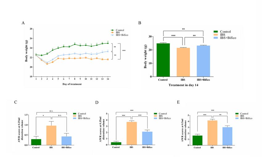
conditions to obtain the high-quality clean tags according to the fqtrim (v0.94). Chimeric sequences were
filtered using Vsearch software (v2.3.4). After dereplication using DADA2, we obtained a feature table and
a feature sequence.

Alpha diversity and beta diversity were calculated by QIIME2, in which the same number of sequences
were extracted randomly through reducing the number of sequences to the minimum of some samples,
and the relative abundance (X bacteria count/total count) was used in bacteria taxonomy. Pictures of
Alpha diversity and Beta diversity were drawn by R (v3.5.2). The sequence alignment of species
annotation was performed by Blast, and the alignment database used was the SILVA and NT-16S.

2.4. Fecal samples preparation for 1H-NMR analysis
The method of fecal sample preparation was described in a previous study [29]. Briefly, 100 mg thawed
stool material were mixed with 0.8 mL PBS containing 10% deuterated water (D2O 99.8%; SIGMA, United
States) and 0.05 mM sodium 3-trimethylsilyl-propionate-d4 (TMSP-2,2,3,3-d4; SIGMA, Untied States) as a
chemical shift reference. The mixture was immersed into ice for 30 min and then dissolved for 10 cycles
(one cycle includes 20 s ultrasound, 10 s crash, and 30 s rest). Then the fecal slurry was centrifuged at
13,000 g for 10 min at 4℃ for twice to obtained supernatants.

2.5. 1H-NMR analysis and data processing
All 1H-NMR spectra were recorded by Bruker 600 MHz AVANCE III spectrometer equipped with a 5mm-
BBFO probe at 25°C. Shimming and proton pulse calibration was performed automatically for each
sample before data acquisition. 1H-NMR spectra were received using NOESYPR 1D pulse sequence with
water suppression. Bruker Topspin 3.2 was used to process the data.

Free induction decays (FIDs) from 1H-NMR of the fecal samples were multiplied by a 0.3 Hz exponential
line broadening prior to Fourier Transformation. All NMR spectra were manually phased, baseline
corrected and referenced to TSP (δ = 0.0) within MestReNova 12 (Mestrelab Research SL, Spain). The
integral region of the spectrum was set between 0.0–9.0 ppm, with a spectral region of 4.5-5.0 ppm to
eliminate the effects of imperfect water suppression. Due to the deviation of metabolite concentration in
the fecal samples of each mouse, each bucket was internally normalized to the total sum of the spectral
integrals prior to pattern recognition analysis. The characteristic peaks of all fecal metabolites were
determined based on related literature [30, 31] and the Biological Magnetic Resonance Bank
(http://www.bmrb.wisc.edu/metabolomics) and Human Metabolome Database (http://www.hmdb.ca/).
2.6. Western blot analysis
Protein extracts were prepared with RIPA Lysis and Extraction Buffer (89901, Thermo Scientific, USA)
supplemented with Protease and Phosphatase Inhibitor Cocktail (78443, Thermo Scientific, USA)
according to the manual. Then proteins were separated on SDS-PAGE gels (10%) followed by transfer to
polyvinyl difluoride (PVDF) membranes (pore size 0.2 µm, 88520, Thermo Scientific, USA). The membrane
was subsequently blotted in 1% bovine serum albumin (BSA, Sigma-Aldrich St. Louis, MO, USA) in PBS

                                                  Page 5/24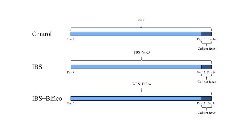
for 2 h and incubated overnight with commercially available primary antibodies against β-actin (1:1000
dilution, 4970S, Cell Signaling Technology, Danvers, MA, USA), IL-6 (1:1000 dilution, 4970S, Cell Signaling
Technology, Danvers, MA, USA) and TNF-a (1:2000 dilution, 41504, Signalway Antibody, Pearland, TX,
USA) at 4°C. After washing three times with PBS containing 0.05% Tween-20, membranes were incubated
with secondary antibodies coupled with HRP (1:4000 dilution, LF102, EpiZyme, Shanghai, China)
followed by washing three times. The images were captured with Bio-Rad gel imaging system and
analyzed by Quantity One software.

2.7. Statistical analysis
The experimental data were processed and analyzed using Graphpad Prism 6 software (version 6.01).
The principal component analysis (PCA) and partial least-squares discriminant analysis (PLS-DA) were
performed by SIMCA software, version 14. The non-metric multidimensional scaling (NMDS) analysis
and classification tree heat map were made using R language (R version 3.5.2). Venn analysis was
pictured by the Bioinformatics website system (http://bioinformatics.psb.ugent.be/webtools/Venn/).
Spearman's correlation analysis was generated by IBM SPSS Statistics 25.0 software. Data was analyzed
to perform normality. The Unpaired Student's two-tailed t-test was used for two sets of data conformed to
the normal distribution. The Kruskal-Wallis test was used for two sets of data and did not conform to the
normal distribution. Analysis of variance (ANOVA) was used to compare multiple groups of data. All
values were expressed as mean ± SEM and P < 0.05 was considered as statistically significant.
Differential metabolomics data must conform to P < 0.05 and VIP > 1 at the same time.

3. Results
3.1. Bifico alleviated visceral hypersensitivity in IBS mice
We adopted a WRS model to simulate symptoms of IBS. During the administration of Bifico, changes in
body weights were recorded daily. The control group,IBS group and IBS + Bifico group weighted 24.03 ±
0.2308, 21.65 ± 0.1131 and 22.45 ± 0.1737 (mean ± SEM), respectively, which meant that body weights of
IBS mice were lower than that of control mice (P < 0.001), after treatment with Bifico, body weights of IBS
+ Bifico mice were still lower than control mice (P < 0.01). However, they were improved compared to IBS
mice (P < 0.01) at the end of the experiment (Fig. 2A and 2B). To evaluate the development of colonic
visceral hypersensitivity, we compared the AWR score at a pressure stimulation of 0.25, 0.35, or 0.5 mL
among three groups. The AWR score of the IBS group was significantly higher than the control group (P <
0.05 at 0.25 ml, P < 0.01 at 0.35 ml and P < 0.01 at 0.35 ml, respectively). After Bifico treatment, the AWR
score under stimulation with 0.25 ml of the Bifico group had no statistical difference compared to the IBS
group or the control group (the IBS + Bifico group vs. the IBS group: P > 0.05 and the IBS + Bifico group vs.
the control group: P > 0.05, respectively). Although the AWR score increased in the Bifico group under
stimulation with 0.35 ml and 0.5 ml compared to the control group (P < 0.001 at 0.35 ml and P < 0.001 at
0.5 ml, respectively), it decreased in the Bifico group under stimulation with 0.35 ml and 0.5 ml compared

                                                  Page 6/24
to the IBS group (P < 0.001 at 0.35 ml and P < 0.01 at 0.5 ml, respectively) (Fig. 2C − 2E). These
information suggested that treatment with Bifico could alleviate visceral hypersensitivity in IBS mice.
3.2. Bifico altered the gut microbiota community in IBS
mice
Fecal samples were obtained from mice at the end of treatment. In order to assess the richness and
diversity of gut microbiota, alpha diversity (Fig. 3A − 3D) was used. Chao1 and Observed_otus both
showed no difference among three groups (Chao1: P > 0.05 and Observed_otus: P > 0.05, respectively). As
for Simpson Evenness and Shannon diversity, the control group had a significant difference compared to
the IBS group or the IBS + Bifico group (Simpson Evenness: Kruskal-Wallis, P < 0.01 and Shannon
diversity: Kruskal-Wallis, P < 0.05, respectively). PCA and NMDS of beta diversity (Fig. 3E and 3F) further
revealed that differences of the gut microbiota community composition between the control group was
significant and the community composition of the IBS + Bifico group was closer to the control group
through treatment with Bifico. These results indicated that the IBS group had lower community diversity
of gut microbiota than the control group and Bifico treatments may not increase gut microbial diversity
but affected the microbial community of IBS mice.
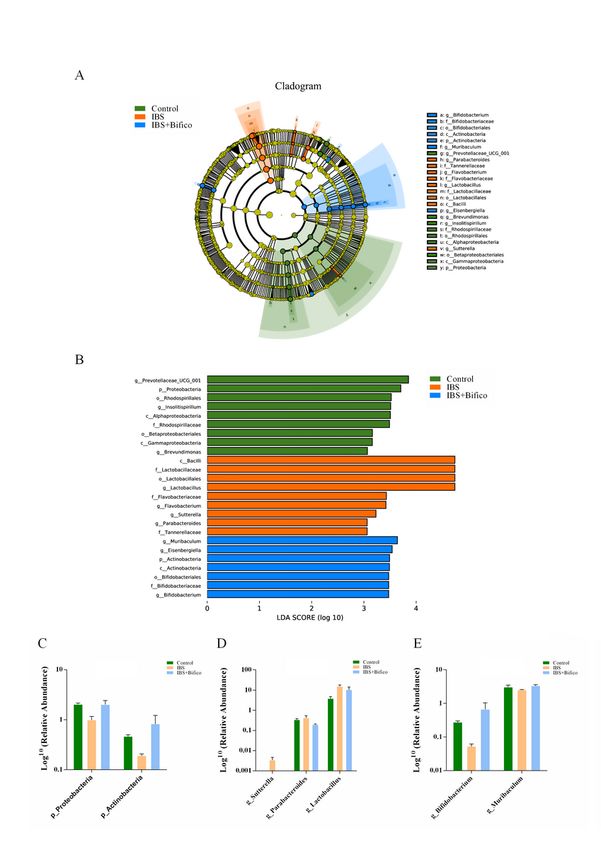
We further investigated the gut microbiota species and their relative abundance through LEfSe (LDA score
(log 10) > 3, P < 0.05). Microbiota in the phylum level, Proteobacteria was predominant in the control
group, while Actinobacteria were enriched in the IBS + Bifico group (Fig. 4A and 4B). Both of them had
reduced relative abundance in IBS mice compared to control mice and administration of Bifico could
elevate their relative abundance (Fig. 4C). In the general level, the control group was characterized by
Prevotellaceae_UCG_001, Insolitispirillum and Brevundimonas. However, Lactobacillus, Flavobacterium,
Sutterella and Parabacteroides were specific for the IBS group. After administration of Bifico, the IBS +
Bifico group could be categorized into Muribaculum, Eisenbergiella and Bifidobacterium (Fig. 4A and 4B).
Expecting four kinds of microbiota including Prevotellaceae_UCG_001, Insolitispirillum, Brevundimonas,
which presented a decreasing trend in the IBS group compared to the control group and Eisenbergiella,
which showed an increasing trend in the IBS group compared to the control group. Treatment of Bifico
could aggravate these trends (data shown in supplementary Fig. S1), as Fig. 4C and 4D showed, and the
relative abundance of Muribaculum genera and Bifidobacterium genera were lower in the IBS group than
in the control group, while Lactobacillus genera, Parabacteroides genera and Sutterella genera were
higher in the IBS group than in the control group. According to the treatment of Bifico, Muribaculum
genera and Bifidobacterium genera had an increased relative abundance compared to the IBS group,
Lactobacillus genera, Parabacteroides genera and Sutterella genera had a decreased relative abundance
compared to the IBS group.
3.3. Bifico changed the gut metabolites in IBS mice
We next explored the potential changes in metabolites related to the gut microbiota. Different enrichment
of metabolites from fecal samples among three groups were observed by 1H-NMR spectroscopy. PCA
Page 7/24showed that the samples from each group were separated from the other two groups, remarkably, the
metabolites of the Bifico group were closer to the metabolites of the control group (Fig. 5A). Moreover,
there was a clear distinction among the three groups in the PLS-DA, indicating that there were significant
differences in the fecal metabolites among the three groups (Fig. 5B). We next verified the credibility and
stability of the model. The model parameters were as shown follows: the IBS group vs. the Control group:
R2Y = 0.721, Q2 = -0.202; the IBS + Bifico group vs. the IBS group: R2Y = 0.759, Q2 = -0.222 (Fig. 5C − 5D).
These results suggested that the models were stable and accurately predictive.
Then we adopted the criteria of VIP > 1 at multivariate statistical analysis and P < 0.05 at univariate
statistics at the same time for screening differential metabolites among groups. Under the criteria, 18 out
of 39 differential metabolites were selected out when the control group was compared to the IBS group.
Meanwhile, 13 out of 39 differential metabolites were found to be similar between the IBS group and the
IBS + Bifico group. Furthermore, a Venn diagram was used to address the overlapping metabolites among
the two collections of differential metabolites (18 and 13, respectively), which marked out 11 metabolites
(Fig. 5E). We expected the relative abundance of choline to be lower in the IBS group compared to the
control group. It was worth noting that it still had a decreased relative abundance after Bifico treatment
compared to the IBS group (data shown in supplementary Fig. S2). There were 10 metabolites including
propionate, butyrate, acetate, valerate, aspartate, glutamate, glycine, trimethylamine, β-glucose and
tryptophan, which showed a decreased tendency in the IBS group compared to the control group, an
elevated tendency in the IBS + Bifico group compared to the IBS group (Fig. 5F), which elucidated that IBS
affected the production of gut metabolites, and some of them were reversed by Bifico administration.
3.4. Bifico reduced the expression of TNF-ɑ and IL-6 in IBS
mice
Because gut microbial dysbiosis is often accompanied by abnormal expression of inflammatory
cytokines, we evaluated the protein levels of TNF-ɑ and IL-6 in colon tissues and confirmed that
expression of TNF-ɑ and IL-6 increased in the IBS group compared with the control group (TNF-ɑ: P < 0.01
and IL-6: P < 0.01, respectively). However, treatment of Bifico could restore protein expression to normal
levels (TNF-ɑ: the IBS group vs. the IBS + Bifico group: P < 0.05, the control group vs. the IBS + Bifico
group: P > 0.05, respectively and IL-6: the IBS group vs. the IBS + Bifico group: P < 0.05, the control group
vs. the IBS + Bifico group: P > 0.05, respectively) (Fig. 6), which indicated that Bifico treatment relieved the
colonic inflammation in IBS mice
3.5. Correlation of gut microbiota, fecal metabolites and
inflammatory cytokines
For a better understanding of the relationship of gut microbiota, fecal metabolites and inflammatory
cytokines which were significantly different among the three groups were analyzed. A heatmap was
calculated by the Spearman's correlation index (Fig. 7). Involving in inflammatory cytokines, we observed
Page 8/24that Actinobacteria phylum and Bifidobacterium genera (belongs to Actinobacteria phylum) were
negatively correlated with IL-6 and TNF-ɑ, whereas Sutterella (belongs to Proteobacteria phylum) genera
were positively correlated with IL-6 and TNF-ɑ. As for metabolites, Bifidobacterium genera and Sutterella
genera were related to metabolites. Bifidobacterium genera were positively correlated with propionate,
butyrate, valerate, aspartate, glutamate, trimethylamine, β-glucose and tryptophan. Dramatically, although
Proteobacteria phylum were positively correlated with valerate, trimethylamine and β-glucose, Sutterella
genera were negatively correlated with propionate, butyrate, valerate, aspartate, trimethylamine and β-
glucose. In addition, Muribaculum genera were positively correlated with glycine, trimethylamine and
valerate. Lactobacillus genera were negatively correlated with β-glucose and trimethylamine.
Parabacteroides genera were positively correlated with choline. These results further supported the
Bifidobacterium genera might be main contributor in the treatment of Bifico.
4. Discussion
IBS is a functional gastrointestinal disorder characterized by visceral hypersensitivity, intestinal immune
activation and gut microbiota dysfunction [32]. Recently, a growing body of evidence has suggested that
gut the microbiome plays a pivotal role in colonic inflammation [33]. As a probiotic mixture, Bifico is
supplied for the treatment of microbiota disorders or alleviating the inflammatory reaction [34, 35].
However, how Bifico treatment functions in IBS is still unclear. Here we analyzed the relationship between
the gut microbiota and the inflammatory cytokines in IBS after treatment with Bifico, which might provide
a theoretical basis for the clinical use of Bifico. Our studies showed that Bifico may relieve the symptoms
of IBS by reducing the protein expression level of IL-6 and TNF-α, altering fecal metabolites and gut
microbiota. Further studies revealed that Bifidobacterium genera may play important roles in treatment.
Previous studies have demonstrated that Bifidobacterium and Lactobacillus could specifically relieve the
symptoms of IBS [36]. Moreover, multispecies probiotics containing strains of more than one genus show
enhanced effects in treating antibiotic-associated diarrhea in children, which suggests that probiotic
mixtures may more be effective than a single-strain [37]. Bifico is a type of multispecies probiotic.
Lactobacillus acidophilus could produce bifidogenic growth factors to stimulate the growth of
Bifidobacterium longum in pure culture [38]. Enterococcus spp. has been used as a probiotic to defend
against gut infection and prevent the colonization of more pathogenic bacteria [39]. In addition,
Enterococcus faecalis could create anaerobic conditions, which might be of benefit for survival of
Bifidobacterium [40].
WRS is an accepted method of creating an acute stress-induced IBS model [24]. In this study, we
observed that treatment of Bifico significantly alleviated intestinal visceral hypersensitivity and reduced
weight loss. These results were consistent with most of the previous studies that effective treatment
could reduce the AWR score of IBS [41–43]. Since IBS often involves gut microbiome dysbiosis, we
collected fecal samples to analyze the gut microbiota. Community richness was measured by Chao1 and
Observed_otus, while community diversity was measured by Simpson Evenness and Shannon diversity.
Results showed that Chao1 and Observed_otus of the IBS group had no significant difference compared
Page 9/24to the control group, but Simpson Evenness and Shannon diversity was lower in the IBS group than in the
control group, were similar to previous findings by Fukui H. et al [44]. Despite treatment, Bifico did not
alter alpha diversity of IBS. In beta diversity, the bacterial composition of the IBS + Bifico group was closer
to the control group through treatment of Bifico. To further analyze the gut microbiota and their relative
abundance according to LEfSe. In the phylum level, the relative abundance of Proteobacteria and
Actinobacteria were markedly lower in the IBS group than in the control group and the IBS + Bifico group.
In the genera level, Parabacteroides and Sutterella were increased in the IBS group compared to the
control group and the IBS + Bifico group. Parabacteroides and Parabacteroides merdae (belongs to
Parabacteroides genera) were considered as potentially pathogenic bacterium that were reported to be
frequently enriched in the hypertensive gut microbiome [45, 46]. Moreover, in infectious diseases,
Parabacteroides merdae is generally considered an opportunistic pathogen, which is able to develop
antimicrobial drug resistance [47]. Sutterella is a controversial bacterium. From a previous review,
Sutterella was related to better outcomes in patients with IBD [48]. Berer K et al. held the opinion that
Sutterella had anti-inflammatory functions in vitro [49]. But some studies have suspected Sutterella plays
a role in the disease progression of IBD [50]. Furthermore, in clinical studies, no difference was observed
in the prevalence of Sutterella spp. between the IBD patients and the healthy subjects [51, 52].
Surprisingly, Lactobacillus expressed higher levels in the IBS group than in the control group and the IBS
+ Bifico group. Similarly, clinical research from Japan also observed that IBS patients had significantly
higher counts of Lactobacillus [53]. Another clinical study considered that Lactobacillus may have no
effect on IBS patients [54]. Compared to the control group and the IBS + Bifico group, the IBS group
showed a significant decrease in the abundance of Muribaculum and Bifidobacterium. Yuan Y et al.
noted that reduction of Muribaculum could result in inflammation, dyslipidemia and glucose intolerance
[55]. As for Bifidobacterium, which belongs to Actinobacteria phylum, numerous studies have shown that
it has benefits in improving epithelial barrier function in mice, act as an anti-inflammatory agent and a
source of SCFAs [56, 57].
SCFAs describe acetic acid, propionic acid, butyric acid, valeric acid and caproic acid, which are the main
byproducts of gut metabolites [58]. In the colon, the proportion of acetate, propionate, and butyrate can
reach 90–95% of SCFAs [59]. It has been widely reported that SCFAs play pivotal roles in anti-
inflammatory and maintenance of intestinal health such as locomotion recovery [60–62]. What’s more,
SCFAs are involved in lipid metabolism and glucose metabolism [63]. According to the altered gut
bacteria, we speculate that colonic metabolites may have changed. Through 1H NMR spectroscopy, we
found that the abundance of acetate, propionate, butyrate and valerate were decreased in the IBS group,
which supported the view of suggesting that patients with IBS had lower levels of SCFA [64]. Treatment of
Bifico could increase their abundance. From Spearman's correlation analysis, we found that
Actinobacteria phylum and Bifidobacterium genera were positively correlated with propionate, butyrate
and valerate, Proteobacteria phylum and Muribaculum genera were positively correlated with valerate.
The Sutterella genera were negatively correlated with propionate, butyrate and valerate.
Page 10/24Changes in the gut microbiota can induce or aggravate inflammation [65]. Recent research regards TNF-α
as a vital inflammatory cytokine in IBS [66]. In addition, IL-6 has reproducibly been detected to be elevated
in IBS patients and rats [67–69]. In our studies, the protein level in colonic IL-6 and TNF-α were higher in
the IBS group than in the control group, further confirmed by the previous results. Zhao HM et al. found
that in the colon of mice with colitis, the level of TNF-α could be significantly reduced by Bifico [70]. Our
results supported the that Bifico could reduce both the protein level of IL-6 and TNF-α in the colon,
alleviating the inflammation to a certain extent. In further correlation analysis, Actinobacteria phylum and
Bifidobacterium genera were negatively correlated with IL-6 and TNF-α, verifying the anti-inflammatory
function reported by Chichlowski M et al. [71]. Interestingly, Sutterella genera were positively correlated
with IL-6 and TNF-α. This result stood suggested that the Sutterella genera had a pro-inflammatory
capacity in the human gastrointestinal tract by Hiippala K et al. [72].
Taken together, using a widely developed IBS mice model, we found that Bifico regulated the gut
microbiota and elevated SCFAs to reduce inflammation and relieve visceral hypersensitivity of IBS mice.
These results hint at the potential clinical utility of treatment with Bifico.
Declarations
Ethics approval and consent to participate
This experiment was supervised and approved by Experimental Animal Ethical Committee of the Zhejiang
Chinese Medical University (Approved No. of ethic committee: ZSLL-2018-014).
Consent for publication
Not applicable
Availability of data and materials
Not yet uploaded to the database.
Competing interests
The authors declare that they have no competing interests.
Funding
This work was supported by National Natural Science Foundation of China (No. 81971600, 81970470
and 81770535), the Zhejiang Provincial Natural Science Foundation of China (LY21H030002), the
Medical Health Science and Technology Project of Zhejiang Provincial Health Commission (2019RC229).
Authors' contributions
Page 11/24Yanlin Zhou and Fan Zhang conceived the study design and wrote the manuscript. Liqi Mao, Tongfei
Feng and Kaijie Wang made data analysis and prepared the figures. Yanlin Zhou, Fan Zhang and Xi
Wang performed the experiments. Maosheng Xu and Bin Lv supervised the scientifific work and revised
the manuscript. All authors have read and agreed to the published version of the manuscript. All authors
read and approved the final manuscript.
Acknowledgements
Not applicable
References
1. Villani AC, Lemire M, Thabane M, et al. Genetic risk factors for post-infectious irritable bowel
syndrome following a waterborne outbreak of gastroenteritis. Gastroenterology. 2010;138:1502–13.
10.1053/j.gastro.2009.12.049.
2. Chen YJ, Wu H, Wu SD, et al. Parasutterella, in association with irritable bowel syndrome and
intestinal chronic inflammation. J Gastroenterol Hepatol. 2018;33:1844–52. 10.1111/jgh.14281.
3. Ford AC, Sperber AD, Corsetti M, et al. Irritable bowel syndrome. Lancet. 2020;396:1675–88.
10.1016/S0140-6736(20)31548-8.
4. Shukla R, Ghoshal U, Ranjan P, et al. Expression of Toll-like Receptors, Pro-, and Anti-inflammatory
Cytokines in Relation to Gut Microbiota in Irritable Bowel Syndrome: The Evidence for Its Micro-
organic Basis. J Neurogastroenterol Motil. 2018;24:628–42. 10.5056/jnm18130.
5. Pimentel M, Lembo A. Microbiome and Its Role in Irritable Bowel Syndrome. Dig Dis Sci.
2020;65:829–39. 10.1007/s10620-020-06109-5.
6. Singh R, Zogg H, Wei L, et al. Gut Microbial Dysbiosis in the Pathogenesis of Gastrointestinal
Dysmotility and Metabolic Disorders. J Neurogastroenterol Motil. 2021;27:19–34.
10.5056/jnm20149.
7. Pusceddu MM, Gareau MG. Visceral pain: gut microbiota, a new hope? J Biomed Sci. 2018;25:73.
10.1186/s12929-018-0476-7.
8. Levy M, Blacher E, Elinav E. Microbiome, metabolites and host immunity. Curr Opin Microbiol.
2017;35:8–15. 10.1016/j.mib.2016.10.003.
9. Cani PD. Gut microbiota: Changes in gut microbes and host metabolism: squaring the circle?, Nat
Rev Gastroenterol Hepatol 13 (2016) 563–564. 10.1038/nrgastro.2016.135.
10. Bhattarai Y, Muniz Pedrogo DA, Kashyap PC. Irritable bowel syndrome: a gut microbiota-related
disorder? Am J Physiol Gastrointest Liver Physiol. 2017;312:G52–62. 10.1152/ajpgi.00338.2016.
11. Sciavilla P, Strati F, Di Paola M, et al. Gut microbiota profiles and characterization of cultivable fungal
isolates in IBS patients. Appl Microbiol Biotechnol. 2021;105:3277–88. 10.1007/s00253-021-11264-
4.
Page 12/2412. Tap J, Derrien M, Tornblom H, et al, Identification of an Intestinal Microbiota Signature Associated
With Severity of Irritable Bowel Syndrome, Gastroenterology 152 (2017) 111–123 e118.
10.1053/j.gastro.2016.09.049.
13. Pretorius L, Smith C. The trace aminergic system: a gender-sensitive therapeutic target for IBS? J
Biomed Sci. 2020;27:95. 10.1186/s12929-020-00688-1.
14. Ford AC, Quigley EM, Lacy BE, et al. Efficacy of prebiotics, probiotics, and synbiotics in irritable bowel
syndrome and chronic idiopathic constipation: systematic review and meta-analysis. Am J
Gastroenterol. 2014;109:1547–61. 10.1038/ajg.2014.202. quiz 1546, 1562.
15. Ghoshal UC, Gwee KA, Holtmann G, et al. The role of the microbiome and the use of probiotics in
gastrointestinal disorders in adults in the Asia-Pacific region - background and recommendations of
a regional consensus meeting. J Gastroenterol Hepatol. 2018;33:57–69. 10.1111/jgh.13840.
16. Choi CH, Jo SY, Park HJ, et al. A randomized, double-blind, placebo-controlled multicenter trial of
saccharomyces boulardii in irritable bowel syndrome: effect on quality of life. J Clin Gastroenterol.
2011;45:679–83. 10.1097/MCG.0b013e318204593e.
17. Begtrup LM, de Muckadell OB, Kjeldsen J, et al. Long-term treatment with probiotics in primary care
patients with irritable bowel syndrome–a randomised, double-blind, placebo controlled trial. Scand J
Gastroenterol. 2013;48:1127–35. 10.3109/00365521.2013.825314.
18. Yu H, Liu L, Chang Z, et al., Genome Sequence of the Bacterium Bifidobacterium longum Strain
CMCC P0001, a Probiotic Strain Used for Treating Gastrointestinal Disease, Genome Announc 1
(2013). 10.1128/genomeA.00716 – 13.
19. Huang NN, Wang GZ, Wang JF, et al. Risk factors for neonatal nosocomial enteric infection and the
effect of intervention with BIFICO. Eur Rev Med Pharmacol Sci. 2016;20:3713–9.
20. Wu J, Gan T, Zhang Y, et al., The prophylactic effects of BIFICO on the antibiotic-induced gut
dysbiosis and gut microbiota, Gut Pathog 12 (2020) 41. 10.1186/s13099-020-00379-0.
21. Cui HH, Chen CL, Wang JD, et al. Effects of probiotic on intestinal mucosa of patients with ulcerative
colitis. World J Gastroenterol. 2004;10:1521–5. 10.3748/wjg.v10.i10.1521.
22. Zhang Y, Zhao X, Zhu Y, et al. Probiotic Mixture Protects Dextran Sulfate Sodium-Induced Colitis by
Altering Tight Junction Protein Expressions and Increasing Tregs. Mediators Inflamm.
2018;2018:9416391. 10.1155/2018/9416391.
23. Yao-Zong Y, Shi-Rong L, Delvaux M. Comparative efficacy of dioctahedral smectite (Smecta) and a
probiotic preparation in chronic functional diarrhoea. Dig Liver Dis. 2004;36:824–8.
10.1016/j.dld.2004.07.012.
24. Lin L, Feng B, Zhou R, et al. Acute stress disrupts intestinal homeostasis via GDNF-RET. Cell Prolif.
2020;53:e12889. 10.1111/cpr.12889.
25. Zhao Q, Yang WR, Wang XH, et al. Clostridium butyricum alleviates intestinal low-grade inflammation
in TNBS-induced irritable bowel syndrome in mice by regulating functional status of lamina propria
dendritic cells. World J Gastroenterol. 2019;25:5469–82. 10.3748/wjg.v25.i36.5469.
Page 13/2426. Al-Chaer ED, Kawasaki M, Pasricha PJ. A new model of chronic visceral hypersensitivity in adult rats
induced by colon irritation during postnatal development. Gastroenterology. 2000;119:1276–85.
10.1053/gast.2000.19576.
27. Wang FY, Su M, Zheng YQ, et al. Herbal prescription Chang'an II repairs intestinal mucosal barrier in
rats with post-inflammation irritable bowel syndrome. Acta Pharmacol Sin. 2015;36:708–15.
10.1038/aps.2014.170.
28. Logue JB, Stedmon CA, Kellerman AM, et al. Experimental insights into the importance of aquatic
bacterial community composition to the degradation of dissolved organic matter. ISME J.
2016;10:533–45. 10.1038/ismej.2015.131.
29. Chen H, Zhang F, Zhang J, et al. A Holistic View of Berberine Inhibiting Intestinal Carcinogenesis in
Conventional Mice Based on Microbiome-Metabolomics Analysis. Front Immunol. 2020;11:588079.
10.3389/fimmu.2020.588079.
30. Swann JR, Garcia-Perez I, Braniste V, et al. Application of (1)H NMR spectroscopy to the metabolic
phenotyping of rodent brain extracts: A metabonomic study of gut microbial influence on host brain
metabolism. J Pharm Biomed Anal. 2017;143:141–6. 10.1016/j.jpba.2017.05.040.
31. Xu M, Ye J, Yang D, et al. Ex-vivo NMR of unprocessed tissue in water: a simplified procedure for
studying intracranial neoplasms. Anal Bioanal Chem. 2007;389:2153–9. 10.1007/s00216-007-1629-
9.
32. Li J, Cui H, Cai Y, et al. Tong-Xie-Yao-Fang Regulates 5-HT Level in Diarrhea Predominant Irritable
Bowel Syndrome Through Gut Microbiota Modulation. Front Pharmacol. 2018;9:1110.
10.3389/fphar.2018.01110.
33. Seksik P, Sokol H, Lepage P, et al. Review article: the role of bacteria in onset and perpetuation of
inflammatory bowel disease. Aliment Pharmacol Ther. 2006;24 Suppl 3:11–8. 10.1111/j.1365-
2036.2006.03053.x.
34. Yu HJ, Liu W, Chang Z, et al. Probiotic BIFICO cocktail ameliorates Helicobacter pylori induced
gastritis. World J Gastroenterol. 2015;21:6561–71. 10.3748/wjg.v21.i21.6561.
35. Jiang XE, Yang SM, Zhou XJ, et al. Effects of mesalazine combined with bifid triple viable on
intestinal flora, immunoglobulin and levels of cal, MMP-9, and MPO in feces of patients with
ulcerative colitis. Eur Rev Med Pharmacol Sci. 2020;24:935–42. 10.26355/eurrev_202001_20079.
36. Brenner DM, Moeller MJ, Chey WD, et al. The utility of probiotics in the treatment of irritable bowel
syndrome: a systematic review. Am J Gastroenterol. 2009;104:1033–49. 10.1038/ajg.2009.25. quiz
1050.
37. Timmerman HM, Koning CJ, Mulder L, et al. Monostrain, multistrain and multispecies probiotics–A
comparison of functionality and efficacy. Int J Food Microbiol. 2004;96:219–33.
10.1016/j.ijfoodmicro.2004.05.012.
38. Warda AK, Clooney AG, Ryan F, et al. A postbiotic consisting of heat-treated lactobacilli has a
bifidogenic effect in pure culture and in human fermented faecal communities. Appl Environ
Microbiol. 2021. 10.1128/AEM.02459-20.
Page 14/2439. Kabwe M, Meehan-Andrews T, Ku H, et al. Lytic Bacteriophage EFA1 Modulates HCT116 Colon
Cancer Cell Growth and Upregulates ROS Production in an Enterococcus faecalis Co-culture System.
Front Microbiol. 2021;12:650849. 10.3389/fmicb.2021.650849.
40. Ya'ari S, Halperin-Sternfeld M, Rosin B, et al., Surface Modification by Nano-Structures Reduces
Viable Bacterial Biofilm in Aerobic and Anaerobic Environments, Int J Mol Sci 21 (2020).
10.3390/ijms21197370.
41. Song YF, Pei LX, Chen L, et al. Electroacupuncture Relieves Irritable Bowel Syndrome by Regulating
IL-18 and Gut Microbial Dysbiosis in a Trinitrobenzene Sulfonic Acid-Induced Post-Inflammatory
Animal Model. Am J Chin Med. 2020;48:77–90. 10.1142/S0192415X20500044.
42. Zhang Y, Zhang H, Zhang W, et al. LncRNA XIST modulates 5-hydroxytrytophan-induced visceral
hypersensitivity by epigenetic silencing of the SERT gene in mice with diarrhea-predominant IBS. Cell
Signal. 2020;73:109674. 10.1016/j.cellsig.2020.109674.
43. Chen BR, Du LJ, He HQ, et al. Fructo-oligosaccharide intensifies visceral hypersensitivity and
intestinal inflammation in a stress-induced irritable bowel syndrome mouse model. World J
Gastroenterol. 2017;23:8321–33. 10.3748/wjg.v23.i47.8321.
44. Fukui H, Nishida A, Matsuda S, et al., Usefulness of Machine Learning-Based Gut Microbiome
Analysis for Identifying Patients with Irritable Bowels Syndrome, J Clin Med 9 (2020).
10.3390/jcm9082403.
45. Hu X, Li H, Zhao X, et al. Multi-omics study reveals that statin therapy is associated with restoration
of gut microbiota homeostasis and improvement in outcomes in patients with acute coronary
syndrome. Theranostics. 2021;11:5778–93. 10.7150/thno.55946.
46. Yan Q, Gu Y, Li X, et al. Alterations of the Gut Microbiome in Hypertension. Front Cell Infect Microbiol.
2017;7:381. 10.3389/fcimb.2017.00381.
47. Boente RF, Ferreira LQ, Falcao LS, et al. Detection of resistance genes and susceptibility patterns in
Bacteroides and Parabacteroides strains. Anaerobe. 2010;16:190–4.
10.1016/j.anaerobe.2010.02.003.
48. Morgan BP, Harris CL. Complement, a target for therapy in inflammatory and degenerative diseases.
Nat Rev Drug Discov. 2015;14:857–77. 10.1038/nrd4657.
49. Berer K, Gerdes LA, Cekanaviciute E, et al. Gut microbiota from multiple sclerosis patients enables
spontaneous autoimmune encephalomyelitis in mice. Proc Natl Acad Sci U S A. 2017;114:10719–24.
10.1073/pnas.1711233114.
50. Lavelle A, Lennon G, O'Sullivan O, et al. Spatial variation of the colonic microbiota in patients with
ulcerative colitis and control volunteers. Gut. 2015;64:1553–61. 10.1136/gutjnl-2014-307873.
51. Mukhopadhya I, Hansen R, Nicholl CE, et al. A comprehensive evaluation of colonic mucosal isolates
of Sutterella wadsworthensis from inflammatory bowel disease. PLoS One. 2011;6:e27076.
10.1371/journal.pone.0027076.
52. Hansen R, Berry SH, Mukhopadhya I, et al. The microaerophilic microbiota of de-novo paediatric
inflammatory bowel disease: the BISCUIT study. PLoS One. 2013;8:e58825.
Page 15/2410.1371/journal.pone.0058825.
53. Tana C, Umesaki Y, Imaoka A, et al. Altered profiles of intestinal microbiota and organic acids may be
the origin of symptoms in irritable bowel syndrome. Neurogastroenterol Motil. 2010;22:512–9.
10.1111/j.1365-2982.2009.01427.x. e114-515.
54. O'Mahony L, McCarthy J, Kelly P, et al. Lactobacillus and bifidobacterium in irritable bowel syndrome:
symptom responses and relationship to cytokine profiles. Gastroenterology. 2005;128:541–51.
10.1053/j.gastro.2004.11.050.
55. Yuan Y, Zhou J, Zheng Y, et al. Beneficial effects of polysaccharide-rich extracts from Apocynum
venetum leaves on hypoglycemic and gut microbiota in type 2 diabetic mice. Biomed Pharmacother.
2020;127:110182. 10.1016/j.biopha.2020.110182.
56. Rahman S, Davids M, van Hamersveld PHP, et al., Dietary Curdlan Enhances Bifidobacteria and
Reduces Intestinal Inflammation in Mice, Nutrients 13 (2021). 10.3390/nu13041305.
57. de la Cuesta-Zuluaga J, Mueller NT, Corrales-Agudelo V, et al. Metformin Is Associated With Higher
Relative Abundance of Mucin-Degrading Akkermansia muciniphila and Several Short-Chain Fatty
Acid-Producing Microbiota in the Gut. Diabetes Care. 2017;40:54–62. 10.2337/dc16-1324.
58. Zietek M, Celewicz Z, Szczuko M. Short-Chain Fatty Acids, Maternal Microbiota and Metabolism in
Pregnancy, Nutrients 13 (2021). 10.3390/nu13041244.
59. Rios-Covian D, Ruas-Madiedo P, Margolles A, et al. Intestinal Short Chain Fatty Acids and their Link
with Diet and Human Health. Front Microbiol. 2016;7:185. 10.3389/fmicb.2016.00185.
60. McBrearty N, Arzumanyan A, Bichenkov E, et al. Short chain fatty acids delay the development of
hepatocellular carcinoma in HBx transgenic mice. Neoplasia. 2021;23:529–38.
10.1016/j.neo.2021.04.004.
61. Carretta MD, Quiroga J, Lopez R, et al. Participation of Short-Chain Fatty Acids and Their Receptors in
Gut Inflammation and Colon Cancer. Front Physiol. 2021;12:662739. 10.3389/fphys.2021.662739.
62. Jing Y, Yu Y, Bai F, et al. Effect of fecal microbiota transplantation on neurological restoration in a
spinal cord injury mouse model: involvement of brain-gut axis. Microbiome. 2021;9:59.
10.1186/s40168-021-01007-y.
63. He J, Zhang P, Shen L, et al., Short-Chain Fatty Acids and Their Association with Signalling Pathways
in Inflammation, Glucose and Lipid Metabolism, Int J Mol Sci 21 (2020). 10.3390/ijms21176356.
64. Goll R, Johnsen PH, Hjerde E, et al. Effects of fecal microbiota transplantation in subjects with
irritable bowel syndrome are mirrored by changes in gut microbiome. Gut Microbes.
2020;12:1794263. 10.1080/19490976.2020.1794263.
65. Shi Y, Zhong L, Li Y, et al. Repulsive Guidance Molecule b Deficiency Induces Gut Microbiota
Dysbiosis and Increases the Susceptibility to Intestinal Inflammation in Mice. Front Microbiol.
2021;12:648915. 10.3389/fmicb.2021.648915.
66. Li Y, Tian X, Li S, et al. Total polysaccharides of adlay bran (Coix lachryma-jobi L.) improve TNF-
alpha induced epithelial barrier dysfunction in Caco-2 cells via inhibition of the inflammatory
response. Food Funct. 2019;10:2906–13. 10.1039/c9fo00590k.
Page 16/2467. Scully P, McKernan DP, Keohane J, et al. Plasma cytokine profiles in females with irritable bowel
syndrome and extra-intestinal co-morbidity. Am J Gastroenterol. 2010;105:2235–43.
10.1038/ajg.2010.159.
68. O'Malley D, Dinan TG, Cryan JF. Altered expression and secretion of colonic interleukin-6 in a stress-
sensitive animal model of brain-gut axis dysfunction. J Neuroimmunol. 2011;235:48–55.
10.1016/j.jneuroim.2011.04.003.
69. O'Malley D, Dinan TG, Cryan JF. Interleukin-6 modulates colonic transepithelial ion transport in the
stress-sensitive wistar kyoto rat. Front Pharmacol. 2012;3:190. 10.3389/fphar.2012.00190.
70. Zhao HM, Huang XY, Zuo ZQ, et al. Probiotics increase T regulatory cells and reduce severity of
experimental colitis in mice. World J Gastroenterol. 2013;19:742–9. 10.3748/wjg.v19.i5.742.
71. Chichlowski M, Shah N, Wampler JL, et al, Bifidobacterium longum Subspecies infantis (B. infantis)
in Pediatric Nutrition: Current State of Knowledge, Nutrients 12 (2020). 10.3390/nu12061581.
72. Hiippala K, Kainulainen V, Kalliomaki M, et al. Mucosal Prevalence and Interactions with the
Epithelium Indicate Commensalism of Sutterella spp. Front Microbiol. 2016;7:1706.
10.3389/fmicb.2016.01706.
Figures
Figure 1
Schematic illustrations of experimental protocols. Intervention timeline for the control group, IBS group,
and IBS + Bifico group. IBS: irritable bowel syndrome. WRS: wrap restraint stress. PBS: phosphate buffer
saline.
Page 17/24Figure 2
Evaluation of treatment efficacy in IBS mice (n=10/group). (A) Body weight of mice during experiments;
(B) Body weight of mice at day 14; AWR scores at a pressure stimulation of 0.25 mL (C), 0.35 mL(D) and
0.5 mL (E). Values were means ± SEM. n.s. represents no significance, *P < 0.05, **P < 0.01, ***P < 0.001
Page 18/24Figure 3
The alpha diversity and beta diversity of gut microbiota in three groups (n=6/group). Alpha diversity of
Chao1 (A) and Observed_otus (B), Simpson Evenness (C) and Shannon diversity (D); Beta diversity of
PCA(E) and NMDS (F). PCA: principal component analysis. NMDS: non-metric multidimensional scaling.
*P < 0.0Microbiome structure in three groups (n=6/group). (A) A Cladogram generated by LEfSe analysis;
Page 19/24(B) LDA of the gut microbiota; Relative abundances of bacterial phyla level (C) and genera level (D-E).
LDA score (log 10) > 3 and P < 0.05 were considered as significant differences.5, **P < 0.01
Figure 4
Microbiome structure in three groups (n=6/group). (A) A Cladogram generated by LEfSe analysis; (B) LDA
of the gut microbiota; Relative abundances of bacterial phyla level (C) and genera level (D-E). LDA score
(log 10) > 3 and P < 0.05 were considered as significant differences.
Page 20/24Figure 5
Analyses of fecal metabolites in three groups (n=8/group). PCA (A) and PLS-DA (B) results of fecal
metabolites among three groups; Validation plot based on the 1H-NMR spectra of fecal samples
observed from the IBS group vs. the Control group (C) and the IBS + Bifico group vs. the IBS group (D),
respectively; (E) Venn diagrams showed the number of altered metabolites between the IBS group and the
Control group (orange), the IBS + Bifico group vs. the IBS group (light blue) and their shared metabolites
Page 21/24(navy blue); (F) Differential metabolites filtered by variable influence on VIP selection according to the
PLS-DA. The filtering conditions VIP > 1 and P < 0.05. PLS-DA: partial least-squares discriminant analysis.
Figure 6
The levels of IL-6 and TNF-ɑ protein levels in mice colon (n=6/group). Quantification of IL-6 (A and B) and
TNF-ɑ (A and C) expression as determined by western blot analysis, normalized to β-actin expression.
Values were means ± SEM. n.s. represents no significance, *P < 0.05, **P < 0.01, ***P < 0.001
Page 22/24Figure 7
Heat map of the relative abundances of differential gut microbiota, fecal metabolites and inflammatory
cytokines identified among groups. Each row represents metabolites or inflammatory cytokines. Each
column represents gut microbiota in the phyla level or the genera level. Red represents that the gut
microbiota was positively correlated with metabolites or inflammation. Blue color represents that the gut
microbiota was negatively correlated with metabolites or inflammation. *P < 0.05, **P < 0.01, ***P < 0.001
Supplementary Files
Page 23/24This is a list of supplementary files associated with this preprint. Click to download.
SupplementaryFigure1.tif
SupplementaryFigure2.tif
Page 24/24You can also read