THE IMPACT OF IGA AND THE MICROBIOTA ON CNS DISEASE - FRONTIERS
←
→
Page content transcription
If your browser does not render page correctly, please read the page content below
MINI REVIEW
                                                                                                                                   published: 15 September 2021
                                                                                                                                 doi: 10.3389/fimmu.2021.742173

                                          The Impact of IgA and the Microbiota
                                          on CNS Disease
                                          Annie Pu , Dennis S. W. Lee , Baweleta Isho , Ikbel Naouar and Jennifer L. Gommerman *

                                          Department of Immunology, Faculty of Medicine, University of Toronto, Toronto, ON, Canada

                                          Although anatomically distant from the central nervous system (CNS), gut-derived signals
                                          can dynamically regulate both peripheral immune cells and CNS-resident glial cells to
                                          modulate disease. Recent discoveries of specific microbial taxa and microbial derived
                                          metabolites that modulate neuroinflammation and neurodegeneration have provided
                                          mechanistic insight into how the gut may modulate the CNS. Furthermore, the
                                          participation of the gut in regulation of peripheral and CNS immune activity introduces a
                                          potential therapeutic target. This review addresses emerging literature on how the
                                          microbiome can affect glia and circulating lymphocytes in preclinical models of human
                           Edited by:
                                          CNS disease. Critically, this review also discusses how the host may in turn influence the
                       Tuan Leng Tay,     microbiome, and how this may impact CNS homeostasis and disease, potentially through
      University of Freiburg, Germany
                                          the production of IgA.
                       Reviewed by:
               Javier Ochoa-Reparaz,      Keywords: IgA, gut microbiome, ageing, multiple sclerosis, neurodegeneration
      Eastern Washington University,
                        United States
                      Iain Comerford,
      University of Adelaide, Australia   INTRODUCTION
                *Correspondence:
             Jennifer L. Gommerman
                                          Complex diseases of the central nervous system (CNS) are caused by a combination of genetic and
        jen.gommerman@utoronto.ca         environmental factors. Human studies and animal models have demonstrated that commensal
                                          microbes residing in a host can influence CNS disease (Figure 1), adding additional complexity to
                  Specialty section:      unraveling the etiology of these diseases. While cheaper and more efficient sequencing technologies
        This article was submitted to     has facilitated the human microbiome project (24), we are only at the beginning of identifying
                     Multiple Sclerosis   disease vs. health-promoting microbes, the environmental and genetic factors that promote such
             and Neuroimmunology,         communities, and the functional output of these communities.
              a section of the journal        Two main factors may modify microbiota. First, the microbiota is highly context-dependent and
             Frontiers in Immunology
                                          modulated by geographic location and diet (25). Strong evidence suggests diet-based alterations
            Received: 15 July 2021        in the microbiome come from extreme diet changes (26) or adoption of new cultural dietary habits
        Accepted: 31 August 2021          (27, 28). There is increasing appreciation for the role of diet-microbiome interactions in CNS
     Published: 15 September 2021
                                          diseases (29). Second, internal factors such as host genetics, age, and sex are also important
                           Citation:      determinants for selecting the gut microbiota (30).
             Pu A, Lee DSW, Isho B,
                                              Herein we review emerging literature linking host-microbiome interactions to lymphocytes and
Naouar I and Gommerman JL (2021)
           The Impact of IgA and the
                                          glial cells in the context of CNS diseases. We describe two key host factors, intestinal IgA and ageing,
        Microbiota on CNS Disease.        that have a profound impact on shaping the microbiome. We also provide examples for how these
         Front. Immunol. 12:742173.       factors impact lymphocytes and glial cells in the context of CNS disease. In summary, we provide a
  doi: 10.3389/fimmu.2021.742173          working model for how interactions between host factors (IgA and ageing) contribute towards

Frontiers in Immunology | www.frontiersin.org                                   1                                  September 2021 | Volume 12 | Article 742173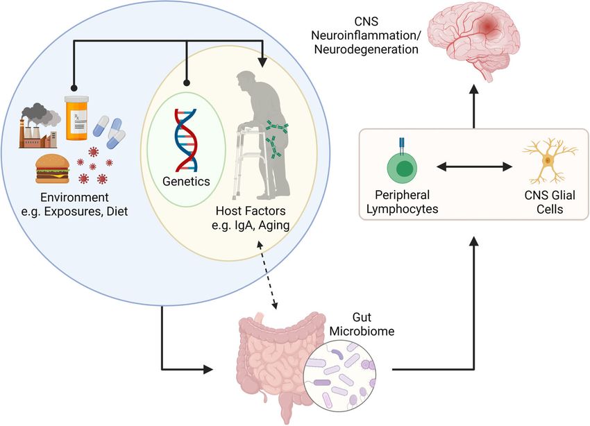
Pu et al. IgA, Microbiota in CNS Disease
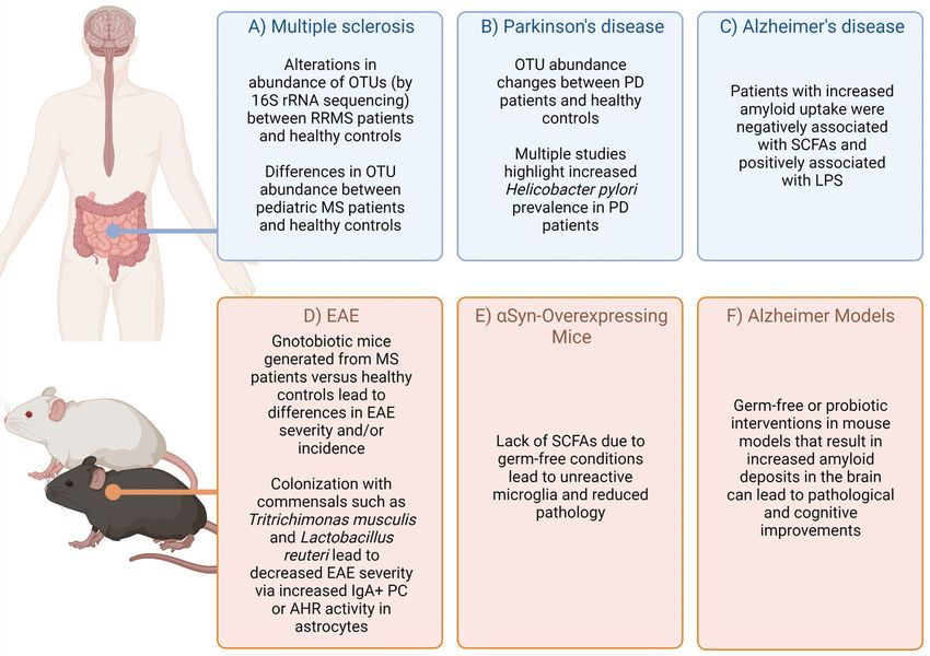
FIGURE 1 | Highlighted evidence for the relationship of microbiota affects in CNS-specific human diseases and animal models. Complex diseases of the CNS are
often difficult to query in humans due to scarcity of tissue samples. However, combining evidence from patients, healthy controls (A–C) (1–14), as well as animal
models (D–F) (15–23) can provide some suggestive evidence on how the microbiota may impact disease. Figure made using (BioRender.com).
shaping the microbiome which in turn can influence lymphocyte induction (36), T cells that are critical in causing pathology in
and glial cell behavior in the context of CNS disease (Figure 2). EAE. Regulatory T cells (Tregs) are similarly sensitive to the gut
microbiome. Their polarization from naïve T cells can be
potentiated by Bacteroides fragilis polysaccharide in EAE (17,
INFLUENCE OF THE MICROBIOTA ON 35). Interestingly, following transplantation of human MS stool
LYMPHOCYTES IN CNS DISEASE samples into mice (fecal microbiome transplant; FMT), several
bacterial species were linked to alterations in TH1 and Treg
Correlative data in MS and mouse models demonstrate a differentiation, and consequently EAE phenotype (37, 38).
bidirectional interaction between the gut and the CNS (15, 16,
31–33); identifying specific contributions of the gut microbiome to B Lymphocytes
CNS disease is imperative for understanding disease pathogenesis. B cells produce antibodies, present antigen to T cells, and secrete
While aberrantly activated lymphocytes are a hallmark of multiple cytokines. When antigen binds to a B cell receptor, these antigen-
sclerosis (MS), this is less studied in Alzheimer’s and Parkinson’s specific B cells are activated and undergo somatic hypermutation
disease (AD, PD). Thus, this section will focus on the impact of and affinity maturation in germinal centers (GC) (39), generating
microbiota on lymphocyte function in MS. high-affinity antigen-specific receptors. GC B cells can also class-
switch to generate different antibody isotypes with specialized
T Lymphocytes effector functions (IgA, IgE, IgG). Mature B cells can also
Although alterations in the microbiome have been reported in differentiate into memory B cells or antibody-secreting cells
MS case-control studies (34), testing causal associations between (ASCs) (40). ASCs comprise both proliferative plasmablasts
these alterations and disease risk requires animal models. Germ- (PBs) and terminally differentiated plasma cells (PCs) (39, 41).
free (GF) mice lack commensal microbiota and thus present a Alterations in microbial abundance have been correlated with
“blank slate” for exploring the impact of commensal microbes on changes in regulatory B cell (Breg) induction (18, 42). Antibiotic
disease. GF mice fail to develop experimental autoimmune treatment enhanced frequencies of IL-10-producing Bregs at
encephalomyelitis (EAE) (35), but when gnotobiotically baseline and in EAE (42). In addition, a role for gut-derived
recolonized or monocolonized with segmented filamentous commensal-reactive IgA+ ASCs in attenuating EAE and possibly
bacteria (SFB), EAE is rescued. SFB enhances TH 17 cell also MS has been shown (18, 19), described below.
Frontiers in Immunology | www.frontiersin.org 2 September 2021 | Volume 12 | Article 742173Pu et al. IgA, Microbiota in CNS Disease
FIGURE 2 | Putative connection between the gut microbiome and CNS neuroinflammation and neurodegeneration. The gut microbiome is shaped by internal (e.g.,
genetics and other host factors such as mucosal IgA and age) and external factors (e.g., environmental exposures, infections, diet, etc.). Recent literature has suggested
that IgA plays a key role in determining the microbes that reside in the gut, but that IgA levels can also be influenced by colonizing microbiota. The gut microbiome is
important for programming of peripheral lymphocytes but can also impact the phenotype and function of CNS resident glial cells (via metabolites such as SCFAs. The
activation (or modulation) of lymphocytes and glial cells can lead to neuroinflammation or neurodegenerative disorders in the CNS. Figure made using (BioRender.com).
INFLUENCE OF THE MICROBIOTA ON given the demyelinating toxin cuprizone (44). Surprisingly,
GLIAL CELLS IN CNS DISEASE supplementing aged mice with probiotic VSL#3 enhanced
serum and fecal SCFAs, but had limited effect post-LPC
Glial cells have been intensively studied in each MS, AD, and PD, administration (44). Indoxyl-3-sulfate (I3S), a product of
and a role for the microbiome in modulating glial cell phenotype microbial tryptophan metabolism, activates aryl hydrocarbon
and function in these diseases is emerging. receptor (AHR) on microglia, augmenting TGFa production to
limit astrocytic inflammation in EAE (45). Feeding whole milk
Microglia promotes AHR ligand and SCFA production and ameliorates
Microglia are CNS-resident macrophages serving key homeostatic EAE in marmosets, although the effect is not attributed solely to
and immune functions in the developing and adult CNS (1). microglia, but overall modulation of inflammation (46).
Maternal microbiota can influence microglial maturation and In a-synuclein-overexpressing (ASO) mice that model PD, the
function during both fetal development and in adulthood, as presence of a gut microbiota promotes the aggregation of a-
demonstrated in GF and antibiotic-treated mice (20, 21). Given synuclein in the caudoputamen and substantia nigra, resulting in
the lack of evidence for a CNS microbiome, presumably microglial activation and motor dysfunction (47). GF ASO mice
microbiota-derived metabolites, such as short-chain fatty acids show significantly decreased levels of aggregated a-synuclein and
(SCFAs), directly or indirectly influence microglial phenotype are protected from development of motor deficits. Re-colonization
(43). Microglia in GF mice are not fully mature, and interestingly, of GF mice with SPF microbiota reversed this rescue effect.
colonization with altered Schaedler’s flora failed to rescue microglial Surprisingly, despite SCFAs being generally thought to be anti-
defects whereas SCFA supplementation reversed the abnormal inflammatory, this study inculpates SCFAs in promoting
phenotype (21), indicating that the presence of SCFA-producing aggregated a-synuclein. SCFA supplementation to GF or
bacteria or a diverse microbiota are necessary for maturation. antibiotic-treated ASO mice increased microglial activation and
In models of MS, antibiotic treatment prior to lysolecithin is sufficient to promote motor impairment. In addition, abundance
(LPC)-induced demyelination decreases microglia activation, of several SCFA-producing enzymes is increased in humanized
indicated by the reduction in intralesional P2ry12loClec7a+ mice that have received an FMT from Parkinson’s disease donors.
inflammatory cells as opposed to P2ry12+Clec7a- homeostatic Like other neurodegenerative diseases, an unhealthy
microglia. Microglial activation was also attenuated in GF mice microbiome is increasingly appreciated as a risk factor for AD.
Frontiers in Immunology | www.frontiersin.org 3 September 2021 | Volume 12 | Article 742173Pu et al. IgA, Microbiota in CNS Disease
Similar to PD, circulating levels of the SCFAs acetate and valerate Host Control of the Microbiota Through IgA
were positively correlated with Ab load in the brains of AD patients Mature B cells primed in Peyer’s patches can differentiate into
(48). Mice treated with probiotic bacteria exhibited ameliorated IgA-producing PCs and home to the intestinal lamina propria
AD-like cognitive decline and decreased Ab aggregation (49). GF (41, 59, 60). The IgA produced by GALT PCs is typically dimeric,
5x Familial Alzheimer’s disease (5xFAD) mice show a decreased joined through the J-chain (41, 61). Secretory IgA is generated
Ab load in the hippocampus compared to conventional 5xFAD when dimeric IgA binds the polymeric-Ig-receptor (pIgR) on the
mice, attributed to the uptake of Ab debris by microglia. A greater basolateral surface of the intestinal epithelium, translocates
number of Iba1+ cells were found in the hippocampus closely through the epithelial cells, and is released into the lumen with
associated with Ab plaques in GF 5xFAD mice, and a higher the secretory component of the pIgR.
percentage of these plaque-associated Iba1+ cells were positive for In mice, IgA both contributes to host control of microbiota and
methoxy-X-O4, an indicator of Ab uptake (50). is responsive to gut microbiota changes. Mice monocolonized with
Bacteroides thetaiotamicron harbor a reduced IgA repertoire
Astrocytes restricted to a single clone against the capsular polysaccharide of
Astrocytes play diverse roles in the homeostatic brain that include the bacterium (62). Oral administration of Lactobacillus casei to
providing trophic support to other CNS cells, regulation of mice resulted in an increase in IgA+ cells in the small intestinal
synaptic activity, and controlling the blood-brain barrier (51– lamina propria (SILP) (63). Exposure of conventional mice to
54). Astrogliosis is also a feature of MS. Unsurprisingly, astrocytes commensal Proteobacteria also resulted in increased serum IgA
play important roles in modulating neuroinflammation, as they levels and induction of IgA+ PC in the bone marrow (64). In
can produce inflammatory cytokines and a host of chemokines contrast, some microbial communities can diminish IgA levels in
that promote chemotaxis of other immune cells. In EAE, astrocytic the lumen due to their ability to degrade the secretory component
inflammation is shown to be directly attenuated by I3S activation (65). Even strain level differences in the microbiome can dictate
of AHR (45). Gut microbiota depletion by antibiotic treatment IgA levels in the host (66). Conversely, the host IgA response can
decreases levels of I3S and worsens EAE disease (55). AHR- dictate the composition of the microbiome. Activation-induced
deficient astrocytes increase expression of several pro- cytidine deaminase-deficient mice (which fail to produce
inflammatory chemokines and cytokines. Importantly, IFNb, a competent IgA), exhibit an expansion of SFB in the small
therapeutic used in some MS patients, works to limit CNS intestine which leads to isolated lymphoid follicle (ILF)
inflammation through this mechanism, as the anti-inflammatory hyperplasia and GC enlargement in secondary lymphoid tissues
effect of IFNb is lost in AHR-deficient astrocytes (55). (67, 68). Restoration of IgA by heterogenetic parabiosis with wild-
type mice reduced SFB populations, ILF protrusion, and spleen
Oligodendrocytes and lymph node size.
Oligodendrocytes were previously thought to be quiescent, In humans, modest alterations in fecal microbiota
myelin-producing cells. However, increasing evidence shows composition are observed in subjects with selective IgA-
that oligodendrocytes actively communicate with and provide deficiency (SIgAd) (69, 70). Compensatory sIgM in SIgAd
metabolic support to neurons (56). Mature oligodendrocytes subjects has a distinct bacterial binding pattern: an unclassified
may also participate in remyelination and are active players Enterobacteriaceae taxon heavily coated by IgA in healthy
during neurodegeneration (57). However, little is known about controls and by IgM in selective IgA-deficient subjects, was
interactions between oligodendrocyte lineage cells and the gut significantly more abundant in SIgAd subjects, demonstrating
microbiome. While treatment of mice with the probiotic VSL#3 that IgA coating specifically restricts expansion of this taxon, and
enhanced SCFA concentrations in feces and serum, there was no the same effect is not achievable by IgM.
effect on remyelination in vivo following LPC-induced Overall, these data indicate that in both mice and humans, a
demyelination (44). Conversely, a separate study found that in bi-directional relationship exists between host IgA and
ex vivo organotypic cerebellar slice cultures demyelinated by gut microbiota.
lysolecithin, the addition of the SCFA member butyrate
enhanced both OPC numbers and mature oligodendrocyte Impact of Ageing on the Microbiome
numbers, indicating a positive effect on remyelination (58). In Growing evidence suggests that the gut microbiota has a critical
summary, the gut microbiota exerts effects not only on impact on the ageing process and is a possible determinant of
peripheral immune compartments, but also act on glial cells, healthy ageing (71–73). Cross-sectional studies have examined
with potential impacts on CNS disease processes. alterations in the microbiota composition across the human
lifespan (74, 75), demonstrating that taxonomical composition
of gut microbiota appears to follow stepwise progression through
HOST FACTORS THAT INFLUENCE THE life. Taxonomic shifts in the microbiota and decrease in
INTESTINAL MICROBIOME – A FOCUS ON microbial richness and diversity are observed in frail older
IGA AND AGEING individuals and associated with worse health outcomes
compared to younger individuals (76–80). Relative abundance
Many external factors influence the gut microbiome such as diet of Ruminococcaceae, Lachnospiraceae, and Bacteroidaceae
and pathogen exposure. In this section, we review host factors families decrease with age, whereas an enrichment and/or
that shape the microbiome, focusing on IgA and ageing. higher prevalence of health-associated genera such as
Frontiers in Immunology | www.frontiersin.org 4 September 2021 | Volume 12 | Article 742173Pu et al. IgA, Microbiota in CNS Disease
Akkermansia, Bifidobacterium, and Christensenellaceae, are microbiota-driven IgA response in human disease. Bacteria
maintained in longevity and extreme longevity (81). Indeed, identified by IgA-seq were differentially expressed in MS
centenarians tend to exhibit indicators of good health, and patients versus healthy controls (19). Stratified by disease
greater gut microbiota complexity (74). The relative abundance activity, MS patients in relapse exhibited decreased percentages
of pathobionts decreases and beneficial commensals, such as of IgA-bound gut bacteria in fecal samples compared to
Akkermansia, are retained (82). Studies that stratify between remitting patients, with corresponding elevation in CSF IgA.
elderly and centenarian status identify changes in taxa associated CNS-infiltrating IgA+ B cells show specificity for gut microbial
with extreme longevity including Odoribacter, Butyricimonas, antigens, indicating the migration of IgA-producing cells from
Desulfovibrio, Bilophila, Oscillospira, and Akkermansia genera, the gut during relapse. IgA is also elevated in cerebrospinal fluid
and the Christensenellaceae and Barnesiellaceae families (75, 81). of MS patients. Importantly, commensal-specific IgA+ ASCs
Similar taxonomic and functional patterns that correlate with have been observed in inflammatory lesions of MS patients
age and frailty in the mouse microbiome have been identified (19). This phenomenon may not be IgA-exclusive, however, as
(83). In aged mice, the ratio of Firmicutes to Bacteroidetes IgG in MS patient CSF has been found to be reactive against MS-
increased ∼9‐fold compared to young mice, indicating associated gut bacterial lysate (93). The implications of these
dysbiosis, although this work was performed in commercially bacteria-reactive IgG in disease have yet to be fully elucidated.
purchased mice rather than mice derived from the same dam Lastly, while IgA+ ASC have been now described in the
(84). Introducing aged microbiota to young mice increased inflamed EAE and MS CNS, it is now appreciated that these
mortality following ischemic stroke, decreased behavioral cells play an important role in homeostasis. Specifically,
outcomes, and increased cytokine levels. Conversely, altering intestinal commensal specific IgA+ ASC have been detected in
the microbiota in aged mice to resemble that of young mice the leptomeninges of healthy mice and humans but are absent in
increased stroke survival and improved recovery (84). Changes GF mice (87). These cells likely maintain barrier integrity near
in the gut microbiota in aged mice were also associated with the dural sinuses; however, it is possible they may also contribute
increased gut permeability and elevations of peripheral to quiescence within the CNS.
inflammation (85, 86). Taken together, except for healthy In summary, in addition to its well appreciated role in shaping
centenarians who resist frailty, ageing is associated with an the microbiome, IgA-producing ASC likewise play important
unhealthy microbiome. roles in the healthy and MS/EAE CNS. The role for these cells in
PD and AD is not yet understood.
Ageing, the Microbiome, and CNS Disease
INFLUENCE OF AGEING AND IGA ON CNS Ageing is the predominant risk factor for neurodegenerative
DISEASE VIA THE MICROBIOME diseases (94), yet in spite of the known role ageing has on the
microbiome, the connection between ageing, the microbiome
Multiple internal host factors impact the microbiome, including and CNS disease has barely been explored.
IgA and ageing. Here we speculate on how these two host factors It is well established that microglia are affected during ageing.
may impact brain health and the trajectory of brain disease via Ageing results in decreased number, uneven distribution, lower
the microbiome. motility, and fewer ramifications, as well as impairment in
phagocytosis and injury responses (2, 95–98). Senescent
IgA, the Microbiome, and CNS Disease microglia increase pro-inflammatory cytokine production (3).
Although IgA+ ASCs can home to the dura mater during An altered microglia morphology and reduced arborization have
homeostasis (87), clonally expanded IgA are absent in steady been observed in the human brain during ageing and age-related
state CNS and only appear during inflammation (88–91). During diseases such as AD (95). This dystrophic morphology is
EAE, a significant reduction in IgA+ ASCs is apparent in the associated with impaired spatial learning (3).
SILP. Additionally, adoptively transferred gut-derived IgA+ Age-related changes in the gut microbiome may have a direct
ASCs were found in the CNS were reactive to mouse-derived impact on microglial function within the CNS. In fact, reduced
gut bacteria and were shown to alleviate neuroinflammation by complexity of microbiota, a feature of ageing, leads to defects in
producing IL-10 at chronic stages of EAE. Over-abundance of microglia maturation and function (21). Recent work
IgA+ ASCs was able to reduce GM-CSF production by T cells, an demonstrated that FMT from aged donor mice into young
important cytokine that promotes neuroinflammation (18). recipients impairs spatial learning and memory in young
Tritrichomonas musculis (T.mu) is a rodent commensal that recipients (4). Conversely, FMT from young donor mice into
promotes IgA production (92). EAE incidence and severity, as aged recipients can rejuvenate age-associated CNS metabolic,
well as spinal cord inflammation and demyelination, are reduced transcriptomic, and behavioral changes (5). Aged into young
in T.mu+ mice (18). T.mu+ mice also exhibited elevated serum FMT induced an altered expression of proteins involved in
and fecal IgA levels and increased frequencies of IgA+ ASCs in synaptic plasticity and neurotransmission in the hippocampus,
the gut, bone marrow, and brain. an area of the CNS known to be affected by the ageing process. A
While the above highlights key findings from animal models, strong reduction of bacteria associated with SCFA production
there is also early evidence suggesting the importance of the (Lachnospiraceae, Faecalibaculum, and Ruminococcaceae) and
Frontiers in Immunology | www.frontiersin.org 5 September 2021 | Volume 12 | Article 742173Pu et al. IgA, Microbiota in CNS Disease
disorders of the CNS (Prevotellaceae and Ruminococcaceae) was CONCLUSIONS
also reported (4). Interestingly, microglia of the hippocampus
acquired an ageing-like phenotype following FMT. Of Chronic, complex diseases of the CNS develop over years.
therapeutic relevance, this age-associated phenotype can be Animal studies conducted under controlled conditions in short
reversed by re-introducing live and complex microbiota or periods miss two large confounders in these diseases – time
microbial metabolites, such as SCFAs (6). (ageing) and the microbiota-IgA axis, with age-associated
The gut microbiota similarly affects astrocytes in both ageing microbiota alterations further complicating this relationship.
and age-associated neurodegenerative diseases (7). Ageing can These are important considerations for animal modelling,
alter the normal function of astrocytes which reduces their given the considerable variability in microbiota composition
ability to properly maintain a healthy CNS environment (8). and gut luminal IgA levels between vivaria (65). In summary,
Astrocyte transcriptomes from multiple mouse brain regions we propose that host factors such as age and intestinal IgA are
have revealed that ageing upregulates genes that eliminate key determinants in how the microbiome impacts lymphocyte
synapses and induces a reactive astrocyte gene signature (9). and glial cell phenotype/function in the context of MS, AD and
Therefore, aged astrocytes may promote synapse elimination and PD (Figure 2).
neuronal damage, contributing to ageing-associated cognitive
decline. Morphological changes in astrocytes have also been
documented in the aged CNS (10–12). Aged astrocytes AUTHOR CONTRIBUTIONS
increase cytokine production, notably CXCL10 (13) that
attracts peripheral immune cells and promotes T cell adhesion AP, DL, BI, and IN all contributed to the writing of this
to endothelial cells (14). CXCR3, which is the CXCL10 receptor, manuscript. AP, DL, and JG contributed to the editing of the
is expressed in microglia, suggesting that astrocytes and text and generation of all figures. All authors contributed to the
microglia communicate during ageing (22, 45). article and approved the submitted version.
12. Robillard KN, Lee KM, Chiu KB, MacLean AG. Glial Cell Morphological and
REFERENCES Density Changes Through the Lifespan of Rhesus Macaques. Brain Behav
1. Nimmerjahn A, Kirchhoff F, Helmchen F. Resting Microglial Immun (2016) 55:60–9. doi: 10.1016/j.bbi.2016.01.006
Cells Are Highly Dynamic Surveillants of Brain Parenchyma In Vivo. 13. Clarke LE, Liddelow SA, Chakraborty C, Munch AE, Heiman M, Barres BA.
Sci (New York NY) (2005) 308(5726):1314–8. doi: 10.1126/science. Normal Aging Induces A1-Like Astrocyte Reactivity. Proc Natl Acad Sci USA
1110647 (2018) 115(8):E1896–905. doi: 10.1073/pnas.1800165115
2. Zoller T, Attaai A, Potru PS, Russ T, Spittau B. Aged Mouse Cortical 14. Sorensen EW, Lian J, Ozga AJ, Miyabe Y, Ji SW, Bromley SK, et al. CXCL10
Microglia Display an Activation Profile Suggesting Immunotolerogenic Stabilizes T Cell-Brain Endothelial Cell Adhesion Leading to the Induction of
Functions. Int J Mol Sci (2018) 19(3):706. doi: 10.3390/ijms19030706 Cerebral Malaria. JCI Insight (2018) 3(8):e98911. doi: 10.1172/jci.insight.98911
3. Niraula A, Sheridan JF, Godbout JP. Microglia Priming With Aging and 15. Miyake S, Kim S, Suda W, Oshima K, Nakamura M, Matsuoka T, et al.
Stress. Neuropsychopharmacology (2017) 42(1):318–33. doi: 10.1038/ Dysbiosis in the Gut Microbiota of Patients With Multiple Sclerosis, With a
npp.2016.185 Striking Depletion of Species Belonging to Clostridia XIVa and IV Clusters.
4. D’Amato A, Di Cesare Mannelli L, Lucarini E, Man AL, Le Gall G, Branca JJV, PloS One (2015) 10(9):e0137429. doi: 10.1371/journal.pone.0137429
et al. Faecal Microbiota Transplant From Aged Donor Mice Affects Spatial 16. Johanson DM, Goertz JE, Marin IA, Costello J, Overall CC, Gaultier A.
Learning and Memory via Modulating Hippocampal Synaptic Plasticity- and Experimental Autoimmune Encephalomyelitis Is Associated With Changes of
Neurotransmission-Related Proteins in Young Recipients. Microbiome (2020) the Microbiota Composition in the Gastrointestinal Tract. Sci Rep (2020) 10
8(1):140. doi: 10.1186/s40168-020-00914-w (1):15183. doi: 10.1038/s41598-020-72197-y
5. Boehme M, Guzzetta KE, Bastiaanssen TFS, van de Wouw M, Moloney GM, 17. Ochoa-Repá raz J, Mielcarz DW, Wang Y, Begum-Haque S, Dasgupta S,
Gual-Grau A, et al. Microbiota From Young Mice Counteracts Selective Age- Kasper DL, et al. A Polysaccharide From the Human Commensal
Associated Behavioral Deficits. Nat Aging (2021) 1(8):666–76. doi: 10.1038/ Bacteroides Fragilis Protects Against CNS Demyelinating Disease. Mucosal
s43587-021-00093-9 Immunol (2010) 3(5):487–95. doi: 10.1038/mi.2010.29
6. Shen P, Roch T, Lampropoulou V, O’Connor RA, Stervbo U, Hilgenberg E, 18. Rojas OL, Probstel AK, Porfilio EA, Wang AA, Charabati M, Sun T, et al.
et al. IL-35-Producing B Cells Are Critical Regulators of Immunity During Recirculating Intestinal IgA-Producing Cells Regulate Neuroinflammation via
Autoimmune and Infectious Diseases. Nature (2014) 507(7492):366–70. doi: IL-10. Cell (2019) 176(3):610–24.e18. doi: 10.1016/j.cell.2018.11.035
10.1038/nature12979 19. Pröbstel A-K, Zhou X, Baumann R, Wischnewski S, Kutza M, Rojas OL, et al.
7. Meldolesi J. Astrocytes: News About Brain Health and Diseases. Biomedicines Gut Microbiota–Specific IgA+ B Cells Traffic to the CNS in Active Multiple
(2020) 8(10):394. doi: 10.3390/biomedicines8100394 Sclerosis. Sci Immunol (2020) 5(53):eabc7191. doi: 10.1126/sciimmunol.abc7191
8. Palmer AL, Ousman SS. Astrocytes and Aging. Front Aging Neurosci (2018) 20. Matcovitch-Natan O, Winter DR, Giladi A, Vargas Aguilar S, Spinrad A,
10:337. doi: 10.3389/fnagi.2018.00337 Sarrazin S, et al. Microglia Development Follows a Stepwise Program to
9. Boisvert MM, Erikson GA, Shokhirev MN, Allen NJ. The Aging Astrocyte Regulate Brain Homeostasis. Sci (New York NY) (2016) 353(6301):aad8670.
Transcriptome From Multiple Regions of the Mouse Brain. Cell Rep (2018) 22 doi: 10.1126/science.aad8670
(1):269–85. doi: 10.1016/j.celrep.2017.12.039 21. Erny D, Hrabe de Angelis AL, Jaitin D, Wieghofer P, Staszewski O, David E, et al.
10. Jyothi HJ, Vidyadhara DJ, Mahadevan A, Philip M, Parmar SK, Manohari SG, Host Microbiota Constantly Control Maturation and Function of Microglia in the
et al. Aging Causes Morphological Alterations in Astrocytes and Microglia in CNS. Nat Neurosci (2015) 18(7):965–77. doi: 10.1038/nn.4030
Human Substantia Nigra Pars Compacta. Neurobiol Aging (2015) 36 22. Liddelow SA, Barres BA. Reactive Astrocytes: Production, Function, and
(12):3321–33. doi: 10.1016/j.neurobiolaging.2015.08.024 Therapeutic Potential. Immunity (2017) 46(6):957–67. doi: 10.1016/
11. Amenta F, Bronzetti E, Sabbatini M, Vega JA. Astrocyte Changes in Aging j.immuni.2017.06.006
Cerebral Cortex and Hippocampus: A Quantitative Immunohistochemical 23. Scheperjans F, Aho V, Pereira PA, Koskinen K, Paulin L, Pekkonen E, et al.
Study. Microsc Res Tech (1998) 43(1):29–33. doi: 10.1002/(SICI)1097-0029 Gut Microbiota Are Related to Parkinson’s Disease and Clinical Phenotype.
(19981001)43:13.0.CO;2-H Mov Disord (2015) 30(3):350–8. doi: 10.1002/mds.26069
Frontiers in Immunology | www.frontiersin.org 6 September 2021 | Volume 12 | Article 742173Pu et al. IgA, Microbiota in CNS Disease
24. Human Microbiome Jumpstart Reference Strains Consortium, Nelson KE, and Immune Regulation and Its Relevance for Inflammatory Bowel Diseases.
Weinstock GM, Highlander SK, Worley KC, Creasy HH, et al. A Catalog of Front Immunol (2019) 10:277. doi: 10.3389/fimmu.2019.00277
Reference Genomes From the Human Microbiome. Sci (New York NY) (2010) 44. McMurran CE, Guzman de la Fuente A, Penalva R, Ben Menachem-Zidon O,
328(5981):994–9. doi: 10.1126/science.1183605 Dombrowski Y, Falconer J, et al. The Microbiota Regulates Murine
25. Deschasaux M, Bouter KE, Prodan A, Levin E, Groen AK, Herrema H, et al. Inflammatory Responses to Toxin-Induced CNS Demyelination But Has
Depicting the Composition of Gut Microbiota in a Population With Varied Minimal Impact on Remyelination. Proc Natl Acad Sci USA (2019) 116
Ethnic Origins But Shared Geography. Nat Med (2018) 24(10):1526–31. doi: (50):25311–21. doi: 10.1073/pnas.1905787116
10.1038/s41591-018-0160-1 45. Rothhammer V, Borucki DM, Tjon EC, Takenaka MC, Chao CC, Ardura-
26. David LA, Maurice CF, Carmody RN, Gootenberg DB, Button JE, Wolfe BE, Fabregat A, et al. Microglial Control of Astrocytes in Response to Microbial
et al. Diet Rapidly and Reproducibly Alters the Human Gut Microbiome. Metabolites. Nature (2018) 557(7707):724–8. doi: 10.1038/s41586-018-0119-x
Nature (2014) 505(7484):559–63. doi: 10.1038/nature12820 46. Kap YS, Bus-Spoor C, van Driel N, Dubbelaar ML, Grit C, Kooistra SM, et al.
27. O’Keefe SJD, Li JV, Lahti L, Ou J, Carbonero F, Mohammed K, et al. Fat, Fibre Targeted Diet Modification Reduces Multiple Sclerosis–Like Disease in Adult
and Cancer Risk in African Americans and Rural Africans. Nat Commun Marmoset Monkeys From an Outbred Colony. J Immunol (2018) 201
(2015) 6(1):6342. doi: 10.1038/ncomms7342 (11):3229. doi: 10.4049/jimmunol.1800822
28. Vangay P, Johnson AJ, Ward TL, Al-Ghalith GA, Shields-Cutler RR, 47. Sampson TR, Debelius JW, Thron T, Janssen S, Shastri GG, Ilhan ZE, et al.
Hillmann BM, et al. US Immigration Westernizes the Human Gut Gut Microbiota Regulate Motor Deficits and Neuroinflammation in a Model
Microbiome. Cell (2018) 175(4):962–72.e10. doi: 10.1016/j.cell.2018.10.029 of Parkinson’s Disease. Cell (2016) 167(6):1469–80.e12. doi: 10.1016/j.cell.
29. Perez-Munoz ME, Sugden S, Harmsen HJM, t Hart BA, Laman JD, Walter J. 2016.11.018
Nutritional and Ecological Perspectives of the Interrelationships Between Diet 48. Marizzoni M, Cattaneo A, Mirabelli P, Festari C, Lopizzo N, Nicolosi V, et al.
and the Gut Microbiome in Multiple Sclerosis: Insights From Marmosets. Short-Chain Fatty Acids and Lipopolysaccharide as Mediators Between Gut
iScience (2021) 24(7):102709. doi: 10.1016/j.isci.2021.102709 Dysbiosis and Amyloid Pathology in Alzheimer’s Disease. J Alzheimers Dis
30. Groot HE, van de Vegte YJ, Verweij N, Lipsic E, Karper JC, van der Harst P. (2020) 78(2):683–97. doi: 10.3233/JAD-200306
Human Genetic Determinants of the Gut Microbiome and Their Associations 49. Bonfili L, Cecarini V, Berardi S, Scarpona S, Suchodolski JS, Nasuti C, et al.
With Health and Disease: A Phenome-Wide Association Study. Sci Rep (2020) Microbiota Modulation Counteracts Alzheimer’s Disease Progression
10(1):14771. doi: 10.1038/s41598-020-70724-5 Influencing Neuronal Proteolysis and Gut Hormones Plasma Levels. Sci Rep
31. Colpitts SL, Kasper EJ, Keever A, Liljenberg C, Kirby T, Magori K, et al. A (2017) 7(1):2426. doi: 10.1038/s41598-017-02587-2
Bidirectional Association Between the Gut Microbiota and CNS Disease in a 50. Mezo C, Dokalis N, Mossad O, Staszewski O, Neuber J, Yilmaz B, et al.
Biphasic Murine Model of Multiple Sclerosis. Gut Microbes (2017) 8(6):561– Different Effects of Constitutive and Induced Microbiota Modulation on
73. doi: 10.1080/19490976.2017.1353843 Microglia in a Mouse Model of Alzheimer’s Disease. Acta Neuropathol
32. Jangi S, Gandhi R, Cox LM, Li N, von Glehn F, Yan R, et al. Alterations of the Commun (2020) 8(1):119. doi: 10.1186/s40478-020-00988-5
Human Gut Microbiome in Multiple Sclerosis. Nat Commun (2016) 7:12015. 51. Abbott NJ, Ronnback L, Hansson E. Astrocyte-Endothelial Interactions at the
doi: 10.1038/ncomms12015 Blood-Brain Barrier. Nat Rev Neurosci (2006) 7(1):41–53. doi: 10.1038/nrn1824
33. Moles L, Egimendia A, Osorio-Querejeta I, Iparraguirre L, Alberro A, Suá rez J, 52. Perea G, Araque A. Properties of Synaptically Evoked Astrocyte Calcium
et al. Gut Microbiota Changes in Experimental Autoimmune Signal Reveal Synaptic Information Processing by Astrocytes. J Neurosci
Encephalomyelitis and Cuprizone Mice Models. ACS Chem Neurosci (2021) (2005) 25(9):2192–203. doi: 10.1523/JNEUROSCI.3965-04.2005
12(5):893–905. doi: 10.1021/acschemneuro.0c00695 53. Bezzi P, Gundersen V, Galbete JL, Seifert G, Steinhauser C, Pilati E, et al.
34. Tremlett H, Bauer KC, Appel-Cresswell S, Finlay BB, Waubant E. The Gut Astrocytes Contain a Vesicular Compartment That Is Competent for
Microbiome in Human Neurological Disease: A Review. Ann Neurol (2017) Regulated Exocytosis of Glutamate. Nat Neurosci (2004) 7(6):613–20. doi:
81(3):369–82. doi: 10.1002/ana.24901 10.1038/nn1246
35. Lee YK, Menezes JS, Umesaki Y, Mazmanian SK. Proinflammatory T-Cell 54. Sofroniew MV, Vinters HV. Astrocytes: Biology and Pathology. Acta
Responses to Gut Microbiota Promote Experimental Autoimmune Neuropathol (2010) 119(1):7–35. doi: 10.1007/s00401-009-0619-8
Encephalomyelitis. Proc Natl Acad Sci USA (2011) 108(Suppl 1):4615–22. 55. Rothhammer V, Mascanfroni ID, Bunse L, Takenaka MC, Kenison JE, Mayo
doi: 10.1073/pnas.1000082107 L, et al. Type I Interferons and Microbial Metabolites of Tryptophan Modulate
36. Ivanov II, Atarashi K, Manel N, Brodie EL, Shima T, Karaoz U, et al. Induction Astrocyte Activity and Central Nervous System Inflammation via the Aryl
of Intestinal Th17 Cells by Segmented Filamentous Bacteria. Cell (2009) 139 Hydrocarbon Receptor. Nat Med (2016) 22(6):586–97. doi: 10.1038/nm.4106
(3):485–98. doi: 10.1016/j.cell.2009.09.033 56. Krasnow AM, Ford MC, Valdivia LE, Wilson SW, Attwell D. Regulation of
37. Berer K, Gerdes LA, Cekanaviciute E, Jia X, Xiao L, Xia Z, et al. Gut Developing Myelin Sheath Elongation by Oligodendrocyte Calcium
Microbiota From Multiple Sclerosis Patients Enables Spontaneous Transients In Vivo. Nat Neurosci (2018) 21(1):24–8. doi: 10.1038/s41593-
Autoimmune Encephalomyelitis in Mice. Proc Natl Acad Sci USA (2017) 017-0031-y
114(40):10719–24. doi: 10.1073/pnas.1711233114 57. Duncan ID, Radcliff AB, Heidari M, Kidd G, August BK, Wierenga LA. The
38. Cekanaviciute E, Yoo BB, Runia TF, Debelius JW, Singh S, Nelson CA, et al. Adult Oligodendrocyte Can Participate in Remyelination. Proc Natl Acad Sci
Gut Bacteria From Multiple Sclerosis Patients Modulate Human T Cells and USA (2018) 115(50):E11807–16. doi: 10.1073/pnas.1808064115
Exacerbate Symptoms in Mouse Models. Proc Natl Acad Sci USA (2017) 114 58. Chen T, Noto D, Hoshino Y, Mizuno M, Miyake S. Butyrate Suppresses
(40):10713–8. doi: 10.1073/pnas.1711235114 Demyelination and Enhances Remyelination. J Neuroinflamm (2019) 16
39. De Silva NS, Klein U. Dynamics of B Cells in Germinal Centres. Nat Rev (1):165. doi: 10.1186/s12974-019-1552-y
Immunol (2015) 15(3):137–48. doi: 10.1038/nri3804 59. Fagarasan S, Kawamoto S, Kanagawa O, Suzuki K. Adaptive Immune
40. Nutt SL, Hodgkin PD, Tarlinton DM, Corcoran LM. The Generation of Regulation in the Gut: T Cell-Dependent and T Cell-Independent IgA
Antibody-Secreting Plasma Cells. Nat Rev Immunol (2015) 15(3):160–71. doi: Synthesis. Annu Rev Immunol (2010) 28:243–73. doi: 10.1146/annurev-
10.1038/nri3795 immunol-030409-101314
41. Brandtzaeg P, Johansen FE. Mucosal B Cells: Phenotypic Characteristics, 60. Wang AA, Gommerman JL, Rojas OL. Plasma Cells: From Cytokine
Transcriptional Regulation, and Homing Properties. Immunol Rev (2005) Production to Regulation in Experimental Autoimmune Encephalomyelitis.
206:32–63. doi: 10.1111/j.0105-2896.2005.00283.x J Mol Biol (2020) 8:166655. doi: 10.1016/j.jmb.2020.09.014
42. Ochoa-Repá raz J, Mielcarz DW, Haque-Begum S, Kasper LH. Induction of a 61. Chung JB, Silverman M, Monroe JG. Transitional B Cells: Step by Step
Regulatory B Cell Population in Experimental Allergic Encephalomyelitis by Towards Immune Competence. Trends Immunol (2003) 24(6):343–9. doi:
Alteration of the Gut Commensal Microflora. Gut Microbes (2010) 1(2):103– 10.1016/S1471-4906(03)00119-4
8. doi: 10.4161/gmic.1.2.11515 62. Peterson DA, McNulty NP, Guruge JL, Gordon JI. IgA Response to Symbiotic
43. Parada Venegas D, de la Fuente MK, Landskron G, Gonzalez MJ, Quera R, Bacteria as a Mediator of Gut Homeostasis. Cell Host Microbe (2007) 2
Dijkstra G, et al. Short Chain Fatty Acids (SCFAs)-Mediated Gut Epithelial (5):328–39. doi: 10.1016/j.chom.2007.09.013
Frontiers in Immunology | www.frontiersin.org 7 September 2021 | Volume 12 | Article 742173Pu et al. IgA, Microbiota in CNS Disease
63. Galdeano CM, Perdigon G. The Probiotic Bacterium Lactobacillus Casei Induces 85. Scott KA, Ida M, Peterson VL, Prenderville JA, Moloney GM, Izumo T, et al.
Activation of the Gut Mucosal Immune System Through Innate Immunity. Clin Revisiting Metchnikoff: Age-Related Alterations in Microbiota-Gut-Brain
Vaccine Immunol (2006) 13(2):219–26. doi: 10.1128/CVI.13.2.219-226.2006 Axis in the Mouse. Brain Behav Immun (2017) 65:20–32. doi: 10.1016/
64. Wilmore JR, Gaudette BT, Gomez Atria D, Hashemi T, Jones DD, Gardner j.bbi.2017.02.004
CA, et al. Commensal Microbes Induce Serum IgA Responses That Protect 86. Thevaranjan N, Puchta A, Schulz C, Naidoo A, Szamosi JC, Verschoor CP,
Against Polymicrobial Sepsis. Cell Host Microbe (2018) 23(3):302–11.e3. doi: et al. Age-Associated Microbial Dysbiosis Promotes Intestinal Permeability,
10.1016/j.chom.2018.01.005 Systemic Inflammation, and Macrophage Dysfunction. Cell Host Microbe
65. Moon C, Baldridge MT, Wallace MA, Carey-Ann D, Burnham , Virgin HW, et al. (2017) 21(4):455–66.e4. doi: 10.1016/j.chom.2017.03.002
Vertically Transmitted Faecal IgA Levels Determine Extra-Chromosomal 87. Fitzpatrick Z, Frazer G, Ferro A, Clare S, Bouladoux N, Ferdinand J, et al. Gut-
Phenotypic Variation. Nature (2015) 521(7550):90–3. doi: 10.1038/nature14139 Educated IgA Plasma Cells Defend the Meningeal Venous Sinuses. Nature
66. Yang C, Mogno I, Contijoch EJ, Borgerding JN, Aggarwala V, Li Z, et al. Fecal (2020) 587(7834):472–6. doi: 10.1038/s41586-020-2886-4
IgA Levels Are Determined by Strain-Level Differences in Bacteroides Ovatus 88. Olsson JE, Link H. Immunoglobulin Abnormalities in Multiple Sclerosis.
and Are Modifiable by Gut Microbiota Manipulation. Cell Host Microbe Relation to Clinical Parameters: Exacerbations and Remissions. Arch Neurol
(2020) 27(3):467–75.e6. doi: 10.1016/j.chom.2020.01.016 (1973) 28(6):392–9. doi: 10.1001/archneur.1973.00490240052009
67. Suzuki K, Meek B, Doi Y, Muramatsu M, Chiba T, Honjo T, et al. Aberrant 89. Link H, Müller R. Immunoglobulins in Multiple Sclerosis and Infections of
Expansion of Segmented Filamentous Bacteria in IgA-Deficient Gut. Proc Natl the Nervous System. Arch Neurol (1971) 25(4):326–44. doi: 10.1001/
Acad Sci USA (2004) 101(7):1981–6. doi: 10.1073/pnas.0307317101 archneur.1971.00490040052007
68. Fagarasan S, Muramatsu M, Suzuki K, Nagaoka H, Hiai H, Honjo T. Critical 90. Omura S, Sato F, Park AM, Fujita M, Khadka S, Nakamura Y, et al.
Roles of Activation-Induced Cytidine Deaminase in the Homeostasis of Gut Bioinformatics Analysis of Gut Microbiota and CNS Transcriptome in
Flora. Sci (New York NY) (2002) 298(5597):1424–7. doi: 10.1126/science.1077336 Virus-Induced Acute Myelitis and Chronic Inflammatory Demyelination;
69. Fadlallah J, El Kafsi H, Sterlin D, Juste C, Parizot C, Dorgham K, et al. Potential Association of Distinct Bacteria With CNS IgA Upregulation. Front
Microbial Ecology Perturbation in Human IgA Deficiency. Sci Transl Med Immunol (2020) 11:1138. doi: 10.3389/fimmu.2020.01138
(2018) 10(439):eaan1217. doi: 10.1126/scitranslmed.aan1217 91. Isho B, Florescu A, Wang AA, Gommerman JL. Fantastic IgA Plasma Cells
70. Catanzaro JR, Strauss JD, Bielecka A, Porto AF, Lobo FM, Urban A, et al. IgA- and Where to Find Them. Immunol Rev (2021) 303:119–37. doi: 10.1111/
Deficient Humans Exhibit Gut Microbiota Dysbiosis Despite Secretion of imr.12980
Compensatory IgM. Sci Rep (2019) 9(1):13574. doi: 10.1038/s41598-019-49923-2 92. Chudnovskiy A, Mortha A, Kana V, Kennard A, Ramirez JD, Rahman A, et al.
71. Claesson MJ, Jeffery IB, Conde S, Power SE, O’Connor EM, Cusack S, et al. Host-Protozoan Interactions Protect From Mucosal Infections Through
Gut Microbiota Composition Correlates With Diet and Health in the Elderly. Activation of the Inflammasome. Cell (2016) 167(2):444–56.e14. doi:
Nature (2012) 488(7410):178–84. doi: 10.1038/nature11319 10.1016/j.cell.2016.08.076
72. Candela M, Biagi E, Brigidi P, O’Toole PW, De Vos WM. Maintenance of a 93. Eckman E, Laman JD, Fischer KF, Lopansri B, Martins TB, Hill HR, et al.
Healthy Trajectory of the Intestinal Microbiome During Aging: A Dietary Spinal Fluid IgG Antibodies From Patients With Demyelinating Diseases
Approach. Mech Ageing Dev (2014) 136-137:70–5. doi: 10.1016/j.mad. Bind Multiple Sclerosis-Associated Bacteria. J Mol Med (Berl) (2021), 1–13.
2013.12.004 doi: 10.1007/s00109-021-02085-z
73. Heintz C, Mair W. You Are What You Host: Microbiome Modulation of the 94. Hou Y, Dan X, Babbar M, Wei Y, Hasselbalch SG, Croteau DL, et al. Ageing as
Aging Process. Cell (2014) 156(3):408–11. doi: 10.1016/j.cell.2014.01.025 a Risk Factor for Neurodegenerative Disease. Nat Rev Neurol (2019) 15
74. Odamaki T, Kato K, Sugahara H, Hashikura N, Takahashi S, J-z X, et al. Age- (10):565–81. doi: 10.1038/s41582-019-0244-7
Related Changes in Gut Microbiota Composition From Newborn to 95. Davies DS, Ma J, Jegathees T, Goldsbury C. Microglia Show Altered
Centenarian: A Cross-Sectional Study. BMC Microbiol (2016) 16(1):90. doi: Morphology and Reduced Arborization in Human Brain During Aging and
10.1186/s12866-016-0708-5 Alzheimer’s Disease. Brain Pathol (2017) 27(6):795–808. doi: 10.1111/
75. Xu C, Zhu H, Qiu P. Aging Progression of Human Gut Microbiota. BMC bpa.12456
Microbiol (2019) 19(1):236. doi: 10.1186/s12866-019-1616-2 96. Soreq LConsortium UKBE and North American Brain Expression C, , Rose J,
76. Claesson MJ, Cusack S, O’Sullivan O, Greene-Diniz R, de Weerd H, Flannery Soreq E, Hardy J, et al. Major Shifts in Glial Regional Identity Are a
E, et al. Composition, Variability, and Temporal Stability of the Intestinal Transcriptional Hallmark of Human Brain Aging. Cell Rep (2017) 18
Microbiota of the Elderly. Proc Natl Acad Sci USA (2011) 108(Suppl 1):4586– (2):557–70. doi: 10.1016/j.celrep.2016.12.011
91. doi: 10.1073/pnas.1000097107 97. Streit WJ. Microglial Senescence: Does the Brain’s Immune System Have an
77. Jackson MA, Jeffery IB, Beaumont M, Bell JT, Clark AG, Ley RE, et al. Expiration Date? Trends Neurosci (2006) 29(9):506–10. doi: 10.1016/
Signatures of Early Frailty in the Gut Microbiota. Genome Med (2016) 8(1):8. j.tins.2006.07.001
doi: 10.1186/s13073-016-0275-2 98. Wasserman JK, Yang H, Schlichter LC. Glial Responses, Neuron Death and
78. O’Toole PW, Jeffery IB. Gut Microbiota and Aging. Sci (New York NY) (2015) Lesion Resolution After Intracerebral Hemorrhage in Young vs. Aged Rats.
350(6265):1214–5. doi: 10.1126/science.aac8469 Eur J Neurosci (2008) 28(7):1316–28. doi: 10.1111/j.1460-9568.
79. O’Toole PW, Jeffery IB. Microbiome-Health Interactions in Older People. Cell 2008.06442.x
Mol Life Sci (2018) 75(1):119–28. doi: 10.1007/s00018-017-2673-z
80. Zapata HJ, Quagliarello VJ. The Microbiota and Microbiome in Aging: Conflict of Interest: The authors declare that the research was conducted in the
Potential Implications in Health and Age-Related Diseases. J Am Geriatr absence of any commercial or financial relationships that could be construed as a
Soc (2015) 63(4):776–81. doi: 10.1111/jgs.13310 potential conflict of interest.
81. Biagi E, Franceschi C, Rampelli S, Severgnini M, Ostan R, Turroni S, et al. Gut
Microbiota and Extreme Longevity. Curr Biology: CB (2016) 26(11):1480–5. Publisher’s Note: All claims expressed in this article are solely those of the authors
doi: 10.1016/j.cub.2016.04.016 and do not necessarily represent those of their affiliated organizations, or those of
82. Ragonnaud E, Biragyn A. Gut Microbiota as the Key Controllers of “Healthy” the publisher, the editors and the reviewers. Any product that may be evaluated in
Aging of Elderly People. Immun Ageing (2021) 18(1):2. doi: 10.1186/s12979- this article, or claim that may be made by its manufacturer, is not guaranteed or
020-00213-w endorsed by the publisher.
83. Langille MG, Meehan CJ, Koenig JE, Dhanani AS, Rose RA, Howlett SE, et al.
Microbial Shifts in the Aging Mouse Gut. Microbiome (2014) 2(1):50. doi: Copyright © 2021 Pu, Lee, Isho, Naouar and Gommerman. This is an open-access
10.1186/s40168-014-0050-9 article distributed under the terms of the Creative Commons Attribution License
84. Spychala MS, Venna VR, Jandzinski M, Doran SJ, Durgan DJ, Ganesh BP, (CC BY). The use, distribution or reproduction in other forums is permitted, provided
et al. Age-Related Changes in the Gut Microbiota Influence Systemic the original author(s) and the copyright owner(s) are credited and that the original
Inflammation and Stroke Outcome. Ann Neurol (2018) 84(1):23–36. doi: publication in this journal is cited, in accordance with accepted academic practice. No
10.1002/ana.25250 use, distribution or reproduction is permitted which does not comply with these terms.
Frontiers in Immunology | www.frontiersin.org 8 September 2021 | Volume 12 | Article 742173You can also read