BFGF and collagen matrix hydrogel attenuates burn wound inflammation through activation of ERK and TRK pathway
←
→
Page content transcription
If your browser does not render page correctly, please read the page content below
www.nature.com/scientificreports
OPEN bFGF and collagen matrix
hydrogel attenuates burn wound
inflammation through activation
of ERK and TRK pathway
Srijita Chakrabarti1,2, Bhaskar Mazumder2, Jadab Rajkonwar1, Manash Pratim Pathak1,
Pompy Patowary1 & Pronobesh Chattopadhyay1*
Burn injuries are most challenging to manage since it causes loss of the integrity of large portions
of the skin leading to major disability or even death. Over the years, hydrogels are considered
as a significant delivery system for wound treatment because of several advantages over other
conventional formulations. We hypothesized that the bFGF-collagen-AgSD incorporated hydrogel
formulation can accelerate the rate of burn healing in animal model and would promote fibroblast
cell proliferation. Neovascularization and re-epithelialization is a hall mark of burn wound healing.
In the present study, histopathological investigation and scanning electron microscopy of skin tissue
of Wistar rats showed almost complete epithelialisation after 16 days in the treatment group. The
developed hydrogel showed significantly accelerated wound closure compared with a standard and
control group. The faster wound closure resulted from increased re-epithelialization and granulation
tissue formation because of the presence of collagen and growth factor. Expressions of proteins
such as TrkA, p- TrkA, ERK1/2, p-ERK1/2, NF-kβ, and p-NF-kβ involved in nerve growth factor (NGF)
signalling pathway were analysed by western blot. All the findings obtained from this study indicated
that the hydrogel can be considered as a promising delivery system against second degree burn by
faster healing.
Skin, the largest organ in the body, is the protective barrier against the environmental hazards and microbial
infections. Post injury complications mainly involve the loss of skin integrity followed by significant disability,
superficial and septic infection or even death1. Burn injuries are indeed among the most challenging ones to man-
age. Tremendous tissue damage and significant fluid loss, resulting from burn injury, impair multiple essential
functions performed by s kin2. The process of burn healing is a complex process involving epidermal regeneration,
fibroblast proliferation, neovascularization, angiogenesis etc. Previous literatures reported that controlled delivery
of growth factors has been recognized as a promising way of accelerating the process of burn wound healing
and promoting cell-induced skin regeneration3–6. Among various growth factors effective for burn healing, basic
fibroblast growth factor (bFGF) is a well-known cytokine for accelerating skin regeneration7,8. This is known as
a potent mitogen and a chemo-attractant for a wide range of cells in vivo and as a growth factor for fibroblasts
and capillary endothelial cells in vitro9. Additionally, bFGF accelerates skin regeneration through promotion of
fibroblast migration and proliferation as well as it also functions in the process of endothelial cell migration and
proliferation thus, promoting angiogenesis which has a potential role in the whole healing process and helps in
granulation tissue formation, re-epithelialization and r emodeling5,10–12. Migrated fibroblasts at the wound site
generate and rearrange the extracellular matrix (ECM) fibres, including collagen which is the major protein in
the ECM. Collagen is a secreted product of cells that form a tissue or organ and provides strength and integrity
to the dermis and other supporting tissues13.
The role of nerve growth factor (NGF) in the processes of inflammation and tissue repair has been studied
earlier14. NGF is produced by many types of cells including fibroblasts and keratinocytes; and NGF produced
at the wounded site regulates the wound h ealing14. Therefore, there is a possibility that proteins such as Tropo-
myosin-receptor kinase A (TrkA), Mitogen-activated protein kinase (MAPK) or extracellular signal-regulated
kinase (ERK) involve in NGF signaling pathways may regulate the wound healing. It is well-known that ERK
pathway is activated by many different stimuli including growth factors and this is a major regulator of the cell
1
Defence Research Laboratory, Tezpur, Assam 784 001, India. 2Department of Pharmaceutical Sciences, Dibrugarh
University, Dibrugarh, Assam 786004, India. *email: chattopadhyay.drl@gmail.com
Scientific Reports | (2021) 11:3357 | https://doi.org/10.1038/s41598-021-82888-9 1
Vol.:(0123456789)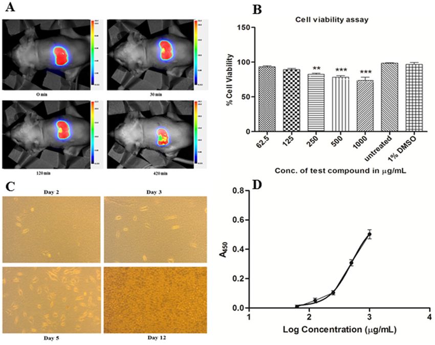
www.nature.com/scientificreports/
Figure 1. (A) Pictorial representation of rat skin after burn injury. (B) Comparative efficacy of different
formulations on partial thickness burn wound healing in Wistar rats. (C) Hydroxyproline content of different
treated groups.
migration and p roliferation15 whereas, TrkA or Tyrosine kinases receptors are the high-affinity cell surface
receptors for many growth factors, cytokines, and hormones. Additionally, silver sulfadiazine (AgSD) has been
considered as the gold standard treatment in topical burn. Therefore in the present study, bFGF, collagen and
AgSD incorporated hydrogel was fabricated and it was hypothesized that this hydrogel could help in epidermal
regeneration, fibroblast proliferation, and neovascularization as well as it could prevent the wound from getting
infected by pathogenic microorganisms. We investigated the ability of the hydrogel to promote wound healing
in vivo using a partial thickness burn wound model in Wistar rats. Further, the fibroblast cell proliferation was
investigated in vitro L929 mouse fibroblast cell line.
Results
bFGF‑collagen‑AgSD hydrogel promotes faster wound healing. To evaluate faster wound healing,
wound contraction and hydroxyproline assay were carried out. Representative photographs of partial thickness
burn wounds in Wistar rats have been presented in Fig. 1A. The wound contraction rates (WCR) of different
groups were compared with control group on different days of treatment and were shown in Fig. 1B. No signifi-
cant (p > 0.05) change was found on 4th day in each groups which means no burn wound healing was visible
externally after 4 days. However, on 8th day, only BOF treated group showed significant % WCR (p˂0.001) and
the % WCR of BOF treated and standard groups changed significantly (p˂0.001) on 12th and 16th day of treat-
ment. Results were subjected to two way ANOVA followed by Bonferroni post-tests (p˂0.001) was considered
significant.
It is well known that collagen helps in providing integrity to the skin tissues and promotes cellular prolifera-
tion and differentiation. Estimation of hydroxyproline, which is a major component of all types of collagen, is
routinely performed to understand the progress of wound healing rate. The rate of wound healing increases
when the amount of hydroxyproline increases in the connective tissue. Hydroxyproline content of the differ-
ent treated groups was given in the Fig. 1C. After 16 days, the highest concentration of hydroxyproline content
(524.03 ± 15.86 μg/10 mg of tissue) was found in hydrogel treated groups which also confirms the highest wound
contraction rate, whereas, the untreated group has the lowest hydroxyproline content (102.30 ± 27.58 μg/10 mg
Scientific Reports | (2021) 11:3357 | https://doi.org/10.1038/s41598-021-82888-9 2
Vol:.(1234567890)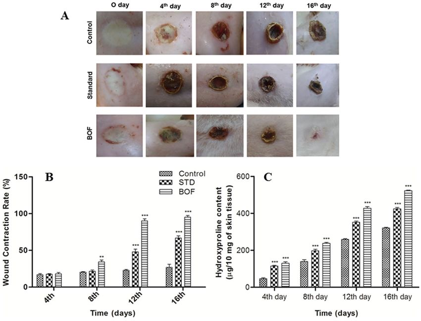
www.nature.com/scientificreports/
of tissue) thus, the lowest wound contraction rate. Hydroxyproline content of standard group was found to be
440.04 ± 18.90 μg/10 mg of tissue. The BOF treated group has the greater hydroxyproline content than the stand-
ard group because BOF contains both collagen and bFGF which help in faster wound healing. Collagen encour-
ages wound repopulation in cells with its regenerative potential and bFGF regulates the tissue repair process.
bFGF‑collagen‑AgSD hydrogel promotes re‑epithelialization, fibroblast proliferation, angio-
genesis. To evaluate the morphological changes of regenerating epidermis during burn healing, histopa-
thology of skin tissues of different groups using hematoxylin–eosin (HE) and Masson-Trichome (MT) stains
has been carried out (Fig. 2A,B). Further, scanning electron microscopy (SEM) of skin tissue of Wistar rats
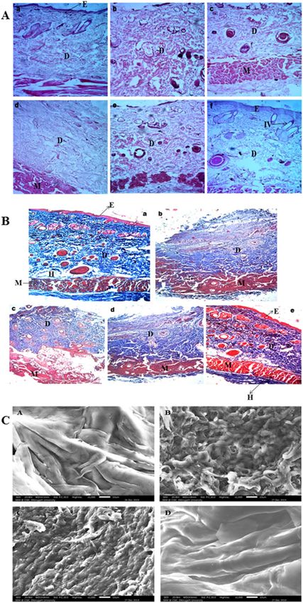
were investigated. In histopathology of skin tissues of untreated group, the epidermis was necrotic and the epi-
thelial cells in the dermal or follicular sheath were disrupted; as a result, the surroundings of the hair follicle
which contain progenitor cells could not maintain and regenerate the dermal papilla, a key component for hair
growth. Thus complete loss of hair shaft follicles was observed. Moreover, disorganized extracellular matrix was
found and complete debridement of epidermal tissue was also observed in partial thickness burn wounds. Burn
wounds treated with hydrogel regained its normal skin structure after 16 days and animals were fully recovered
with a dense and uniform neo-tissue structure, increased deposition of collagen fibres, macrophage and fibro-
blast proliferation, multiplication of fibrous connective tissue in the dermis, and promoted angiogenesis. In
standard group, the burn wounds of animals were not fully recovered and the commercial product was unable
to regain its normal skin structure. The epithelium was well-formed but with low deposition of collagen fibres.
Additionally, SEM images of control rat skin section showed smooth, intact and homogeneous skin surface
which is similar to previous finding16 whether; the morphology was changed in the untreated rat skin section
as compared to the SEM image of the control rat skin (Fig. 2C). The untreated rat skin section showed irregular
and discontinuous structure with loose arrangement of skin. Animals of standard group also showed irregular
skin structure however, it was less discrete than the structure of untreated rat skin. Our findings concur with
the observations of previous authors who investigated scanning electron microscopic characterization of wound
healing of rat s kin17 and ligament18. Additionally, BOF treated rat skin section showed similar surface topogra-
phy as compared to the control rat skin section therefore; reformation of the skin architecture revealed that the
developed hydrogel formulation helped in re-epithelialization of the burn wound rat skin which can be corre-
lated with the histopathology findings.
Pro and anti‑inflammatory cytokines in wound repair. It has been previously reported that pro-
inflammatory cytokines such as interleukins (IL)-1α, IL-1β, IL-6, and TNF-α and anti-inflammatory cytokines
including IL-10 play an important role in wound repair. Pro-inflammatory cytokines influence various processes
at the wound site, including stimulation of keratinocyte and fibroblast proliferation, synthesis and breakdown
of extracellular matrix proteins, fibroblast chemotaxis, and regulation of the immune response4. In this present
study, expressions of IL-1β, IL-6, and TNF-α were shown to be down regulated (Fig. 3A) during the inflamma-
tory phase of healing in the BOF treated group compared to untreated group. Moreover, as reported earlier,
the anti-inflammatory cytokine, IL-10, plays a major role in the limitation and termination of inflammatory
responses and regulates growth and/or differentiation of various immune cells, keratinocytes and endothelial
cells4. In our study, IL-10 initially increased in untreated group compared to BOF treated group and standard
group. A decrease in the level of pro-inflammatory cytokines after 16 days in BOF treated group indicated that
the inflammation gradually reduced; because cytokines are the primary mediators of the inflammatory reaction
to burn injury19. Therefore, the use of developed hydrogel in partial thickness burn might able to reduce the
inflammation.
bFGF‑collagen‑AgSD hydrogel regulates burn healing via NGF signaling pathway. Previous lit-
erature suggested that topical exogenous nerve growth factor (NGF) may have roles to increase wound closure20.
To investigate the therapeutic efficacy of the formulated hydrogel against partial thickness burn wounds, the
expression levels of some proteins involve in NGF signaling pathways have been evaluated in burn granulation
tissue of all treatment groups at 8th and 16th day after burn injury. We evaluated the expressions of some pro-
teins such as Tropomyosin-receptor kinase A (TrkA), p-TrkA, Extracellular Regulated Kinase 1 and 2 (ERK1/2),
p-ERK1/2, NF-kβ, and p-NF-kβ, involved in NGF signaling pathway, by western blotting. Expressions of TrkA,
p-TrkA, ERK1/2, & p-ERK1/2 relative to β-actin were up-regulated and NF-kβ, p-NF-kβ were down-regulated
in the treatment groups in a time dependent manner as compared to the control group (Fig. 3B). The expressions
of NF-kβ and p-NF-kβ were down-regulated because it is one of the most important regulators of pro-inflam-
matory gene e xpression21. Further, activation of NF-kβ leads to the activation of transcription of various genes
and thereby regulates inflammation22. Therefore, down-regulation of this protein indicated reduction of inflam-
mation in burn granulation tissue. Additionally, the expressions of Trk A, p-Trk A, ERK1/2, and p-ERK1/2 were
up-regulated which indicated that the developed hydrogel might activate the NGF signaling pathway and this
might contribute to its ability to accelerate the rate of burn wound healing in rats. This finding is similar to a
previous finding where it was stated that NGF produced in the wound may induce regeneration of fibroblasts in
granulation tissue and keratinocytes at the wound e dges14. Results were subjected to one way ANOVA followed
by Dunnett’s test was considered significant. Statistical analysis was performed using GraphPad Prism statistical
software version 5.01 (San Diego, California, USA).
Bio‑distribution study by in vivo imaging. Representative photographs of bio-distribution of AgSD
through partial thickness burn wounds of Wistar rats have been presented in Fig. 4A. After 7 hr, the intensity of
the formulation was reduced but there was no distribution of AgSD into any tissues or organs which indicated
Scientific Reports | (2021) 11:3357 | https://doi.org/10.1038/s41598-021-82888-9 3
Vol.:(0123456789)www.nature.com/scientificreports/
Figure 2. (A) Histopathology of different treated groups; (a) normal skin of rat, (b) untreated burned skin, (c)
Rat skin of standard group on 8th day and (d) on 16th day, (e) BOF treated skin on 8th day and (f) on 16th day
by using HE stain. (B) Histopathology of different treated groups; a) normal skin of rat, (b) untreated burned
skin, (c) Rat skin of standard group on 16th day, (d) BOF treated skin on 8th day and (e) on 16th day by using
MT stain. (C) Scanning electron microscopy of skin tissue of Wistar rats; (A) normal skin of rat, (B) untreated
burned skin, (C) standard group, (D) BOF treated.
Scientific Reports | (2021) 11:3357 | https://doi.org/10.1038/s41598-021-82888-9 4
Vol:.(1234567890)www.nature.com/scientificreports/
Figure 3. (A) Estimation of pro- and anti-inflammatory cytokines (A) IL-1β, (B) IL-6, (C) Rat TNF and (D)
IL-10 after burn wound on 4th, 8th, 12th and 16th day by using rat serum. Data are expressed as mean ± SEM
(n = 3). *** indicates p˂0.001, ** indicates p˂0.01, * indicates p˂0.05 as compared to the control group and
### indicates p˂0.001, ## indicates p˂0.01, # indicates p˂0.05 as compared to the standard group. (B) Western
blotting analysis of β actin, Trk A, p-Trk A, ERK1/2, p-ERK1/2 NF-kβ, p-NF-kβ expressions in burn skin tissue
of rats from different treatment groups. Data are expressed as mean ± SEM (n = 3). *** indicates p˂0.001, **
indicates p˂0.01, * indicates p˂0.05 as compared to the negative control group.
that it was retained by the superficial layer of the skin. Thus the toxicity of the AgSD might be minimized and
hence the adverse effects related to AgSD was reduced which is one of the important criteria of any pharma-
ceutical formulations. This finding is similar with the previous finding where it was stated that the absorption
of AgSD was negligible through both superficial and deep dermal burn surfaces as well as through the normal
skin23. Moreover, other previous studies about absorption of silver from partial-thickness and full-thickness
burn wounds (5% body surface area) reported that most of the silver was associated with the superficial eschar
and very little was absorbed into deeper layers of skin24,25. However, significant absorption of silver was observed
from patients with extensive burns (> 40% body surface) when treated topically with AgSD, so there is the pos-
sibility of silver toxicity occurring26–28.
Cytotoxicity study of hydrogel using MTT assay. Dermal toxicity of hydrogel formulation was carried
out using MTT assay on mouse L929 fibroblast cell line (Fig. 4B). According to the assay, the hydrogel showed
negligible cytotoxicity although cell viability reduced slightly with the increase concentration of the formulation.
The cytotoxicity assay showed that % cell viability of L929 cells at the highest concentration of the formulation
(1000 μg/mL) was found to be 73.42 ± 4.77. Therefore, the formulation did not inhibit cell proliferation on L929
cell line. This result indicated that the hydrogel formulation has a negligible cytotoxicity on mouse fibroblast
cells and could be further assessed in burn wound. % cell viability was plotted against concentrations of test
samples. Experiments were performed in triplicate and the data were presented as mean ± SD (n = 3). Further,
it was evaluated by one-way analysis of variance (ANOVA) followed by Dunnett’s test. Statistical analysis was
performed using GraphPad Prism statistical software version 5.01 (San Diego, California, USA).
Scientific Reports | (2021) 11:3357 | https://doi.org/10.1038/s41598-021-82888-9 5
Vol.:(0123456789)www.nature.com/scientificreports/
Figure 4. (A) Bio-distribution study of AgSD in partial thickness burn wounds of Wistar rats. (B) The
cytotoxicity effect of hydrogel formulation on L929 cell line. Untreated cells were considered as negative control
and 1% DMSO as vehicle control (***p˂0.0001). (C) Representative photographs of L929 cells maintained in
culture medium. (D) Treatment of L929 cells with hydrogel formulation increases cell proliferation as detected
by BrdU cell proliferation assay.
bFGF‑collagen‑AgSD hydrogel promotes fibroblast cell proliferation. Figure 4C displayed L929
cells showed active proliferation and fibroblast shaped cells after 2, 3, 5 and 12 days. BrdU cell proliferation assay
was performed to investigate the cytotoxicity of AgSD or quantify the cell proliferation induced by bFGF. The
result of this assay was illustrated in Fig. 4D. It showed that the absorbance at 450 nm was increased with the
increasing concentration (62.5, 125, 250, 500 and 1000 μg/ml) of the hydrogel formulation which means BrdU
incorporation was increased in L929 fibroblast cells treated with hydrogel formulation thus, the cell proliferation
was increased. Absorbance at 450 nm (A450) was plotted against log concentrations of test samples. Experiments
were performed in triplicate and the data were presented as mean ± SD (n = 3). Further, it was evaluated by dose
response curve using GraphPad Prism statistical software version 5.01 (San Diego, California, USA).
Discussion
The process of burn healing is a complex process involving epidermal regeneration, fibroblast proliferation,
neovascularization, angiogenesis etc. Over the past few years, several investigations have been made to improve
wound closure rate and healing time because there are numerous antimicrobial topical formulations which are
sufficient to prevent the pathogenic infections but cannot able to achieve faster wound closure. In order to over-
come these drawbacks, researchers were inspired by the concept of exogenous application of growth factors found
to be promising in burn wound treatment as it may decrease the healing p eriod29,30. Therefore, in the present
research, AgSD and bFGF loaded collagen based hydrogel formulation has been developed in our laboratory for
the first time to facilitate rapid burn wound healing simultaneously preventing pathogenic infections.
The wound contraction rate and the hydroxyproline content found maximum in the BOF treated group in
comparison to untreated and standard groups. It might happen due to the presence of bFGF and collagen. bFGF
promotes many cells such as dermal fibroblasts, keratinocytes, endothelial cells, melanocytes etc.; and induces
Scientific Reports | (2021) 11:3357 | https://doi.org/10.1038/s41598-021-82888-9 6
Vol:.(1234567890)www.nature.com/scientificreports/
tissue remodeling, wound healing and neovascularization due to its mitogenic and angiogenic c haracteristics31.
Despite of several advantages including a multifunctional role in stimulation of cell growth and tissue repair;
bFGF has a very short biological half-life when injected and is unstable in solution; moreover, rapid enzymatic
degradation make it difficult to be applied in the free form, thus unable to achieve effective concentrations to
treat wound h ealing30. Therefore, as per previous literatures, to overcome these problems, bFGF was encapsu-
lated within c ollagen32–34. Collagen plays an important role in the formation of tissues and also can form fibers
with extra strength and stability through its self-aggregation and cross-linking. Thus it can be widely used in
biomedical application35. Additionally, histopathology and SEM images revealed that burn wounds treated with
BOF regained its normal skin structure after 16 days and animals were fully recovered with re-epithelialization,
fibroblast proliferation, and promoted angiogenesis. Our findings are similar to other published evidences which
encourage the use of growth factors in burn healing. For example, Fu X et al., (2000) reported that the topical
applications of recombinant bovine bFGF to burn wounds accelerated the rate of formation of granulation tissue
as well as healing times36. Additionally, it was also reported that in animal systems, bFGF induces cell migra-
tion, neovascularization, and granulation tissue formation; moreover, it stimulates the healing of partial- and
full thickness wounds of the skin, cornea, cartilage and b rain37,38. Another study reported that bFGF stimulates
endothelial migration, proliferation and capillary like tube formation, promoting new vessel growth in vivo and
in vitro39. Moreover, bFGF potentiates leukocyte recruitment to inflammation sites in skin and improves dermal
wound healing outcomes upon direct delivery, either through a targeted peptide delivery system, or alongside
tissue engineering scaffold. It has also been reported earlier that for partial-thickness skin defect such as second-
degree burn, bFGF should be started at the earliest possible after such an injury31.
The pro-inflammatory cytokines such as IL-1β, IL-6, and TNF-α were found down regulated during the
inflammatory phase of healing in the BOF treated group compared to untreated group. A decrease in the level
of pro-inflammatory cytokines after 16 days in BOF treated group indicated that the inflammation gradually
reduced; because cytokines are the primary mediators of the inflammatory reaction to burn injury19. Therefore,
the use of bFGF-collagen-AgSD hydrogel in partial thickness burn might able to reduce the inflammation.
In western blotting, the expression levels of various proteins involve in NGF signaling pathways have been
assessed in burn granulation tissue. The expressions of NF-kβ and p-NF-kβ were down-regulated in the treatment
groups in a time dependent manner as compared to the control group because it is one of the most important
regulators of pro-inflammatory gene expression21. Activation of NF-kβ leads to the activation of transcription
of various genes and thereby regulates inflammation22. Therefore, down-regulation of this protein indicated
reduction of inflammation in burn granulation tissue. Additionally, the expressions of proteins such as Trk A,
p-Trk A, ERK1/2, and p-ERK1/2 were up-regulated which indicated that the developed hydrogel might activate
the NGF signaling pathway and this might contribute to its ability to accelerate the rate of burn wound heal-
ing in rats. As NGF produced in the wound may induce regeneration of fibroblasts in granulation tissue and
keratinocytes at the wound e dges14.
In bio-distribution study, no distribution of AgSD was found into any tissues or organs which indicated that
it was retained by the superficial layer of the skin. Thus the toxicity of the AgSD might be minimized and hence
the adverse effects related to AgSD was reduced which is one of the important criteria of any pharmaceutical
formulations.
Further, the cytotoxic effect of the hydrogel formulation was investigated on dermal cells using MTT assay.
It is well-known that MTT assay is based on the reduction of yellow tetrazolium MTT to a purple formazan dye
by mitochondrial succinate dehydrogenase40. A typical wound healing process encompasses complex cellular
changes that include inflammation, re-epithelialization, angiogenesis, granulation tissue formation, migration
and proliferation of keratinocytes and fibroblasts and remodeling of E CM41. In the early stages of wound heal-
ing, fibroblasts play an important role by actively proliferating, migrating to wound area and transforming
into myofibroblasts which facilitate wound contraction p rocess42. Additionally, fibroblasts are involved in the
synthesis of ECM components, immature collagen, developing mechanical forces and remodeling the scar43,44.
The hydrogel showed minimal toxicity on L929 mouse fibroblast cell line indicating negligible cytotoxic effect
of the developed formulation on dermal cells. Since the developed formulation contains bFGF along with AgSD,
it may be possible that the bFGF may have negated the cytotoxic effect of AgSD. This can be supported by the
findings of McCauley et al.44 who reported that growth factors including bFGF may have a role of cyto-protection
to cells responsible for wound healing and help in initiation and modulation of the process of wound h ealing45.
Further, treatment of L929 cells with hydrogel formulation increases cell proliferation as detected by BrdU cell
proliferation assay and it may be possible that the bFGF may increase the proliferation because of its role in
cell proliferation. As per previous literatures, bFGF is an important member of a heparin-binding protein fam-
ily, which controls the proliferation, differentiation, and migration of different cells. Additionally, it is a potent
regulator of cell proliferation, differentiation and function and is critically important in normal development,
tissue maintenance, wound repair and a ngiogenesis5,12,46.
Finally, from the present findings it can be concluded that the developed hydrogel formulation is not cytotoxic
and helps in cell proliferation however, this claim is limited to the highest concentration of the formulation and
the specific cell line used in this study. Hence, the formulation needs to be studied further in partial thickness
burn wound using different cell lines with higher concentrations. However, our study is one step forward to the
application of this product in burn wound care.
Scientific Reports | (2021) 11:3357 | https://doi.org/10.1038/s41598-021-82888-9 7
Vol.:(0123456789)www.nature.com/scientificreports/
Materials and methods
Study design. We hypothesized that the bFGF-collagen-AgSD incorporated hydrogel formulation would
accelerate the rate of burn healing in animal model and would promote fibroblast cell proliferation as well as
prevent the wound from getting infected by pathogenic microorganisms. The experimental ranges were selected
on the basis of previous findings30,47,48.
Animals, burn model and treatment. Healthy, adult Wistar albino rats were obtained from Central
Animal facility, Defence Research Laboratory, Tezpur, Assam, India. Animals were kept in polypropylene cage
providing free access of water and standard rodent chow (Pranav Agro Industries Limited, Sangli, Maharash-
tra, India) ad libitum. General conditions of captivity were maintained in simulated atmospheric conditions 25
(± 2) ˚C temperature; 70% RH; 12 h light/dark cycle. All experimenting protocols using animal were performed
according to the “Principles of Laboratory Animal care” (NIH publication 85–23, revised 1985) and approved
by the Institutional Animal Ethical Committee (IAEC) of Defence Research Laboratory, Tezpur, Assam, India
(approval no. IAEC/DRL/05/2016).
The required amount of collagen, PVA, AgSD, and bFGF were added in phosphate buffer solution (PBS,
pH—7.4) followed by thorough mixing with the help of magnetic s tirrer49,50. The temperature was maintained
at 4ºC during the preparation and the pH of the formulation was adjusted to 5.0.
Animals were divided into three groups i.e. control (burned but untreated), standard (burned and treated
by commercial product; 1% w/w silver sulfadiazine and 0.2% w/w Chlorhexidine gluconate), and BOF (best
optimized hydrogel formulation) treated group (burned and treated with bFGF-collagen-AgSD hydrogel). All
rats were anaesthetized with sodium phenobarbitone. Then the trauma was performed by holding a cylindrical
shaped bar with the radius of 1 cm on the shaved back skin of rats and hot water (98 ± 1ºC) was poured into this
bar and held for 15 s ecs51. After the formation of burns, the rats were housed individually and treatments were
given once daily until complete epithelialization. The progressive changes of burned area were photographed on
day 0, 4th day, 8th day, 12th day, and 16th day. Then all images were evaluated by using size analysis software-
Image J as indicated in the Results and figures.
Received: 5 April 2020; Accepted: 25 January 2021
References
1. Singer, A. J. & Clark, R. A. Cutaneous wound healing. N. Engl. J. Med. 341, 738–746 (1999).
2. Madaghiele, M., Demitri, C., Sannino, A. & Ambrosio, L. Polymeric hydrogels for burn wound care: advanced skin wound dress-
ings and regenerative templates. Burns Trauma. 2, 153 (2014).
3. Babensee, J. E., McIntire, L. V. & Mikos, A. G. Growth factor delivery for tissue engineering. Pharm. Res. 17, 497–504 (2000).
4. Werner, S. & Grose, R. Regulation of wound healing by growth factors and cytokines. Physiol. Rev. 83, 835–870 (2003).
5. Barrientos, S., Stojadinovic, O., Golinko, M. S., Brem, H. & Tomic-Canic, M. Growth factors and cytokines in wound healing.
Wound Repair Regen. 16, 585–601 (2008).
6. Shamloo, A., Sarmadi, M., Aghababaie, Z. & Vossoughi, M. Accelerated full-thickness wound healing via sustained bFGF delivery
based on a PVA/chitosan/gelatin hydrogel incorporating PCL microspheres. Int. J. Pharm. 537, 278–289 (2018).
7. Abraham, J. A. et al. Nucleotide sequence of a bovine clone encoding the angiogenic protein, basic fibroblast growth factor. Science
233, 545–548 (1986).
8. Gospodarowicz, D. 3 fibroblast growth factor and its involvement in developmental processes. Curr. Top. Dev. Biol. 24, 57–93
(1990).
9. Miyoshi, M. et al. Effects of bFGF incorporated into a gelatin sheet on wound healing. J. Biomater. Sci. Polym. Ed. 16, 893–907
(2005).
10. Montesano, R., Vassalli, J. D., Baird, A., Guillemin, R. & Orci, L. Basic fibroblast growth factor induces angiogenesis in vitro. PNAS
83, 7297–7301 (1986).
11. Ahrendt, G., Chickering, D. E. & Ranieri, J. P. Angiogenic growth factors: a review for tissue engineering. Tissue Eng. 4, 117–130
(1998).
12. Silva, A. K. A., Richard, C., Bessodes, M., Scherman, D. & Merten, O. W. Growth factor delivery approaches in hydrogels. Biomac-
romol 10, 9–18 (2008).
13. Landis, W. J., Silver, F. H. & Freeman, J. W. Collagen as a scaffold for biomimetic mineralization of vertebrate tissues. J. Mater.
Chem. 16, 1495–1503 (2006).
14. Matsuda, H. et al. Role of nerve growth factor in cutaneous wound healing: accelerating effects in normal and healing-impaired
diabetic mice. J. Exp. Med. 187, 297–306 (1998).
15. Lee, S. et al. ERK activating peptide, AES16-2M promotes wound healing through accelerating migration of keratinocytes. Sci.
Rep. 8, 1–10 (2018).
16. Abuarra, A. et al. The effects of different laser doses on skin. Int. J. Phys. Sci. 7, 400–407 (2012).
17. Forrester, J. C., Hunt, T. K., Hayes, T. L. & Pease, R. F. W. Scanning electron microscopy of healing wounds. Nature 221, 373–374
(1969).
18. Hurschler, C., Provenzano, P. P. & Vanderby, R. Jr. Scanning electron microscopic characterization of healing and normal rat liga-
ment microstructure under slack and loaded conditions. Connect Tissue Res. 44, 59–68 (2003).
19. Nyhlén, K., Gautam, C., Andersson, R. & Srinivas, U. Modulation of cytokine-induced production of IL-8 in vitro by interferons
and glucocorticosteroids. Inflammation. 28, 77–88 (2004).
20. Muangman, P. et al. Nerve growth factor accelerates wound healing in diabetic mice. Wound Repair Regen. 12, 44–52 (2004).
21. Tak, P. P. & Firestein, G. S. NF-κB: a key role in inflammatory diseases. J. Clin. Investig. 107, 7–11 (2001).
22. Liu, T., Zhang, L., Joo, D. & Sun, S. C. NF-κB signaling in inflammation. Signal Transduct. Tar. 2, 17023 (2017).
23. Sano, S., Fujimori, R., Takashima, M. & Itokawa, Y. Absorption, excretion and tissue distribution of silver sulphadiazine. Burns.
8, 278–285 (1982).
24. Harrison, H. N. Pharmacology of sulfadiazine silver: its attachment to burned human and rat skin and studies of gastrointestinal
absorption and extension. Arch. Surg. 114, 281–285 (1979).
Scientific Reports | (2021) 11:3357 | https://doi.org/10.1038/s41598-021-82888-9 8
Vol:.(1234567890)www.nature.com/scientificreports/
25. Tsipouras, N., Rix, C. J. & Brady, P. H. Solubility of silver sulfadiazine in physiological media and relevance to treatment of thermal
burns with silver sulfadiazine cream. Clin. Chem. 41, 87–91 (1995).
26. Wang, X. W., Wang, N. Z., Zhang, O. Z., Zapata-Sirvent, R. L. & Davies, J. W. L. Tissue deposition of silver following topical use
of silver sulphadiazine in extensive burns. Burns. 11, 197–201 (1985).
27. Boosalis, M. G., McCall, J. T., Ahrenholz, D. H., Solem, L. D. & McClain, C. J. Serum and urinary silver levels in thermal injury
patients. Surgery. 101, 40–43 (1987).
28. Wan, A. T., Conyers, R. A., Coombs, C. J. & Masterton, J. P. Determination of silver in blood, urine, and tissues of volunteers and
burn patients. Clin. Chem. 37, 1683–1687 (1991).
29. Alemdaroglu, C. et al. An investigation on burn wound healing in rats with chitosan gel formulation containing epidermal growth
factor. Burns. 32, 319–327 (2006).
30. Losi, P. et al. Fibrin-based scaffold incorporating VEGF-and bFGF-loaded nanoparticles stimulates wound healing in diabetic
mice. Acta Biomater 9, 7814–7821 (2013).
31. Akita, S., Akino, K. & Hirano, A. Basic fibroblast growth factor in scarless wound healing. Adv. Wound Care. 2, 44–49 (2013).
32. Downs, E. C., Robertson, N. E., Riss, T. L. & Plunkett, M. L. Calcium alginate beads as a slow-release system for delivering angio-
genic molecules in vivo and in vitro. J. Cell. Physiol. 152, 422–429 (1992).
33. Edelman, E. R., Nugent, M. A. & Karnovsky, M. J. Perivascular and intravenous administration of basic fibroblast growth factor:
vascular and solid organ deposition. PNAS 90, 1513–1517 (1993).
34. Iwakura, A., Tabata, Y., Tamura, N., Doi, K., Nishimura, K., Nakamura, T., Shimizu, Y., Fujita, M. & Komeda, M. Gelatin sheet
incorporating basic fibroblast growth factor enhances healing of devascularized sternum in diabetic rats. Circulation, 104(suppl_1),
I-325 (2001).
35. Lee, C. H., Singla, A. & Lee, Y. Biomedical applications of collagen. Int. J. Pharm. 221, 1–22 (2001).
36. Fu, X. et al. Recombinant bovine basic fibroblast growth factor accelerates wound healing in patients with burns, donor sites and
chronic dermal ulcers. Chin. Med. J. 113, 367–371 (2000).
37. Grazul-Bilska, A. T. et al. Wound healing: the role of growth factors. Drugs Today (Barc) 39, 787–800 (2003).
38. Lawrence, W. T. & Diegelmann, R. F. Growth factors in wound healing. Clin. Dermato.l, 12, 157–169 (1994).
39. Herdon, D. N. Growth hormones and factors in surgical patients. Adv. Surg. 25, 65–97 (1992).
40. Shahneh, F. Z., Valiyari, S., Azadmehr, A., Hajiaghaee, R., Yaripour, S., Bandehagh, A. & Baradaran, B. Inhibition of growth and
induction of apoptosis in fibrosarcoma cell lines by Echinophora platyloba DC: in vitro analysis. Adv. Pharmacol. Sci. 2013 (2013)
41. Guo, S. A. & DiPietro, L. A. Factors affecting wound healing. J. Dent. Res. 89, 219–229 (2010).
42. Li, W. et al. Mechanism of human dermal fibroblast migration driven by type I collagen and platelet-derived growth factor-BB.
Mol. Biol. Cell. 15, 294–309 (2004).
43. Werner, S., Krieg, T. & Smola, H. Keratinocyte-fibroblast interactions in wound healing. J. Investig. Dermatol. 127, 998–1008 (2007).
44. Kumar, P. M. & Ghosh, A. Development and evaluation of silver sulfadiazine loaded microsponge based gel for partial thickness
(second degree) burn wounds. Eur. J. Pharm. Sci. 96, 243–254 (2017).
45. McCauley, R. L., Li, Y. Y., Chopra, V., Herndon, D. N. & Robson, M. C. Cytoprotection of human dermal fibroblasts against silver
sulfadiazine using recombinant growth factors. J. Surg. Res. 56, 378–384 (1994).
46. Welham, N. V. et al. A rat excised larynx model of vocal fold scar. J. Speech Lang. Hear. Res. https://doi.org/10.1044/1092-
4388(2009/08-0049 (2009).
47. Fraser, J. F., Bodman, J., Sturgess, R., Faoagali, J. & Kimble, R. M. An in vitro study of the anti-microbial efficacy of a 1% silver
sulphadiazine and 0.2% chlorhexidine digluconate cream, 1% silver sulphadiazine cream and a silver coated dressing. Burns. 30,
35–41 (2004).
48. Namviriyachote, N., Aramwit, P. & Muangman, P. PSS3 the efficacy of epidermal growth factor in burn wound treatment. Value
Health 15, A568 (2012).
49. Puttawibul, P., Meesane J. & Benjakul, S. Preparation and characterization of collagen/PVA hybrid biomimetic hydrogels scaffold
for wound healing. In Biomedical Enginering International Conference (2012).
50. Sengupta, S., Banerjee, S., Sinha, B. & Mukherjee, B. Improved skin penetration using in situ nanoparticulate diclofenac diethyl-
amine in hydrogel systems: in vitro and in vivo studies. AAPS PharmaSciTech. 17, 307–317 (2016).
51. Degim, Z. et al. Evaluation of chitosan gel containing liposome-loaded epidermal growth factor on burn wound healing. Int. Wound
J. 8, 343–354 (2011).
Acknowledgements
One of the authors, Srijita Chakrabarti extends her gratitude to Defence Research Laboratory (DRL), Defence
Research and Development Organization (DRDO), Ministry of Defence, Govt. of India for providing necessary
facilities. Author also gratefully acknowledges Department of Pharmaceutical Sciences, Dibrugarh University
for providing administrative support to carry out her PhD work.
Author contributions
P.C., S.C., and B.M: conception or design of the work. S.C: wrote the main manuscript text. J.R., M.P.P., and
P.P: acquisition, analysis, and interpretation of data. S.C., and J.R: prepared figures. All authors reviewed the
manuscript.
Competing interests
The authors declare no competing interests.
Additional information
Supplementary Information The online version contains supplementary material available at https://doi.
org/10.1038/s41598-021-82888-9.
Correspondence and requests for materials should be addressed to P.C.
Reprints and permissions information is available at www.nature.com/reprints.
Publisher’s note Springer Nature remains neutral with regard to jurisdictional claims in published maps and
institutional affiliations.
Scientific Reports | (2021) 11:3357 | https://doi.org/10.1038/s41598-021-82888-9 9
Vol.:(0123456789)www.nature.com/scientificreports/
Open Access This article is licensed under a Creative Commons Attribution 4.0 International
License, which permits use, sharing, adaptation, distribution and reproduction in any medium or
format, as long as you give appropriate credit to the original author(s) and the source, provide a link to the
Creative Commons licence, and indicate if changes were made. The images or other third party material in this
article are included in the article’s Creative Commons licence, unless indicated otherwise in a credit line to the
material. If material is not included in the article’s Creative Commons licence and your intended use is not
permitted by statutory regulation or exceeds the permitted use, you will need to obtain permission directly from
the copyright holder. To view a copy of this licence, visit http://creativecommons.org/licenses/by/4.0/.
© The Author(s) 2021
Scientific Reports | (2021) 11:3357 | https://doi.org/10.1038/s41598-021-82888-9 10
Vol:.(1234567890)You can also read