Autonomic dysreflexia associated with cervical spinal cord gliofibroma: case report
←
→
Page content transcription
If your browser does not render page correctly, please read the page content below
Mizuno et al. BMC Neurology      (2021) 21:252
https://doi.org/10.1186/s12883-021-02271-z

 CASE REPORT                                                                                                                                         Open Access

Autonomic dysreflexia associated with
cervical spinal cord gliofibroma: case report
Hiroyuki Mizuno1*, Fumiaki Honda1, Hayato Ikota2 and Yuhei Yoshimoto1

  Abstract
  Background: Autonomic dysreflexia (AD) is an abnormal reflex of the autonomic nervous system normally
  observed in patients with spinal cord injury from the sixth thoracic vertebra and above. AD causes various
  symptoms including paroxysmal hypertension due to stimulus. Here, we report a case of recurrent AD associated
  with cervical spinal cord tumor.
  Case presentation: The patient was a 57-year-old man. Magnetic resonance imaging revealed an intramedullary
  lesion in the C2, C6, and high Th12 levels. During the course of treatment, sudden loss of consciousness occurred
  together with abnormal paroxysmal hypertension, marked facial sweating, left upward conjugate gaze deviation,
  ankylosis of both upper and lower extremities, and mydriasis. Seizures repeatedly occurred, with symptoms
  disappearing after approximately 30 min. AD associated with cervical spinal cord tumor was diagnosed. Histological
  examination by tumor biopsy confirmed the diagnosis of gliofibroma. Radiotherapy was performed targeting the
  entire brain and spinal cord. The patient died approximately 3 months after treatment was started.
  Conclusions: AD is rarely associated with spinal cord tumor, and this is the first case associated with cervical spinal
  cord gliofibroma. AD is important to recognize, since immediate and appropriate response is required.
  Keywords: Autonomic dysreflexia, Gliofibroma, Cervical spinal cord tumor

Background                                                                              gradually worsened and movement became difficult over a
Autonomic dysreflexia (AD) is an abnormal reflex of the                                 period of 4 months, so he was transferred by ambulance.
autonomic nervous system that is primarily observed in                                  On admission, he was aware and conscious, and com-
patients with spinal cord injury located at the sixth thor-                             plained of nervous pain in the right lower leg. Physical
acic vertebra and above [1]. AD is well known to cause                                  examination found mild paralysis of manual muscle test
various symptoms including paroxysmal hypertension                                      4/5 and increased deep tendon reflex in the left half of the
due to stimulus [2, 3]. Here, we report a rare case of re-                              body. Orthostatic hypotension was observed in which sys-
current AD associated with a cervical spinal cord tumor.                                tolic blood pressure decreased to approximately 70 mmHg
                                                                                        when sitting. In addition, mild urination and defecation
Case presentation                                                                       disorders were noted. Cerebrospinal fluid examination in-
History and examination                                                                 dicated elevated initial pressure of 27 cmH2O and high
A 57-year-old man presented with right lower extremity                                  protein level of 605 mg/dl. No tumor cells were detected
pain and light headedness when standing up. He had no                                   in the cerebrospinal fluid, and collagen-related antibody
related family history or medical history. His right leg pain                           levels were normal. Magnetic resonance imaging revealed
                                                                                        contrast-enhanced lesions at the posterior intramedullary
* Correspondence: m09201080@gunma-u.ac.jp                                               C2 and high C6 levels, left posterior intramedullary Th12
1
 Departments of Neurosurgery, Gunma University Graduate School of                       level, medullary cone surface, and left middle cerebellar
Medicine, 3-39-22 Showa-machi, Gunma 371-8511 Maebashi, Japan
Full list of author information is available at the end of the article                  peduncle (Fig. 1).

                                         © The Author(s). 2021 Open Access This article is licensed under a Creative Commons Attribution 4.0 International License,
                                         which permits use, sharing, adaptation, distribution and reproduction in any medium or format, as long as you give
                                         appropriate credit to the original author(s) and the source, provide a link to the Creative Commons licence, and indicate if
                                         changes were made. The images or other third party material in this article are included in the article's Creative Commons
                                         licence, unless indicated otherwise in a credit line to the material. If material is not included in the article's Creative Commons
                                         licence and your intended use is not permitted by statutory regulation or exceeds the permitted use, you will need to obtain
                                         permission directly from the copyright holder. To view a copy of this licence, visit http://creativecommons.org/licenses/by/4.0/.
                                         The Creative Commons Public Domain Dedication waiver (http://creativecommons.org/publicdomain/zero/1.0/) applies to the
                                         data made available in this article, unless otherwise stated in a credit line to the data.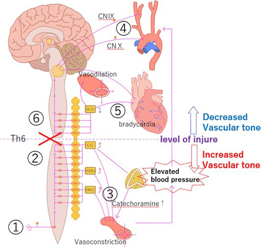
Mizuno et al. BMC Neurology       (2021) 21:252                                                                                         Page 2 of 7

 Fig. 1 T1-weighted magnetic resonance image with gadolinium on admission showing multiple contrast-enhanced lesions in the posterior
 intramedullary spinal cord at the C2 and C6 levels and on the spinal cord surface at the C7 and C8 levels (A, B), and in the left posterior
 intramedullary spinal cord at the Th12 level and on the surface of the spinal cone (C, D)

Postadmission course                                                        symptoms gradually disappeared after approximately
On the day after admission, the patient suddenly lost                       30 min to one hour, and the patient’s consciousness dis-
consciousness and systolic blood pressure rose to around                    turbance was ameliorated. Such attacks repeatedly oc-
230 mmHg, with marked facial sweating, upper conju-                         curred 1 to 5 times daily and were mainly triggered by
gate deviation of the left eye, ankylosis of both upper                     pain during physical activity and daily nursing care. Elec-
and lower limbs, and dilated pupils. His head was imme-                     troencephalography in the resting state recorded on day
diately elevated and head computed tomography/mag-                          2 showed α waves predominantly originating from the
netic resonance imaging was performed but found no                          occipital lobe at 8–10 Hz, but no laterality was observed.
abnormalities. Intravenous injection of nicardipine                         Complex partial epileptic seizures were suspected, so
hydrochloride was performed to reduce his blood pres-                       oral administration of lacosamide (100 mg/day) was ini-
sure, but his blood pressure was refractory and difficult                   tiated on the same day for prophylaxis. However, the fre-
to reduce immediately. This neurological symptom                            quency of seizures remained unchanged.
could not be explained by a transient ischemic attack, so                      Sudden onset of symptoms of sympathetic hyperactiv-
antithrombotic therapy was not started. Epileptic sei-                      ity accompanied by consciousness disorder triggered by
zures were suspected, and diazepam (10 mg) and fosphe-                      pain indicated AD associated with an intramedullary
nytoin sodium hydrate (750 mg) were administered                            tumor. Initially, the number of therapeutic agents was
intravenously. Conjugate deviation of the eye and anky-                     increased and more care was taken to minimize the
losis in both upper and lower extremities were alleviated,                  stimulation caused when changing postures and moving
but the hypertension, facial sweating, and dilated pupils                   the bed, which acted as triggers. However, non-steroidal
showed almost no improvement. However, these                                anti-inflammatory drugs had little analgesic effect on the
Mizuno et al. BMC Neurology      (2021) 21:252                                                                          Page 3 of 7

resting right lower extremity pain, so oral administration            interstitial tissue. No increase in mitotic figures, micro-
of carbamazepine 400 mg/day was initiated, together                   vessel proliferation, or necrosis was observed. Immuno-
with administration of tofisopam (150 mg/day) and                     histochemical examination showed the tumor cells were
gabapentin (1200 mg/day). The patient had presented                   S-100 protein (+), glial fibrillary acidic protein (+), Olig2
with bladder-rectal disorder at the time of admission, so             (+), mIDH1-R132H (-), p53 (+, less than 10 %), and
catheterization of the urethra was performed and oral                 ATRX (+), with MIB-1 labeling index of 2 % (Fig. 3).
administration of laxatives and prokinetic agents for
constipation was initiated. However, the seizures repeat-
                                                                      Postoperative course
edly occurred, so early treatment was planned. Tumor
                                                                      The frequency of AD was gradually reduced by adjusting
biopsy was performed at the Th10-11 level on hospital
                                                                      the nursing system, alleviating pain, and initiating oral
day 3.
                                                                      pharmacotherapy (Fig. 4). The histological diagnosis was
                                                                      gliofibroma, with few findings suggesting malignancy.
Operative findings
                                                                      However, imaging revealed disseminated lesions spread-
No adhesion of the tumor to the dura was observed.
                                                                      ing from the entire spinal cord to the posterior fossa. Ir-
The tumor was white on the surface, elastic hard with a
                                                                      radiation 36 Gy was administered to the whole spinal
capsule, and hypovascular. The capsule structure disap-
                                                                      cord and posterior fossa, and 12.6 Gy to the local lesion
peared in the deep part of the tumor, and the boundary
                                                                      in the cerebellum. However, the tumors showed little
between the tumor and the normal spinal cord was un-
                                                                      change, and the disseminated lesions gradually spread.
clear. Therefore, the tumor appeared to be intramedul-
                                                                      The patient died approximately 3 months after the
lary (Fig. 2). The operation was completed after only
                                                                      course of treatment was completed.
collecting tissue for diagnosis.

Histopathological findings                                            Discussion and conclusion
Tumor cells with short spindle-shaped nuclei and eo-                  Autonomic dysreflexia
sinophilic cytoplasm proliferated in bundles, forming an              AD is a paroxysmal abnormal reflex of the autonomic
intricate pattern along with collagenous fibrous                      nerves occurring in patients with spinal cord injury of
                                                                      Th6 and above, initially reported in 1917 [4]. AD has
                                                                      been defined as uncontrollable transient increase in
                                                                      blood pressure of 20 mmHg or more [5, 6]. The clinical
                                                                      manifestations are diverse, and include abnormal auto-
                                                                      nomic hyperactivity symptoms such as sudden hyperten-
                                                                      sion, dilated pupils, headache, and bradycardia, in
                                                                      addition to sweating, piloerection, rubefaction, nasal
                                                                      congestion of non-paralyzed skin, chest pain, nausea,
                                                                      and vomiting [2, 7]. The compensatory mechanism fails
                                                                      with severe seizures and hyperperfusion occurs, causing
                                                                      consciousness disorder/convulsive seizures, occasional
                                                                      hypertensive encephalopathy, and cerebral hemorrhage
                                                                      [8]. Other serious life-threatening complications such as
                                                                      fatal arrhythmia require urgent diagnosis and treatment
                                                                      [9, 10]. Complications during the chronic stage may lead
                                                                      to deterioration of cardiac function, cognitive impair-
                                                                      ment, and other pulmonary, retinal, and renal disorders,
                                                                      so the prognosis is poor [11–13].
                                                                        Onset is generally reported to occur most frequently
                                                                      during the chronic phase after spinal cord injury (3–6
                                                                      months after injury) [3], with up to 40 seizures daily
                                                                      [14]. AD rarely occurs in patients with spinal cord tumor
 Fig. 2 Intraoperative photographs showing the tumor was elastic      and non-traumatic spinal cord disease such as multiple
 hard with a capsule, white surface, and no adhesion to the dura,     sclerosis [15, 16]. Only one case of AD has been re-
 and extended exophytically from the left side of the dorsal column   ported in cervical intramedullary astrocytoma with onset
 of the spinal cord (Upper). The tumor was relatively hypovascular    associated with spinal cord tumor [15]. The most com-
 and the boundary between the tumor tissue and normal spinal cord
                                                                      mon seizure-inducing factor is bladder/rectal tension
 was not clear (Lower)
                                                                      due to bladder-rectal dysfunction associated with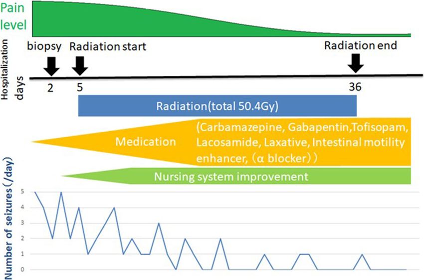
Mizuno et al. BMC Neurology (2021) 21:252 Page 4 of 7
Fig. 3 Pathological findings. A: Hematoxylin and eosin staining showing tumor cells with short spindle-shaped nucleus and eosinophilic
cytoplasm proliferated in bundles and intricate shapes with collagenous interstitium. Original magnification x100. B and C: Immunohistochemical
staining showing positive results for glial fibrillary acidic protein in the cytoplasm (B) and for Olig2 in the nucleus (C). D: MIB-1 staining showing
labeling index of about 2 %
autonomic neuropathy, but various other stimuli may the sympathetic nerve system (greater splanchnic nerve)
also be inducing factors [2, 17, 18]. branching from the Th4-L2 levels, resulting in sympa-
thetic nerve-mediated vasoconstriction and increased
Mechanism blood pressure. This systemic increase in blood pressure
The mechanism for onset is as follows [19]. The trigger caused by the pain trigger stimulates the baroreceptors
stimulus passes up the spinal cord via the higher sensory of the carotid sinus and aortic arch, resulting in vagal
branches to reach the brain, and is also transmitted to bradycardia and central vasodilation of the vasomotor
Fig. 4 Treatment course, pain level, and number of seizures. The pain and seizure frequency gradually decreased with drug treatment,
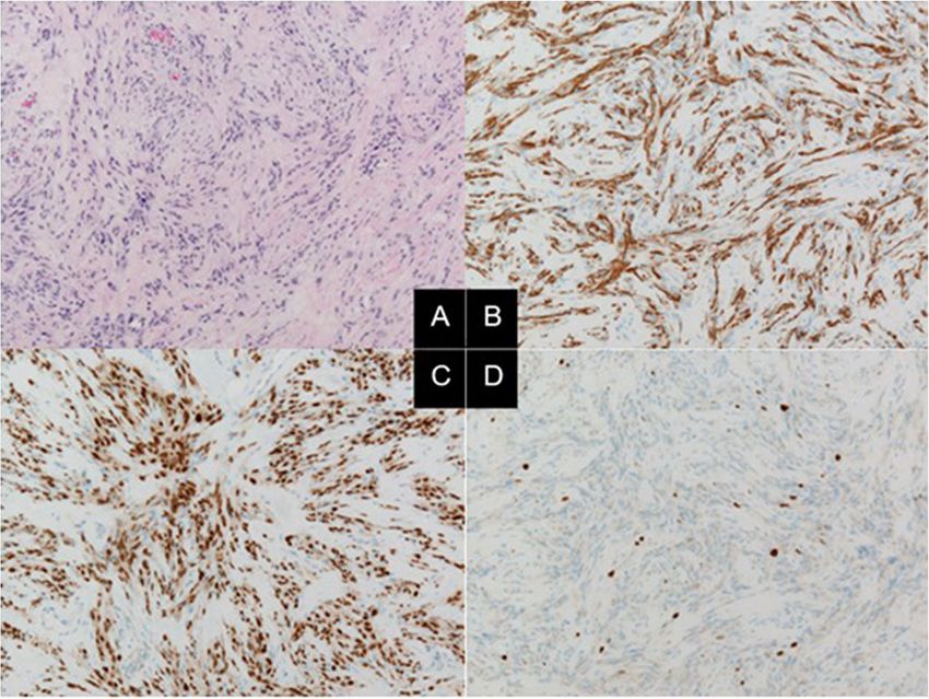
improvement of the nursing system, and start of radiation therapy, but the seizures could not be completely suppressedMizuno et al. BMC Neurology (2021) 21:252 Page 5 of 7 nerves. Such increase in blood pressure is immediately hypersensitivity contributes to the severity of the hyper- controlled by this series of compensatory mechanisms in tension [21]. healthy individuals. However, the vagal descending path- way is disrupted in patients with spinal cord injury, so Treatment that no inhibitory signal is transmitted below the level of Elimination of the cause and prophylaxis the spinal cord injury and consequently the vasocon- AD-induced hypertension is an abnormal reflex arc which striction continues. Blood vessels above the level of is not relieved unless the causative stimulus is eliminated. spinal cord injury undergo vasodilation as a strong in- Therefore, identification of the cause is the most import- hibitory signal is transmitted. Therefore, vasoconstric- ant treatment step [22]. However, the seizure-inducing tion below the injury level and vasodilation above the factors differ from case to case, so individual analysis of injury level occur simultaneously, and the vascular tone the causes is important to treat individual patients. Blad- gap results in abnormal hypertension, dilated pupils, der/rectal tightness due to bladder-rectal injury is the headache, sweating, and flushing. This abnormal reflex most common symptom, so urinary retention should be arc is not resolved and hypertension persists unless the confirmed, or catheter obstruction should be excluded if stimulus ceases (Fig. 5). At the same time, the sympa- an indwelling urethral catheter has been inserted [18]. thetic ganglia also act on the adrenal cortex, resulting in Oral administration of anticholinergic drugs and gabapen- raised serum adrenaline and noradrenaline concentra- tin is effective for preventing intestinal tract/bladder ten- tions, and promotion of hypertension [20]. The α1 and sion [23, 24], and botox injection into the bladder/rectal α2 receptors are up-regulated in the peripheral blood sphincter and surgical treatment were successful in severe vessels of AD patients and vascular hypersensitivity to cases [25–28]. However, the onset of AD cannot be com- catecholamines is increased, and this higher pletely inhibited only by elimination and prevention of the Fig. 5 Seizure mechanism. Vasoconstriction below the injury level and vasodilation above the injury level was caused by t created using Adobe Illustrator. he spinal cord injury above the Th6 cord. This vascular tone gap caused abnormal hypertension, dilated pupils, headaches, sweating, and flushing. This figure was created using Adobe Illustrator
Mizuno et al. BMC Neurology   (2021) 21:252                                                                                     Page 6 of 7

causes, so oral pharmacotherapy and preparation for re-       was neuropathic pain, gabapentin, which is administered
sponse to seizures are necessary.                             for neuropathic pain and may have protective effects
                                                              against AD, was orally administered in addition to carba-
Oral pharmacotherapy                                          mazepine, resulting in gradual alleviation of the pain. In
Nifedipine, a typical Ca blocker, is the most commonly        addition, we reviewed the nursing system and minimized
used oral drug to relieve seizures [12, 29, 30], but re-      the irritation associated with movement. Oral adminis-
quires care in long-term administration because AD pa-        tration of laxatives and prokinetic agents reduced blad-
tients are hypotensive due to autonomic neuropathy [31,       der/intestinal    irritation.  In    addition,    internal
32]. Prazosin, a selective alpha-blocker, does not exert a    administration of tofisopam and lacosamide was started.
rapid inhibitory effect on cardiac function and resting       This combined treatment achieved gradual decrease in
blood pressure, so can significantly reduce the severity      seizure frequency, but not complete inhibition. Adminis-
of AD-related seizures [33, 34]. In addition, oral admin-     tration of an alpha-blocker might also be appropriate to
istration of nitroglycerin, hydralazine, captopril, prosta-   consider.
glandin E2, terazosin, clonidine, and hydralazine reduce
the frequency and severity of seizures [29, 35–37]. The
                                                              Conclusions
sympatholytic drugs, guanethidine and tofisopam, nor-
                                                              The present case of gliofibroma manifested with ex-
mally used for autonomic neuropathy, and gabapentin,
                                                              tremely rare sympathetic hypertonic reflex associated
sometimes used for neuropathic pain, are also reported
                                                              with cervical spinal cord tumor. AD is rarely associated
to reduce the frequency of seizures [38].
                                                              with spinal cord tumor, and this is the first case associ-
                                                              ated with cervical spinal cord gliofibroma. AD is import-
Emergency response
                                                              ant to recognize, since immediate and appropriate
Emergent reduction of cerebral perfusion pressure can
                                                              response is required.
be achieved by raising the head of the patient [34]. Anti-
hypertensive agents are administered as needed [18, 39].      Abbreviations
Nitroglycerin, hydralazine, captopril, and other agents       AD: Autonomic dysreflexia; ATRX: α-Thalassemia/mental retardation
have been recommended as antihypertensive agents in           syndrome X-linked; MIB-1: Mind bomb homolog-1; mIDH1: Mutations in
                                                              isocitrate dehydrogenase 1; Olig2: Oligodendrocyte lineage transcription
emergencies [10, 30, 40]. Frequent blood pressure moni-       factor 2
toring (every 2–5 min) is important because blood pres-
sure may change rapidly [41].                                 Authors’ contributions
                                                              HM, FH contributed to the concept, drafting, and reporting of the case. HI
Gliofibroma                                                   assisted in the preparation of the manuscript. YY contributed to the critical
                                                              revision of the manuscript for important intellectual content. FH, HI, and YY
Gliofibroma is a rare histological type defined in the 4th    reviewed the manuscript. All authors have read and approved the final
edition of the Classification of Tumours of the Central       manuscript.
Nervous System of the World Health Organization in
2007. Gliofibroma consists of both glial and mesenchy-        Funding
mal components. The age of onset is 11 to 54 years (me-       This study was not funded.

dian: 14 years), and the male-female ratio is 2:3. The
most common sites are the cerebrum (36 %) and spinal          Availability of data and materials
                                                              Not applicable.
cord (28 %). The glioma component may be low or high
grade, but the mesenchymal component has no malig-            Declarations
nant characteristics. The histological types that should
be excluded during differential diagnosis include desmo-      Ethics approval and consent to participate
                                                              Not applicable.
plastic infantile astrocytoma and ganglioglioma, pleo-
morphic      xanthoastrocytoma,       and     gliosarcoma.
                                                              Consent to publication
Gliofibroma has a good prognosis, but dissemination           A written informed consent was obtained from the relatives of the patient
and death have been reported in cases with anaplastic         for publication of this Case report and any accompanying images.
glioma components and increased mitotic Figs. [42, 43].
                                                              Competing interests
Present case                                                  The authors report no conflict of interest concerning the materials or
                                                              methods used in this study or the findings specified in this paper.
The present patient had gliofibroma located in high
spinal positions, at the C2, C6, and Th12 levels. Severe      Author details
                                                              1
AD repeatedly occurred due to neural pain in the right         Departments of Neurosurgery, Gunma University Graduate School of
                                                              Medicine, 3-39-22 Showa-machi, Gunma 371-8511 Maebashi, Japan.
buttock. Radiotherapy was initiated immediately after bi-     2
                                                               Departments of Human Pathology, Gunma University Graduate School of
opsy was performed. Since the primary trigger for AD          Medicine, Gunma, Maebashi, Japan.
Mizuno et al. BMC Neurology (2021) 21:252 Page 7 of 7
Received: 25 November 2020 Accepted: 8 June 2021 24. Rabchevsky AG, Patel SP, Duale H, Lyttle TS, O’Dell CR, Kitzman PH.
Gabapentin for spasticity and autonomic dysreflexia after severe spinal cord
injury. Spinal Cord. 2011;49(1):99–105.
25. Barton CH, Khonsari F, Vaziri ND, Byrne C, Gordon S, Friis R. The effect of
References modified transurethral sphincterotomy on autonomic dysreflexia. J Urol.
1. Thompson CE, Witham AC. Paroxysmal hypertension in spinal cord injuries. 1986;135(1):83–5.
N Engl J Med. 1948;239(8):291–4. 26. Dykstra DD, Sidi AA, Scott AB, Pagel JM, Goldish GD. Effects of botulinum A
2. Karlsson AK. Autonomic dysreflexia. Spinal Cord. 1999;37(6):383–91. toxin on detrusor-sphincter dyssynergia in spinal cord injury patients. J Urol.
3. Lindan R, Joiner E, Freehafer AA, Hazel C. Incidence and clinical features of 1988;139(5):919–22.
autonomic dysreflexia in patients with spinal cord injury. Paraplegia. 1980; 27. Schurch B, Stöhrer M, Kramer G, Schmid DM, Gaul G, Hauri D. Botulinum-A
18(5):285–92. toxin for treating detrusor hyperreflexia in spinal cord injured patients: a
4. Head H, Riddoch G. The autonomic bladder, excessive sweating and some new alternative to anticholinergic drugs? Preliminary results. J Urol. 2000;
other reflex condition, in gross injuries of the spinal cord. Brain. 1917;40(2– 164(3 Pt 1):692–7.
3):188–263. 28. Sidi AA, Becher EF, Reddy PK, Dykstra DD. Augmentation enterocystoplasty
5. Phillips AA, Elliott SL, Zheng MM, Krassioukov AV. Selective alpha adrenergic for the management of voiding dysfunction in spinal cord injury patients. J
antagonist reduces severity of transient hypertension during sexual Urol. 1990;143(1):83–5.
stimulation after spinal cord injury. J Neurotrauma. 2015;32(6):392–6. 29. Braddom RL, Rocco JF. Autonomic dysreflexia. A survey of current
6. Teasell RW, Arnold JM, Krassioukov A, Delaney GA. Cardiovascular treatment. Am J Phys Med Rehabil. 1991;70(5):234–41.
consequences of loss of supraspinal control of the sympathetic nervous 30. Esmail Z, Shalansky KF, Sunderji R, Anton H, Chambers K, Fish W. Evaluation
system after spinal cord injury. Arch Phys Med Rehabil. 2000;81(4):506–16. of captopril for the management of hypertension in autonomic dysreflexia:
7. Kirshblum SC, House JG, O’connor KC. Silent autonomic dysreflexia during a a pilot study. Arch Phys Med Rehabil. 2002;83(5):604–8.
routine bowel program in persons with traumatic spinal cord injury: a 31. Furlan JC, Fehlings MG. Cardiovascular complications after acute spinal cord
preliminary study. Arch Phys Med Rehabil. 2002;83(12):1774–6. injury: pathophysiology, diagnosis, and management. Neurosurg Focus.
8. Phillips AA, Ainslie PN, Warburton DE, Krassioukov AV. Cerebral blood flow 2008;25(5):E13.
responses to autonomic dysreflexia in humans with spinal cord injury. J 32. Zheng MM, Phillips AA, Elliott SL, Krassioukov AV. Prazosin: a potential new
Neurotrauma. 2016;33(3):315–8. management tool for iatrogenic autonomic dysreflexia in individuals with
9. Jain A, Ghai B, Jain K, Makkar JK, Mangal K, Sampley S. Severe autonomic spinal cord injury? Neural Regen Res. 2015;10(4):557–8.
dysreflexia induced cardiac arrest under isoflurane anesthesia in a patient 33. Jaillon P. Clinical pharmacokinetics of prazosin. Clin Pharmacokinet. 1980;
with lower thoracic spine injury. J Anaesthesiol Clin Pharmacol. 2013;29(2): 5(4):365–76.
241–3. 34. Krassioukov AV, Harkema SJ. Effect of harness application and postural
10. Vallès M, Benito J, Portell E, Vidal J. Cerebral hemorrhage due to autonomic changes on cardiovascular parameters of individuals with spinal cord injury.
dysreflexia in a spinal cord injury patient. Spinal Cord. 2005;43(12):738–40. Spinal Cord. 2006;44(12):780–6.
35. Crassous PA, Denis C, Paris H, Sénard JM. Interest of alpha2-adrenergic
11. Cragg JJ, Noonan VK, Krassioukov A, Borisoff J. Cardiovascular disease and
agonists and antagonists in clinical practice: background, facts and
spinal cord injury: results from a national population health survey.
perspectives. Curr Top Med Chem. 2007;7(2):187–94.
Neurology. 2013;81(8):723–8.
36. Eldahan KC, Rabchevsky AG. Autonomic dysreflexia after spinal cord injury:
12. Krassioukov A, Warburton DE, Teasell R, Eng JJ, Spinal Cord Injury
Systemic pathophysiology and methods of management. Auton Neurosci.
Rehabilitation Evidence Research Team. A systematic review of the
2018;209:59–70.
management of autonomic dysreflexia after spinal cord injury. Arch Phys
37. Vaidyanathan S, Soni BM, Sett P, Watt JW, Oo T, Bingley J. Pathophysiology
Med Rehabil. 2009;90(4):682–95.
of autonomic dysreflexia: long-term treatment with terazosin in adult and
13. Phillips AA, Warburton DE, Ainslie PN, Krassioukov AV. Regional
paediatric spinal cord injury patients manifesting recurrent dysreflexic
neurovascular coupling and cognitive performance in those with low blood
episodes. Spinal Cord. 1998;36(11):61–770.
pressure secondary to high-level spinal cord injury: improved by alpha-1
38. Otani T, Kondo A, Kobayashi M. [Clinical studies of autonomic hyperreflexia].
agonist midodrine hydrochloride. J Cereb Blood Flow Metab. 2014;34(5):
Acta Urologica Japonica. 1985;31(7):1143–9. Japanese.
794–801.
39. Showkathali R, Antionios TF. Autonomic dysreflexia; a medical emergency. J
14. Hubli M, Gee CM, Krassioukov AV. Refined assessment of blood pressure
R Soc Med. 2007;100(8):382–3.
instability after spinal cord injury. Am J Hypertens. 2015;28(2):173–81.
40. Squair JW, Phillips AA, Harmon M, Krassioukov AV. Emergency management
15. Furlan JC, Fehlings MG, Halliday W, Krassioukov AV. Autonomic dysreflexia
of autonomic dysreflexia with neurologic complications. CMAJ. 2016;188(15):
associated with intramedullary astrocytoma of the spinal cord. Lancet
1100–3.
Oncol. 2003;4(9):574–5.
41. Consortium for Spinal Cord Medicine. Acute management of autonomic
16. Kulcu DG, Akbas B, Citci B, Cihangiroglus M. Autonomic dysreflexia in a man
dysreflexia: individuals with spinal cord injury presenting to health-care
with multiple sclerosis. J Spinal Cord Med. 2009;32(2):198–203.
facilities. J Spinal Cord Med. 2002;25(Suppl 1):67–88.
17. Canon S, Shera A, Phan NM, Lapicz L, Scheidweiler T, Batchelor L, et al.
42. Friede RL. Gliofibroma. A peculiar neoplasia of collagen forming glia-like
Autonomic dysreflexia during urodynamics in children and adolescents with
cells. J Neuropathol Exp Neurol. 1978;37(3):300–13.
spinal cord injury or severe neurologic disease. J Pediatr Urol. 2015;11(1):32.
43. Vasquez M, Miller DC, Epstein F, Allen JC, Budzilovich GN. Glioneurofibroma:
e1-4.
renaming the pediatric “gliofibroma”: a neoplasm composed of Schwann
18. Shergill IS, Arya M, Hamid R, Khastgir J, Patel HR, Shah PJ. The importance of
cells and astrocytes. Mod Pathol. 1991;4(4):519–23.
autonomic dysreflexia to the urologist. BJU Int. 2004;93(7):923–6.
19. Kurnick NB. Autonomic hyperreflexia and its control in patients with spinal
cord lesions. Ann Intern Med. 1956;44(4):678–86. Publisher’s Note
20. Leman S, Sequeira H. Activation of adrenal preganglionic neurons during Springer Nature remains neutral with regard to jurisdictional claims in
autonomic dysreflexia in the chronic spinal cord-injured rat. Auton Neurosci. published maps and institutional affiliations.
2002;98(1–2):94–8.
21. Krum H, Louis WJ, Brown DJ, Howes LG. A study of the alpha-1
adrenoceptor blocker prazosin in the prophylactic management of
autonomic dysreflexia in high spinal cord injury patients. Clin Auton Res.
1992;2(2):83–8.
22. Guyenet P, Cabot JB. Inhibition of sympathetic preganglionic neurons by
catecholamines and clonidine: mediation by an alpha-adrenergic receptor. J
Neurosci. 1981;1(8):908–17.
23. Giannantoni A, Di Stasi SM, Scivoletto G, Mollo A, Silecchia A, Fuoco U, et al.
Autonomic dysreflexia during urodynamics. Spinal Cord. 1998;36(11):756–60.You can also read