The expression and role of tenascin C in abdominal aortic aneurysm formation and progression
←
→
Page content transcription
If your browser does not render page correctly, please read the page content below
Interactive CardioVascular and Thoracic Surgery 34 (2022) 841–848                                                                            ORIGINAL ARTICLE
https://doi.org/10.1093/icvts/ivac018 Advance Access publication 8 February 2022

    Cite this article as: Nagel F, Schaefer A-K, Gonçalves IF, Acar E, Oszwald A, Kaiser P et al. The expression and role of tenascin C in abdominal aortic aneurysm forma-
    tion and progression. Interact CardioVasc Thorac Surg 2022;34:841–8.

    The expression and role of tenascin C in abdominal aortic aneurysm
                        formation and progression
Felix Nagela,b, Anne-Kristin Schaefer a, Inês Fonseca Gonçalvesa, Eylem Acara, Andre Oszwaldc, Philipp Kaisera,
 Renate Kainc, Karola Treschera,b, Wolf H. Eilenbergd, Christine Brostjan d, David Santera,e, Attila Kiss a and
                                            Bruno K. Podesser a,b,*
a

                                                                                                                                                                               Downloaded from https://academic.oup.com/icvts/article/34/5/841/6524594 by guest on 20 August 2022
    Ludwig Boltzmann Institute for Cardiovascular Research, Center for Biomedical Research, Medical University of Vienna, Vienna, Austria
b
    Department of Cardiac Surgery, University Hospital St. Pölten, Karl Landsteiner University, St. Pölten, Austria
c
    Department of Pathology, Medical University of Vienna, Vienna, Austria
d
    Department of Vascular Surgery, Medical University of Vienna, Vienna, Austria
e
    Department of Cardiac Surgery, University Hospital Basel, Basel, Switzerland

* Corresponding author. Ludwig Boltzmann Institute for Cardiovascular Research, Center for Biomedical Research, Medical University of Vienna, Waehringer Guertel

                                                                                                                                                                               EXPERIMENTAL
  18-20, Leitstelle 1Q, 1090 Vienna, Austria. Tel: +43-140400-52210; fax: +43-140400-52290; e-mail: bruno.podesser@meduniwien.ac.at (B.K. Podesser).

Received 8 December 2021; accepted 18 January 2021

Abstract
OBJECTIVES: Up-regulation of tenascin C (TNC), a matricellular protein, produced mainly by vascular smooth muscle cells (VSMC), is
associated with the progression and dilation of abdominal aortic aneurysms (AAA). The aims of this study were (i) to evaluate whether
serum levels of TNC in patients with AAA patients correlate with aortic diameter and (ii) to clarify the role of TNC in formation and
progression of AAA in a murine model.
METHODS: In 15 patients with AAA serum levels of TNC were measured and correlated with aortic diameters. Moreover, in a murine
calcium chloride AAA model, the impact of TNC deficiency on AAA diameter was evaluated. Finally, human VSMC were incubated with
TNC to clarify its regulating potential.

C The Author(s) 2022. Published by Oxford University Press on behalf of the European Association for Cardio-Thoracic Surgery.
V
This is an Open Access article distributed under the terms of the Creative Commons Attribution License (https://creativecommons.org/licenses/by/4.0/), which
permits unrestricted reuse, distribution, and reproduction in any medium, provided the original work is properly cited.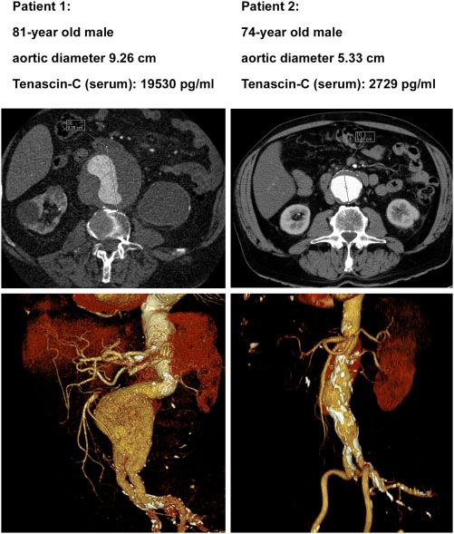
842                                         F. Nagel et al. / Interactive CardioVascular and Thoracic Surgery

RESULTS: In the clinical cohort, there was a trend of correlation between serum TNC levels and AAA diameter (P = 0.055). TNC knock
out mice with AAA showed significantly lower diameter ratios compared to the wild-type group (WT) 3 weeks (P < 0.05) and 10 weeks
(P < 0.05) after AAA induction. Immunohistochemistry revealed increased TNC expression in aortic tissue from WT with AAA as com-
pared sham-operated mice. Furthermore, WT with AAA showed a more disrupted Elastin structure than TNC knock out mice 10 weeks
after AAA induction. In human aortic VSMC, TNC incubation induced expression of remodelling associated proteins.
CONCLUSIONS: TNC might play a causative role in the formation, dilation and progression of AAA. Our results indicate that TNC might
be a biomarker as well as a potential therapeutic target in the treatment of AAA.
Keywords: Abdominal aortic aneurysm • Tenascin C • Serum marker • Extracellular matrix

                                                                               preceding 5 years [8–10]. TNC levels in serum were measured
 ABBREVIATIONS
                                                                               by enzyme-linked immunosorbent assay (ab213831, Abcam,
 AAA    Abdominal aortic aneurysms
                                                                               Cambridge, UK) according to the manufacturer’s recommen-
 Ang II Angiotensin II

                                                                                                                                                     Downloaded from https://academic.oup.com/icvts/article/34/5/841/6524594 by guest on 20 August 2022
 CaCl2  Calcium chloride                                                       dations. Syngo.via—CT vascular software (V. 5.1, Siemens
 KO     Knock out mice                                                         Healthcare GmbH, Erlangen, Germany) was used for diameter
 TLR-4  Toll-like receptor 4                                                   measurements as well as three-dimensional reconstructions.
 TNC    Tenascin C                                                             The maximum aortic diameter in computed tomography angi-
 VSMC Vascular smooth muscle cells                                             ography was measured perpendicularly to the aortic axis after
 WT     Wild-type mice                                                         multiplanar reformation.

                                                                               Murine abdominal aortic aneurysm model and
INTRODUCTION                                                                   organ preparation
Abdominal aortic aneurysms (AAA) are defined as dilations of the               The experimental protocol was approved by the Ethics
abdominal aorta with more than 30 mm diameter in men and                       Committee for Laboratory Animal Experiments at the Medical
are a risk factor for rupture [1]. Therefore, in asymptomatic AAA              University of Vienna and the Austrian Ministry of Science
patients with no additional risk factors, an abdominal aorta diam-             Research and Economy (BMWFW-66.009/0278-II/3b/2012) as
eter of more than 50–55 mm is recommended as indication for                    well as conforms with the Guide for the Care and Use of
surgical or endovascular repair [1]. While management of risk fac-             Laboratory Animals published by the US National Institutes of
tors e.g. arterial hypertension and smoking is standard of care, no            Health (NIH Publication No. 85-23, revised 1996).
causal medical treatment has yet proven to be an advantage in                     In male, 9-week-old A/J wild type (WT) and A/J TNC knock
randomized placebo-controlled trials [1, 2].                                   out mice [A/J-TgH(Tnc), RBRC00007, RIKEN BioResource
   Mechanistically, chronic inflammation is associated with the                Center, Tsukuba, Ibaraki, Japan] AAA were induced by peri-
development of AAA leading to vascular smooth muscle cells                     aortic calcium chloride (CaCl2) application, as described by
(VSMC) proliferation and apoptosis as well as extracellular matrix             Chiou et al. [11–14]. Shortly, after subcutaneous administration
dysregulation [2]. More recently, experimental studies demon-                  of buprenorphine (0.1 mg/kg body weight) and sedation with
strated that the up-regulation of tenascin C (TNC), a matricellular            4% isoflurane, mice were intubated. Under continuous ventila-
glycoprotein, correlates with AAA progression [3, 4]. Moreover,                tion with 2% isoflurane, median laparotomy was performed.
our group showed that an elevated TNC expression in patients                   After preparation of the infrarenal aorta, its diameter was mea-
with acute Type A dissection of the ascending aorta, probably                  sured with the operating microscope, as described by Chiou
acts as a factor of destabilization [5]. Similarly, previous preclinical
                                                                               et al. [14]. Subsequently, a 5  10 mm piece of cotton gauze
and clinical studies provided further evidence that pathophysio-
                                                                               soaked with 0.5 M CaCl2 (Amresco E506-100ML CaCl2 1 M
logic adverse remodelling has been improved in TNC deficient
                                                                               Sterile Solution, VWR International GmbH, Darmstadt,
mice with pressure overload induced left ventricular hypertrophy
                                                                               Germany; diluted 1:1 with 0.9% sodium chloride) was applied
or myocardial infarction [6, 7].
                                                                               on the surface of the aorta for 15 min. The gauze was removed
   The present study aimed (i) to evaluate whether TNC serum
                                                                               and the laparotomy was closed. After recovery of spontaneous
levels correlate with aortic diameters in patients with AAA and (ii)
                                                                               breathing, mice were extubated. Piritramide (30 mg piritra-
to clarify the causative role of TNC on AAA formation in a murine
                                                                               mide diluted in 10 ml Glucose 10% + 250 ml Water) in the
TNC knockout model as well as human aortic VSMC culture.
                                                                               drinking water was used for postoperative analgesia. Sham
                                                                               groups were treated identically, but sodium chloride (0.9%)
MATERIALS AND METHODS                                                          was used instead of CaCl2 application.
                                                                                  Aortic diameter measurement and tissue harvesting were per-
Patient cohort of abdominal aortic aneurysms                                   formed 3 and 10 weeks after AAA induction under deep sedoanal-
                                                                               gesia with intraperitoneal administration of ketamine (100 mg/kg
The study was approved by the institutional ethics committee                   body weight) and xylazine (5 mg/kg body weight). Again, the
at the Medical University of Vienna (1729/2014) and per-                       infrarenal aorta was prepared and the aortic diameter was mea-
formed according to the Declaration of Helsinki. Due to the                    sured with the operating microscope (Supplementary Material,
confounding effects on TNC expression, exclusion criteria                      Fig. S1). The primary endpoint was the ratio between the aortic di-
were: active infections and carcinoma diagnosed during the                     ameter at AAA induction and at organ harvesting [14].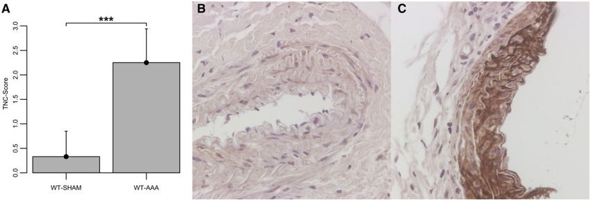
F. Nagel et al. / Interactive CardioVascular and Thoracic Surgery 843
Tenascin C immunohistochemistry considered significant. Levels of significance are shown as follows:
*P < 0.05, **P < 0.01 and ***P < 0.001.
Formalin-fixed, paraffin-embedded samples harvested 3 weeks Data analysis and visualization were performed in R for Mac (R
after AAA induction were sectioned at 2 mm thickness and stained 3.0.2, The R foundation for Statistical Computing, Vienna,
according to instructions provided for the ABC staining kit Austria). The following R packages were used: Hmisc, plotrix and
(Vectastain Elite, Vector laboratories). Importantly, antigen re- splines.
trieval was performed by immersing rehydrated sections in Tris-
HCL buffer, pH 7.4, containing protease (Type XIV from
Streptomyces griseus) at a concentration of 1.9 mg/ml at 37 C for
RESULTS
13 min. Incubation with primary antibody against TNC (ab19011,
Abcam, 1:1000 dilution) was performed overnight at 4 C.
Serum tenascin C levels in patients with abdominal
Sections were imaged using an AxioImager.M2 microscope aortic aneurysms
equipped with an AxioCam 512 colour camera (both Zeiss) at
10 magnification, and staining intensity was semi-quantitatively A cohort of 18 patients with AAA was screened, of which 15
patients were included. Patient characteristics are shown in
Downloaded from https://academic.oup.com/icvts/article/34/5/841/6524594 by guest on 20 August 2022
scored by an observer blinded to experimental conditions.
Table 1. The patients showed an average maximal aortic diameter
of 60.4 ± 11.8 mm and average TNC levels of 8744 ± 5836 pg/ml.
Elastica van Gieson staining and analysis The serum level of TNC showed a trend of positive correlation
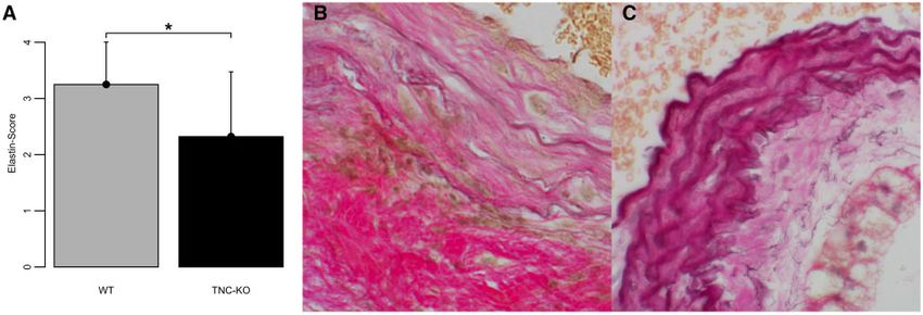
with the maximal aortic dimension (Fig. 1; r = 0.505, P = 0.055).
Formalin-fixed, paraffin-embedded samples harvested 10 weeks Representative computed tomography images and three-dimen-
EXPERIMENTAL
after AAA induction were sectioned at 4 mm thickness and were sional reconstructions with abdominal aorta dimension as well as
stained according to the manufacturer’s (Morphisto GmbH, serum TNC levels of 2 patients are shown in Fig. 2.
Frankfurt am Main, Germany) recommendations. The degree of
elastin degradation was semi-quantitatively scored by 2 observ-
ers blinded to experimental conditions as previously described
Animal characteristics and aorta diameter in mice
[15]. An average was calculated for each section.
In total, 81 animals were used (WT-SHAM: n = 17, WT-AAA:
n = 23, TNC KO-SHAM: n = 18, TNC KO-AAA: n = 23). No deaths
Human aortic vascular smooth muscle cell culture due to aortic rupture or dissection were observed. The animals
did not show any significant differences in body weight prior
Human aortic VSMC were cultured using M199 complete media to AAA induction (WT-SHAM: 25.2 ± 1.8 g, WT-AAA: 24.8 ± 1.8
supplemented with 20% foetal bovine serum and 1% Penicillin g, TNC KO-SHAM: 26.6 ± 3.1 g, TNC KO-AAA: 25.8 ± 2.5 g; n.s.).
and Streptomycin. Cells were treated for 24 h under following In WT and TNC KO mice, AAA groups showed a significant in-
conditions: (i) Control, (ii) 0.1 mM angiotensin II (Ang II) (Merck, crease in aortic diameter ratio compared to sham-operated
Darmstadt, Germany), (iii) 10 mg/ml TNC (Merck, Darmstadt, mice 3 and 10 weeks after AAA induction (3 weeks: WT-SHAM
Germany) and (iv) 10 mg/ml TNC in addition to 50 nM TAK-242 versus WT-AAA: P < 0.001, TNC KO-SHAM versus TNC KO-
(Merck, Darmstadt, Germany). After the treatment, total RNA was AAA: P < 0.05; 10 weeks: WT-SHAM versus WT-AAA: P < 0.001,
isolated using RNeasy Mini kit (Qiagen, Hilden, Germany) and ex- TNC KO-SHAM versus TNC KO-AAA: P < 0.05, Fig. 3). Whereas
pression of target genes (related to AAA formation and linked to no significant changes in diameter ratios were found in sham
TNC [16], Supplementary Material, Table S1) were assessed by re- groups (3 weeks: WT-SHAM: 0.96 ± 0.22, TNC KO-SHAM:
verse transcription and quantitative polymerase chain reaction.
Reverse transcription and quantitative polymerase Table 1: Patient characteristics
chain reaction
Age (years) 67.0 ± 8.0
Gender (male) 15 (100%)
Total RNA was transcribed into cDNA using QuantiTect reverse Body mass index 26.8 ± 2.8
transcription kit (Qiagen, Hilden, Germany). Samples were mea- Serum tenascin C (pg/ml) 8744 ± 5836
sured in duplicates to a final reaction volume of 20 ll per well. Maximum AAA diameter [mm] 60.4 ± 11.8
The initial denaturation step of 5 min at 95 C was followed by 40 Aneurysm morphology
Fusiform 12 (80%)
cycles of 15 s 95 C, 30 s 50 C and 30 s 72 C, using ROTOR-Gene Saccular 3 (20%)
Q (Qiagen, Hilden, Germany) and Rotor-Gene Q series software Positive family history 2 (13.3%)
for computed tomography value analysis. GAPDH was used as Arterial hypertension 13 (86.7%)
housekeeping gene to normalize yielded Ct-values. Relative gene Hyperlipidaemia 11 (73.3%)
Diabetes mellitus 2 (13.3%)
expression was calculated using 2-DDCt method. Peripheral artery disease 3 (20%)
Coronary artery disease 3 (20%)
Stroke 0
Data acquisition and statistical analysis History of smoking 15 (100%)
Packyears 48.4 ± 29.6
All data are shown as mean ± standard deviation. One-way COPD 6 (40%)
ANOVA with Tukey-HSD post hoc analysis as well as unpaired t- Data are presented as n (%) or mean plus standard deviation (n = 15).
tests was used to compare means in different groups. Pearson AAA: abdominal aortic aneurysms; COPD: chronic obstructive pulmonary
correlation coefficient was calculated. P-values of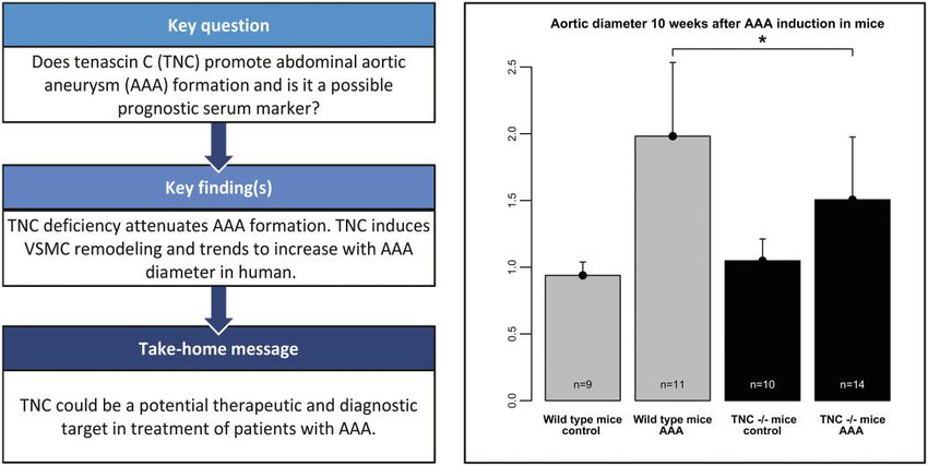
844                                             F. Nagel et al. / Interactive CardioVascular and Thoracic Surgery

                                                                                   and 10 weeks (TNC KO: 1.51 ± 0.47, WT: 1.98 ± 0.55, P < 0.05,
                                                                                   Fig. 3B) after AAA induction, respectively.

                                                                                   Tenascin C immunochemistry
                                                                                   TNC expression was markedly increased 3 weeks after AAA in-
                                                                                   duction in WT mice compared to sham-operated mice (WT-
                                                                                   SHAM: 0.33 ± 0.52, WT-AAA: 2.25 ± 0.69, P < 0.001, Fig. 4A). In
                                                                                   WT mice with AAA TNC was mainly expressed in the tunica me-
                                                                                   dia. In addition, sham-operated WT mice and both sham-oper-
                                                                                   ated as well as AAA TNC KO mice did not show any specific TNC
                                                                                   staining.

                                                                                   Elastin structure

                                                                                                                                                           Downloaded from https://academic.oup.com/icvts/article/34/5/841/6524594 by guest on 20 August 2022
                                                                                   Sham-operated groups showed no signs of elastin degradation.
                                                                                   AAA in WT mice had more degraded elastin fibres as well as focal
                                                                                   infiltrates of leukocytes compared to TNC KO mice with AAA
                                                                                   showing more dilated and less degraded elastin fibres (Fig. 5A–C;
                                                                                   WT-AAA: 3.25 ± 0.75, TNC KO-AAA: 2.32 ± 1.15, P < 0.05).

                                                                                   The effect of tenascin C on human aortic vascular
Figure 1: Correlation between aortic diameter (mm) and serum tenascin C lev-
                                                                                   smooth muscle cells
els (pg/ml) in patient cohort of AAA (n = 15).
                                                                                   To further explain the role of TNC in AAA, human aortic VSMC
                                                                                   were incubated with TNC or Ang II. Both conditions resulted in a
                                                                                   massive up-regulation of matrix metalloproteinase 2 (Fig. 6A;
                                                                                   Control: 0.94 ± 0.23, TNC: 2.02 ± 0.64, TNC+TAK242: 1.05 ± 0.23;
                                                                                   Ang II: 1.69 ± 0.44; Control versus Ang II: P < 0.05, Control versus
                                                                                   TNC: P < 0.01, TNC versus TNC+TAK242: P < 0.001) and COL3
                                                                                   (Fig. 6B; Control: 0.80 ± 0.30, TNC: 1.90 ± 0.64, TNC+TAK242:
                                                                                   0.84 ± 0.21; Ang II: 2.02 ± 0.33; Control versus Ang II: P < 0.001,
                                                                                   Control versus TNC: P < 0.01, TNC versus TNC+TAK242: P <
                                                                                   0.01). More interestingly, administration of TNC as well as Ang II
                                                                                   further increased the expression of TNC (Fig. 6D; Control:
                                                                                   1.02 ± 0.22, TNC: 1.78 ± 0.59, TNC+TAK242: 0.78 ± 0.20; Ang II:
                                                                                   1.89 ± 0.31; Control versus Ang II: P < 0.01, Control versus TNC: P
                                                                                   < 0.05, TNC versus TNC+TAK242: P < 0.001) and angiotensin-
                                                                                   converting enzyme 1 (Fig. 6E; Control: 0.77 ± 0.39, TNC:
                                                                                   1.53 ± 0.51, TNC+TAK242: 0.82 ± 0.11; Ang II: 1.66 ± 0.28; Control
                                                                                   versus Ang II: P < 0.01, Control versus TNC: P < 0.05, TNC versus
                                                                                   TNC+TAK242: P < 0.05) in VSMC. The effect of TNC depended on
                                                                                   Toll-like receptor 4 (TLR-4) activation. Similar to that, the up-reg-
                                                                                   ulation of matrix metalloproteinase 2 and Col 3 by TNC was
                                                                                   markedly declined in presence of the TLR-4 inhibitor TAK242.
                                                                                   The expression of Elastin was significantly down-regulated after
                                                                                   TNC and Ang II incubation. Hereby, the effect of TNC was not re-
                                                                                   versible by TLR-4 inhibition (Fig. 6C; Control: 1.24 ± 0.39, TNC:
                                                                                   0.67 ± 0.31, TNC+TAK242: 0.46 ± 0.09; Ang II: 0.86 ± 0.14; Control
                                                                                   versus Ang II: P < 0.05, Control versus TNC: P < 0.01, TNC versus
                                                                                   TNC+TAK242: n.s.).

Figure 2: Representative three-dimensional reconstructed computed tomogra-
phy images as well as tenascin C serum levels obtained from 2 abdominal aortic
aneurysms patients.
                                                                                   DISCUSSION
                                                                                   Selective and sensitive biomarkers for longitudinal monitoring of
0.92 ± 0.08, n.s.; 10 weeks: WT-SHAM: 0.94 ± 0.10, TNC KO-                         AAA disease activity are missing. Moreover, despite successful
SHAM: 1.05 ± 0.16, n.s.), TNC KO mice with AAA showed a sig-                       surgical and interventional repair, no causative pharmacological
nificantly lower diameter ratio compared to the WT group 3                         treatment for patients with AAA has been proven in randomized
weeks (TNC KO: 1.39 ± 0.25, WT: 1.67 ± 0.22, P < 0.05, Fig. 3A)                    placebo-controlled trials. According to the results of our present
F. Nagel et al. / Interactive CardioVascular and Thoracic Surgery 845
Downloaded from https://academic.oup.com/icvts/article/34/5/841/6524594 by guest on 20 August 2022
Figure 3: Aortic diameter ratio between the aortic size at AAA induction and during organ harvesting at 3 weeks (A) and 10 weeks (B) post-induction. Data are shown
as mean plus standard deviation. *P < 0.05, for the level of significance in the comparison between WT-AAA and tenascin C KO-AAA (one-way ANOVA with Tukey-
HSD post hoc test). AAA: abdominal aortic aneurysms; KO: knock out; WT: wild-type mice.
EXPERIMENTAL
Figure 4: Expression of TNC 3 weeks after AAA induction (A) as well as representative images of TNC immunohistochemistry of the WT-SHAM (B) and the WT-AAA
group (C). Data are shown as mean plus standard deviation. ***P < 0.001, for the level of significance in the comparison between WT-SHAM and WT-AAA (unpaired
T-test). WT-SHAM: n = 6, WT-AAA: n = 6. Original magnification, 200. AAA: abdominal aortic aneurysms; TNC: tenascin C; WT: wild-type mice.
Figure 5: Scoring of elastin structure in Elastica van Gieson staining 10 weeks after AAA induction (A). Representative images of the WT-AAA group with severe elastin
degradation and inflammatory cell infiltrates (B) and the TNC KO-AAA group with mild distention (C). Data are shown as mean plus standard deviation. *P < 0.05, for
the level of significance in the comparison between WT-AAA and TNC KO-AAA (unpaired T-test). WT-AAA: n = 10, TNC KO-AAA: n = 14. Original magnification, 200.
AAA: abdominal aortic aneurysms; KO: knock out; TNC: tenascin C; WT: wild-type mice.
study, we observed a consistent trend towards correlation be- experimental model of CaCl2-induced AAA, TNC deficient mice
tween abdominal aortic diameter and TNC serum levels in showed a significantly reduced AAA formation and progression
patients with AAA. In line with this clinical finding in an as well as markedly reduced elastin disruption compared to WT846 F. Nagel et al. / Interactive CardioVascular and Thoracic Surgery
Downloaded from https://academic.oup.com/icvts/article/34/5/841/6524594 by guest on 20 August 2022
Figure 6: Expression matrix metalloproteinase 2 (A), Col3 (B), Elastin (C), TNC (D) as well as angiotensin-converting enzyme 1 (E) in human aortic vascular smooth
muscle cells culture supernatant measured by reverse transcription and quantitative polymerase chain reaction after incubation with TNC. TNC+ TAK242 and Ang II.
*P < 0.05, **P < 0.01 and ***P < 0.001, for the level of significance in the comparison between Control and TNC, TNC and TNC+TAK242 (1-way ANOVA with Tukey-
HSD post hoc test) as well as Control and Ang II (unpaired T-test). TNC: tenascin C.
mice. Mechanistically, TNC like Ang II induces the expression of area and showed a massive up-regulation [3]. Previous investiga-
ECM remodelling associated proteins as well as positive feedback tions have already indicated that TNC was mostly expressed
loops in human aortic VSMC. This effect was partially reversed by within mononuclear inflammatory cell infiltrates in human AAA
TLR-4 inhibition. associated with chronic inflammation and linked to neovasculari-
TNC is a matricellular protein, which is expressed during em- zation [23].
bryogenesis, cancer and various remodelling processes. Its ex- In our study, TNC deficient mice showed an attenuated AAA
pression is modulated by cytokines, other matricellular proteins formation 3 and 10 weeks after periaortic CaCl2 application
as well as mechanical stress [8]. VSMCs are the main source of linked to a reduced elastin degradation. Our findings demon-
TNC in the aorta. TNC expression is associated with the forma- strate contradictory results with the data of Kimura et al. [24].
tion of AAA [3]. Furthermore, TNC was mainly expressed in the Kimura et al. reported an increased rate of suprarenal dissections
border zones of the AAA, mostly affected by inflammation and and ruptures in TNC KO mice in a model of CaCl2 induced AAA
thereby may regulate the progression of AAA [3, 4, 17]. combined with Ang II infusions. The authors conclude that TNC
In the present study, we measured the levels of serum TNC in 15 acted protective on increased haemodynamic stress on the
patients with AAA. We could observe a trend towards a correlation descending thoracic aorta due to CaCl2 induced abdominal aor-
between TNC levels and AAA diameter. Importantly, there are no tic stiffening and Ang II infusions. Moreover, no significant differ-
evidence-based studies assessing serum TNC as prognostic marker ences between AAA diameter in TNC-KO and WT mice were
for AAA. However, in patients with Type A dissection, we and others found 6 weeks after CaCl2 induced AAA induction. Of impor-
have demonstrated that TNC levels in plasma as well as in aortic tis- tance, aortic size measurements were conducted ex vivo by his-
sue were elevated [5, 18, 19]. Furthermore, higher serum TNC levels tology and not compared to the baseline diameter in each
were associated with increased in-hospital mortality after acute Type animal [3]. It is important to stress that in the present study, we
A and Type B dissection, suggesting the maladaptive role of TNC followed the animals for 3 and 10 weeks after CaCl2 induced AAA
[20, 21]. These findings contradict another study, where elevated se- [24]. There was an increase in diameter over time and a signifi-
rum TNC levels on Day 7 after acute Type B dissection were associ- cant difference between the aortic diameter in TNC KO animals
ated with a lower risk for chronic aortic enlargement [22]. Further as compared to WT after AAA induction.
studies on larger populations need to elucidate the role of TNC as a In human aortic VSMC similarly to Ang II, incubation with TNC
marker for AAA progression as well as its time-dependent role in led to up-regulation of matrix metalloproteinase 2 and Col3 as well
AAA and other aortic diseases. as to a down-regulation of Elastin. In combination with the in-
Next, we wanted to test the hypothesis that TNC is involved in creased Elastin degradation observed in WT mice with AAA, TNC
the progression of AAA. We used a model of CaCl2-induced AAA could promote adverse aortic remodelling [25]. As a sign of positive
in WT and TNC-KO mice. In line with previous studies, TNC ex- feedback loop, Ang II as well as TNC induced TNC and angiotensin-
pression in AAA in WT mice was markedly localized in the border converting enzyme 1 expression. Comparable effects of TNC couldF. Nagel et al. / Interactive CardioVascular and Thoracic Surgery 847
be observed in cardiac fibroblasts by our group [16]. These changes Conflict of interest: none declared.
were partially reversed by the application of the TLR-4 antagonist
TAK242. TNC is a known ligand of integrins as well as TLR-4 and
subsequently interferes with matricellular proteins such as fibronec- Author contributions
tin facilitating cell migration [4, 26]. Additionally, it induces the NFjB
signalling pathway via TLR-4 activation in various cell types including Felix Nagel: Conceptualization; Data curation; Investigation; Methodology;
Project administration; Visualization; Writing—original draft. Anne-Kristin
macrophages and fibroblasts, and consequently accelerates proin- Schaefer: Data curation; Investigation; Project administration. Inês Fonseca
flammatory cytokine expression which also play a central role in Gonçalves: Data curation; Investigation. Eylem Acar: Data curation;
AAA pathophysiology [2, 27, 28]. Based on these experimental results Investigation. Andre Oszwald: Data curation; Investigation. Philipp Kaiser:
and the trend in our clinical cohort, we assume that TNC might play Data curation; Investigation. Renate Kain: Data curation; Investigation. Karola
Trescher: Conceptualization. Wolf Hans Eilenberg: Data curation. Christine
a maladaptive rather than beneficial role in the formation and pro-
Brostjan: Data curation; Writing—review & editing. David Santer:
gression of AAA. Conceptualization; Investigation; Methodology; Project administration;
Writing—review & editing. Attila Kiss: Conceptualization; Investigation;
Methodology; Project administration; Writing—review & editing. Bruno Karl
Limitations Podesser: Conceptualization; Funding acquisition; Investigation; Methodology;
Downloaded from https://academic.oup.com/icvts/article/34/5/841/6524594 by guest on 20 August 2022
Writing—review & editing.
Certain limitations need to be acknowledged. First, our patient
cohort was small including only AAA patients, men only and
therefore underpowered to detect a significant correlation be- Reviewer information
tween AAA diameter and TNC serum levels. This takes into ac-
Interactive CardioVascular and Thoracic Surgery thanks George J. Arnaoutakis,
EXPERIMENTAL
count that men show an up to 3- to 6-fold increased prevalence
of AAA [1, 29]. Moreover, only male animals were included. Luca Bertoglio, Mario Giovanni Gerardo D’Oria and the other anonymous
reviewer(s) for their contribution to the peer review process of this article.
Second, CaCl2 induced AAA show aortic wall thickening and do
not develop intraluminal thromboses compared to other models
like the elastase model. However, the CaCl2 model does not re- REFERENCES
quire aortic clamping as well as an aortotomy and is therefore
less invasive as well as less susceptible for induction flaws. [1] Wanhainen A, Verzini F, Van Herzeele I, Allaire E, Bown M, Cohnert T et
Additionally, using the CaCl2 model, mice do not develop rup- al. Editor’s choice—European Society for Vascular Surgery (ESVS) 2019
tures or aortic dissections. No further in vivo aortic size measure- clinical practice guidelines on the management of abdominal aorto-iliac
artery aneurysms. Eur J Vasc Endovasc Surg 2019;57:8–93.
ments (e.g. ultrasound) have been performed in addition to the
[2] Golledge J. Abdominal aortic aneurysm: update on pathogenesis and
intraoperative size evaluation [14, 30]. Third, we were not able to medical treatments. Nat Rev Cardiol 2019;16:225–42.
perform reverse transcription and quantitative polymerase chain [3] Kimura T, Yoshimura K, Aoki H, Imanaka-Yoshida K, Yoshida T, Ikeda Y
reactions or other methods including MMP zymography on mu- et al. Tenascin-C is expressed in abdominal aortic aneurysm tissue with
rine aortic samples because of the limited tissue. This limitation an active degradation process. Pathol Int 2011;61:559–64.
[4] Imanaka-Yoshida K, Matsumoto K. Multiple roles of tenascins in homeo-
will be addressed in future studies. stasis and pathophysiology of aorta. Ann Vasc Dis 2018;11:169–80.
[5] Trescher K, Thometich B, Demyanets S, Kassal H, Sedivy R, Bittner R
et al. Type A dissection and chronic dilatation: tenascin-C as a key factor
CONCLUSION in destabilization of the aortic wall. Interact CardioVasc Thorac Surg
2013;17:365–70.
[6] Podesser BK, Kreibich M, Dzilic E, Santer D, Förster L, Trojanek S et al.
Higher serum TNC in patients with AAA might be associated with
Tenascin-C promotes chronic pressure overload-induced cardiac dys-
increased disease activity and aortic wall instability. Due to the function, hypertrophy and myocardial fibrosis. Journal of Hypertension
preclinical observation with a trend of correlation between TNC 2018;36:847–56. https://doi.org/10.1097/HJH.0000000000001628.
expression and progression of AAA diameter, we suggest to pre- [7] Santer D, Nagel F, Gonçalves IF, Kaun C, Wojta J, Fagyas M et al.
pare a larger clinical study to clarify the role of TNC as prognostic Tenascin-C aggravates ventricular dilatation and angiotensin-converting
enzyme activity after myocardial infarction in mice. ESC Heart Failure
marker in AAA. 2020;7:2113–22. https://doi.org/10.1002/ehf2.12794.
[8] Midwood KS, Hussenet T, Langlois B, Orend G. Advances in tenascin-C
biology. Cell Mol Life Sci 2011;68:3175–99.
SUPPLEMENTARY MATERIAL [9] Yuan W, Zhang W, Yang X, Zhou L, Hanghua Z, Xu K. Clinical signifi-
cance and prognosis of serum tenascin-C in patients with sepsis. BMC
Anesthesiol 2018;18:170.
Supplementary material is available at ICVTS online. [10] Piccinini AM, Midwood KS. Endogenous control of immunity against
infection: tenascin-C regulates TLR4-mediated inflammation via
microRNA-155. Cell Rep 2012;2:914–26.
ACKNOWLEDGEMENTS [11] Koyama Y, Kusubata M, Yoshiki A, Hiraiwa N, Ohashi T, Irie S et al. Effect
of tenascin-C deficiency on chemically induced dermatitis in the mouse.
J Invest Dermatol 1998;111:930–5.
The authors thank the staff of the Center for Biomedical Research,
[12] Nakao N, Hiraiwa N, Yoshiki A, Ike F, Kusakabe M. Tenascin-C promotes
Medical University of Vienna for animal care and Milat Inci as a healing of Habu-snake venom-induced glomerulonephritis: studies in
valuable laboratory technician. knockout congenic mice and in culture. Am J Pathol 1998;152:1237–45.
[13] Matsuda A, Yoshiki A, Tagawa Y, Matsuda H, Kusakabe M. Corneal
wound healing in tenascin knockout mouse. Invest Ophthalmol Vis Sci
Funding 1999;40:1071–80.
[14] Chiou AC, Chiu B, Pearce WH. Murine aortic aneurysm produced by
periarterial application of calcium chloride. J Surg Res 2001;99:371–6.
This work was supported by the Ludwig Boltzmann Institute for [15] Krishna SM, Seto SW, Jose RJ, Biros E, Moran CS, Wang Y et al. A peptide
Cardiovascular Research, REM 2017/20, Vienna, Austria. antagonist of thrombospondin-1 promotes abdominal aortic aneurysm848 F. Nagel et al. / Interactive CardioVascular and Thoracic Surgery
progression in the angiotensin II-infused apolipoprotein-E-deficient [23] Satta J, Soini Y, Pöllänen R, Pääkkö P, Juvonen T. Tenascin expression is
mouse. Arterioscler Thromb Vasc Biol 2015;35:389–98. associated with a chronic inflammatory process in abdominal aortic
[16] Perera-Gonzalez M, Kiss A, Kaiser P, Holzweber M, Nagel F, Watzinger S aneurysms. J Vasc Surg 1997;26:670–5.
et al. The role of tenascin C in cardiac reverse remodeling following [24] Kimura T, Shiraishi K, Furusho A, Ito S, Hirakata S, Nishida N et al.
banding-debanding of the ascending aorta. Int J Mol Sci 2021;22:2023. Tenascin C protects aorta from acute dissection in mice. Sci Rep 2014;4:
[17] Didangelos A, Yin X, Mandal K, Saje A, Smith A, Xu Q et al. Extracellular ma- 4051.
trix composition and remodeling in human abdominal aortic aneurysms: a [25] Rabkin SW. The role matrix metalloproteinases in the production of aor-
proteomics approach. Mol Cell Proteomics 2011;10:M111.008128. tic aneurysm. Prog Mol Biol Transl Sci 2017;147:239–65.
[18] Matsumoto K, Satoh K, Maniwa T, Tanaka T, Okunishi H, Oda T. [26] Imanaka-Yoshida K, Hiroe M, Yoshida T. Interaction between cell and
Proteomic comparison between abdominal and thoracic aortic aneur- extracellular matrix in heart disease: multiple roles of tenascin-C in tissue
ysms. Int J Mol Med 2014;33:1035–47. remodeling. Histol Histopathol 2004;19:517–25.
[19] Majumdar R, Miller DV, Ballman KV, Unnikrishnan G, McKellar SH, Sarkar [27] Midwood K, Sacre S, Piccinini AM, Inglis J, Trebaul A, Chan E et al.
G et al. Elevated expressions of osteopontin and tenascin C in ascending Tenascin-C is an endogenous activator of Toll-like receptor 4 that is es-
aortic aneurysms are associated with trileaflet aortic valves as compared sential for maintaining inflammation in arthritic joint disease. Nat Med
with bicuspid aortic valves. Cardiovasc Pathol 2007;16:144–50. 2009;15:774–80.
[20] Nozato T, Sato A, Hirose S, Hikita H, Takahashi A, Endo H et al. [28] Midwood KS, Chiquet M, Tucker RP, Orend G. Tenascin-C at a glance.
Preliminary study of serum tenascin-C levels as a diagnostic or prognostic J Cell Sci 2016;129:4321–7.
biomarker of type B acute aortic dissection. Int J Cardiol 2013;168:4267–9. [29] Ulug P, Powell JT, Sweeting MJ, Bown MJ, Thompson SG; SWAN
Downloaded from https://academic.oup.com/icvts/article/34/5/841/6524594 by guest on 20 August 2022
[21] Guo T, Zhou X, Zhu A, Peng W, Zhong Y, Chai X. The role of serum Collaborative Group. Meta-analysis of the current prevalence of screen-
tenascin-C in predicting in-hospital death in acute aortic dissection. Int detected abdominal aortic aneurysm in women. Br J Surg 2016;103:
Heart J 2019;60:919–23. 1097–104.
[22] Nozato T, Sato A, Hikita H, Takahashi A, Imanaka-Yoshida K, Yoshida T et [30] Busch A, Bleichert S, Ibrahim N, Wortmann M, Eckstein H-H, Brostjan C
al. Impact of serum tenascin-C on the aortic healing process during the et al. Translating mouse models of abdominal aortic aneurysm to the
chronic stage of type B acute aortic dissection. Int J Cardiol 2015;191:97–9. translational needs of vascular surgery. JVS Vasc Sci 2021;2:219–34.You can also read