Automated Non-invasive Skin Cancer Detection using Dermoscopic Images
←
→
Page content transcription
If your browser does not render page correctly, please read the page content below
ITM Web of Conferences 40, 03044 (2021)                                                                  https://doi.org/10.1051/itmconf/20214003044
ICACC-2021

         Automated Non-invasive Skin Cancer Detection using
         Dermoscopic Images
         Shruti Kale1, Reema Kharat2, Sagarika Kalyankar3, Sangita Chaudhari4, Apurva Shinde5

         1Dept. of Computer Engineering, Ramrao Adik Institute of Technology, Nerul, Navi Mumbai
         2Dept. of Computer Engineering, Ramrao Adik Institute of Technology, Nerul, Navi Mumbai
         3Dept. of Computer Engineering, Ramrao Adik Institute of Technology, Nerul, Navi Mumbai
         4Dept. of Computer Engineering, Ramrao Adik Institute of Technology, Nerul, Navi Mumbai
         5Dept. of Computer Engineering, Ramrao Adik Institute of Technology, Nerul, Navi Mumbai

                       Abstract. Skin Cancer is resulting from the growth of the harmful tumour of the melanocytes the rates are
                       rising to another level. The medical business is advancing with the innovation of recent technologies; newer
                       tending technology and treatment procedures are being developed. The early detection of skin cancer can
                       help the chance of increase in its growth in other parts of body. In recent years, medical practitioners tend to
                       use non invasive Computer aided system to detect the skin cancers in early phase of its spreading instead of
                       relying on traditional skin biopsy methods. Convolution neural network model is proposed and used for
                       early detection of the cancer, and it type. The proposed model could classify the dermoscopic images into
                       correct type with accuracy 91.2%.

                       Keywords- tumour, melanocytes, medical practitioners, non-invasive, biopsy, dermoscopic.

       1 Introduction                                                         Cancer (MSC). BCC is because of skin openness
                                                                              particularly found in the sun uncovered regions like the
       Skin Cancer rates are rising to another level. In 2018,                face, head, neck, arms, and legs. SCC is the disease of
       287,723 new incidents were reported according to                       keratinocyte cells found on the external surface of the
       intervals the planet and so the vary of deaths happened                skin. It seems like a red firm knocks layered patches.
       because of skin cancer is 60,712. at intervals the U.S.A.,             Melanoma emerges from melanocytes, specific
       almost 96,480 new malignant skin cancers area unit                     pigmented cells that are discovered. Risk can be reduced
       aiming to be identified. The harsh UV radiation from the               by preventive or avoiding revelation to ultraviolet (UV)
       Sunlight affects melanoma. Malignant melanomas                         radiation. Inspecting skin for doubtful changes can help
       invariably tend to unfold into surroundings but if treated             to detect skin cancer at its earliest along with some
       at its early-stage dangers can be avoided. Therefore,                  computer aided diagnosis system. Early detection of skin
       early identification and treatment of skin cancer is                   cancer is an important step toward its fast growth in
       particularly important and crucial task. Generally, in                 other parts of the body. Early detection of cancers and its
       automated system it can be done in two parts: (1) Pre-                 type can be detected by dermatologists with the help of
       processing of input and effective feature extraction and               computer aided system. Thus, the objective of this paper
       (2) Accurate classification of the image.                              is to construct a Computer Aided Diagnosis system that
                                                                              assists medical professionals detect skin cancer without
       Skin cancer is the irregular growth of skin cells and its              the need of any expensive tools.
       most frequently grows on skin exposed to the sun. But
       this form of cancer can also happen on areas of your skin              2 Related Works
       not normally unprotected to sunlight. Skin cancer is
       broadly categorised into Basal Cell Carcinoma (BCC),                   About 1 million cases of non-cancerous and 288,000 of
       Squamous Cell Carcinoma (SCC) and Melanoma Skin                        cancerous cases have been detected in 2018. The impact

         *
             Corresponding author: reema97.kharat@gmail.com

  © The Authors, published by EDP Sciences. This is an open access article distributed under the terms of the Creative Commons Attribution License 4.0
  (http://creativecommons.org/licenses/by/4.0/).
ITM Web of Conferences 40, 03044 (2021) https://doi.org/10.1051/itmconf/20214003044
ICACC-2021
of biological theory on continuation and value more With al, laboratory studies reportable a clinical
stress the previously exhaust aid structure and lift the sensitivity, a difference which could be credited to the
query of economic property. Carcinoma and particularly standard of the dataset input, thus version knowledge as
metric linear unit early spotting is difficult for each new. Lately, a primary potential scientific empiric
dermatologists. Dermatoscope is reserved into version education reportable on a 2-step method, count a second
the excellence of care, however in objective test layer of sonification (visual information was noises) to a
dermatologist come through a restricted diagnostic decilitre classifier to enhance accuracy of detection. This
sensitivity of 400 metric linear unit detection Thanks to twin decilitre utilised a sophisticated dermoscope, a
the quality of graphic ideas rooted in an exceedingly comparatively dear device, and a way extremely keen
dermoscopic image. General doctors appear to profit about medico expertise translation it fewer appropriate
from use of a dermoscopic course, whereas several 51 for extensive medical care doctor’s use. Accordingly,
accurately diagnosed lesions necessitate more the influence of copy superiority on correctness of
enhancements. Likewise, specificity of diagnosing by diagnosing has been suspected. It had been determined
dermatologists necessitate an extra improvement, as to check a cheap device secret by its builder as a skin
mirrored by a range of 28:1 to 9:1 variety of operations scientific instrument with polarized lightweight (SMP).
that require to be removed to spot one skin cancer and a Picture’s quality paternal by SMP preclude in most cases
3:1 magnitude relation for general carcinoma. a particular clinical diagnosing thanks to haziness and
lack of fine high level dermoscopic patterns and
Deep learning (DL) classifiers area unit an auspicious diagnostic structures. Table 2.1 shows summary of
applicant for discovery of different types of skin cancers. existing approaches.
Table 2.1: Survey of Existing Systems
Authors Image Image Pre- Image Feature extracted Classification
Acquisition processing Segmentation
Ichim and Elimination of Texture features, shape and Neural network, Back
Popescu noise, Dull Razor - colour features propogation,
[3] ISIC and PH2 hair removal, AlexNet and ResNet
contrast and
normalisation
Demyanov K Means clustering and sparse SVM with RBF
et al. [4] ISIC - - coding with visual features Kernel and CNN
from Dense SIFT and SURF
Majumder PH2 Dataset Dull-Razor algo- Otsu’s Asymmetry score across x and ANN with
and to remove dark thresholding y-axis, Area/perimeter ratio, backpropagation
Ullah[5] hair from images. method Compactness index, Average algorithm
Median filter is diameter of lesion
also applied
Jain et PH2 Dataset Image resizing Otsu’s Asymmetry score across x and Backpropagation
al.[6] and contrast thresholding y-axis, ABCD features, Neural Network
adjustment. method is variegation and lesion diameter (BNN)
Applied applied variation
averaging filter to
RGB input
image.
Eltayef et 200 8-bit RGB Directional Gabor Fuzzy c-means Asymmetry, border, Color Not specified
al.[7] color images with filters are and MRF is variegation and lesion diameter
a resolution of implemented to used
768*560 extract the hairs
Isasi et 40 RGB OF Pattern Globular, ABCD feature alongwith Classification is done
al.[8] 500x500 pixels recognition - Recticular and features extracted from pattern by ABCD rule
image catalogue globular blue-vein recognition algo.
by certified
dermatologistes
She et Not specified High pass Snake-based Asymmetry score across x and Principal component
al.[9] filtering and detection y-axis, Area/perimeter ratio, analysis is used for
gradients technique is Compactness index, Average classification
application used to diameter of lesion.
determine lesion
boundry
2ITM Web of Conferences 40, 03044 (2021) https://doi.org/10.1051/itmconf/20214003044
ICACC-2021
3. Proposed System neural networks, the hidden layers of a CNN have a
particular design. In regular neural networks, every layer
Various attempts made by researchers to detect skin is made by a collection of vegetative cells and one
cancer in its early stage using image processing vegetative cell of a layer is connected to every neuron of
techniques, machine learning and deep learning the previous layer. The design of hidden layers in a very
techniques. As the problem of existing system was that CNN is slightly completely different.
the output was not accurate. Even if it’s cancerous cell it This restriction to native connections and extra merge
was detected as non-cancerous or vice-vera. System layers sums up native vegetative cell product into one
Flow Diagram predicting malignant lesions is depicted worth leads to Translation in variant options. This leads
in figure 3.1. It consists of four steps: (1) image pre- to an easier coaching procedure and a lower model
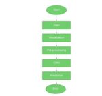
processing; (2) image segmentation; (3) feature quality. Figure 3.3 shows the steps followed in the
extraction; (4) classification. The input image can have proposed system. Data: Data is extracted for analysis
noise and other artefacts such as hairs, air bubbles, towards skin cancer. In our project we have used over
varied colour, and textures of the skin. To get rid of 8000 images. To test our system.
these factors, pre-processing need to be applied on input Data Visualization: It is a graphical presentation of
image before passing it to segmentation or feature knowledge of data. By applying perceptible segments
extraction phase so that eventually classifier can classify like charts, graphs, maps to provide an approachable way
these input lesions in correct classifications as malignant to see and acknowledge direction, outliers, and patterns
or benign and the type of cancer. in data.
Image Pre-Processing: Is a procedure of making the rare
information and creation it appropriate for a machine
learning model. A filter is used to enhance the objects
such as hair, marks in bright background.
CNN: Over here the system predicts if the image is
cancerous or not, and if its cancerous then it predicts
which type of cancer it is.
Figure 3.1: Skin Cancer Detection Process
In our proposed system, we are utilising modified
Convolutional Neural Network (CNN). CNN consist of
sequence of several convolutional layer with filters,
pooling layers, fully connected layer. Convolutional
layer is the initial layer which extract feature from an
image. Pooling layers minimize the size of the activation
map. Fully connected layer operates on a flattened input
where each input is connected to all neurons. Figure 3.2
shows architecture of CNN.
Figure 3.2: CNN Architecture
CNNs are convolution neural networks that are appeared Figure 3.3: Flowchart for Proposed System
to be terribly strong in areas like image recognition and
classification. CNNs area unit a supervised learning Performance Metrics for classifiers: A classifier set
technique and area unit thus instruct victimisation apart every object to a category. This piece of work is
knowledge tagged with the various categories. primarily, mostly not excellent, and entity is also assigned to the
CNNs grasp the link in the middle of the input items and incorrect category to gauge the classification quality, the
the category tags and contains 2 elements: the invisible category—the category allocated by the classifier is
layers during which the options area unit drawn out and, likened with the class. this permits the substances to be
at the top of the process, the totally connected layers that alienated into the subsequent four subsections:
area unit used for the classification task. not like regular 1. True positive (TP): Positive Class.
*
Corresponding author: reema97.kharat@gmail.com
3ITM Web of Conferences 40, 03044 (2021) https://doi.org/10.1051/itmconf/20214003044
ICACC-2021
2. True negative (TN): Negative Class.
3. False positive (FP): Incorrect Positive Class.
4. False Negative (FN): Incorrect Negative Class.
4. Results and Discussion
The diagnostic system proposed in this work has been
implemented in Python. To testing the system, a dataset
of approximately 10000 dermoscopic images from PH2 Figure 4.3: Localization of lesions
dataset are considered including mixed lesion images for
Benign keratosis, melanocytic nevi, dermatofibroma,
melanoma, vascular lesions, basal cell carcinoma, actinic
keratoses [10].
The system could correctly classify the images as
’Benign’ or ’Malignant’ as well as the type of skin
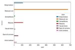
cancer as BCC, SCC or MSC. Figure 4.1 shows the
sample statistics for different dermoscopic images.
Figure 4.2 shows sample images for various types of
skin cancer. Figure 4.3 and 4.4 shows localization and Figure 4.4: Characteristics of lesions
characteristics of lesion for some sample input images.
Figure 4.5 show the user interface of the system and the
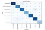
predicted output as the type of skin cancer. There is 91.2
accuracy achieved at epoch as 19 for the CNN model.
Figure 4.6 shows the confusion matrix for 200 sample
which are used in testing the model.
Figure 4.1 : Satistics of dermoscipic images used in
experimentation
Figure 4.5: The predicted skin cancer type
Figure 4.2 : Skin cancer dermoscopic images
4ITM Web of Conferences 40, 03044 (2021) https://doi.org/10.1051/itmconf/20214003044
ICACC-2021
[2]. World Health Organization on Climate Change and
Human Health.
https://www.who.int/globalchange/climate/summar
y/en/index7.html
[3]. Ichim, Loretta, and Dan Popescu. "Melanoma
Detection Using an Objective System Based on
Multiple Connected Neural Networks." IEEE
Access 8 (2020): 179189-179202.
[4]. Demyanov, Sergey, et al. "Classification of
dermoscopy patterns using deep convolutional
neural networks." 2016 IEEE 13th International
Symposium on Biomedical Imaging (ISBI). IEEE,
2016.
[5]. Majumder, Sharmin, and Muhammad Ahsan Ullah.
"Feature extraction from dermoscopy images for an
Figure 4.5: Accuracy assessment of the predicted effective diagnosis of melanoma skin cancer." 2018
result 10th International Conference on Electrical and
Computer Engineering (ICECE). IEEE, 2018.
5. Conclusion
[6]. Eltayef, Khalid, Yongmin Li, and Xiaohui Liu.
In recent years, tremendous growth has been observed in "Detection of melanoma skin cancer in
different type of skin cancer in Asian as well as other dermoscopy images." Journal of Physics:
countries. Non-invasive approaches are gaining Conference Series. Vol. 787. No. 1. IOP
popularity for early detection of skin cancers. A well- Publishing, 2017.
trained computer aided diagnosis system can correctly [7]. Jain, Shivangi, and Nitin Pise. "Computer aided
detect presence of skin cancer and the type of cancer. In melanoma skin cancer detection using image
the proposed system some pre-processing is applied on processing." Procedia Computer Science 48
input images to enhance the quality of images and to (2015): 735-740.
remove hairs from those images. CNN model is utilised
which could be able to correctly detect the skin cancer [8]. Isasi, A. Gola, B. García Zapirain, and A. Méndez
and classify it as one of the three types of cancer with an Zorrilla. "Melanomas non-invasive diagnosis
accuracy of 91.2%. However, overall accuracy can be application based on the ABCD rule and pattern
affected in case of dermoscopic image of dark-skinned recognition image processing algorithms."
patients. Also, the large size dermoscopic image dataset Computers in Biology and Medicine 41.9 (2011):
would be used further to improve the classification 742-755.
results. [9]. She, Zhishun, Y. Liu, and A. Damatoa.
"Combination of features from skin pattern and
References ABCD analysis for lesion classification." Skin
Research and Technology 13.1 (2007): 25-33.
[1]. World Cancer Research Fund on Skin Cancer
[10]. PH2 Database. https://www.fc.up.pt/addi/ph2\
Statistics.
%20database.html
https://www.wcrf.org/dietandcancer/cancer-
trends/skin-cancer-statistics.
5You can also read


