Age related differences for expression of the nerve specific proteins after peripheral nerve injury
←
→
Page content transcription
If your browser does not render page correctly, please read the page content below
EXPERIMENTAL AND THERAPEUTIC MEDICINE 24: 682, 2022
Age‑related differences for expression of the
nerve‑specific proteins after peripheral nerve injury
HIROYUKI OBATA1,2, KIYOHITO NAITO1,2, AYAKA KIKUI1,2, SHINJI NAKAMURA3,
KAORI SUZUKI4, SO KAWAKITA1,2, TAKAMARU SUZUKI1,2, KENJI GOTO2, NANA NAGURA2,
YOICHI SUGIYAMA2, ISAO NAGAOKA4,5 and MUNEAKI ISHIJIMA1,2
1
Department of Medicine for Orthopaedics and Motor Organ, Juntendo University Graduate School of Medicine;
2
Department of Orthopaedics, Juntendo University Faculty of Medicine, Tokyo 113‑8421;
3
Laboratory of Morphology and Image Analysis, Research Support Center; 4Department of Biochemistry and
Systems Biomedicine, Juntendo University Graduate School of Medicine, Tokyo 113‑8421;
5
Faculty of Medical Science, Juntendo University, Urayasu, Chiba 279‑0013, Japan
Received June 29, 2022; Accepted September 6, 2022
DOI: 10.3892/etm.2022.11618
Abstract. The effects of aging on axon regeneration currently nerve‑specific proteins in aged mice. Based on the results of
remain unclear. In addition, the up‑regulated expression of the present study, compensatory changes induced by periph‑
neurotrophic factors that occurs within one week of periph‑ eral nerve injury were initiated by the up‑regulated expression
eral nerve injury has been shown to play an important role of REST/NRSF in young mice, but not in aged mice.
in the axon regeneration. To investigate the effects of aging
on axon regeneration, the expression of nerve‑specific proteins Introduction
immediately after peripheral nerve injury were compared
between young and aged mice. A mouse peripheral nerve In the central nervous system, repressor element‑1
injury model was prepared using the sciatic nerve compression silencing transcription/neuron‑restrictive silencer factor
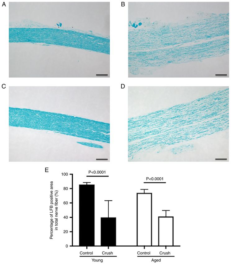
method. In each group, Luxol fast blue staining and immuno‑ (REST/NRSF) maintains homeostasis by suppressing the
fluorescence staining were performed to assess the degree of apoptosis of neurons (1‑3). Its expression increases with
Wallerian degeneration in the sciatic nerve, and to evaluate age and protects nerves against aging stress (3). We previ‑
the expression of repressor element 1‑silencing transcription ously reported the up‑regulated expression of REST/NRSF
factor (REST)/neuron‑restrictive silencer factor (NRSF), in neurons in the peripheral and central nervous systems
brain‑derived neurotrophic factor (BDNF), neurotrophin with aging (4). However, the effects of REST/NRSF on axon
3 (NT3), nerve growth factor (NGF), and semaphorin 3A regeneration following peripheral nerve injury currently
(Sema3A) in the dorsal root ganglion, respectively. Wallerian remain unclear.
degeneration was observed in both young and aged mice after The ability to regenerate axons damaged by peripheral
peripheral nerve injury. Significant increases were observed in nerve injury decreases with age (5,6). Our previous find‑
the expression of REST/NRSF (P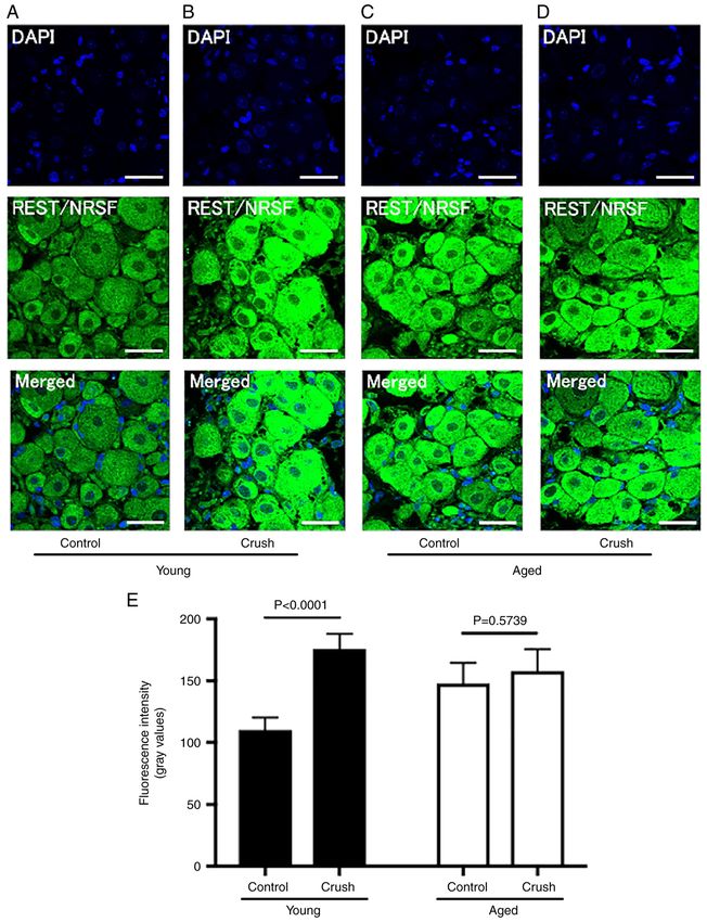
2 OBATA et al: AGING AND NERVE‑SPECIFIC PROTEIN EXPRESSION
which currently remain unclear, were discussed based on the three areas in the Control group without peripheral nerve
expression of nerve‑specific proteins. injury. The area distal to the crushed site was used in the
present study because this was the site at which Wallerian
Materials and methods degeneration occurred. Using a light microscope (Carl Zeiss,
KS400), the percentage of the area stained by LFB to the total
Animal Model. The present study was approved by the Animal nerve fiber area was calculated in both groups (16,17).
Care Committee of Juntendo University, Tokyo, Japan (regis‑
tration no. 1555; approval no. 2021312). Histochemical assessment of the expression of nerve‑specific
Forty male C57BL/6 mice (Young group: 10‑week‑old proteins after peripheral nerve injury. Immunofluorescence
mice, n=20; Aged group: 70‑week‑old mice, n=20) purchased staining was performed to assess the expression of nerve‑specific
from Japan SLC, Inc. (Shizuoka, Japan) were used. Mice were proteins after peripheral nerve injury, as described previous
housed at 5 animals/cage in a sterile environment controlled by Goto et al (4). Tissue sections were prepared by cutting
at a temperature of 22±2˚C, humidity of 40‑60%, and 12‑h the DRG at a thickness of 3 µm. Samples were deparaffinized
light and dark cycle, and were given water that was CRF‑1 and autoclaved at 121˚C for 10 min for antigen retrieval. After
gamma‑ray irradiated at 15 kGy (Oriental Yeast Co., Ltd.) a treatment with True View™ (SP‑8400, Vector) to suppress
ad libitum. There are some reports that low estrogen affects autofluorescence, samples were blocked using 2% bovine
peripheral neuropathy (10,11). Because estrogen decrease with serum albumin (A2153; Sigma-Aldrich; Merck KGaA) in
age (12,13), males with less estrogen fluctuations and suscep‑ PBS containing 0.05% Tween‑20 (PBS‑Tween) for 30 min.
tible to estrogen were used in this study. Samples were then reacted with antibodies against the target
proteins at 4˚C for 15 h. After washing with PBS‑Tween, a
Peripheral nerve injury model. The Young group (n=20) and goat anti‑mouse IgG antibody labeled with Alexa Fluor 488
Aged group (n=20) were divided into Control and Crush groups (A11001; Thermo Fisher Scientific, Inc.) was used as a
to create four groups (A: Young control (n=10), B: Young crush secondary antibody, and a rabbit IgG monoclonal antibody as
(n=10), C: Aged control (n=10), and D: Aged crush (n=10)). a negative control. The intensity of fluorescence in each section
Chronic constriction injury (CCI) is a partial nerve injury that was quantified in the photon counting mode using a fluores‑
is mostly used in rodents and is performed using a hemostatic cence imaging microscope (Leica, TCSSP5). The antibodies
forceps (14). It induces incomplete nerve injury. In the present used in the present study were against REST/NRSF, a tran‑
study, this CCI model was used and defined as the peripheral scription factor that regulates the expression of nerve‑specific
nerve injury group (Crush group). proteins, neurotrophin 3 (NT3), brain‑derived neurotrophic
Under general anesthesia with isoflurane inhalation factor (BDNF), and nerve growth factor (NGF), which are
anesthetic solution (4% isoflurane for induction and 2% for neurotrophic factors, and semaphorin 3A (Sema3A), an axon
maintenance) (7), a skin incision was made on the lateral side guidance factor. The following antibodies were obtained from
of the right hindlimb. A light microscope (Zeiss, Axioskop2, commercial sources: rabbit polyclonal anti‑REST/NRSF
magnification, x40) was used to manipulate the sciatic nerve. (1:200, 22242‑1‑AP; ProteinTech), rabbit polyclonal anti‑NT3
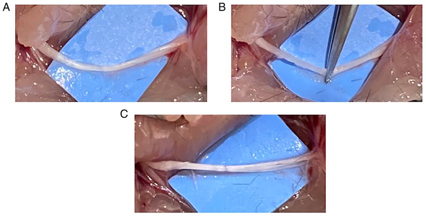
The sciatic nerve was dissected from the surrounding tissues (1:50, 12369‑1‑AP; ProteinTech), rabbit polyclonal anti‑BDNF
(Fig. 1A) and crushed for 30 sec using the hemostatic forceps (1:1,000, GTX132621; GNT), rabbit polyclonal anti‑NGF
with sufficient strength to flatten the sciatic nerve to caused (1:50, ab6199; Abcam), and rabbit polyclonal anti‑Sema3A
Wallerian degeneration (Fig. 1B and C), according to a previ‑ (1:50, ab23393; Abcam).
ously reported method (14). The postoperative activity of In the photon counting mode, fluorescence intensity was
mice was not limited, and they were maintained in the same measured at 20 randomly selected sites from the perikaryon in
environment as that before the procedure. Under general anes‑ a region of interest (ROI) set in a fluorescence‑emitting area,
thesia, the right sciatic nerve and L3‑5 dorsal root ganglion and mean fluorescence intensity was calculated. Fluorescence
(DRG) of each group were harvested (15). Control group were intensity measured using each antibody was compared
only harvested the samples, and samples from the Crush group between the four groups.
were harvested one week after surgery. Mice were sacrificed
by cervical dislocation on the day the sciatic nerves and DRG Statistical analysis. Data are presented as the mean ± standard
were harvesting. The harvested sciatic nerve and DRG were deviation (SD) and were analyzed for significant differences
fixed in 4% paraformaldehyde at room temperature for 72 h using a two‑way ANOVA with age and nerve injury set as two
and paraffin blocks were prepared. independent variables (Prism 7; GraphPad Software). After
the two‑way ANOVA, Turkey's multiple comparisons test was
Histochemical assessment of the degree of Wallerian degener- used as a post hoc test. Differences were considered to be
ation in peripheral nerves. Luxol fast blue (LFB) staining was significant at P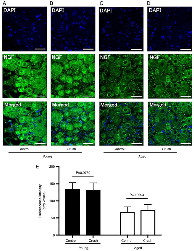
EXPERIMENTAL AND THERAPEUTIC MEDICINE 24: 682, 2022                                                            3

Figure 1. Surgical procedure to create the peripheral nerve injury model. (A) Mouse right sciatic nerve was dissected from the surrounding tissue. (B) The
sciatic nerve was crushed for 30 sec using a hemostatic forceps. (C) The chronic constriction injury was made by a hemostatic forceps.

of the myelin sheath to the total nerve fiber area were 85.7±2.6               group (Aged crush group: 123.8±35.9, P=0.9802) (Fig. 5B,
and 74.0±4.5% in the Young control and Aged control groups,                    D and E). The fluorescence intensity of NGF was significantly
respectively, and did not significantly differ (P=0.1891).                     higher in the Young control group (135.1±17.2) than in the Aged
Following peripheral nerve injury, sciatic nerve fiber swelling,               control group (68.1±13.7) (P
4                                      OBATA et al: AGING AND NERVE‑SPECIFIC PROTEIN EXPRESSION

Table I. Comparison of the expression of the nerve‑specific proteins between Young and Aged group.

Nerve‑specific proteins                                       Young                                     Aged                                   P‑value

REST/NRSF (gray values)                                    110.2±9.5                                 147.8±15.8                               P
EXPERIMENTAL AND THERAPEUTIC MEDICINE 24: 682, 2022                                                         5

Figure 3. Histochemical assessment of the expression of REST/NRSF in the DRG by immunofluorescence staining. The expression of nerve‑specific proteins
was quantified by immunofluorescence staining using a REST/NRSF antibody. DRG in the Young group (10 weeks old) and Aged group (70 weeks old)
were used and fluorescence intensity was compared. REST/NRSF stained green; DAPI stained blue; scale bar, 20.0 µm. (A) Young control group. (B) Young
crush group. (C) Aged control group. (D) Aged crush group. (E) The fluorescence intensity of REST/NRSF. DRG, dorsal root ganglion; REST, repressor
element 1‑silencing transcription factor; NRSF, neuron‑restrictive silencer factor.

Young and Aged groups. However, the effects of aging on axon                     Peripheral nerve injury always induces inflammation. It is
regeneration after peripheral nerve injury currently remain                  a complex series of molecular and cellular events through the
unknown (20‑22).                                                             recruitment of circulating proteins and leukocytes to the injury
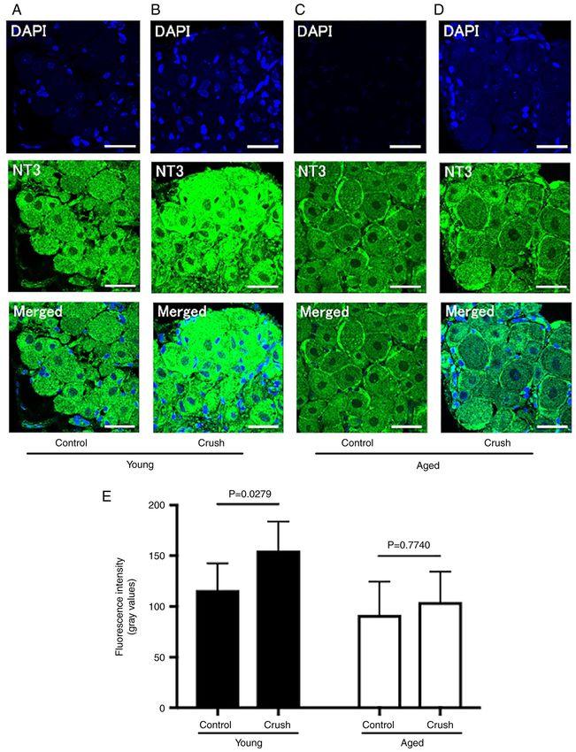
6                                     OBATA et al: AGING AND NERVE‑SPECIFIC PROTEIN EXPRESSION

Figure 4. Histochemical assessment of the expression of NT3 in DRG by immunofluorescence staining. The expression of nerve‑specific proteins was quanti‑
fied by immunofluorescence staining using a NT3 antibody. In the present study, DRG in the Young group (10 weeks old) and Aged group (70 weeks old) were
used and fluorescence intensity was compared (NT3 stained green; DAPI stained blue; scale bar, 20.0 µm). (A) Young control group. (B) Young crush group.
(C) Aged control group. (D) Aged crush group. (E) The fluorescence intensity of NT3. NT3, neurotrophin 3; DRG, dorsal root ganglion.

site within hours to days after peripheral nerve injury (23). These           injury, we quantified the expression of nerve‑specific proteins
reactions associated with inflammation are thought to have a                  in young and aged mice with similarly degenerated peripheral
significant effect on axon regeneration (24,25). To investigate               nerves. In peripheral nerve injury, the expression of most of
the effects of aging on axon regeneration after peripheral nerve              the genes required for axon regeneration and synaptogenesis is
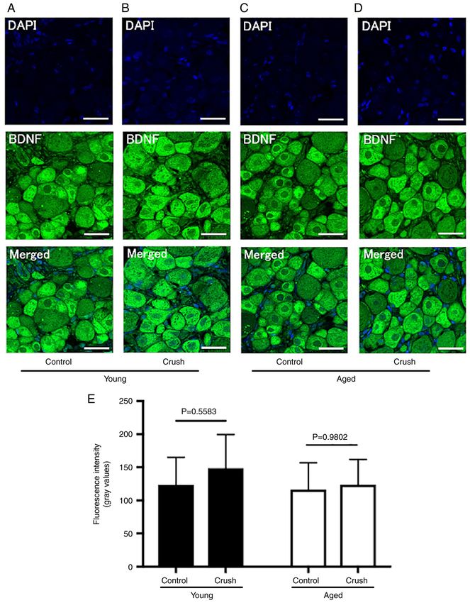
EXPERIMENTAL AND THERAPEUTIC MEDICINE 24: 682, 2022                                                           7

Figure 5. Histochemical assessment of BDNF expression in DRG by immunofluorescence staining. The expression of nerve‑specific proteins was quantified
by immunofluorescence staining using a BDNF antibody. In the present study, DRG in the Young group (10 weeks) and Aged group (70 weeks) were used and
fluorescence intensity was compared (BDNF stained green; DAPI stained blue; scale bar, 20.0 µm). (A) Young control group. (B) Young crush group. (C) Aged
control group. (D) Aged crush group. (E) The fluorescence intensity of BDNF. BDNF, brain‑derived neurotrophic factor; DRG, dorsal root ganglion.

regulated by REST/NRSF (26‑28), and REST/NRSF possesses                       of neurons (1‑3). We also previously demonstrated that the
a wide number of functions through its regulation of more than                expression of REST/NRSF increased with age in peripheral
1000 target genes (3,29‑31). In the central nervous system, the               nerves (4). In the present study, the expression of REST/NRSF
expression of REST/NRSF was shown to increase with age,                       was significantly higher in the Aged control group than in the
and maintained homeostasis by suppressing the apoptosis                       Young control group, consistent with the previous studies.
8                                     OBATA et al: AGING AND NERVE‑SPECIFIC PROTEIN EXPRESSION

Figure 6. Histochemical assessment of the expression of NGF in DRG by immunofluorescence staining. The expression of nerve‑specific proteins was quanti‑
fied by immunofluorescence staining using a NGF antibody. In the present study, DRG in the Young group (10 weeks) and Aged group (70 weeks) were used
and fluorescence intensity was compared (NGF stained green; DAPI stained blue; scale bar, 20.0 µm. (A) Young control group. (B) Young crush group.
(C) Aged control group. (D) Aged crush group. (E) The fluorescence intensity of NGF. NGF, nerve growth factor; DRG, dorsal root ganglion.

The up‑regulated expression of REST/NRSF after peripheral                         The expression of neurotrophic factors has been shown to
nerve injury has also been demonstrated (32‑35), and indicates                play an important role in axon regeneration after peripheral
the initiation of axon regeneration in the injured peripheral                 nerve injury (8,9). NT3 is a necessary factor for Schwann cell
nerve (32). In the peripheral nerve injury model used in the                  survival and differentiation in the absence of axons (9,36),
present study, the expression of REST/NRSF was significantly                  BDNF maintains peripheral nerve homeostasis by regulating
increased in the Young group, but not in the Aged group,                      neuron survival maintenance, neurite outgrowth promotion,
suggesting that axon regeneration was initiated immediately                   and synaptogenesis (9,37‑40), and NGF promotes axon elonga‑
after peripheral nerve injury in the Young group, but not in the              tion in peripheral nerves (41‑44). Therefore, the neurotrophic
Aged group.                                                                   factors investigated in the present study are markers for
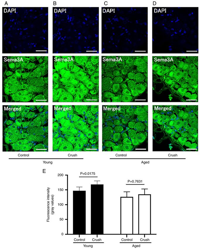
EXPERIMENTAL AND THERAPEUTIC MEDICINE 24: 682, 2022                                                           9

Figure 7. Histochemical assessment of the expression of Sema3A in DRG by immunofluorescence staining. The expression of nerve‑specific proteins was
quantified by immunofluorescence staining using a Sema3A antibody. In the present study, DRG in the Young group (10 weeks) and Aged group (70 weeks)
were used and fluorescence intensity was compared (Sema3A stained green; DAPI stained blue; scale bar, 20.0 µm. (A) Young control group. (B) Young crush
group. (C) Aged control group. (D) Aged crush group. (E) The fluorescence intensity of Sema3A. Sema3A, semaphorin 3A; DRG, dorsal root ganglion.

Schwann cell migration (NT3), myelination (BDNF), and                         the expression of NGF was significantly lower in the Aged
axon elongation (NGF). To investigate the effects of aging on                 control group than in the Young control group, suggesting
the expression of these neurotrophic factors, their expression                that Schwann cell migration may not be affected by aging
was compared between the Young and Aged control groups.                       in the Control group in the presence of axons. Furthermore,
The results obtained showed no significant differences in the                 the results on the expression of BDNF suggest that myelina‑
expression of NT3 or BDNF between these groups, while                         tion was not affected by aging. However, the results on the
10                                 OBATA et al: AGING AND NERVE‑SPECIFIC PROTEIN EXPRESSION

expression of NGF indicate that axon elongation decreased             is highly meaningful to investigate the expression of these
with aging. These results are consistent with our previous find‑      nerve‑specific proteins at one week after peripheral nerve
ings showing that the ability to regenerate axons in peripheral       injury. Because the up‑regulated expression of neurotrophic
nerve is decreased with age (7). Moreover, we investigated            factors and Schwann cell migration, an early stage of axon
changes in the expression of neurotrophic factors character‑          regeneration, occur within one week after nerve injury (52).
ized by age‑related changes in the Young and Aged groups              Therefore, in this study, we performed evaluation at one week
following peripheral nerve injury. In comparisons with the            after peripheral nerve injury and discussed it.
respective Control groups, the expression of BDNF and NGF                 Based on the results of present study, while compensa‑
did not significantly differ between the Young and Aged crush         tory changes for peripheral nerve injury were initiated by the
groups, while the expression of NT3 significantly increased in        upregulation of the REST/NRSF, followed by Schwann cell
the Young group, but not in the Aged group. In other words,           migration in the Young group, these compensatory changes did
Wallerian degeneration occurred and axons were absent in              not occur in the Aged group. The regulation of REST/NRSF
injured peripheral nerves, axon regeneration was initiated by         expression appears to be essential for axon regeneration
the increased expression of REST/NRSF, and the migration              when peripheral nerves are exposed to stress. In peripheral
of Schwann cells, which is the initial stage of axon regenera‑        nerves, aging‑associated functional and electrophysiological
tion, was induced by the upregulation of NT3 in the Young             disorders have been reported in clinical study (6). This study
group. However, based on the results for the expression of            and our previous study showed the REST/NRSF expression is
REST/NRSF and NT3, axon regeneration was not initiated                increased with age (4), and the expression is also increased by
in the Aged group following peripheral nerve injury. The              nerve injury according to the results of this study. Therefore,
expression of BDNF and NGF, which have been suggested to              focusing on the expression of REST/NRSF is expected to
play a role in myelination and axon elongation after Schwann          elucidate the pathology of nerve injury, and is also expected
cell migration for axon regeneration, was not increased in the        to contribute significantly to the treatment of age‑related
Young or Aged group one week after peripheral nerve injury            peripheral nerve injury.
in the present study.                                                     In conclusion, the present results suggested that Wallerian
    There are several limitations in this study. First, in creating   degeneration occurred after peripheral nerve injury in the
the CCI model for peripheral nerve injury, the compression            Young and Aged groups. On the other hand, compensatory
method with a hemostatic forceps was used, so the degree              changes for peripheral nerve injury and Schwann cell migration
of nerve injury may be different with each mice. It has been          were initiated in the Young group, but not in the Aged group.
reported that the expression of neurotrophic factor varies
depending on the degree of nerve injury (Neurapraxia,                 Acknowledgements
Axonotmesis, Neurotmesis) (9). However, there were no
complication (death, disability, etc). Next, in this study, we        Not applicable.
performed evaluation the expression of nerve‑specific proteins
such as REST/NRSF and discussed how they affect axon                  Funding
regeneration. On the other hand, although inflammatory
cytokines are up‑regulated with aging or nerve injury (45,46),        No funding was recieved.
age‑related differences in inflammatory cytokine levels after
peripheral nerve injury are unclear (47). However, the degree         Availability of data and materials
of inflammation after nerve injury was not investigated in
this sturdy. In a future study, to investigate the degree of          The datasets used and/or analyzed during the current study are
inflammation and age‑related differences in inflammatory              available from the corresponding author on reasonable request.
cytokine levels after peripheral nerve injury may provide a
more detailed understanding of the effects of aging on axon           Authors' contributions
regeneration. Moreover, we also investigated the expression
of nerve‑specific proteins in the cytoplasm and the nucleus in        HO mainly wrote the manuscript and acquired, analyzed and
the perikaryon of DRG, but there was no significant differ‑           interpretated the data. KN wrote the manuscript and made
ence among them. Therefore, the fluorescence intensity of             substantial contributions to conception and design of the
the whole cell was measured by the method of this study.              study, and interpretation of data. SN and KS contributed to
Therefore, since all DRG samples were used by histochemical           acquisition, analysis and interpretation of data. SK, TS KG,
assessment in this study, the quantification of nerve‑specific        AK, NN and YS contributed to acquisition of data. SK and TS
proteins by western blot were not able to be performed. This is       confirm the authenticity of all the raw data. IN made substan‑
the limitation of this study and a future issue. Last, since this     tial contributions to conception and design. MI contributed to
study is a fixed‑point observation one week after peripheral          the analysis and interpretation of data. All authors read and
nerve injury, it only evaluates the effect of peripheral nerve        approved the final manuscript.
injury and aging in a limited manner. It has been reported
that the expression of nerve‑specific proteins varies greatly         Ethics approval and consent to participate
depending on the time after nerve injury (9,48‑51). We believe
that it may be possible to evaluate the effect of peripheral nerve    The present study was approved by the Animal Care
injury and aging in more detail by evaluating with time course.       Committee of Juntendo University (Tokyo, Japan; registration
However, in assessment of the axon regeneration process, it           no. 1555; approval no. 2021312).
EXPERIMENTAL AND THERAPEUTIC MEDICINE 24: 682, 2022 11
Patient consent for publication 19. Calkins DJ: Age‑related changes in the visual pathways: Blame
it on the axon. Invest Ophthalmol Vis Sci 54: ORSF37‑41, 2013.
20. Bland JD: The relationship of obesity, age, and carpal tunnel
Not applicable. syndrome: More complex than was thought? Muscle Nerve 32:
527‑532, 2005.
21. Di Caprio F, Meringolo R, Shehab Eddine M and Ponziani L:
Competing interests Morton's interdigital neuroma of the foot: A literature review.
Foot Ankle Surg 24: 92‑98, 2018.
The authors declare that they have no competing interests. 22. Martínez‑Aparicio C, Jääskeläinen SK, Puksa L, Reche‑Lorite F,
Torné‑Poyatos P, Paniagua Soto J and Falck B: Constitutional
risk factors for focal neuropathies in patients referred for electro‑
References myography. Eur J Neurol 27: 529‑535, 2020.
23. Ransohoff RM: Chemokines and chemokine receptors:
1. Lu T, Aron L, Zullo J, Pan Y, Kim H, Chen Y, Yang TH, Kim HM, Standing at the crossroads of immunobiology and neurobiology.
Drake D, Liu XS, et al: REST and stress resistance in ageing and Immunity 31: 711‑721, 2009.
Alzheimer's disease. Nature 507: 448‑454, 2014. 24. Benowitz LI and Popovich PG: Inflammation and axon regenera‑
2. Kawamura M, Sato S, Matsumoto G, Fukuda T, Shiba- tion. Curr Opin Neurol 24: 577‑583, 2011.
Fukushima K, Noda S, Takanashi M, Mori N and Hattori N: 25. Vargas ME, Watanabe J, Singh SJ, Robinson WH and Barres BA:
Loss of nuclear REST/NRSF in aged‑dopaminergic neurons in Endogenous antibodies promote rapid myelin clearance and
Parkinson's disease patients. Neurosci Lett 699: 59‑63, 2019. effective axon regeneration after nerve injury. Proc Natl Acad
3. Mampay M and Sheridan GK: REST: An epigenetic regulator of Sci USA 107: 11993‑11998, 2010.
neuronal stress responses in the young and ageing brain. Front 26. Schoenherr CJ and Anderson DJ: The neuron‑restrictive silencer
Neuroendocrinol 53: 100744, 2019. factor (NRSF): A coordinate repressor of multiple neuron‑specific
4. Goto K, Naito K, Nakamura S, Nagura N, Sugiyama Y, Obata H, genes. Science 267: 1360‑1363, 1995.
Kaneko A and Kaneko K: Protective mechanism against 27. Lunyak VV and Rosenfeld MG: No rest for REST: REST/NRSF
age‑associated changes in the peripheral nerves. Life Sci 253: regulation of reurogenesis. Cell 121: 499‑501, 2005.
117744, 2020. 28. Ballas N and Mandel G: The many faces of REST oversee epigen‑
5. Büttner R, Schulz A, Reuter M, Akula AK, Mindos T, Carlstedt A, etic programming of neuronal genes. Curr Opin Neurobiol 15:
Riecken LB, Baader SL, Bauer R and Morrison H: Inflammaging 500‑506, 2005.
impairs peripheral nerve maintenance and regeneration. Aging 29. Bruce AW, Donaldson IJ, Wood IC, Yerbury SA, Sadowski MI,
Cell 17: e12833, 2018. Chapman M, Göttgens B and Buckley NJ: Genome‑wide
6. Verdú E, Ceballos D, Vilches JJ and Navarro X: Influence of analysis of repressor element 1 silencing transcription
aging on peripheral nerve function and regeneration. J Peripher factor/neuron‑restrictive silencing factor (REST/NRSF) target
Nerv Syst 5: 191‑208, 2000. genes. Proc Natl Acad Sci USA 101: 10458‑10463, 2004.
7. Kaneko A, Naito K, Nakamura S, Miyahara K, Goto K, Obata H, 30. Chong JA, Tapia‑Ramírez J, Kim S, Toledo‑AraI JJ, Zheng Y,
Nagura N, Sugiyama Y, Kaneko K and Ishijima M: Influence of Boutros MC, Altshuller YM, Frohman MA, Kraner SD and
aging on the peripheral nerve repair process using an artificial Mandel G: REST: A mammalian silencer protein that restricts
nerve conduit. Exp Ther Med 21: 168, 2021. sodium channel gene expression to neurons. Cell 80: 949‑957, 1995.
8. Chan JR, Cosgaya JM, Wu YJ and Shooter EM: Neurotrophins 31. Zhao Y, Zhu M, Yu Y, Qiu L, Zhang Y, He L and Zhang J: Brain
are key mediators of the myelination program in the peripheral REST/NRSF is not only a silent repressor but also an active
nervous system. Proc Natl Acad Sci USA 98: 14661‑14668, protector. Mol Neurobiol 54: 541‑550, 2017.
2001. 32. Oh YM, Mahar M, Ewan EE, Leahy KM, Zhao G and Cavalli V:
9. Omura T, Sano M, Omura K, Hasegawa T, Doi M, Sawada T and Epigenetic regulator UHRF1 inactivates REST and growth
Nagano A: Different expressions of BDNF, NT3, and NT4 in suppressor gene expression via DNA methylation to promote
muscle and nerve after various types of peripheral nerve injuries. axon regeneration. Proc Natl Acad Sci USA 115: E12417‑E12426,
J Peripher Nerv Syst 10: 293‑300, 2005. 2018.
10. Kim JK, Hann HJ, Kim MJ and Kim JS: The expression of 33. Zhang F, Gigout S, Liu Y, Wang Y, Hao H, Buckley NJ, Zhang H,
estrogen receptors in the tenosynovium of postmenopausal Wood IC and Gamper N: Repressor element 1‑silencing tran‑
women with idiopathic carpal tunnel syndrome. J Orthop Res 28: scription factor drives the development of chronic pain states.
1469‑1474, 2010. Pain 160: 2398‑2408, 2019.
11. Al‑Rousan T, Sparks JA, Pettinger M, Chlebowski R, Manson JE, 34. Zhang J, Chen SR, Chen H and Pan HL: RE1‑silencing tran‑
Kauntiz AM and Wallace R: Menopausal hormone therapy and scription factor controls the acute‑to‑chronic neuropathic pain
the incidence of carpal tunnel syndrome in postmenopausal transition and Chrm2 receptor gene expression in primary
women: Findings from the Women's Health Initiative. PLoS sensory neurons. J Biol Chem 293: 19078‑19091, 2018.
One 13: e0207509, 2018. 35. Ueda H, Kurita JI, Neyama H, Hirao Y, Kouji H, Mishina T,
12. Lee AR, Pechenino AS, Dong H, Hammock BD and Kasai M, Nakano H, Yoshimori A and Nishimura Y: A mimetic
Knowlton AA: Aging, estrogen loss and epoxyeicosatrienoic of the mSin3‑binding helix of NRSF/REST ameliorates abnormal
acids (EETs). PLoS One 8: e70719, 2013. pain behavior in chronic pain models. Bioorg Med Chem Lett 27:
13. Shifren JL and Schiff IJ: The aging ovary. J Womens Health 4705‑4709, 2017.
Gend Based Med 9 (Suppl 1): S3‑S7, 2000. 36. Richner M, Ulrichsen M, Elmegaard SL, Dieu R, Pallesen LT
14. Schram S, Chuang D, Schmidt G, Piponov H, Helder C, Kerns J, and Vaegter CB: Peripheral nerve injury modulates neurotrophin
Gonzalez M, Song F and Loeb JA: Mutant SOD1 prevents signaling in the peripheral and central nervous system. Mol
normal functional recovery through enhanced glial activation Neurobiol 50: 945‑970, 2014.
and loss of motor neuron innervation after peripheral nerve 37. Park H and Poo MM: Neurotrophin regulation of neural circuit
injury. Neurobiol Dis 12: 469‑478, 2019. development and function. Nat Rev Neurosci 14: 7‑23, 2013.
15. Gallaher ZR and Steward O: Modest enhancement of sensory 38. Huang EJ and Reichardt LF: Neurotrophins: Roles in neuronal
axon regeneration in the sciatic nerve with conditional co‑dele‑ development and function. Annu Rev Neurosci 24: 677‑736,
tion of PTEN and SOCS3 in the dorsal root ganglia of adult mice. 2001.
Exp Neurol 303: 120‑133, 2018. 39. Miranda M, Morici JF, Zanoni MB and Bekinschtein P:
16. Lindborg JA, Mack M and Zigmond RE: Neutrophils are Brain‑derived neurotrophic factor: A key molecule for memory
critical for myelin removal in a peripheral nerve injury model of in the healthy and the pathological brain. Front Cell Neurosci 13:
Wallerian degeneration. J Neurosci 37: 10258‑10277, 2017. 363, 2019.
17. Niemi JP, DeFrancesco‑Lisowitz A, Roldán‑Hernández L, 40. Yang J, Harte‑Hargrove LC, Siao CJ, Marinic T, Clarke R, Ma Q,
Lindborg JA, Mandell D and Zigmond RE: A critical role for Jing D, Lafrancois JJ, Bath KG, Mark W, et al: Hempstead,
macrophages near axotomized neuronal cell bodies in stimu‑ proBDNF negatively regulates neuronal remodeling, synaptic
lating nerve regeneration. J Neurosci 33: 16236‑16248, 2013. transmission, and synaptic plasticity in hippocampus. Cell Rep 7:
18. Apel PJ, Ma J, Callahan M, Northam CN, Alton TB, Sonntag WE 796‑806, 2014.
and Li Z: Effect of locally delivered IGF‑1 on nerve regeneration 41. Bhang SH, Jeon O, Choi CY, Kwon YH and Kim BS: Controlled
during aging: An experimental study in rats. Muscle Nerve 41: release of nerve growth factor from fibrin gel. J Biomed Mater
335‑341, 2010. Res A 80: 998‑1002, 2007.12 OBATA et al: AGING AND NERVE‑SPECIFIC PROTEIN EXPRESSION
42. Chen ZW and Wang MS: Effects of nerve growth factor on 49. Rose K, Ooi L, Dalle C, Robertson B, Wood IC and Gamper N:
crushed sciatic nerve regeneration in rats. Microsurgery 16: Transcriptional repression of the M channel subunit Kv7.2 in
547‑551, 1995. chronic nerve injury. Pain 152: 742‑754, 2011.
43. Kemp SW, Webb AA, Dhaliwal S, Dhaliwal S, Syed S, Walsh SK 50. Uchida S, Hara K, Kobayashi A, Funato H, Hobara T, Otsuki K,
and Midha R: Dose and duration of nerve growth factor (NGF) Yamagata H, McEwen BS and Watanabe Y: Early life stress
administration determine the extent of behavioral recovery enhances behavioral vulnerability to stress through the activation
following peripheral nerve injury in the rat. Exp Neurol 229: of REST4‑mediated gene transcription in the medial prefrontal
460‑470, 2011. cortex of rodents. J Neurosci 30: 15007‑15018, 2010.
44. Moattari M, Kouchesfehani HM, Kaka G, Sadraie SH and 51. Gervasi NM, Dimtchev A, Clark DM, Dingle M, Pisarchik AV
Naghdi M: Evaluation of nerve growth factor (NGF) treated mesen‑ and Nesti LJ: C‑terminal domain small phosphatase 1 (CTDSP1)
chymal stem cells for recovery in neurotmesis model of peripheral regulates growth factor expression and axonal regeneration in
nerve injury. J Craniomaxillofac Surg 46: 898‑904, 2018. peripheral nerve tissue. Sci Rep 11: 14462, 2021.
45. Wu D, Ren Z, Pae M, Guo W, Cui X, Merrill AH and Meydani SN: 52. Yin Q, Kemp GJ, Yu LG, Wagstaff SC and Frostick SP: Expression
Aging up‑regulates expression of inflammatory mediators in of Schwann cell‑specific proteins and low‑molecular‑weight
mouse adipose tissue. J Immunol 179: 4829‑4839, 2007. neurofilament protein during regeneration of sciatic nerve treated
46. Gaudet AD, Popovich PG and Ramer MS: Wallerian degenera‑ with neurotrophin‑4. Neuroscience 105: 779‑783, 2001.
tion: Gaining perspective on inflammatory events after peripheral
nerve injury. J Neuroinflammation 30: 110, 2011. This work is licensed under a Creative Commons
47. Fitzgerald M and McKelvey R: Nerve injury and neuropathic Attribution-NonCommercial-NoDerivatives 4.0
pain‑A question of age. Exp Neurol 275 Pt 2: 296‑302, 2016. International (CC BY-NC-ND 4.0) License.
48. Shudo Y, Shimojo M, Fukunaga M and Ito S: Pituitary adenylate
cyclase‑activating polypeptide is regulated by alternative splicing
of transcriptional repressor REST/NRSF in nerve injury. Life
Sci 143: 174‑181, 2015.You can also read