World Journal of Clinical Cases - World J Clin Cases 2022 May 26; 10(15): 4713-5123 - NET
←
→
Page content transcription
If your browser does not render page correctly, please read the page content below
ISSN 2307-8960 (online)

World Journal of
Clinical Cases
World J Clin Cases 2022 May 26; 10(15): 4713-5123

                                                    Published by Baishideng Publishing Group Inc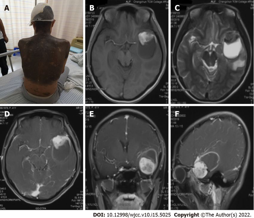
World Journal of
WJ C C                                                    Clinical Cases
Contents                                                  Thrice Monthly Volume 10 Number 15 May 26, 2022

          EDITORIAL
 4713     Diet and intestinal bacterial overgrowth: Is there evidence?
          Souza C, Rocha R, Cotrim HP

          MINIREVIEWS
 4717     Definition and classification of acute-on-chronic liver diseases
          Zhang YY, Meng ZJ

 4726     Management of neurosurgical patients during coronavirus disease 2019 pandemics: The Ljubljana,
          Slovenia experience
          Velnar T, Bosnjak R

          ORIGINAL ARTICLE
          Clinical and Translational Research
 4737     Glycolytic and fatty acid oxidation genes affect the treatment and prognosis of liver cancer
          Zou JY, Huang YJ, He J, Tang ZX, Qin L

 4761     Detection of a novel panel of 24 genes with high frequencies of mutation in gastric cancer based on next-
          generation sequencing
          Zeng HH, Yang Z, Qiu YB, Bashir S, Li Y, Xu M

          Case Control Study
 4776     Outcomes of cervical degenerative disc disease treated by anterior cervical discectomy and fusion with
          self-locking fusion cage
          Zhang B, Jiang YZ, Song QP, An Y

 4785     Impact of COVID-19 pandemic on clinicopathological features of transplant recipients with hepatocellular
          carcinoma: A case-control study
          Akbulut S, Sahin TT, Ince V, Yilmaz S

          Retrospective Study
 4799     Risk factors and optimal predictive scoring system of mortality for children with acute paraquat poisoning
          Song Y, Wang H, Tao YH

 4810     Application effect of thoracoscopic tricuspid valvuloplasty in geriatric patients with tricuspid valve
          disease
          Jiang W, Long XM, Wei KQ, Li SC, Zhang Z, He BF, Li H

 4818     Endoscopic ultrasonography in the evaluation of condition and prognosis of ulcerative colitis
          Jin RF, Chen YM, Chen RP, Ye HJ

        WJCC    https://www.wjgnet.com                      I                   May 26, 2022    Volume 10   Issue 15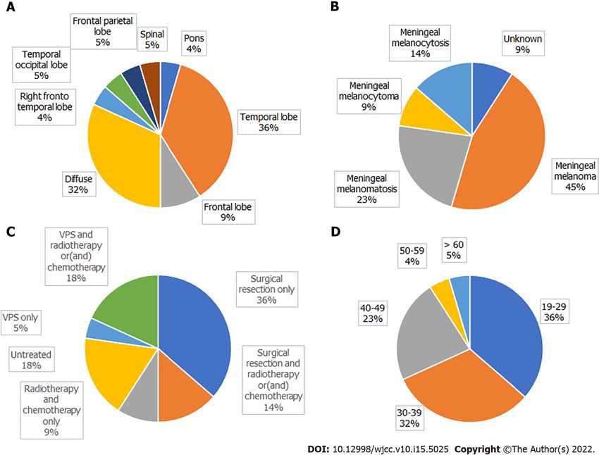
World Journal of Clinical Cases
Contents
                                                           Thrice Monthly Volume 10 Number 15 May 26, 2022

 4827     Dynamic interaction nursing intervention on functional rehabilitation and self-care ability of patients after
          aneurysm surgery
          Xie YE, Huang WC, Li YP, Deng JH, Huang JT

          Clinical Trials Study
 4836     Validations of new cut-offs for surgical drains management and use of computerized tomography scan
          after pancreatoduodenectomy: The DALCUT trial
          Caputo D, Coppola A, La Vaccara V, Passa R, Carbone L, Ciccozzi M, Angeletti S, Coppola R

          Observational Study
 4843     Psychosocial adaptation and influencing factors among patients with chemotherapy-induced peripheral
          neuropathy
          Zhou X, Wang DY, Ding CY, Liu H, Sun ZQ

          META-ANALYSIS
 4856     Outcome of the efficacy of Chinese herbal medicine for functional constipation: A systematic review and
          meta-analysis
          Lyu Z, Fan Y, Bai Y, Liu T, Zhong LL, Liang HF

          CASE REPORT
 4878     Familial gastrointestinal stromal tumors with KIT germline mutation in a Chinese family: A case report
          Yuan W, Huang W, Ren L, Xu C, Luan LJ, Huang J, Xue AW, Fang Y, Gao XD, Shen KT, Lv JH, Hou YY

 4886     Nonfunctional pancreatic neuroendocrine tumours misdiagnosed as autoimmune pancreatitis: A case
          report and review of literature
          Lin ZQ, Li X, Yang Y, Wang Y, Zhang XY, Zhang XX, Guo J

 4895     Sudden deafness as a prodrome of cerebellar artery infarction: Three case reports
          Li BL, Xu JY, Lin S

 4904     Importance of abdominal X-ray to confirm the position of levonorgestrel-releasing intrauterine system: A
          case report
          Maebayashi A, Kato K, Hayashi N, Nagaishi M, Kawana K

 4911     Bedside ultrasonic localization of the nasogastric tube in a patient with severe COVID-19: A case report
          Zhu XJ, Liu SX, Li QT, Jiang YJ

 4917     Paradoxical herniation after decompressive craniectomy provoked by mannitol: A case report
          Du C, Tang HJ, Fan SM

 4923     Targeted next-generation sequencing identifies a novel nonsense mutation in ANK1 for hereditary
          spherocytosis: A case report
          Fu P, Jiao YY, Chen K, Shao JB, Liao XL, Yang JW, Jiang SY

 4929     Nonfunctional bladder paraganglioma misdiagnosed as hemangioma: A case report
          Chen J, Yang HF

        WJCC    https://www.wjgnet.com                       II                       May 26, 2022    Volume 10   Issue 15
World Journal of Clinical Cases
Contents
                                                         Thrice Monthly Volume 10 Number 15 May 26, 2022

 4935     Special type of Wernekink syndrome in midbrain infarction: Four case reports
          Yang YZ, Hu WX, Zhai HJ

 4942     Primary extraskeletal Ewing’s sarcoma of the lumbar nerve root: A case report
          Lei LH, Li F, Wu T

 4949     Yellow nail syndrome accompanied by minimal-change nephrotic syndrome: A case report
          Zhang YN, Wang MH, Yu WC, Cheng W, Cong JP, Huang XP, Wang FF

 4957     Total femur replacement with 18 years of follow-up: A case report
          Yang YH, Chen JX, Chen QY, Wang Y, Zhou YB, Wang HW, Yuan T, Sun HP, Xie L, Yao ZH, Yang ZZ

 4964     Male metaplastic breast cancer with poor prognosis: A case report
          Kim HY, Lee S, Kim DI, Jung CS, Kim JY, Nam KJ, Choo KS, Jung YJ

 4971     CD8-positive indolent T-Cell lymphoproliferative disorder of the gastrointestinal tract: A case report and
          review of literature
          Weng CY, Ye C, Fan YH, Lv B, Zhang CL, Li M

 4985     Bone flare after initiation of novel hormonal therapy in patients with metastatic hormone-sensitive
          prostate cancer: A case report
          Li KH, Du YC, Yang DY, Yu XY, Zhang XP, Li YX, Qiao L

 4991     Postoperative infection of the skull base surgical site due to suppurative parotitis: A case report
          Zhao Y, Zhao Y, Zhang LQ, Feng GD

 4998     Blunt aortic injury–traumatic aortic isthmus pseudoaneurysm with right iliac artery dissection aneurysm:
          A case report
          Fang XX, Wu XH, Chen XF

 5005     Extensive complex thoracoabdominal aortic aneurysm salvaged by surgical graft providing landing zone
          for endovascular graft: A case report
          Jang AY, Oh PC, Kang JM, Park CH, Kang WC

 5012     Gastric heterotopia of colon found cancer workup in liver abscess: A case report
          Park JG, Suh JI, Kim YU

 5018     Clinical manifestations and gene analysis of Hutchinson-Gilford progeria syndrome: A case report
          Zhang SL, Lin SZ, Zhou YQ, Wang WQ, Li JY, Wang C, Pang QM

 5025     Neurocutaneous melanosis with an intracranial cystic-solid meningeal melanoma in an adult: A case
          report and review of literature
          Liu BC, Wang YB, Liu Z, Jiao Y, Zhang XF

 5036     Metastasis of liver cancer to the thyroid after surgery: A case report
          Zhong HC, Sun ZW, Cao GH, Zhao W, Ma K, Zhang BY, Feng YJ

        WJCC    https://www.wjgnet.com                      III                    May 26, 2022   Volume 10     Issue 15
World Journal of Clinical Cases
Contents
Thrice Monthly Volume 10 Number 15 May 26, 2022
5042 Spontaneous liver rupture following SARS-CoV-2 infection in late pregnancy: A case report
Ambrož R, Stašek M, Molnár J, Špička P, Klos D, Hambálek J, Skanderová D
5051 Carotid blowout syndrome caused by chronic infection: A case report
Xie TH, Zhao WJ, Li XL, Hou Y, Wang X, Zhang J, An XH, Liu LT
5057 Is repeat wide excision plus radiotherapy of localized rectal melanoma another choice before
abdominoperineal resection? A case report
Chiu HT, Pu TW, Yen H, Liu T, Wen CC
5064 Metaplastic breast cancer with chondrosarcomatous differentiation combined with concurrent bilateral
breast cancer: A case report
Yang SY, Li Y, Nie JY, Yang ST, Yang XJ, Wang MH, Zhang J
5072 Rare solitary splenic metastasis from a thymic carcinoma detected on fluorodeoxyglucose-positron
emission tomography: A case report
Tsai YH, Lin KH, Huang TW
5077 Type A aortic dissection following heart transplantation: A case report
Zeng Z, Yang LJ, Zhang C, Xu F
5082 Catheter-related infections caused by Mycobacterium abscessus in a patient with motor neurone disease: A
case report
Pan SF, Zhang YY, Wang XZ, Sun JJ, Song SL, Tang YR, Wang JL
5088 Clear aligner treatment for a four-year-old patient with anterior cross-bite and facial asymmetry: A case
report
Zou YR, Gan ZQ, Zhao LX
5097 Knot impingement after arthroscopic rotator cuff repair mimicking infection: A case report
Kim DH, Jeon JH, Choi BC, Cho CH
5103 Solitary primary pulmonary synovial sarcoma: A case report
He WW, Huang ZX, Wang WJ, Li YL, Xia QY, Qiu YB, Shi Y, Sun HM
5111 Anesthetic management for intraoperative acute pulmonary embolism during inferior vena cava tumor
thrombus surgery: A case report
Hsu PY, Wu EB
5119 Delayed diagnosis of arytenoid cartilage dislocation after tracheal intubation in the intensive care unit: A
case report
Yan WQ, Li C, Chen Z
WJCC https://www.wjgnet.com IX May 26, 2022 Volume 10 Issue 15World Journal of Clinical Cases
Contents
Thrice Monthly Volume 10 Number 15 May 26, 2022
ABOUT COVER
Editorial Board Member of World Journal of Clinical Cases, Jing Yang, MD, Associate Professor, Department of the
First General Surgery, Gansu Provincial Hospital, Lanzhou 730000, Gansu Province, China. 21634604@qq.com
AIMS AND SCOPE
The primary aim of World Journal of Clinical Cases (WJCC, World J Clin Cases) is to provide scholars and readers from
various fields of clinical medicine with a platform to publish high-quality clinical research articles and
communicate their research findings online.
WJCC mainly publishes articles reporting research results and findings obtained in the field of clinical medicine
and covering a wide range of topics, including case control studies, retrospective cohort studies, retrospective
studies, clinical trials studies, observational studies, prospective studies, randomized controlled trials, randomized
clinical trials, systematic reviews, meta-analysis, and case reports.
INDEXING/ABSTRACTING
The WJCC is now indexed in Science Citation Index Expanded (also known as SciSearch ®), Journal Citation
Reports/Science Edition, Scopus, PubMed, and PubMed Central. The 2021 Edition of Journal Citation Reports®
cites the 2020 impact factor (IF) for WJCC as 1.337; IF without journal self cites: 1.301; 5-year IF: 1.742; Journal
Citation Indicator: 0.33; Ranking: 119 among 169 journals in medicine, general and internal; and Quartile category:
Q3. The WJCC's CiteScore for 2020 is 0.8 and Scopus CiteScore rank 2020: General Medicine is 493/793.
RESPONSIBLE EDITORS FOR THIS ISSUE
Production Editor: Ying-Yi Yuan; Production Department Director: Xiang Li; Editorial Office Director: Jin-Lei Wang.
NAME OF JOURNAL INSTRUCTIONS TO AUTHORS
World Journal of Clinical Cases https://www.wjgnet.com/bpg/gerinfo/204
ISSN GUIDELINES FOR ETHICS DOCUMENTS
ISSN 2307-8960 (online) https://www.wjgnet.com/bpg/GerInfo/287
LAUNCH DATE GUIDELINES FOR NON-NATIVE SPEAKERS OF ENGLISH
April 16, 2013 https://www.wjgnet.com/bpg/gerinfo/240
FREQUENCY PUBLICATION ETHICS
Thrice Monthly https://www.wjgnet.com/bpg/GerInfo/288
EDITORS-IN-CHIEF PUBLICATION MISCONDUCT
Bao-Gan Peng, Jerzy Tadeusz Chudek, George Kontogeorgos, Maurizio Serati, Ja https://www.wjgnet.com/bpg/gerinfo/208
Hyeon Ku
EDITORIAL BOARD MEMBERS ARTICLE PROCESSING CHARGE
https://www.wjgnet.com/2307-8960/editorialboard.htm https://www.wjgnet.com/bpg/gerinfo/242
PUBLICATION DATE STEPS FOR SUBMITTING MANUSCRIPTS
May 26, 2022 https://www.wjgnet.com/bpg/GerInfo/239
COPYRIGHT ONLINE SUBMISSION
© 2022 Baishideng Publishing Group Inc https://www.f6publishing.com
© 2022 Baishideng Publishing Group Inc. All rights reserved. 7041 Koll Center Parkway, Suite 160, Pleasanton, CA 94566, USA
E-mail: bpgoffice@wjgnet.com https://www.wjgnet.com
WJCC https://www.wjgnet.com X May 26, 2022 Volume 10 Issue 15World Journal of
WJ C C Clinical Cases
Submit a Manuscript: https://www.f6publishing.com World J Clin Cases 2022 May 26; 10(15): 5025-5035
DOI: 10.12998/wjcc.v10.i15.5025 ISSN 2307-8960 (online)
CASE REPORT
Neurocutaneous melanosis with an intracranial cystic-solid
meningeal melanoma in an adult: A case report and review of
literature
Bo-Chuan Liu, Yu-Bo Wang, Zhuang Liu, Yan Jiao, Xian-Feng Zhang
Specialty type: Neurosciences Bo-Chuan Liu, Department of Neurosurgery, The Second Affiliated Hospital of Xi'an Medical
University, Xi'an 710038, Shaanxi Province, China
Provenance and peer review:
Unsolicited article; Externally peer Yu-Bo Wang, Xian-Feng Zhang, Department of Neurosurgery, First Hospital of Jilin University,
reviewed. Changchun 130021, Jilin Province, China
Peer-review model: Single blind Zhuang Liu, Department of Neurosurgery, Dongfeng County Hospital, Liaoyuan 136300, Jilin
Province, China
Peer-review report’s scientific
quality classification Yan Jiao, Department of Hepatobiliary and Pancreatic Surgery, First Hospital of Jilin
University, Changchun 130021, Jilin Province, China
Grade A (Excellent): 0
Grade B (Very good): B, B Corresponding author: Xian-Feng Zhang, PhD, Full Professor, Department of Neurosurgery,
Grade C (Good): 0 First Hospital of Jilin University, No. 71 Xinmin Street, Changchun 130021, Jilin Province,
Grade D (Fair): 0 China. zhangxianf@jlu.edu.cn
Grade E (Poor): 0
P-Reviewer: Choudhery MS,
Pakistan; Saito R, Japan
Abstract
BACKGROUND
Received: December 7, 2021 Neurocutaneous melanosis (NCM) is a rare congenital, nonhereditary neurocu-
Peer-review started: December 7, taneous syndrome that mainly occurs in children; adult NCM is very rare. Due to
2021 its rarity, the clinical features and treatment strategies for NCM remain unclear.
First decision: January 25, 2022 The purpose of this study was to explore the clinical features, diagnosis, treatment
Revised: February 12, 2022 and prognosis of NCM in adults. Most intracranial meningeal melanomas are
Accepted: March 26, 2022 solid masses, and cystic-solid malignant melanomas are very rare. Due to the lack
Article in press: March 26, 2022 of data, the cause of cystic changes and the effect on prognosis are unknown.
Published online: May 26, 2022 CASE SUMMARY
A 41-year-old woman was admitted to the hospital with intermittent headache for
1 mo. Magnetic resonance imaging (MRI) showed a 4.7 cm × 3.6 cm cystic-solid
mass in the left temporal lobe with peritumoral edema. The entire mass was
removed, and postoperative pathology indicated malignant melanoma.
CONCLUSION
MRI is the first-choice imaging approach for diagnosing central nervous system
diseases in NCM patients, although cerebrospinal fluid may also be used. At
present, there is no optimal treatment plan; gross total resection combined with
BRAF inhibitors and MEK inhibitors might be the most beneficial treatment.
WJCC https://www.wjgnet.com 5025 May 26, 2022 Volume 10 Issue 15Liu BC et al. NCM in an adult
Key Words: Neurocutaneous melanosis; Meningeal melanoma; Central nervous system disease; Adult;
Cystic-solid tumor; Case report
©The Author(s) 2022. Published by Baishideng Publishing Group Inc. All rights reserved.
Core Tip: Neurocutaneous melanosis (NCM) is a rare congenital, nonhereditary neurocutaneous syndrome
that mainly occurs in children, while adult NCM is very rare. Due to its rarity, clinical features and
treatment strategies for NCM remain unclear. Herein, we report a case and review such reports and
explore the clinical features, diagnosis, treatment and prognosis of NCM in adults.
Citation: Liu BC, Wang YB, Liu Z, Jiao Y, Zhang XF. Neurocutaneous melanosis with an intracranial cystic-solid
meningeal melanoma in an adult: A case report and review of literature. World J Clin Cases 2022; 10(15): 5025-
5035
URL: https://www.wjgnet.com/2307-8960/full/v10/i15/5025.htm
DOI: https://dx.doi.org/10.12998/wjcc.v10.i15.5025
INTRODUCTION
Neurocutaneous melanosis (NCM) is a rare congenital, nonhereditary neurocutaneous syndrome that
mainly occurs in children; adult NCM is very rare. Specifically, NCM is a rare combined abnormality of
the skin and central nervous system (CNS)[1], mainly presenting as congenital melanocytic nevi with
benign or malignant melanoma of the central nervous system[2]. NCM is a neuroectodermal dysplasia
believed to arise from an embryological defect in the migration of melanoblasts from the neural crest to
the leptomeninges and skin[3]; it was first described by Rokitanki in 1861[4]. Patients with NCM usually
have neurological symptoms at an early age. Most patients have a poor prognosis once symptoms occur,
regardless of whether the CNS tumors are benign or malignant; in most cases, the disease is fatal within
3 years of onset[5]. Primary malignant melanoma in the central nervous system is usually a single solid
mass, and melanoma with large cystic changes is extremely rare. Here, we describe a rare adult NCM
complicated with intracranial cystic-solid malignant melanoma and review the relevant literature to
discuss the clinical features, diagnosis, treatment and prognosis.
CASE PRESENTATION
Chief complaints
The patient was admitted to our department with intermittent headache for 1 mo.
History of present illness
A 41-year-old woman was admitted to the hospital with intermittent headache for 1 mo. The headache
could not be relieved by rest. There were no obvious symptoms of nausea, vomiting and epilepsy.
History of past illness
The patient was born with diffuse melanosis of the skin, mainly covering the back of the trunk,
shoulders and neck, with multiple satellite nevi on the limbs and a few hairs on the surface of the nevi
in the first few years after birth (Figure 1A). The lesion was considered to be a birthmark and had not
been treated. No malignant clinical signs, such as enlargement, ulcers, bleeding or itching, were
observed in the pigmented area as the patient aged.
Personal and family history
There was no past history of skin or CNS disease or malignant tumor. The patient had no history of
surgery, and her parents and children had no history of congenital nevi or melanoma.
Physical examination
The patient has diffuse melanosis of the skin, mainly covering the back of the trunk, shoulders and neck,
with multiple satellite nevi on the limbs and a few hairs on the surface of the nevi. Examination of her
nervous system revealed no obvious abnormalities.
WJCC https://www.wjgnet.com 5026 May 26, 2022 Volume 10 Issue 15Liu BC et al. NCM in an adult
Figure 1 Images and physical examination. A: Physical examination revealed a giant skin nevus; B: T1-weighted; C: T2-weighted; D-F: After the injection of a
contrast agent.
Laboratory examinations
Laboratory examinations such as routine blood tests, routine urine tests and blood biochemistry were
normal. The pathological results showed malignant melanoma. Immunohistochemistry results were as
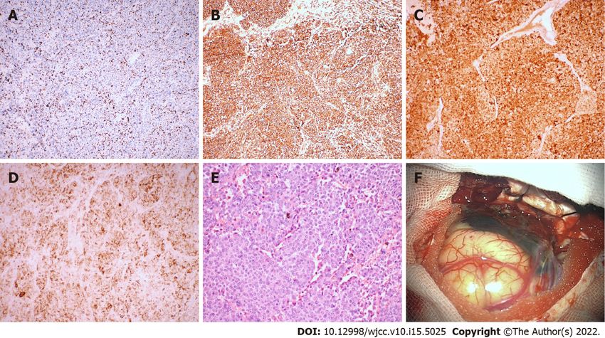
follows: Ki-67 (+30%), S-100 (+), HMB45 (+), Vimentin (+); all other markers were negative (Figure 2).
Imaging examinations
MRI examination of the head revealed a mass with intramural cysts located on the left temporal lobe.
The solid part of the tumor was hyperintense on T1-weighted images and hypointense on T2-weighted
images, and the cystic components showed the opposite. The lesion had a clear boundary of approx-
imately 4.7 cm x 3.6 cm and was surrounded by peritumoral edema (Figure 1B and C). After the
injection of a contrast agent (Gd-DTPA), obvious heterogeneous enhancement was observed in the solid
part, while the cystic part showed abnormal ring-shaped enhancement (Figure 1D-F).
FINAL DIAGNOSIS
We ultimately diagnosed this patient with NCM with meningeal melanoma.
TREATMENT
The patient underwent surgery to remove the intracranial mass after admission. During the operation,
the tumor appeared blackish brown (the peripheral leptomeninges were also stained black), with light-
yellow sac fluid inside, and the blood supply to the tumor was extremely rich. First, the cystic fluid was
drawn out, and the lesion was then gradually separated along its periphery until it was completely
removed. The pathological results showed malignant melanoma. Immunohistochemistry results were as
follows: Ki-67 (+30%), S-100 (+), HMB45 (+), Vimentin (+); all other markers were negative (Figure 2).
WJCC https://www.wjgnet.com 5027 May 26, 2022 Volume 10 Issue 15Liu BC et al. NCM in an adult
Figure 2 Immunohistochemistry and direct observation of the tumor. A: Ki-67 (+30%); B: Vimentin (+); C: S-100 (+); D: HMB45 (+); E: × 200, HE; F:
The tumor appeared blackish brown, had no boundary or capsule, and was soft; the peripheral leptomeninges were also stained black.
OUTCOME AND FOLLOW-UP
The patient was in good condition after the operation; she refused further radiotherapy and
chemotherapy and was discharged on the tenth day after the operation. Five months after the operation,
the patient went to a local hospital due to unconsciousness after severe headache and died of ineffective
treatment. Her death was considered to be due to obstructive hydrocephalus from tumor progression.
DISCUSSION
We report an adult female patient with a congenital melanocytic nevus (CMN) on her neck, shoulder
and back. A space-occupying lesion in the left temporal lobe was found during a magnetic resonance
imaging (MRI) scan of her head. The tumor was resected en bloc under microscopy, and subsequent
histopathology and immunohistochemistry analyses revealed malignant melanoma. Adult NCM and
cystic-solid malignant melanomas are very rare. Our present case report may provide an additional
reference that can serve as a potential guide for clinicians and radiologists.
In PubMed, we searched for adult NCM cases published between 1990 and 2020. “Neurocutaneous
melanosis” was used as the key word and “Adult: 19 + years” as the qualification, and reports without
detailed clinical data were excluded. In addition, references cited in selected articles were manually
searched and reviewed to identify other potentially eligible studies. Patient data were extracted, and the
clinical data were described and analyzed.
A search of the literature revealed 22 adult NCM cases, including our patient (Table 1); there were 13
males and 9 females, with a male to female ratio of 1.444:1. The mean patient age was 34.13 ± 11.41 years
(range: 19-67 years). Congenital melanocytic nevus was most commonly located in the trunk (71.4%),
followed by multiple congenital nevi (18.1%). Intracranial hypertension (45%) and epileptic seizures
(40%) were the most common first clinical symptoms; others included disturbances of consciousness,
movement disorders, and paresthesia. Imaging showed that the lesions were mainly located in the
temporal lobe, frontal lobe, and pons. Cystic changes in intracranial melanoma tumors occurred in two
patients.
Subtotal resection of tumors occurred in 6 cases, gross total resection in 5 cases, ventriculoperitoneal
shunt in 4 cases, gross total tumor resection plus ventriculoperitoneal shunt in 1 case, and subtotal
tumor resection plus ventriculoperitoneal shunt in 1 case. Three patients received postoperative
chemotherapy, 2 received postoperative radiotherapy plus chemotherapy, and 2 received chemotherapy
without surgery; 4 did not receive treatment. Among those receiving chemotherapy, 5 patients were
treated with temozolomide[5-9], 1 patient received combined immunochemotherapy[10], and 1 patient
received adjuvant chemotherapy with fotemustine[11]. According to the pathological results, 9 cases of
meningeal melanoma, 5 cases of meningeal melanomatosis, 2 cases of meningeal melanocytoma, 3 cases
of meningeal melanocytosis, and 3 cases of unknown pathology were diagnosed (Figure 3).
WJCC https://www.wjgnet.com 5028 May 26, 2022 Volume 10 Issue 15Liu BC et al. NCM in an adult
Table 1 Clinical data from the published cases of neurocutaneous melanosis in adults
Neurological
Age CMN Neurological Follow-
Ref. tumor Hydrocephalus Surgery Pathology Chemotherapy/radiotherapy
(yr)/sex location symptoms up
location
Frisoni et al 20/M Back, Weakness of Left pons No None Unknown None Unknown
[28] neck, right lower
trunk limb
Vadoud- 50/F Back, Depression, Right Yes STR Meningeal None 3 wk,
Seyedi et al neck, intracranial temporal lobe melanoma death
[29] trunk, hypertension
limbs for 4 mo
Arunkumar 24/M Face Epilepsy for 5 Left frontal Yes STR Meningeal None 48 h,
et al[30] yr lobe, right melanoma death
sphenoid
Peretti- 19/M Back, hip Intracranial Left fronto- Yes None Meningeal None 2 mo,
Viton et al hypertension parietal melanocytosis death
[31] for 4 mo temporal
lobe, diffuse
Shinno et al 35/M Sporadic Intracranial Right fronto- No VPS Meningeal Chemotherapy 30 mo,
[10] hypertension temporal melanomatosis death
for 2 mo lobe, diffuse
de Andrade 21/F Lower Epilepsy Bilateral No None Unknown None > 10 yr
et al[32] back started at the temporal
age of 7 lobe, pons
Tartler et al 36/M Lower Epilepsy for 1 Left temporal No STR, Meningeal Radiotherapy, chemotherapy Unknown
[8] back, hip yr lobe VPS melanoma
Kang et al 29/M Sporadic Intracranial Bilateral No STR Meningeal None Unknown
[15] hypertension temporal lobe melanocytoma
for 3 yr,
Dandy-
Walker
syndrome
Kiecker et 42/M Head Right limb Left temporal No GTR Meningeal Chemotherapy 8 mo,
al[11] weakness, occipital lobe melanoma relapse
hemianopia,
aphasia for 3
wk
Chute et al 43/M Head, Epilepsy for 5 Left temporal No None Meningeal None 5 yr,
[33] trunk, yr lobe melanoma death
limbs
Zhang et al 25/M Abdomen, Intracranial Diffuse, Yes VPS Meningeal None 4 mo,
[34] chest, hypertension sulcus melanocytosis death
back for 4 mo
Walbert et 30/F Sporadic Epilepsy for 5 Diffuse Yes VPS, Meningeal Radiotherapy, chemotherapy > 12 mo
al[6] yr, Dandy- GTR melanoma
Walker
syndrome
Ge et al[35] 37/M Left arm Intracranial Right frontal No STR Meningeal None 8 d, death
hypertension parietal lobe melanoma
for 1 yr
Kurokawa 40/F Trunk Left leg feels T10 cord No GTR Meningeal None > 4 yr
et al[36] abnormal and melanocytoma
weak for 2 yr
Oliveira et 29/F Trunk Epilepsy for 4 Diffuse No STR Meningeal None 21 d,
al[37] mo melanomatosis death
Matsumura 44/M Trunk, Epilepsy, Brain and Yes None Meningeal Chemotherapy 6 mo,
et al[7] lower memory loss spine, diffuse melanomatosis death
limbs
Bhatia et al 35/M Trunk, Intracranial Brain and Yes VPS Meningeal Chemotherapy Unknown
[5] back hypertension spine, diffuse melanomatosis
for 6 mo
Kolin et al 67/M Trunk, Epilepsy for 6 Meninges, Yes None Meningeal Radiotherapy Unknown
[25] back mo diffuse melanomatosis
WJCC https://www.wjgnet.com 5029 May 26, 2022 Volume 10 Issue 15Liu BC et al. NCM in an adult
Ma et al[16] 34/F Trunk, Intracranial Left temporal No GTR Meningeal None 22 mo,
abdomen, hypertension lobe melanoma death
head, for 1 wk
lower
limbs
Alessandro 20/F Sporadic Epilepsy for Right Yes VPS Meningeal Chemotherapy 1 mo,
et al[9] 18 mo temporal melanocytosis death
lobe, diffuse
Present 41/F Trunk, Intracranial Left temporal No GTR Meningeal None 5 mo,
report back, neck hypertension lobe melanoma death
for 1 mo
Araújo et al 30/F Back, left Impaired Frontal lobe, No STR Meningeal Radiotherapy 7 mo,
[38] thigh vision, cervical melanoma death
intracranial vertebrae
hypertension
for 1 wk
F: Female; M: Male; GTR: Gross total resection; STR: Subtotal resection; VPS: Ventricular-peritoneal shunt.
Figure 3 Distribution of patients. A: The location of the tumor; B: Pathological type; C: Treatment; D: Age.
The survival time of 3 patients was not mentioned. There was one special case of a patient who had
seizures at the age of 7 and was still alive at the age of 21. The survival period of patients after the
appearance of symptoms was 4.75 mo to more than 6 years, and 14 patients (82%) died within 3 years.
According to previous reports, patients with NCM generally develop neurological symptoms at an
early age, and most of them die within 3 years after the onset of symptoms. Adult NCM is very rare[5].
The vast majority of patients with NCM are sporadic cases, with few familial cases being reported.
Approximately 2/3 of patients with NCM have a giant CMN, and the remaining 1/3 have multiple
small lesions[12]. Patients with a giant CMN located on the posterior axis of the trunk and multiple
satellite nevi are at higher risk for NCM[13]. The current diagnostic criteria for NCM were revised by
Kadonaga et al[14] in 1991 as follows: (1) Large or multiple congenital nevi are observed in association
with meningeal melanosis or melanoma. Large refers to a lesion size equal to or greater than 20 cm in
WJCC https://www.wjgnet.com 5030 May 26, 2022 Volume 10 Issue 15Liu BC et al. NCM in an adult
diameter in an adult; in neonates and infants, large is defined as a size of 9 cm in the head or 6 cm in the
body. Multiple signifies three or more lesions; (2) There is no evidence of cutaneous melanoma, except
in patients in whom the examined areas of the meningeal lesions were histologically benign; and (3)
There is no evidence of meningeal melanoma, except in patients in whom the cutaneous lesions were
histologically benign. According to the above criteria, our patient was diagnosed with NCM. As NCM is
very rare, there is no specific incidence. Men seem to be more susceptible to the disease than women, or
it is possible that male patients have a greater chance of surviving to adulthood than female patients.
Most intracranial meningeal melanomas are solid masses, and some tumors may have small necrotic
cysts inside; cystic-solid malignant melanoma is very rare. In the literature, there were 2 patients with
similar lesions. Kang et al[15] reported a patient with NCM who had a cystic-solid meningeal melano-
cytoma in the left temporal lobe, a dermoid cyst in the right temporal lobe, and Dandy-Walker
syndrome. Ma et al[16] reported a patient with NCM who had cystic-solid malignant melanoma in the
left temporal lobe and survived for 22 mo after partial resection of the tumor. Combined with our
patient, melanoma with large cystic degeneration occurred only in adult patients, and whether tumors
were benign or malignant did not seem to have an absolute relationship with cystic degeneration. Due
to limited data, it is impossible to clarify the impact of cystic changes in NCM intracranial tumors on the
prognosis of patients.
In general, when a cystic mass develops in the central nervous system, the cystic component is caused
by tumor necrosis or degeneration. The tumor cyst of our patient was mainly filled with a light-yellow
liquid. In the literature, we found that the cysts of some tumors may constitute a nutrient reservoir for
brain tumors to ensure that tumor metabolism and tumor cell synthesis can be carried out. Through a
component analysis, serum was reported as one of the possible sources of nutrients in most cystic fluids,
which may be due to the destruction of the blood–brain barrier by the tumor[17].
Clinical presentations of NCM are nonspecific and vary depending on the tumor site, size and
invasion site; these mainly include epilepsy, intracranial hypertension, dyskinesia, and hydrocephalus
[9]. Hydrocephalus has been reported in approximately two-thirds of cases and occurs as a result of the
obstruction of cerebrospinal fluid (CSF) flow due to melanotic infiltration of arachnoid villi and
leptomeninges[12]. Based on our data, the incidence of hydrocephalus in adults was 42.8%. According
to this set of data, the peak of neurological symptoms in adult patients is in the third 10 years of life. In
addition, the association between Dandy-Walker syndrome and NCM is rare and complex. It has been
reported that approximately 8%-10% of cases of Dandy-Walker syndrome are related to NCM[18]. There
are currently two theories describing the connection between NCM and Dandy-Walker syndrome. It is
thought that melanotic infiltration interferes with the capacity of primitive meningeal cells to induce the
normal migration of neurons, deposition of extracellular matrix, and development of normal CSF
pathways, leading to posterior fossa cyst formation and vermian dysgenesis. Alternatively, Omar et al
[19] suggested that melanosis interferes with the ectodermal-mesodermal interaction, causing an
abnormal formation of the fourth ventricle and cerebellum.
Some literature reports have shown that when intracranial lesions of NCM manifest as solid
melanoma foci, they are usually asymptomatic[20]. When solid lesions involve the amygdala, pons, and
cerebellum, most have a stable or benign course until they develop into malignant melanoma[21]. This
may be one reason why NCM does not develop in some patients until adulthood.
At present, MRI is the first-choice imaging approach for diagnosing central nervous system diseases
in NCM patients, although it cannot distinguish benign and malignant intracranial tumors. In fact, as
melanin pigment is inherently paramagnetic, typical NCM lesions usually exhibit high intensity on T1-
weighted images and low intensity to isointensity on T2-weighted images[22]. Due to differences in the
melanin content, intracranial melanomas are divided into the melanin type, nonmelanin type, mixed
type (melanin type plus nonmelanin type) and blood type according to MRI characteristics[23]. Because
NCM does not have a unique clinical manifestation, patients with a giant CMN without neurological
symptoms should be examined regularly to rule out the disease. It has been reported that the most
common melanin deposits occur in the temporal lobe (especially the amygdala), cerebellum and pons
[24]. However, lesions were found in the frontal lobe of some patients, as revealed by our analysis.
There are reports in the literature indicating that CSF can also be used to diagnose NCM. Kolin et al
[25] reported on a patient they diagnosed with CSF, which they claimed was the first case report of a
diagnosis of primary leptomeningeal melanomatosis (PLM) with an NRAS mutation using a CSF
sample for both confirmatory immunocytochemistry and molecular testing. However, in regard to the
diagnosis of NCM, we believe that imaging examinations have the advantages of convenience and
noninvasiveness. As a result, diagnostic imaging is still the first choice, and CSF examination can be
used to further determine whether the lesion is benign or malignant.
Due to the rarity of NCM, a standard treatment approach for this disease has not yet been
established. The current treatment options are mainly microsurgery, chemotherapy, immunotherapy,
gene therapy, ventricular-peritoneal shunt and comprehensive treatment. However, melanoma in some
patients is diffuse and cannot be removed surgically, and it seems that the effects of radiotherapy and
chemotherapy cannot effectively extend the life of patients. In the current study, mutations in codon 61
of the NRAS gene were found in most cases. However, there are currently no established targeted
therapies for patients with NRAS-mutated melanomas; BRAF inhibitors and MEK inhibitors may be
used for treatment, but their effects and safety remain to be determined[26].
WJCC https://www.wjgnet.com 5031 May 26, 2022 Volume 10 Issue 15Liu BC et al. NCM in an adult
Table 2 KM curve
Age (yr) Sex Multifocla or diffuse Hydrocephalus Tumor resection Chemotherapy Radiotherapy follow up (d) Vital status
37 M No No Yes No No 8 Dead
50 F No Yes Yes No No 21 Dead
29 F Yes No Yes No No 21 Dead
20 F Yes Yes No Yes No 30 Dead
19 M Yes Yes No No No 60 Dead
25 M Yes Yes No No No 120 Dead
41 F No No Yes No No 150 Dead
44 M Yes Yes No Yes No 182 Dead
30 F Yes No Yes No Yes 210 Dead
42 M No No Yes Yes No 240 Alive
30 F Yes Yes Yes Yes Yes 365 Alive
34 F No No Yes No No 660 Dead
35 M Yes No No Yes No 900 Dead
40 F No No Yes No No 1460 Alive
21 F Yes No No No No 1825 Alive
43 M No No No No No 1825 Dead
Table 3 Log rank
Variables P value
Sex 0.669
Multifocal or diffuse 0.752
Hydrocephalus 0.086
Tumor resection 0.788
Chemotherapy 0.636
Radiotherapy 0.614
Neurocutaneous melanosis usually has a very poor prognosis. The 2016 classification by the World
Health Organization categorizes central nervous system melanocyte lesions into meningeal melano-
cytosis, meningeal melanomatosis, meningeal melanocytoma and meningeal melanoma, where
meningeal melanocytosis is considered benign and meningeal melanocytoma is considered relatively
benign. The prognosis of meningeal melanoma is very poor. Moreover, the prognosis of patients with
NCM is poor, even if the intracranial lesion is diagnosed as benign melanocytosis[16]. According to the
literature, most patients die of benign melanocyte overgrowth or malignant transformation within 3
years after symptom onset[27]. In the literature, 15 patients died during follow-up (71%), the survival
status of 3 patients was unknown, and 3 patients survived for a long time.
We have tried to analyze the survival data. We excluded the patients without follow-up and the
patients died within 3 d after the surgery, 16 patients were included in the survival analysis. Then we
divided the patients into different groups (Table 2) and use Kaplan-Meier graph to estimate the overall
survival in different groups. The differences between the curves were analyzed by log-rank test,
statistical significance was defined as P < 0.05. Unfortunately, we have not found any independent
prognostic factors associated with overall survival (Table 3).
CONCLUSION
In summary, we report an adult NCM patient and summarize 21 known cases. NCM is relatively rare,
especially in adult patients. Among adult patients, the proportion of male patients is higher, and most
patients are found to have CMN in the trunk of the body. Therefore, patients with CMN should remain
WJCC https://www.wjgnet.com 5032 May 26, 2022 Volume 10 Issue 15Liu BC et al. NCM in an adult
vigilant and undergo regular imaging examinations. At present, MRI is the first-choice imaging
approach for diagnosing central nervous system diseases in NCM patients. Most patients develop
symptoms when they are young. Although some patients may not have central nervous system
symptoms until adulthood, once symptoms appear, the prognosis is poor, regardless of whether the
intracranial lesions are benign or malignant. Currently, effective treatments are still lacking. Thus, more
case reports describing NCM, as well as long-term follow-up studies, are warranted to fully understand
NCM in the adult population. To address these limitations, our present case report provides an
additional reference among the few available that can serve as a potential guide for clinicians and
radiologists.
FOOTNOTES
Author contributions: Liu BC and Wang YB contributed equally to this work; Liu BC and Zhang XF diagnosed the
patient; Liu BC analyzed the literature data; and All authors wrote and revised the manuscript and issued the final
approval for the version to be submitted.
Informed consent statement: Informed written consent has been obtained from the patient for publication of this
report and any accompanying images.
Conflict-of-interest statement: All authors declare that there are no any conflicts of interest.
CARE Checklist (2016) statement: The authors have read the CARE Checklist(2016), and the manuscript was
prepared and revised according to the CARE Checklist(2016).
Open-Access: This article is an open-access article that was selected by an in-house editor and fully peer-reviewed by
external reviewers. It is distributed in accordance with the Creative Commons Attribution NonCommercial (CC BY-
NC 4.0) license, which permits others to distribute, remix, adapt, build upon this work non-commercially, and license
their derivative works on different terms, provided the original work is properly cited and the use is non-
commercial. See: https://creativecommons.org/Licenses/by-nc/4.0/
Country/Territory of origin: China
ORCID number: Bo-Chuan Liu 0000-0003-4933-2295; Yu-Bo Wang 0000-0002-3404-0225; Zhuang Liu 0000-0003-4065-
6920; Yan Jiao 0000-0002-2471-6260; Xian-Feng Zhang 0000-0001-8958-2585.
S-Editor: Ma YJ
L-Editor: A
P-Editor: Ma YJ
REFERENCES
1 Gönül M, Soylu S, Gül U, Aslan E, Unal T, Ergül G. Giant congenital melanocytic naevus associated with Dandy-Walker
malformation, lipomatosis and hemihypertrophy of the leg. Clin Exp Dermatol 2009; 34: e106-e109 [PMID: 19438567
DOI: 10.1111/j.1365-2230.2008.03191.x]
2 Agero AL, Benvenuto-Andrade C, Dusza SW, Halpern AC, Marghoob AA. Asymptomatic neurocutaneous melanocytosis
in patients with large congenital melanocytic nevi: a study of cases from an Internet-based registry. J Am Acad Dermatol
2005; 53: 959-965 [PMID: 16310055 DOI: 10.1016/j.jaad.2005.07.046]
3 Scattolin MA, Lin J, Peruchi MM, Rocha AJ, Masruha MR, Vilanova LC. Neurocutaneous melanosis: follow-up and
literature review. J Neuroradiol 2011; 38: 313-318 [PMID: 21489630 DOI: 10.1016/j.neurad.2011.02.007]
4 Taboada-Suárez A, Brea-García B, Suárez-Peñaranda JM, Couto-González I. Neurocutaneous melanosis in association
with proliferative skin nodules. Pediatr Dermatol 2011; 28: 681-684 [PMID: 21995739 DOI:
10.1111/j.1525-1470.2011.01383.x]
5 Bhatia R, Kataria V, Vibha D, Kakkar A, Prasad K, Mathur S, Garg A, Bakhshi S. Mystery Case: Neurocutaneous
melanosis with diffuse leptomeningeal malignant melanoma in an adult. Neurology 2016; 86: e75-e79 [PMID: 26903493
DOI: 10.1212/WNL.0000000000002396]
6 Walbert T, Sloan AE, Cohen ML, Koubeissi MZ. Symptomatic neurocutaneous melanosis and Dandy-Walker
malformation in an adult. J Clin Oncol 2009; 27: 2886-2887 [PMID: 19398571 DOI: 10.1200/JCO.2008.21.5830]
7 Matsumura M, Okudela K, Tateishi Y, Umeda S, Mitsui H, Suzuki T, Nakayama T, Inayama Y, Ohashi K.
Leptomeningeal melanomatosis associated with neurocutaneous melanosis: an autopsy case report. Pathol Int 2015; 65:
100-105 [PMID: 25521302 DOI: 10.1111/pin.12238]
8 Tartler U, Mang R, Schulte KW, Hengge U, Megahed M, Reifenberger J. [Neurocutaneous melanosis and malignant
melanoma]. Hautarzt 2004; 55: 971-974 [PMID: 15365641 DOI: 10.1007/s00105-004-0807-3]
9 Alessandro L, Blaquier JB, Bártoli J, Diez B. Diagnostic and therapeutic approach for neurocutaneous melanosis in a
young adult. Neurologia (Engl Ed) 2019; 34: 336-338 [PMID: 27939117 DOI: 10.1016/j.nrl.2016.09.008]
WJCC https://www.wjgnet.com 5033 May 26, 2022 Volume 10 Issue 15Liu BC et al. NCM in an adult
10 Shinno K, Nagahiro S, Uno M, Kannuki S, Nakaiso M, Sano N, Horiguchi H. Neurocutaneous melanosis associated with
malignant leptomeningeal melanoma in an adult: clinical significance of 5-S-cysteinyldopa in the cerebrospinal fluid---case
report. Neurol Med Chir (Tokyo) 2003; 43: 619-625 [PMID: 14723271 DOI: 10.2176/nmc.43.619]
11 Kiecker F, Hofmann MA, Audring H, Brenner A, Labitzke C, Sterry W, Trefzer U. Large primary meningeal melanoma in
an adult patient with neurocutaneous melanosis. Clin Neurol Neurosurg 2007; 109: 448-451 [PMID: 17382464 DOI:
10.1016/j.clineuro.2007.02.004]
12 Sharouf F, Zaben M, Lammie A, Leach P, Bhatti MI. Neurocutaneous melanosis presenting with hydrocephalus and
malignant transformation: case-based update. Childs Nerv Syst 2018; 34: 1471-1477 [PMID: 29948137 DOI:
10.1007/s00381-018-3851-5]
13 Jakchairoongruang K, Khakoo Y, Beckwith M, Barkovich AJ. New insights into neurocutaneous melanosis. Pediatr
Radiol 2018; 48: 1786-1796 [PMID: 30074086 DOI: 10.1007/s00247-018-4205-x]
14 Kadonaga JN, Frieden IJ. Neurocutaneous melanosis: definition and review of the literature. J Am Acad Dermatol 1991;
24: 747-755 [PMID: 1869648 DOI: 10.1016/0190-9622(91)70115-i]
15 Kang SG, Yoo DS, Cho KS, Kim DS, Chang ED, Huh PW, Kim MC. Coexisting intracranial meningeal melanocytoma,
dermoid tumor, and Dandy-Walker cyst in a patient with neurocutaneous melanosis. Case report. J Neurosurg 2006; 104:
444-447 [PMID: 16572661 DOI: 10.3171/jns.2006.104.3.444]
16 Ma M, Ding ZL, Cheng ZQ, Wu G, Tang XY, Deng P, Wu JD. Neurocutaneous Melanosis in an Adult Patient with
Intracranial Primary Malignant Melanoma: Case Report and Review of the Literature. World Neurosurg 2018; 114: 76-83
[PMID: 29530698 DOI: 10.1016/j.wneu.2018.02.007]
17 Dahlberg D, Struys EA, Jansen EE, Mørkrid L, Midttun Ø, Hassel B. Cyst Fluid From Cystic, Malignant Brain Tumors: A
Reservoir of Nutrients, Including Growth Factor-Like Nutrients, for Tumor Cells. Neurosurgery 2017; 80: 917-924 [PMID:
28327992 DOI: 10.1093/neuros/nyw101]
18 Danial-Mamlouk C, Mamlouk MD, Handwerker J, Hasso AN. Case 220: Neurocutaneous Melanosis. Radiology 2015;
276: 609-613 [PMID: 26203712 DOI: 10.1148/radiol.2015131288]
19 Omar AT, Bagnas MAC, Del Rosario-Blasco KAR, Diestro JDB, Khu KJO. Shunt Surgery for Neurocutaneous Melanosis
with Hydrocephalus: Case Report and Review of the Literature. World Neurosurg 2018; 120: 583-589.e3 [PMID:
30205217 DOI: 10.1016/j.wneu.2018.09.002]
20 Bekiesińska-Figatowska M, Sawicka E, Żak K, Szczygielski O. Age related changes in brain MR appearance in the course
of neurocutaneous melanosis. Eur J Radiol 2016; 85: 1427-1431 [PMID: 27423683 DOI: 10.1016/j.ejrad.2016.05.014]
21 Kim SJ, Kim JH, Son B, Yoo C. A giant congenital melanocytic nevus associated with neurocutaneous melanosis. Clin
Neuroradiol 2014; 24: 177-184 [PMID: 24173435 DOI: 10.1007/s00062-013-0217-y]
22 Striano P, Consales A, Severino M, Prato G, Occella C, Rossi A, Cama A, Nozza P, Baglietto MG. A 3-year-old boy with
drug-resistant complex partial seizures. Brain Pathol 2012; 22: 725-728 [PMID: 22925082 DOI:
10.1111/j.1750-3639.2012.00619.x]
23 Isiklar I, Leeds NE, Fuller GN, Kumar AJ. Intracranial metastatic melanoma: correlation between MR imaging
characteristics and melanin content. AJR Am J Roentgenol 1995; 165: 1503-1512 [PMID: 7484597 DOI:
10.2214/ajr.165.6.7484597]
24 Taylor DR, Wait SD, Wheless JW, Boop FA. Amygdalar neuromelanosis intractable epilepsy without leptomeningeal
involvement. J Neurosurg Pediatr 2013; 12: 21-24 [PMID: 23641959 DOI: 10.3171/2013.3.PEDS12502]
25 Kolin DL, Geddie WR, Ko HM. CSF cytology diagnosis of NRAS-mutated primary leptomeningeal melanomatosis with
neurocutaneous melanosis. Cytopathology 2017; 28: 235-238 [PMID: 27696542 DOI: 10.1111/cyt.12366]
26 Küsters-Vandevelde HV, Willemsen AE, Groenen PJ, Küsters B, Lammens M, Wesseling P, Djafarihamedani M, Rijntjes
J, Delye H, Willemsen MA, van Herpen CM, Blokx WA. Experimental treatment of NRAS-mutated neurocutaneous
melanocytosis with MEK162, a MEK-inhibitor. Acta Neuropathol Commun 2014; 2: 41 [PMID: 24713450 DOI:
10.1186/2051-5960-2-41]
27 Furtado S, Furtado SV, Ghosal N, Hegde AS. Fatal leptomeningeal melanoma in neurocutaneous melanosis. Pediatr
Dermatol 2012; 29: 358-361 [PMID: 21906139 DOI: 10.1111/j.1525-1470.2011.01424.x]
28 Frisoni GB, Gasparotti R, Di Monda V. Giant congenital nevus and chronic progressive ascending hemiparesis (Mills
syndrome). Report of a case. Ital J Neurol Sci 1992; 13: 259-263 [PMID: 1624284 DOI: 10.1007/BF02224400]
29 Vadoud-Seyedi R, Heenen M. Neurocutaneous melanosis. Dermatology 1994; 188: 62-65 [PMID: 8305762 DOI:
10.1159/000247089]
30 Arunkumar MJ, Ranjan A, Jacob M, Rajshekhar V. Neurocutaneous melanosis: a case of primary intracranial melanoma
with metastasis. Clin Oncol (R Coll Radiol) 2001; 13: 52-54 [PMID: 11292138 DOI: 10.1053/clon.2001.9215]
31 Peretti-Viton P, Gorincour G, Feuillet L, Lambot K, Brunel H, Raybaud C, Pellissier JF, Chérif AA. Neurocutaneous
melanosis: radiological-pathological correlation. Eur Radiol 2002; 12: 1349-1353 [PMID: 12042938 DOI:
10.1007/s00330-001-1195-z]
32 de Andrade DO, Dravet C, Raybaud C, Broglin D, Laguitton V, Girard N. An unusual case of neurocutaneous melanosis.
Epileptic Disord 2004; 6: 145-152 [PMID: 15504713]
33 Chute DJ, Reiber K. Three unusual neuropathologic-related causes of sudden death. J Forensic Sci 2008; 53: 734-738
[PMID: 18471225 DOI: 10.1111/j.1556-4029.2008.00696.x]
34 Zhang W, Miao J, Li Q, Liu R, Li Z. Neurocutaneous melanosis in an adult patient with diffuse leptomeningeal melanosis
and a rapidly deteriorating course: case report and review of the literature. Clin Neurol Neurosurg 2008; 110: 609-613
[PMID: 18407407 DOI: 10.1016/j.clineuro.2008.02.022]
35 Ge P, Wang H, Zhong Y, Chen B, Ling F, Luo Y. Rare presentation in an adult patient with neurocutaneous melanosis. Int
J Dermatol 2010; 49: 1311-1313 [PMID: 20964655 DOI: 10.1111/j.1365-4632.2009.04347.x]
36 Kurokawa R, Kim P, Kawamoto T, Matsuda H, Hayashi S, Yamazaki S, Hatamochi A, Mori S, Shimoda M, Kubota K.
Intramedullary and retroperitoneal melanocytic tumor associated with congenital blue nevus and nevus flammeus: an
uncommon combination of neurocutaneous melanosis and phacomatosis pigmentovascularis--case report. Neurol Med Chir
(Tokyo) 2013; 53: 730-734 [PMID: 24077274 DOI: 10.2176/nmc.cr2012-0241]
WJCC https://www.wjgnet.com 5034 May 26, 2022 Volume 10 Issue 15Liu BC et al. NCM in an adult
37 Oliveira RS, Carvalho AP, Noro F, Melo AS, Monteiro R, Guimarães R, Landeiro JA. Neurocutaneous melanosis. Arq
Neuropsiquiatr 2013; 71: 130-131 [PMID: 23392328 DOI: 10.1590/s0004-282x2013000200016]
38 Araújo C, Resende C, Pardal F, Brito C. Giant congenital melanocytic nevi and neurocutaneous melanosis. Case Rep Med
2015; 2015: 545603 [PMID: 25722729 DOI: 10.1155/2015/545603]
WJCC https://www.wjgnet.com 5035 May 26, 2022 Volume 10 Issue 15Published by Baishideng Publishing Group Inc
7041 Koll Center Parkway, Suite 160, Pleasanton, CA 94566, USA
Telephone: +1-925-3991568
E-mail: bpgoffice@wjgnet.com
Help Desk: https://www.f6publishing.com/helpdesk
https://www.wjgnet.com
© 2022 Baishideng Publishing Group Inc. All rights reserved.You can also read