Using Molecular Tools to Understand Microbial Carbonates - MDPI
←
→
Page content transcription
If your browser does not render page correctly, please read the page content below
geosciences

Review
Using Molecular Tools to Understand Microbial Carbonates
Elise M. Cutts 1 , Matthew J. Baldes 1 , Emilie J. Skoog 1 , James Hall 1 , Jian Gong 1 , Kelsey R. Moore 2
and Tanja Bosak 1, *

                                          1   Department of Earth, Atmospheric and Planetary Sciences, Massachusetts Institute of Technology,
                                              Cambridge, MA 02139, USA; ecutts@mit.edu (E.M.C.); mbaldes@mit.edu (M.J.B.); skoog@mit.edu (E.J.S.);
                                              jayhall@mit.edu (J.H.); gojian@mit.edu (J.G.)
                                          2   Division of Geological and Planetary Sciences, California Institute of Technology, Pasadena, CA 91125, USA;
                                              krmoore@caltech.edu
                                          *   Correspondence: tbosak@mit.edu; Tel.: +1-617-324-3959

                                          Abstract: Here we review the application of molecular biological approaches to mineral precipitation
                                          in modern marine microbialites. The review focuses on the nearly two decades of nucleotide sequenc-
                                          ing studies of the microbialites of Shark Bay, Australia; and The Bahamas. Molecular methods have
                                          successfully characterized the overall community composition of mats, pinpointed microbes involved
                                          in key metabolisms, and revealed patterns in the distributions of microbial groups and functional
                                          genes. Molecular tools have become widely accessible, and we can now aim to establish firmer links
                                          between microbes and mineralization. Two promising future directions include “zooming in” to
                                          assess the roles of specific organisms, microbial groups, and surfaces in carbonate biomineralization
                                          and “zooming out” to consider broader spans of space and time. A middle ground between the two
                                          can include model systems that contain representatives of important microbial groups, processes, and
                                          metabolisms in mats and simplify hypothesis testing. These directions will benefit from expanding
Citation: Cutts, E.M.; Baldes, M.J.;
                                          reference datasets of marine microbes and enzymes and enrichments of representative microbes
Skoog, E.J.; Hall, J.; Gong, J.; Moore,
                                          from mats. Such applications of molecular tools should improve our ability to interpret ancient
K.R.; Bosak, T. Using Molecular Tools
to Understand Microbial Carbonates.
                                          and modern microbialites and increase the utility of these rocks as long-term recorders of microbial
Geosciences 2022, 12, 185. https://       processes and environmental chemistry.
doi.org/10.3390/geosciences12050185
                                          Keywords: microbialites; microbial carbonates; photosynthesis; exopolymeric substances; molecular
Academic Editors:
                                          approaches; carbonate textures
Christophe Dupraz, Pieter T. Visscher,
Kimberley L Gallagher, Brendan
Paul Burns, Michael Rogerson and
Jesus Martinez-Frias
                                          1. Introduction
Received: 11 March 2022
                                                Fossilized microbial mats are the earliest record of life on Earth [1–3]. Even though
Accepted: 23 April 2022
                                          the identities of individual organisms in fossilized mats remain unknown, the macroscopic
Published: 25 April 2022
                                          sizes of lithified microbial mats—microbialites—tell us that a myriad organisms must have
Publisher’s Note: MDPI stays neutral      come together to bind sediments, drape rock surfaces, form cohesive layers, and promote
with regard to jurisdictional claims in   mineral precipitation (Figure 1). Most microbialites were preserved by carbonate minerals,
published maps and institutional affil-   but exceptional preservation of textures, organic matter, and microbial fossils in some
iations.                                  Archean and many Proterozoic laminated microbialites and stromatolites also required
                                          more localized precipitation of silica (Figure 1). Studies of modern microbial mats and
                                          their fossilized counterparts in the 20th and the 21st century revealed the dependence of
                                          macroscopic microbialite morphologies, as well as the microscopic textures and microbial
Copyright: © 2022 by the authors.
                                          fossils in carbonate microbialites on environmental physics and chemistry; the distribution
Licensee MDPI, Basel, Switzerland.
                                          and nature of different primary producers, such as coccoidal vs. filamentous cyanobacteria;
This article is an open access article
                                          the types of microbial surfaces present; and the small-scale gradients in microbial activity
distributed under the terms and
conditions of the Creative Commons
                                          (see References [4–24]).
Attribution (CC BY) license (https://
creativecommons.org/licenses/by/
4.0/).

Geosciences 2022, 12, 185. https://doi.org/10.3390/geosciences12050185                                     https://www.mdpi.com/journal/geosciences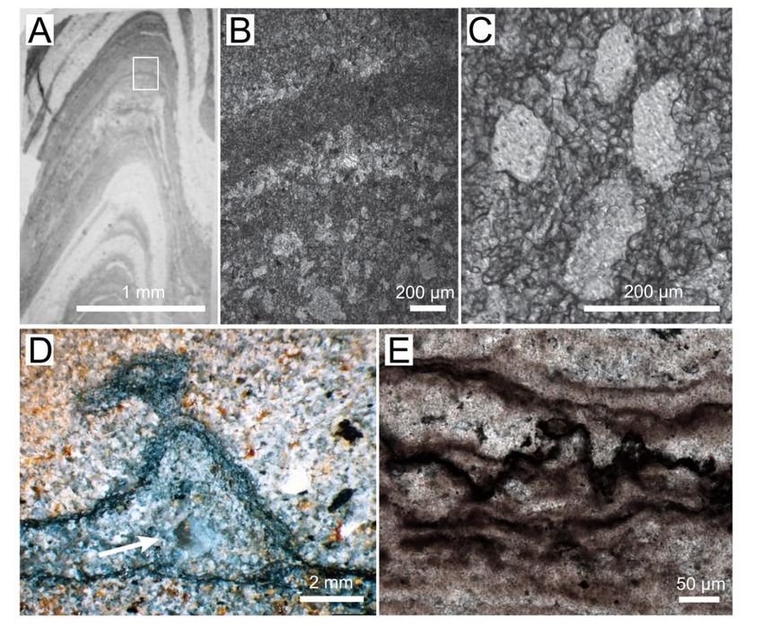
Geosciences
Geosciences2022,
2022,12,
12,x185
FOR PEER REVIEW 2 of
2 of19
18
Figure1.1.Textures
Figure Texturesin inArchaean
Archaeanand andProterozoic
Proterozoicmicrobial
microbialmats
matsand
andstromatolites.
stromatolites.(A)(A)Photograph
Photograph
ofofaathin
thin section ofof conical
conicalstromatolite
stromatolitefrom
from the
the 3 Ga
3 Ga Chobeni
Chobeni Formation,
Formation, South
South Africa.
Africa. The The
lightlight
areas
areas
containcontain
silica,silica,
and the anddark
the laminae
dark laminae contain
contain dolomite.
dolomite. White White rectangle
rectangle outlinesoutlines
the areathe area en-in
enlarged
larged in panel
panel (B). B. (B) Photomicrograph
(B) Photomicrograph of laminae
of laminae preserved
preserved by microcrystalline
by microcrystalline and microsparitic
and microsparitic dolomite
dolomite (dark). The lighter laminae contain pores filled by silica. (C) Enlarged
(dark). The lighter laminae contain pores filled by silica. (C) Enlarged view of silica-filled view of silica-filled
pores
pores surrounded by 10–100 mm–thick dolomite laminae. (D) Photograph of a polished slab show-
surrounded by 10–100 mm–thick dolomite laminae. (D) Photograph of a polished slab showing a
ing a tufted silicified mat in a sandstone from the Archean Moodies Group, South Africa [25]. The
tufted silicified mat in a sandstone from the Archean Moodies Group, South Africa [25]. The arrow
arrow points to a silica-filled void. (Reprinted with permission from ref. [25]. Copyright 2015 Else-
vier) (E)to
points a silica-filled void.
Photomicrograph (Reprinted
of the with permission
dark, organic-rich layers infrom ref. [25].
a pustular Copyright
silicified 2015the
mat from Elsevier)
~1600
(E)Proterozoic
Ma Photomicrograph
Balbirini of Dolomite,
the dark, organic-rich
Australia. layers in a pustular silicified mat from the ~1600 Ma
Proterozoic Balbirini Dolomite, Australia.
Most of the microbial constituents in marine carbonate-precipitating mats are not
readilyMost of the microbial
identifiable constituents
by eye [7,26,27], in marine carbonate-precipitating
but Cyanobacteria, the primary producersmats are not
in modern
readily identifiable by eye [7,26,27], but Cyanobacteria, the primary producers
microbialites, excrete copious extracellular polymeric substances (EPS) that bind sedi- in modern
microbialites, excrete copious extracellular polymeric substances (EPS) that bind sediments
ments (Figure 2) [7,24]. Sediment-trapping alone cannot build stromatolites—carbonate
(Figure 2) [7,24]. Sediment-trapping alone cannot build stromatolites—carbonate minerals
minerals precipitated in situ cement the microbialites and create some of their textures
precipitated in situ cement the microbialites and create some of their textures (Figure 2C,D),
(Figure 2C,D), and processes within mats could potentially influence the growth or disso-
and processes within mats could potentially influence the growth or dissolution of trapped
lution of trapped grains, as well. Microchemical measurements and visualization tech-
grains, as well. Microchemical measurements and visualization techniques combined with
niques combined with isotope labeling and microscopy have linked the activity of sulfate
isotope labeling and microscopy have linked the activity of sulfate reducing bacteria and
reducing bacteria and cyanobacteria to carbonate precipitation and cementation in mod-
cyanobacteria to carbonate precipitation and cementation in modern microbialites and
ern microbialites and carbonate grains (see References [7,8,28–39]). However, these ap-
carbonate grains (see References [7,8,28–39]). However, these approaches alone cannot
proaches alone cannot explain the mind-boggling diversity of microbes in modern mats
explain the mind-boggling diversity of microbes in modern mats [40–45], confirm specific
[40–45], confirm specific microbial interactions that effect mineralization, or predict which
microbial interactions that effect mineralization, or predict which textures and microbialite
textures and microbialite
morphologies will form. morphologies
Consequently,will form. Consequently,
the incomplete the incomplete
understanding of modernunder-
micro-
standing of modern microbialites impacts what we can learn from Archean
bialites impacts what we can learn from Archean and Proterozoic stromatolites, andwhich
Protero-
are
zoic stromatolites,
characterized by awhich aregreater
notably characterized
diversityby
ofastromatolite
notably greater diversity
textures of stromatolite
compared to modern
textures compared
stromatolites to modern
(Figure 1). stromatolites (Figure 1).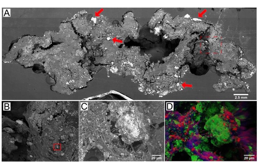
Geosciences 2022, 12, x FOR PEER REVIEW 3 of 19
Geosciences 2022, 12, 185 3 of 18
Figure 2. SEM and EDS images of microbial–carbonate interactions in a microbial mat. (A) SEM
Figure 2. SEM and EDS images of microbial–carbonate interactions in a microbial mat. (A) SEM
of a peritidal pustular mat from Shark Bay, Australia. Arrows point to the aragonite grains trapped
of a peritidal pustular mat from Shark Bay, Australia. Arrows point to the aragonite grains trapped
by the mat. Red box (dashed) shows the area enlarged in panel (B). (B) Enlarged view of the area
by the mat.
outlined by Red boxbox
the red (dashed) shows
in panel the area
(A). Red enlarged
box shows theinarea
panel (B). (B)inEnlarged
enlarged view
panel (C). (C) of the area
Microcrys-
outlined by the redthat
talline aragonite boxprecipitates
in panel (A).in
Red boxsome
EPS, showscyanobacterial
the area enlarged in panelare
filaments (C). (C)visible.
also Microcrystalline
(D) EDS
aragonite that precipitates
map of carbon (red), calciumin (green),
EPS, someandcyanobacterial
sulfur (blue) offilaments
the regionareshown
also visible. (D)
in panel EDS map of
(C).
carbon (red), calcium (green), and sulfur (blue) of the region shown in panel (C).
The tools of molecular biology can address biomineralization mechanisms at very
The tools
fine scales of molecular
to reveal biology
processes can address
by which specificbiomineralization
organisms, genes, mechanisms at very
biomolecules, and fine
mi-
scales to reveal processes by which specific organisms, genes, biomolecules, and
crobial interactions influence the precipitation of carbonates and other minerals. Broadlymicrobial
interactions influence
defined molecular the precipitation
techniques encompassof carbonates
all methods andthat
otherelucidate
minerals.the
Broadly defined
structure and
molecular techniques encompass all methods that elucidate the structure and function
function of biomolecules (namely nucleic acids, proteins, lipids, or carbohydrates) or har- of
biomolecules (namely nucleic acids, proteins, lipids, or carbohydrates) or harness
ness the properties of biomolecules to observe biological processes. Over the past three the proper-
ties of biomolecules to observe biological processes. Over the past three decades, molecular
decades, molecular tools—specifically those based on nucleic acid sequencing—have re-
tools—specifically those based on nucleic acid sequencing—have revealed substantial micro-
vealed substantial microbial, gene, metabolic, and organic diversity in microbial mats.
bial, gene, metabolic, and organic diversity in microbial mats.
Here, we review the application of molecular biological approaches to the study of
Here, we review the application of molecular biological approaches to the study of
mineral precipitation in modern microbialites. Although the diversity of biomineralizing
mineral precipitation in modern microbialites. Although the diversity of biomineralizing
microbial ecosystems and biomolecules is vast, this review specifically focuses on the use
microbial ecosystems and biomolecules is vast, this review specifically focuses on the
of nucleic acid–based methods to investigate processes in modern marine carbonate pre-
use of nucleic acid–based methods to investigate processes in modern marine carbonate
cipitating microbialites in Shark Bay, Australia; and Highbourne Cay, The Bahamas, as
precipitating microbialites in Shark Bay, Australia; and Highbourne Cay, The Bahamas,
two extensively studied modern sites. We primarily consider nucleic acid sequencing
as two extensively studied modern sites. We primarily consider nucleic acid sequencing
techniques because they are at once powerful and accessible, and because they have been
techniques because they are at once powerful and accessible, and because they have been
applied commonly in some of the most studied modern microbial carbonate systems.
applied commonly in some of the most studied modern microbial carbonate systems.
2. Microbial
2. Influences on
Microbial Influences on Mineral
Mineral Precipitation
Precipitation inin Microbialites
Microbialites
Molecular techniques
Molecular techniques are are one
one powerful
powerful means
means ofof observing
observing and
and characterizing
characterizing the
the
biological factors
biological factors at
at play
play inin mineralization
mineralization (Figure
(Figure 3).
3). These
These biological
biological factors
factors fall
fall broadly
broadly
into two categories: metabolic activities that change the chemical environment to promote
or inhibit
inhibitmineralization;
mineralization;and and organic
organic compounds
compounds that influence
that influence mineralmineral nucleation,
nucleation, growth,
growth, ordering
ordering and shape.and shape.
The The precipitation
precipitation and dissolution
and dissolution of minerals
of carbonate carbonatedepend
mineralson de-
the
pend on
degree to the degree
which to which
carbonate carbonate
minerals minerals are or
are oversaturated oversaturated
undersaturatedor undersaturated
in solution, whichin
solution,
itself whichonitself
depends depends
carbonate on carbonate
alkalinity alkalinity
[4,31,46]. Microbial[4,31,46]. Microbial
communities communities
locally influence
locally
the influence
alkalinity andthethealkalinity
saturationand theofsaturation
state carbonatestate of carbonate
minerals minerals
via metabolic via metabolic
activities that act
activities
as that actor
direct sources assinks
directfor
sources or sinks
carbonate ions for carbonate
or induce ions or
changes induce
in the pH. changes in the pH.
An understanding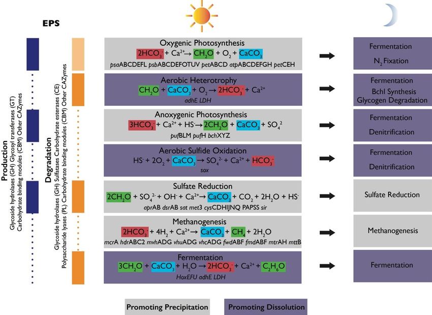
Geosciences 2022, 12, 185                                                                                                                               4 of 18
       Geosciences 2022, 12, x FOR PEER REVIEW                                                                                         4 of 19

                                       ofAncommunity
                                            understandingstructure    and the
                                                            of community         effects
                                                                             structure  andofthe
                                                                                              various    metabolic
                                                                                                 effects of          guilds enables
                                                                                                            various metabolic guilds en-predictions of
                                       the  netpredictions
                                        ables    effect of of
                                                           this
                                                              thealkalinity
                                                                  net effect ofengine   (Figure
                                                                                this alkalinity   4). (Figure 4).
                                                                                                engine

                                         Figure 3. Summary schematic of potential and known factors contributing to mineralization in
                                          Figure   3. Summary schematic of potential and known factors contributing to mineralization in
                                           microbial mats. Environmental stresses enhance EPS production by photosynthetic microbes and
                                          microbial          Environmental
                                                       mats.Negatively
                                           other organisms.             charged stresses
                                                                                 functionalenhance   EPS
                                                                                            groups trap  andproduction    by photosynthetic
                                                                                                             bind free cations on the surface of microbes and
                                           EPS. Microbial
                                          other  organisms.metabolic processes
                                                              Negatively       that contribute
                                                                            charged            to EPS
                                                                                       functional      production
                                                                                                    groups          and degradation
                                                                                                              trap and               (alkalinity
                                                                                                                         bind free cations    on the surface of
                                           engine) can influence the concentrations of carbonate ions, release cations, and modify organic sur-
                                          EPS.  Microbial   metabolic    processes   that  contribute    to EPS   production
                                           faces or produce organic compounds that influence mineral nucleation and growth.
                                                                                                                                and  degradation     (alkalinity
                                          engine) can influence the concentrations of carbonate ions, release cations, and modify organic
            Geosciences 2022, 12, x FOR PEER REVIEW                                                                                          5 of 19
                                          surfaces or produce organic compounds that influence mineral nucleation and growth.

                                             Figure 4. Schematic of metabolic guilds commonly present in microbial mat communities and their
                                             contributions to the alkalinity engine (adapted from Reference [4]). Each group includes a simplified
                                             equation representing its major metabolism as it relates to the precipitation of carbonate minerals,
                                             alkalinity, and the production or degradation of organic matter [46]. Relevant functional genes as-
                                             sociated with some of these metabolisms were detected in Shark Bay mats [47–50]. Some metabo-
                                             lisms promote carbonate precipitation (gray boxes) or dissolution (purple boxes). Shifts in metabolic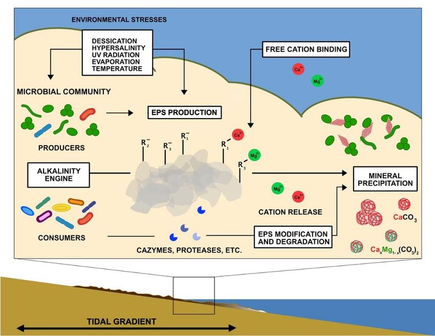
Geosciences 2022, 12, 185                                                                                                   5 of 18

                            Figure 4. Schematic of metabolic guilds commonly present in microbial mat communities and their
                            contributions to the alkalinity engine (adapted from Reference [4]). Each group includes a simplified
                            equation representing its major metabolism as it relates to the precipitation of carbonate minerals,
                            alkalinity, and the production or degradation of organic matter [46]. Relevant functional genes associ-
                            ated with some of these metabolisms were detected in Shark Bay mats [47–50]. Some metabolisms
                            promote carbonate precipitation (gray boxes) or dissolution (purple boxes). Shifts in metabolic
                            activity from day to night can alter the net effects of metabolisms. Left column shows the contribution
                            of each group to EPS cycling and lists some gene families associated with production (blue) and
                            degradation (orange). Light orange box corresponding to oxygenic photosynthesis indicates that
                            oxygenic phototrophs (Cyanobacteria) are known to recycle some of their own EPS but are generally
                            considered net producers of EPS. Vertical purple and orange boxes shown known contributions of
                            some microbial groups to EPS cycling. Dotted lines show that there is still much to learn about EPS
                            cycling in mats and the contribution of various groups of organisms.

                                  Phototrophic CO2 fixation favors the net precipitation of carbonate minerals. Oxygenic
                            photosynthesis takes in HCO3 - and releases OH− ions to increase carbonate alkalinity and pH.
                            Anoxygenic photosynthetic oxidation of HS− decreases alkalinity; however, simultaneous
                            increases in pH maintain favorable conditions for precipitation. These processes are restricted
                            to the upper illuminated parts of the mat and are offset by aerobic respiration, which favors
                            carbonate dissolution by increasing pCO2 . The effects of oxygenic photosynthesis and aer-
                            obic respiration are balanced in time. At night, oxygenic and anoxygenic phototrophs can
                            ferment, fix N2 , and contribute to denitrification— all processes that produce protons and
                            favor dissolution. For these reasons, carbonate minerals in modern microbialites precipitate
                            more extensively in the zones of microbial sulfate reduction and extensive organic degrada-
                            tion [8,32,51] and typically do not preserve the finely laminated photosynthetic textures, such
                            as those shown in Figure 1. The activity of sulfate-reducing bacteria (SRB) is less constrained
                            in time and space. The diversity of organic substrates, the completeness of substrate oxidation,
                            environmental buffering, and the extent of sulfate reduction determine the degree to which
                            sulfate reduction increases alkalinity [52,53]. Some sulfide produced by SRB can feed back into
                            anoxygenic photosynthesis and light-independent sulfide oxidation, with the latter process
                            particularly favoring carbonate dissolution (see References [45,54,55]). Other processes, such
                            as iron reduction or the release of ammonia due to protein degradation, increase alkalinity
                            and may contribute to mineral precipitation (see References [56,57]). Deeper in the mat,
                            methanogenesis and fermentation favor carbonate precipitation and dissolution, respectively
                            (see References [37,58,59]).
                                  The initial nucleation of carbonate mineral phases often imposes the largest kinetic
                            barrier to the precipitation of these minerals from oversaturated solutions. Organic molecules
                            appear to drastically reduce this barrier and control the mineral phase and ordering (see
                            References [60–67]). Incipient carbonate precipitates in modern microbial systems occur
                            as nanometer-sized grains within the EPS and on cell surfaces [21–23,32,68,69] (Figure 2).
                            Different cell or viral surfaces and microbial EPS can influence mineral grain sizes, shapes, and
                            crystal ordering [64,70–74]. Thus, organic surfaces within microbial mats are just as critical to
                            mineral nucleation, maturation, and diagenesis in microbial mats, as are major metabolisms,
                            such as photosynthesis and sulfate reducers. These surfaces include both cells themselves,
                            which can have different membrane or cell-wall characteristics (e.g., Gram-negative vs. Gram-
                            positive) relevant to mineralization [75], and their extracellular secretions.
                                  Microorganisms that live in benthic environments, and especially cyanobacteria, se-
                            crete copious amounts of EPS. The negatively charged functional groups on organic sur-
                            faces (e.g., carboxyl, acetyl, hydroxyl, succinyl, and others) can bind Ca2+ and Mg2+ and
                            are thought to be the most important mineral nucleation sites [61,74]. These functional
                            groups—in particular, carboxylic acids, phosphates, and amines—are typically assumed to
                            be present in EPS by the models used to characterize EPS using titration data [76]. Such
                            models are yet to incorporate the reports of abundant sulfate groups produced by sulfate-
                            reducing bacteria [77], pustule-forming cyanobacteria [50,78,79], and other cyanobacte-
Geosciences 2022, 12, 185 6 of 18
ria [80]. Sulfate-rich EPS produced by pustule-forming cyanobacteria binds magnesium to
promote microbial silicification in solutions that contain more silica compared to modern-
day seawater [78,79]. In fact, the spatial proximity of fossiliferous cation-rich chert and
texture-preserving dolomite has generated testable hypotheses about the contribution of
Mg-binding microbial surfaces in the formation of both chert and Mg-rich carbonates (see
Figure 1; see References [78,80].
EPS degradation changes the chemical structure of the extracellular matrix. This
process modifies and removes functional groups [50], changes the acidity of the surfaces,
renders smaller fragments of macromolecules, and releases multivalent cations back into
the environment, where they can form minerals [19,20,29,30,35,74,77,81]. The downstream
degradation of smaller insoluble and soluble products of EPS degradation may further
promote this precipitation by fueling sulfate reduction and the alkalinity engine [34].
Accordingly, extensive carbonate mineral precipitation in modern microbialites occurs
primarily in the zones of organic degradation, in voids, and outside of the active photo-
synthetic layer. Therefore, characterizing, tracking, and quantifying the cycling of EPS in
different regions of microbial mats is critical for our understanding of biomineralization in
microbial systems.
3. Using Molecular Tools to Link Microbes to Mineralization in Microbialites from
Shark Bay, Australia; and Highbourne Cay, Bahamas
The stromatolites and mats from hypersaline Shark Bay, Australia (Figure 5), and
marine stromatolites and thrombolites in The Bahamas are among the world’s most promi-
nent and well-studied microbialites [42,43]. While a comprehensive summary of past
sequencing-based studies of the many extant microbial carbonate systems is beyond the
scope of this review, these two microbialite systems are excellent case studies of how molec-
ular techniques—principally SSU 16/18S rRNA amplicon sequencing, metagenomics, and
transcriptomics—have been applied to investigate mechanisms that result in different mat
morphologies and textures and influence carbonate alkalinity in marine microbial mats.
To date, these studies have described the compositions of microbial communities in mats
of different morphological types, at different geographical locations, and within different
mat layers, and characterized the diversity of Cyanobacteria and sulfate reducers—the
two functional clades with the most explicitly established links to the alkalinity engine
and carbonate precipitation. More recent studies have expanded on this by measuring the
expression of genes associated with photosynthesis, sulfate reduction, and EPS production
and degradation in time and space.
The Shark Bay mats have been the subject of molecular ecology studies since the
early 2000s [82,83]. Burns, Goh, Allen, and Neilan [83] created the first 16S rDNA clone
libraries of Shark Bay stromatolites, and within one year, Papineau et al. [84] had provided
the first quantitative description of Shark Bay microbial community structure based on
16S rRNA amplicon data. Molecular studies began a few years later in The Bahamas, where
a robust collection of the literature on the biogeochemical and microscopic observations
had already established strong links between certain types of microbial activity—primarily
photosynthesis, sulfate reduction, and EPS cycling—and mat lithification [7,8,35,40]. The
first amplicon-based studies of Bahamian mats characterized the diversity of Cyanobacte-
ria [85–88] and sulfate reducers [36]. Laboratory studies of whole mat samples and isolates
of mat organisms from The Bahamas used nucleic acid sequencing to assess microbial diver-
sity [89–91]. Within one decade of the earliest molecular studies, the microbial community
compositions of Shark Bay [42,48,92,93] and Bahamian [94–99] mats and microbialites had
been extensively described, using small-subunit rRNA sequencing. Broadly speaking, these
studies found abundant (>10%) Cyanobacteria and Proteobacteria in the Bahamian mats
regardless of mat type. In contrast, Cyanobacteria typically accounted for only ~5% of the
mat community in Shark Bay, where Proteobacteria, Bacteroidetes, Planctomycetes, and
Firmicutes were more abundant. The reasons behind these differences remain unclear andand transcriptomics—have been applied to investigate mechanisms that result in different
mat morphologies and textures and influence carbonate alkalinity in marine microbial
mats. To date, these studies have described the compositions of microbial communities in
mats of different morphological types, at different geographical locations, and within dif-
Geosciences 2022, 12, 185 ferent mat layers, and characterized the diversity of Cyanobacteria and sulfate reducers— 7 of 18
the two functional clades with the most explicitly established links to the alkalinity engine
and carbonate precipitation. More recent studies have expanded on this by measuring the
expression of genes
invite questions associated
about with photosynthesis,
the functional sulfate
roles of organisms reduction,
from all theseand EPSinproduc-
groups different
tion and degradation
microbial mats. in time and space.
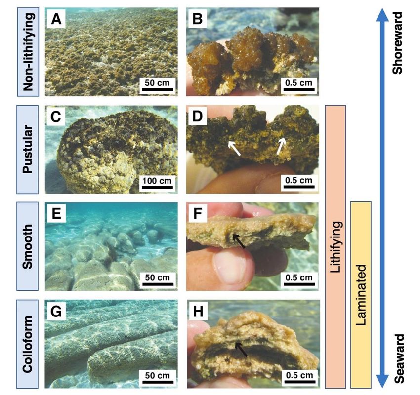
Figure
Figure 5. 5.Modern
Modernmatmattypes
types in
in Hamelin
HamelinPool, Pool,Shark
SharkBay,
Bay,Australia.
Australia.Adapted
Adaptedfromfrom
Babilonia, Conesa,
Babilonia,
Casaburi,
Conesa, Pereira,Pereira,
Casaburi, Louyakis, Reid, and
Louyakis, Foster
Reid, and[47], under
Foster [47],the license
under theCC-BY
license4.0. (A) Sheet
CC-BY of EPS-rich
4.0. (A) Sheet
of non-lithifying
EPS-rich non-lithifying
pustular matspustular
in the mats in the
intertidal intertidal
zone. zone. (B) of
(B) Cross-section Cross-section of EPS-richpustular
EPS-rich non-lithifying non-
lithifying pustular mat. (C) Pustular stromatolite-forming mat. (D) Cross-section of lithified
mat. (C) Pustular stromatolite-forming mat. (D) Cross-section of lithified pustular mat. (E) Lithified pustu-
larsmooth
mat. (E) Lithified
mats smooth mats
(F) Cross-section (F) Cross-section
of smooth mat showingof smooth mat showing
laminations. laminations.
(G) Lithified colloform (G)
matsLithi-
have a
fied colloform mats have a bumpier surface texture and less fine laminations than smooth mats. (H)
bumpier surface texture and less fine laminations than smooth mats. (H) Cross-section of a colloform
Cross-section of a colloform mat domal structure. Arrows in panels (D,F,H) indicate the cyanobac-
mat domal structure. Arrows in panels (D,F,H) indicate the cyanobacterial layers.
terial layers.
Even before metagenomics and transcriptomics opened their respective windows
on The Shark Bay
functional mats have
potential and been
gene the subject of
expression inmolecular ecology studies
mats, researchers since the early
began attempting to as-
2000s
sociate the molecular ecology of mats with functions, mat textures, andclone
[82,83]. Burns, Goh, Allen, and Neilan [83] created the first 16S rDNA libraries
mineralization
ofpotential.
Shark BayEarly
stromatolites, andinwithin
fluorescence one year, Papineau
situ microscopy targetingettheal.16S
[84]rRNA
had provided the first
of sulfate-reducing
quantitative description of Shark Bay microbial community structure
bacteria showed that these presumed anaerobes were actually active in oxygenatedbased on 16S rRNA mat
amplicon data.
layers and inMolecular studies
close contact withbegan a few years later
oxygen-producing in The Bahamas,
cyanobacteria [36].where a robustmi-
The greater
collection of the literature
crobial diversity on the
of the more biogeochemical
lithified Bahamian and mat microscopic observations
types was tentatively had al-to
attributed
ready established strong links between certain types of microbial activity—primarily
a greater metabolic diversity and biogeochemical conditions that would favor mineral-
ization [96,97]. Similarly, the differences in the abundances and types of Cyanobacteria
and other community components across the major classes of Bahamian stromatolites and
thrombolites were hypothesized to either drive or reflect some morphological and biogeo-
chemical differences [94,96,97]. Low eukaryotic diversity in five thrombolitic mats was
interpreted as evidence against thrombolites being simply “bioturbated stromatolites” [94].
A combination of phylogenetic analysis and culture experiments testing for antagonistic in-
teractions between microbes isolated from Bahamian mats suggested that such interactionsGeosciences 2022, 12, 185 8 of 18
between heterotrophs, including Gammaproteobacteria, Firmicutes, Bacillus, Halobacillus, or
Exigunobacterium, in Bahamian mats could drive lamination [100].
Niche differentiation refers to the spatial distribution of microbes within mats based
on amplicon-based microbial community composition. This concept is often used to un-
derstand mineralization in Shark Bay and some other environments [101,102]. In arguably
the most comprehensive study addressing niche differentiation in Shark Bay, Wong, Smith,
Visscher, and Burns [42] combined depth-resolved 16S rRNA amplicon sequencing with bio-
geochemical data including oxygen depth profiles and measurements of sulfate reduction
rates in two Shark Bay mat types. This study hypothesized that the differences in lithifica-
tion between the lithifying smooth mats and the non-lithifying pustular mats depended
on the lower abundances of Cyanobacteria and Deltaproteobacteria the latter [42]. The
same study also used the co-occurrence of certain taxonomic groups to motivate ecological-
functional hypotheses. Specifically, the detection of Bacteroidetes, Proteobacteria, and
Cyanobacteria in the upper layers of mats was interpreted as possible evidence for the
“phototrophic consortia” that drive primary production and motivated an early argument
for Bacteroidetes as degraders of EPS in mats [42].
As sequencing became less expensive, metagenomic studies enabled researchers to move
beyond arguments based on niche differentiation and taxonomy and attempt to connect min-
eralization to the functional potential of mat microbes more directly [38,41,47,48,86,103–106].
Building on previous metagenomic work in other mats [10,11], the first metagenomic studies
of lithifying mats in the Bahamas [86,103] and Shark Bay [38] confirmed many of the core find-
ings of earlier 16S/18S rRNA-based community profiles and the presence of genes encoding
for photosynthesis and sulfate reduction in the mat metagenome.
Some studies also proposed new hypotheses that linked the mat microbial community
to carbonate precipitation. Khodadad and Foster [103] attributed the primary difference in
functional potential between non-lithifying and lithifying stromatolitic mats in the Bahamas
to the enrichment of carbohydrate-processing genes in lithifying mats. They interpreted
this finding as a potential indication of enhanced EPS degradation in lithifying mats
combined with the faster consumption of a wider variety of organic and inorganic sulfur-
containing substrates (e.g., sulfate, thiosulfate, etc.) [103]. Sulfate commonly modifies the
EPS of pustular mats from Shark Bay, Western Australia, where a combination of culturing
and metagenomic analyses identified Cyanobacteria as the main producers of sulfated
polysaccharides [50]. The same study proposed a connection among the ecology, chemical
properties and biogeochemical cycles in lithifying pustular mats by detecting sulfatases,
enzymes required to degrade sulfated polysaccharides, in a number of metagenome-
assembled genomes, quantifying sulfatase activity and associating carbonate precipitates
with areas with fewer cyanobacteria and less sulfate-rich EPS.
Comparisons among the metagenomes of different mat types in Shark Bay and The
Bahamas motivated qualitative hypotheses regarding the relative importance of photo-
synthesis and heterotrophy in driving the carbonate alkalinity engine. A combination of
metagenomic and biogeochemical data from Bahamian thrombolites pointed to photosyn-
thesis as the most important driver of mineralization in thrombolites [105]. In contrast,
Ruvindy, White, Neilan, and Burns [38] argued that the greater prevalence of predicted
photosynthesis genes in the metagenomes of Bahamian mats compared to Shark Bay mats
could indicate that Shark Bay biomineralization is driven more by heterotrophy than by
photosynthesis. Analyses of functional genes and microscopy in a gradient of stabilized
to high-energy mobilized oolitic sands were used to argue for the roles of diverse mi-
crobial metabolisms, including the degradation of EPS, in the precipitation of carbonate
ooids [107,108]. Stable isotope analyses of the mat and ooid carbonate provided additional
support for photosynthesis, rather than heterotrophy, as the primary driver of mineraliza-
tion in Bahamian mats [38] and isotopic imprints of photosynthetic processes in marine
ooids [39]. Because the relative abundance of genes associated with photosynthesis was
greater in pustular lithifying mats from shallower waters in Shark Bay compared to the
smooth and colloform mats at greater depths, Babilonia and co-authors [47] argued thatGeosciences 2022, 12, 185 9 of 18
lithification in shallower mats might be controlled more by photosynthesis than heterotro-
phy, and vice versa for deeper mats. Different abundances of genes from different functional
pathways in the metagenomes from adjacent stromatolites and thrombolites in Highbourne
Cay and Shark Bay were also interpreted as evidence that some as-yet opaque aspect of
microbial community metabolism could underpin their differing mineral fabrics [109].
Most recently, transcriptomics has allowed researchers to track gene expression in mats,
particularly those associated with photosynthesis and heterotrophy [49,105,109,110]. Depth-
resolved metatranscriptomics of Bahamian thrombolites characterized gene expression at
midday and, unsurprisingly, found more gene transcripts in pathways related to photosyn-
thesis relative to anaerobic respiration [109]. Most of the photosynthesis gene transcripts
were associated with cyanobacterial genera closely related to Dichotrix sp. (order Nosto-
cales), the dominant cyanobacterial clade in Bahamian mats [109]. A subsequent year-long
transcriptomic survey identified the filamentous Rivulaceae and coccoidal Xenococcaceae
as the most active members of the community in the Bahamian thrombolites [104].
The metatranscriptomic analyses of the microbial activity over seasonal and diel cycles
and in multiple mat types in Shark Bay that included cyclone-derived materials (EPS-rich
cobbles and sludge) found abundant and active Bacteroidetes and sulfate-reducing bacteria
in the EPS-rich cobbles [49]. This study argued that heterotrophic degradation of EPS by
Bacteroidetes coupled to sulfate reduction could explain the greater amount of carbonate
found in cobbles relative to sludge. This may be the first time that an explicit link between
carbonate alkalinity and the activity of Bacteroidetes—an abundant phylum in Shark
Bay and Highbourne Cay—has been proposed. Elevated transcription of sialic acid and
aTMP–rhamnose synthesis pathways in the cobbles was also interpreted as an indication
that there could be two different kinds of EPS present in cobbles, each contributing to
the matrix’s resilience against degradation or cohesive properties in different ways that
could potentially impact the shape and preservation of cobbles [110]. The high functional
potential of Bacteroidetes, Planctomycetes, Verrucomicrobia, Chloroflexi, Myxococcota,
and a few other microbial groups from pustular mats in Shark Bay for the degradation of
sulfated EPS supports inferences from the transcriptomic data [50]. Molecular tools can
now proceed to identify organisms involved in these pathways and their connections to the
activities of Cyanobacteria, sulfate-reducing bacteria, and the many other microbial groups
in the mats.
4. What Next?
After nearly two decades of molecular studies, the community compositions of the
carbonate-precipitating mats from Shark Bay and The Bahamas have been described thor-
oughly, with studies based on different methods and performed by different researchers
yielding broadly consistent findings. Likewise, numerous metagenomic studies and some
transcriptomic studies have described the presence and taxonomic distribution of genes
and gene transcripts from broadly defined functional categories (e.g., elemental cycling
pathways, “carbohydrate metabolism”, etc.) in mats. Even so, we still do not understand
why microbial mats host many different microbial groups, genes, and metabolisms and
how the diverse microbes participate in building mats, shield the community from en-
vironmental stresses, or influence mineral precipitation. The puzzle of relating mineral
textures and morphologies preserved in fossil microbialites to biological processes that can
be observed in extant microbialites is more confounding still. Ancient mats preserved in
fossil microbialites have undergone extensive information loss, from the decay of highly
diagnostic biomolecules, such as nucleic acids, proteins, and lipids, to the alteration of orig-
inal textures by diagenesis and metamorphism [5]. Future studies of modern microbialites
should solidify evidence for the connections between molecular data and biogeochemi-
cal and microscopic observations of carbonate precipitation, identify testable hypotheses
about mechanisms that drive these connections, and then test them in situ or in simplified
model systems.Geosciences 2022, 12, 185 10 of 18
Molecular tools yield bewildering amounts of data. Today, a metagenomic dataset
is within reach of any researcher with a few thousand dollars to spare on nucleotide se-
quencing. Data analyses—and good hypotheses to explore—have displaced technical
complexities and cost as the limiting resources. More data and more powerful techniques
will surely come. Given the granularity of molecular data, which can track the presence of
individual species and their genes in space and time, how do we ensure that we do not
miss the mat for the genes? Molecular studies are also only as good as the bioinformatic
tools, reference databases, and biochemical studies that inform them. Given the large
existing molecular datasets that describe many modern microbial carbonates, one promis-
ing avenue of future molecular research is to “zoom in”, narrowing analyses to simple
testable hypotheses about the contributions of specific organisms, groups of organisms, and
microbial interactions to carbonate precipitation and the formation of microbial textures.
This hypothesis-driven approach is necessary to understand how modern marine micro-
bial carbonates form, how different microbes interact in zones of carbonate precipitation,
and how these interactions depend on environmental stresses or chemistry (Figure 3). A
process-oriented understanding of carbonate precipitating microbial systems under a range
of environmental chemical conditions relevant to the past is necessary to understand the
textural differences between modern and Archean or Proterozoic microbialites (see Figure 1;
see Reference [6]).
The precipitation of dolomite is an example of a geological puzzle that will require
consideration of the roles of specific strains, genes, microbial surfaces, and metabolisms
to solve. This mineral preserved exquisite microbial textures in rocks of Archean and
Proterozoic ages (see Figure 1; see References [5,111–113]), but is uncommon or not fabric-
retentive in more recent rocks [114–116]. The abiotic formation of dolomite is kinetically
limited [117,118], so it has been proposed that microbes and/or organic compounds could
mediate its formation. Indeed, culture studies and molecular analyses linked various
bacteria to the precipitation of protodolomite, including Desulfovibrio brasiliensis [119,120],
Virgibacillus sp. [68,76], and Desulfobulbus mediterraneus [67]. The phases precipitated with
microbial or organic compounds tend to be disordered (proto)dolomite [60,65,76,121], with
only minor amounts of dolomite present [60]. Thus, the formation of ordered dolomite
may require additional factors, such as manganese cations [64]. In the future, molecular
methods could be used to understand the roles of other microbes in dolomite precipitation,
as well as the role of different microbial EPSs and their degradation in the nucleation of
nanocrystalline dolomite. Anoxygenic phototrophs may require special attention. Recent
16S rRNA amplicon sequencing revealed a seasonal shift in the hypersaline sabkhas of Qatar
from oxygenic phototrophs to anoxygenic phototrophs. Because this shift was accompanied
by a change in EPS functional groups, anoxygenic phototrophs were hypothesized to be
responsible for dolomite precipitation [122]. Studies of microbial biofilms in Archean-
analog solutions also documented the formation of ordered dolomite on the surfaces of
the green sulfur bacterium Chlorobium limicola and nanocrystalline dolomite on EPS in
Chlorobium-containing biofilms [64,123]. Cyanobacteria, too, have been shown to influence
the crystallinity and morphology of magnesium carbonates. They are associated with the
globular precipitates of hydromagnesite in lake Salda, Turkey; and dypingite in the alkaline
wetlands of Atlin, British Columbia [124,125]. It is possible that these precipitates coalesce
to form the larger-scale clotted textures that are common in these environments [73,124],
but more targeted molecular studies are needed to elucidate the contribution of specific
microbes to mineral textures in these and other magnesium-rich carbonate structures.
Molecular tools, field observations, and experiments could also illuminate the under-
studied contributions of viruses to mat lithification. Viruses could potentially impact miner-
alization by providing mineral nucleation sites, modulating microbial community composi-
tion via lysis and transferring auxiliary metabolic genes that alter host metabolism [126].
To date, the molecular diversity of viruses in carbonate-precipitating systems has received
the most attention [127,128], whereas viral contributions to lithification have been explored
less frequently [129,130]. In one such study, Pacton et al. [131] suggested that mineral pre-Geosciences 2022, 12, 185 11 of 18
cipitation can occur directly on viral surfaces and on cell debris created from cell lysis. The
inferred viral particles in this study were first permineralized by amorphous magnesium
silicates that were subsequently converted to magnesium carbonate nanospheres [131]. A
recent study [129] reported potential viruses embedded in high-Mg calcite grains in calci-
fying microbial mats from the hypersaline lake La Salada de Chiprana, Spain. Molecular
methods for virus characterization in lithifying systems combined with mineral growth
experiments could evaluate the potential of viruses to nucleate Mg-rich carbonate minerals
and distinguish viruses from other biomineralizing surfaces [132,133], such as membrane
vesicles that can become coated by dolomite [63,121].
The narrowed scope of “zoomed in” future studies could also explore specific functions
or metabolisms, such as EPS degradation. A number of studies of bulk mat biogeochemistry
and local EPS properties described above identified EPS cycling as key to biomineralization,
but molecular techniques have barely begun to investigate EPS cycling and the role of this
cycling in carbonate precipitation [5,12,13,50]. Thus far, molecular tools have identified
Bacteroidetes, Alpha- and Gammaproteobacteria, and Planctomycetes as some of the
most abundant taxa across many microbial mats [42,48,49,102]. The reasons behind these
abundances and the connections of these taxa to EPS cycling, the modification of organic
surfaces, and biomineralization remain to be established. This knowledge gap persists
due to the difficulties associated with characterizing the chemical composition of EPS and
narrowing down the molecular data to a certain function or metabolism. To address these
challenges, future studies could tractably focus on microbes that possess or express large
numbers of diverse carbohydrate-active enzymes (CAZymes); connect their activities to
EPS modification [50] and degradation; track the flow of carbon from these to other mat
organisms; and establish stronger links between the diversity of organisms, genes, and
compounds present in the zones of active carbonate nucleation. Proteomic tools could also
characterize the protein fraction of EPS to reveal any matrix-bound enzymes involved in
the degradation of EPS. Given that such enzymes are “available” for use not only by the
organism that excreted them but also by its neighbors—which, in many cases, are also
bound to the matrix—these enzymes may be key to understanding the relationships among
organisms involved in EPS cycling. The organisms whose genomes encode EPS-degrading
enzymes may serve as libraries of polysaccharide and protein-degrading potential for
the entire community, enabling organisms without their own extracellular CAZymes
to play important roles in downstream EPS degradation. Future studies that focus on
this question will benefit from the improvement and further development of databases
and annotation tools for carbohydrate-active enzymes and sulfatases, such as the CAZy
database [134], the dbCAN CAZyme-annotation web server [135,136], and the SulfAtlas
sulfatase database [137]. Likewise, obtaining more reference genomes for isolated and
characterized marine polysaccharide degraders and more references sequences for isolated
and characterized proteins that degrade marine polysaccharides, of which there are very
few, will improve our ability to interpret molecular data related to EPS degradation and tie
genes to function.
In parallel, future studies could “zoom out” to explore microbial influences on carbon-
ate precipitation over broader spans of time and space. For example, metatranscriptomic
analyses of calcifying cobbles in Shark Bay found a greater abundance of genes associated
with heterotrophic and anaerobic metabolisms after the cyclone and elevated transcription
of genes associated with the production and protection of EPS [110]. Another study of
seasonal changes related the growth of subtidal microbial mats during the summer season
to the preferential stabilization of sediments in Shark Bay [15], with consequences yet to
be determined for lithification. These studies highlight the necessity of frequent sampling
across environmental gradients and multiple seasons to better relate lithification to the
microbial response to changes in their environment.
Molecular techniques beyond the sequencing-based methods that have already been
widely applied to microbial carbonate systems offer yet more opportunities to explore
biological processes linked to mineralization. Proteomics [138] and lipidomics [139], asGeosciences 2022, 12, 185 12 of 18
well as the determination of the structures of EPS polysaccharides [140], will be vital for
understanding the biochemical environment in mats and how this environment ultimately
shapes mineral textures and fabrics. Likewise, a targeted analysis of mat composition using
spatially resolved methods such as Mass Spec Imaging (MSI) and desorption electrospray
ionization (DESI) could enable spatially resolved, real-time observation of biomolecules in
mats [141,142]. Nucleic acid sequencing information will be essential in identifying and
tracking the sources and sinks of the biomolecules detectable by these other techniques.
Representative cultures of relevant organisms can help circumvent at least some issues
that arise from the inherent complexity of natural EPS and tackle numerous hypotheses
that arise from -omic information. Defined or less diverse cultures and laboratory exper-
iments can focus directly onto the roles of specific organisms, metabolic or biosynthetic
pathways and related genes, and partnerships among all of these in carbonate precipitation.
Laboratory experiments can also establish environments that might not be represented
naturally on Earth today but are analogs of conditions in the past and on other planets.
Because mineral nucleation and growth in mats begins at scales smaller than or compa-
rable to individual cells, studies of lithification in microbial systems will benefit from
molecular techniques that can track gene expression and the synthesis of proteins and
polysaccharides in defined systems [143–145]. These methods, as well as spatially resolved
transcriptomics [146,147], FISH, and NanoSIMS with isotope or other types of labels can
then characterize the spatial distributions of the known, as well as the currently understud-
ied, organisms [36,148–151] and visualize microbial activity, localization, metabolisms, and
matrix properties in complex biofilms and microbe–mineral systems [152–157].
Although many of these studies focus on individual microbes, genes, and processes,
they can eventually be scaled up to multi-microbe model systems to better quantify the
effects of microbial interactions on biomineralization and texture formation, starting with
model systems, expanding insights to natural mats, engineering and decarbonization
applications [158]. Integrating the insights gained from molecular studies in modern
microbialites and laboratory model systems with other approaches will be critical for
connecting studies of modern mats to mineral textures and mat morphologies in ancient
microbialites. Through these multi-pronged approaches, we can start to develop predictions
of carbonate microbialites and microbial textures that integrate processes controlled by
different genes, organic surfaces, and microbial metabolisms and superimpose them onto
the background of environmental evolution to reconstruct the rich record of microbial
textures preserved in carbonate microbialites from the Archean Eon onward.
Author Contributions: Conceptualization, E.M.C. and T.B.; writing—original draft preparation,
E.M.C., M.J.B., J.G., J.H., K.R.M., E.J.S. and T.B.; writing—review and editing, E.M.C. and T.B.;
visualization, E.M.C., E.J.S., M.J.B., J.G. and K.R.M.; supervision, T.B.; project administration, E.M.C.
and T.B.; funding acquisition, T.B. and E.M.C. All authors have read and agreed to the published
version of the manuscript.
Funding: This research was funded by The Simons Foundation Collaboration on the Origins of Life
(SCOL) grant number 327126 to TB and the NASA 80NSSC20K0234 grant to TB. The APC was funded
by the John V. Jarve MIT Internal Award to TB. This material is based upon work supported by the
National Science Foundation Graduate Research Fellowship under Grant No. 1745302 to EC. Any
opinion, findings, and conclusions or recommendations expressed in this material are those of the
authors(s) and do not necessarily reflect the views of the National Science Foundation.
Data Availability Statement: Not applicable.
Acknowledgments: The authors acknowledge the Center for Nanoscale Systems at Harvard Univer-
sity for providing facilities to perform the SEM/EDS analyses.
Conflicts of Interest: The authors declare no conflict of interest.Geosciences 2022, 12, 185 13 of 18
References
1. Allwood, A.C.; Walter, M.R.; Kamber, B.S.; Marshall, C.P.; Burch, I.W. Stromatolite reef from the Early Archaean era of Australia.
Nature 2006, 441, 714–718. [CrossRef] [PubMed]
2. Tice, M.M.; Lowe, D.R. Photosynthetic microbial mats in the 3416-Myr-old ocean. Nature 2004, 431, 549–552. [CrossRef] [PubMed]
3. Homann, M. Earliest life on earth: Evidence from the Barberton Greenstone Belt, South Africa. Earth-Sci. Rev. 2019, 196, 102888.
[CrossRef]
4. Dupraz, C.; Reid, R.P.; Braissant, O.; Decho, A.W.; Norman, R.S.; Visscher, P.T. Processes of carbonate precipitation in modern
microbial mats. Earth-Sci. Rev. 2009, 96, 141–162. [CrossRef]
5. Bosak, T.; Knoll, A.H.; Petroff, A.P. The meaning of stromatolites. Annu. Rev. Earth Planet. Sci. 2013, 41, 21–44. [CrossRef]
6. Grotzinger, J.P.; Knoll, A.H. Stromatolites in Precambrian carbonates: Evolutionary mileposts or environmental dipsticks? Annu.
Rev. Earth Planet. Sci. 1999, 27, 313–358. [CrossRef]
7. Reid, R.P.; Visscher, P.T.; Decho, A.W.; Stolz, J.F.; Bebout, B.M.; Dupraz, C.; Macintyre, L.G.; Paerl, H.W.; Pinckney, J.L.; Prufert-
Bebout, L.; et al. The role of microbes in accretion, lamination and early lithification of modern marine stromatolites. Nature 2000,
406, 989–992. [CrossRef]
8. Visscher, P.T.; Reid, R.P.; Bebout, B.M. Microscale observations of sulfate reduction: Correlation of microbial activity with lithified
micritic laminae in modern marine stromatolites. Geology 2000, 28, 919–922. [CrossRef]
9. Petroff, A.; Beukes, N.; Rothman, D.; Bosak, T. Biofilm growth and fossil form. Phys. Rev. X 2013, 3, 041012. [CrossRef]
10. Petroff, A.P.; Sim, M.S.; Maslov, A.; Krupenin, M.; Rothman, D.H.; Bosak, T. Biophysical basis for the geometry of conical
stromatolites. Proc. Natl. Acad. Sci. USA 2010, 107, 9956–9961. [CrossRef]
11. Grotzinger, J.P.; Rothman, D.H. An abiotic model for stromatolite morphogenesis. Nature 1996, 383, 423–425. [CrossRef]
12. Walter, M.R.; Bauld, J.; Brock, T.D. Microbiology and morphogenesis of columnar stromatolites (Conophyton, Vacerrilla) from hot
springs in Yellowstone National Park. In Stromatolites; Walter, M.R., Ed.; Developments in Sedimentology; Elsevier: Amsterdam,
The Netherlands, 1976; Volume 20, pp. 273–310.
13. Hoffman, P. Environmental diversity of Middle Precambrian stromatolites. In Stromatolites; Walter, M.R., Ed.; Elsevier Scientific
Publishing Company: Amsterdam, The Netherlands, 1976; Volume 20, pp. 599–612.
14. Hoffman, P.F. Stromatolite morphogenesis in Shark Bay, Western Australia. In Stromatolites; Walter, M.R., Ed.; Developments in
Sedimentology; Elsevier: Amsterdam, The Netherlands, 1976; Volume 20, pp. 261–272.
15. Murshid, S.; Mariotti, G.; Pruss, S.B.; Bosak, T.; Suosaari, E.P. Seasonal changes in sediment erodibility in a sandy carbonate
environment detected from turbidity time series. Mar. Geol. 2021, 439, 106570. [CrossRef]
16. Suosaari, E.P.; Reid, R.P.; Araujo, T.A.A.; Playford, P.E.; Holley, D.K.; McNamara, K.J.; Eberli, G.P. Environmental pressures
influencing living stromatolites in Hamelin Pool, Shark Bay, Western Australia. Palaios 2016, 31, 483–496. [CrossRef]
17. Suosaari, E.; Reid, R.; Playford, P.; Foster, J.; Stolz, J.; Casaburi, G.; Hagan, P.; Chirayath, V.; Macintyre, I.; Planavsky, N. New
multi-scale perspectives on the stromatolites of Shark Bay, Western Australia. Sci. Rep. 2016, 6, 20557. [CrossRef] [PubMed]
18. Altermann, W. Accretion, trapping and binding of sediment in Archean stromatolites—Morphological expression of the antiquity
of life. Space Sci. Rev. 2008, 135, 55–79. [CrossRef]
19. Arp, G.; Reimer, A.; Reitner, J. Calcification in cyanobacterial biofilms of alkaline salt lakes. Eur. J. Phycol. 1999, 34, 393–403.
[CrossRef]
20. Arp, G.; Thiel, V.; Reimer, A.; Michaelis, W.; Reitner, J. Biofilm exopolymers control microbialite formation at thermal springs
discharging into the alkaline Pyramid Lake, Nevada, USA. Sediment. Geol. 1999, 126, 159–176. [CrossRef]
21. Couradeau, E.; Benzerara, K.; Gérard, E.; Estève, I.; Moreira, D.; Tavera, R.; López-García, P. Cyanobacterial calcification in
modern microbialites at the submicrometer scale. Biogeosciences 2013, 10, 5255–5266. [CrossRef]
22. Gautret, P.; Camoin, G.; Golubic, S.; Sprachta, S. Biochemical control of calcium carbonate precipitation in modern lagoonal
microbialites, Tikehau Atoll, French Polynesia. J. Sediment. Res. 2004, 74, 462–478. [CrossRef]
23. Sprachta, S.; Camoin, G.; Golubic, S.; Le Campion, T. Microbialites in a modern lagoonal environment: Nature and distribution,
Tikehau atoll (French Polynesia). Palaeogeogr. Palaeoclimatol. Palaeoecol. 2001, 175, 103–124. [CrossRef]
24. Neumann, A.C.; Gebelein, C.D.; Scoffin, T.P. The composition, structure and erodability of subtidal mats, Abaco, Bahamas.
J. Sediment. Petrol. 1970, 40, 274–297.
25. Homann, M.; Heubeck, C.; Airo, A.; Tice, M.M. Morphological adaptations of 3.22 Ga-old tufted microbial mats to Archean
coastal habitats (Moodies Group, Barberton Greenstone Belt, South Africa). Precambrian Res. 2015, 266, 47–64. [CrossRef]
26. Jahnert, R.J.; Collins, L.B. Characteristics, distribution and morphogenesis of subtidal microbial systems in Shark Bay, Australia.
Mar. Geol. 2012, 303, 115–136. [CrossRef]
27. Reid, R.P.; James, N.P.; Macintyre, I.G.; Dupraz, C.P.; Burne, R.V. Shark Bay stromatolites: Microfabrics and reinterpretation of
origins. Facies 2003, 49, 299–324. [CrossRef]
28. Pages, A.; Welsh, D.T.; Teasdale, P.R.; Grice, K.; Vacher, M.; Bennett, W.W.; Visscher, P.T. Diel fluctuations in solute distributions
and biogeochemical cycling in a hypersaline microbial mat from Shark Bay, WA. Mar. Chem. 2014, 167, 102–112. [CrossRef]
29. Arp, G.; Helms, G.; Karlinska, K.; Schumann, G.; Reimer, A.; Reitner, J.; Trichet, J. Photosynthesis versus exopolymer degradation
in the formation of microbialites on the atoll of Kiritimati, Republic of Kiribati, Central Pacific. Geomicrobiol. J. 2012, 29, 29–65.
[CrossRef]You can also read


