Update on the management of infant and toddler developmental dysplasia of the hip - RACGP
←
→
Page content transcription
If your browser does not render page correctly, please read the page content below

Clinical

Update on the management of
infant and toddler developmental
dysplasia of the hip

Brian Loh, Emily Woollett                                      THE HUMAN ACETABULUM is shallower            total hip replacements in Australia are
                                                               at birth than at any other time during       directly attributable to DDH.5
                                                               development. The embryonic acetabulum           The emphasis of this article is on the
This article is the third in a commissioned                    begins as a deeply set cavity that almost    management of DDH in infancy and the
series on paediatric orthopaedics.                             entirely encloses the femoral head but       early years; management of DDH during
Background                                                     gradually becomes shallower as birth         adolescence and adulthood is not included.
Developmental dysplasia of the hip                             approaches. This trend reverses after
(DDH) encompasses the pathological                             birth, with the acetabulum deepening,
spectrum of hip instability that produces                      femoral head coverage increasing and hip     Establishing the diagnosis
subluxation or dislocation and                                 morphology improving.1                       As previously stated by Williams, ‘repeated,
radiological features of abnormal
                                                                  Teleologically, it is possible that a     carefully performed clinical examinations of
acetabular development. It is important
                                                               temporarily increased hip range of           the paediatric hip from newborn to walking
to approach DDH with sound prior
knowledge to recognise, diagnose and                           motion in the third trimester is required    age remain the best method for early
manage its variety of presentations.                           to accommodate the lower limb                detection of DDH’.6 This, combined with an
                                                               within the limited intrauterine space.       understanding of the risk factors, will help
Objective
                                                               Postnatally, the sphericity of the femoral   in the early diagnosis and management of
The aim of this article is to summarise
contemporary practice in DDH with
                                                               head and deepening of the acetabulum         children with DDH (Table 1).
an emphasis on recent changes in                               are expected to increase progressively          The priority is differentiating between
diagnosis, surveillance and treatment                          through childhood.1,2                        neonatal hip instability, acetabular
recommendations for general                                       The wide spectrum of pathology            dysplasia and subluxation or dislocation
practitioners (GPs), who see infants                           affecting this process is named              of the hip, as severity of involvement
and toddlers in practice.                                      developmental dysplasia of the hip (DDH).    (Figure 1) determines the treatment
Discussion                                                     Presentations can vary from an infant born   required to obtain a concentric reduction.
The management of DDH requires                                 with a fixed, irreducible dislocated hip        Neonatal hip instability is laxity limited
accurate diagnosis of the pathoanatomy                         through to mild acetabular dysplasia only    to the hip joint capsule and is influenced
and depends on the age of the child. GPs                       diagnosed in adulthood.3 Management          by hormonal and genetic factors.7–10 This
are essential for the early detection of                       of this wide continuum of pathology          laxity has a natural tendency to correct
DDH, which then allows for harnessing
                                                               requires careful consideration, because      itself, with 95.3% of hips returning to
of the remodelling potential of the hip
cartilaginous anlage to achieve a stable                       management varies greatly as the age of      normal morphology by 12 weeks of age.11
and mature hip. GPs also play an                               the child increases.4                           The spectrum continues to more
important part in surveillance, and                               DDH and its sequelae continue to          severe forms of dysplasia with worsening
this article provides an update on the                         have a major impact on healthcare, with      hip instability, to the established hip
pivotal points.                                                the Australian Orthopaedic Association       subluxation with significant hip dysplasia,
                                                               National Joint Replacement Registry          some of which may then progress to frank
                                                               (AOANJRR) reporting that nearly 7% of        hip dislocation.3,4,11,12

© The Royal Australian College of General Practitioners 2021                                                    Reprinted from AJGP Vol. 50, No. 4, April 2021   207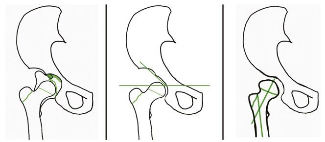
Clinical                                                                            Update on the management of infant and toddler developmental dysplasia of the hip

Imaging for DDH                                        (IHDI) classification method, which                    long-term goal of a congruent, spherical
Ultrasonography                                        demonstrates excellent interexaminer                   hip at skeletal maturity.12,19 Reduction
In the presence of risk factors or with                reliability even when ossific nuclei are               of the hip is relatively straightforward
an equivocal clinical examination,                     absent or delayed.18                                   in neonatal hip instability, but if the hip
ultrasonography is the reference standard                                                                     subluxates and remains incongruent while
in children aged 6 months

DDH, developmental dysplasia of the hip
Adapted from Department of Orthopaedics, Referral guidelines: Developmental dysplasia of the hip – DDH, Melbourne, Vic: Royal Children’s Hospital, 2020,
Available at www.rch.org.au/ortho/for_health_professionals/Developmental_dysplasia_of_the_hip_%E2%80%93_DDH/ [Accessed 4 February 2021].

208   Reprinted from AJGP Vol. 50, No. 4, April 2021                                                               © The Royal Australian College of General Practitioners 2021
Update on the management of infant and toddler developmental dysplasia of the hip                                                                             Clinical

reduced, stable and developmentally                            stabilisation.20,25,26 Care must be taken at   weeks to confirm concentric reduction both
appropriate hip while aiming to avoid                          each step of treatment to minimise the risk    clinically and radiologically. If reduction is
the complications of growth disturbance                        of iatrogenic injury.                          not achieved within this timeframe, bracing
and osteonecrosis.22,23 Early reduction                                                                       must be reconsidered and an alternative
facilitates normal acetabular development                                                                     treatment method chosen.4,23,24
and reduces the need for later pelvic                          Treatment options                                 Complications of bracing include:
surgery. A concentric reduction normalises                     Hip abduction bracing                          failure to obtain and maintain reduction,
the joint forces, which stimulates                             In a child aged
Clinical                                                                          Update on the management of infant and toddler developmental dysplasia of the hip

with a proximal tenotomy of the                        applied prior to an MRI confirmation of                 Although uncommon, re-dislocation
adductor longus.                                       concentric reduction.                                following an open reduction is a serious
   After closed or open reduction of the                                                                    complication and can markedly increase
femoral head, the hip is held in flexion,              Open reduction                                       the rate of osteonecrosis. Recent
abduction and internal rotation.18 The                 The indications for an open reduction are            modifications in technique have included
degree of abduction must be maintained                 failed closed reduction, a non-concentric            transferring the ligamentum teres as a
in the ‘safe zone’, as insufficient                    reduction and excessive hip abduction                tenodesis20,30 and the development of
abduction predisposes to re-dislocation,               in the spica cast. Open reductions can               arthroscopy-assisted techniques.31
and excessive abduction potentially                    be performed medially or anteriorly and
increases the risk of osteonecrosis.4,12,20,26         must manage all factors obstructing a                Femoral osteotomy
The stability of hip post-reduction is                 stable concentric reduction. The patient             The abnormal proximal femoral anatomy in
assessed clinically and radiologically. If             is then immobilised in a hip spica cast              DDH may predicate the need for a femoral
the safe zone is achievable within an arc              prior to a transition into a hip abduction           osteotomy to achieve reduction, reduce
of 20–60° of abduction, a spica cast is                brace at the six-week mark.20,29,30                  tension and correct the joint forces around
                                                                                                            the hip.32–34 Fixation is achieved with a
                                                                                                            blade plate and can be performed to varise,
                                                                                                            de-rotate or shorten the proximal femur.

                                                                                                            Acetabular osteotomy
                                                                                                            Insufficient coverage of the femoral head
                                                                                                            or acetabular dysplasia may require pelvic
                                                                                                            osteotomy, especially in the late-presenting
                                                                                                            child, because of the unpredictable
                                                                                                            remodelling potential of the acetabulum.
                                                                                                            Techniques have been developed to
                                                                                                            address the problems encountered in
                                                                                                            acetabular dysplasia: an abnormally
                                                                                                            oriented acetabulum with adequate
                                                                                                            capacity or an overly capacious acetabulum
                                                                                                            resulting in a dislocatable hip.35–38

                                                                                                            Conclusion
Figure 2. Intra-articular obstacles to concentric reduction of the hip in patients with                     Recent advances in surveillance, diagnosis
developmental dysplasia of the hip                                                                          and management of the DDH spectrum
                                                                                                            (Table 3) have improved outcomes for
                                                                                                            many patients, but much remains to be
                                                                                                            discovered.25,39 In 2010, the IHDI initiated
                                                                                                            an international, multicentre prospective
                                                                                                            study at nine centres across Australia,
                                                                                                            North America and Europe in response to
                                                                                                            these evidence gaps in the management of
                                                                                                            infant and toddler DDH.
                                                                                                               In the meantime, the universal tenet in
                                                                                                            DDH care remains that prompt diagnosis
                                                                                                            and management are the most important
                                                                                                            factors related to outcome; it is important
                                                                                                            to be cognisant about developmental
                                                                                                            hip pathology in asymptomatic patients
 A                                        B                             C                                   through infancy and childhood.40

Figure 3. Potential causes of residual instability in developmental dysplasia of the hip
                                                                                                            Authors
a. Patulous and redundant capsule; b. Acetabular dysplasia; c. Proximal femoral anteversion
                                                                                                            Brian Loh MBBS, BMedSci, PGradDipAnat, FRACS,
and coxa valga                                                                                              FAOrthA, Orthopaedic Surgeon, Royal Children’s
                                                                                                            Hospital, Melbourne, Vic

210   Reprinted from AJGP Vol. 50, No. 4, April 2021                                                             © The Royal Australian College of General Practitioners 2021
Update on the management of infant and toddler developmental dysplasia of the hip Clinical
19. Ramsey PL, Lasser S, MacEwen GD. Congenital
dislocation of the hip: Use of the Pavlik harness in
Table 3. Recent advances in the care of patients with developmental dysplasia
the child during the first six months of life. J Bone
of the hip Joint Surg Am 1976;58(7):1000–04.
20. Bache CE, Graham HK, Dickens DR, et al.
Pathoanatomy • Evolving understanding of the affected anatomy in DDH Ligamentum teres tenodesis in medial approach
• Emerging knowledge about the genetic basis of DDH open reduction for developmental dislocation
of the hip. J Pediatr Orthop 2008;28(6):607–13.
doi: 10.1097/bpo.0b013e318184202c.
Surveillance • Improving sonographic techniques and better interpretation
21. Sarban S, Ozturk A, Tabur H, Isikan UE.
of ultrasonography
Anteversion of the acetabulum and femoral neck
• Increasing use of MRI for confirmation of reduction in early walking age patients with developmental
dysplasia of the hip. J Pediatr Orthop B
• Development of International Hip Dysplasia Registry 2005;14(6):410–14. doi: 10.1097/01202412-
200511000-00003.
Management • Increased availability of bracing care
22. Mladenov K, Dora C, Wicart P, Seringe R. Natural
• Ligamentum tenodesis to minimise re-dislocation rate history of hips with borderline acetabular index
and acetabular dysplasia in infants. J Pediatr
• Development of arthroscopy-assisted techniques
Orthop 2002;22(5):607–12. doi: 10.1097/01241398-
• Gore-Tex and water-resistant spica cast liners 200209000-00008.
23. Alexiev VA, Harcke HT, Kumar SJ. Residual
Prevention • Increased awareness of safe swaddling techniques dysplasia after successful Pavlik harness
treatment: Early ultrasound predictors. J Pediatr
• Evolution of baby carrier slings to improve infant hip
Orthop 2006;26(1):16–23. doi: 10.1097/01.
abduction bpo.0000187995.02140.c7.
24. Upasani VV, Bomar JD, Matheney TH, et al.
DDH, developmental dysplasia of the hip; MRI, magnetic resonance imaging
Evaluation of brace treatment for infant hip
dislocation in a prospective cohort: Defining
the success rate and variables associated with
failure. J Bone Joint Surg Am 2016;98(14):1215–21.
Emily Woollett BAppSc (ClinSc), MHSc (Osteo), 10. Carroll KL, Schiffern AN, Murray KA, doi: 10.2106/JBJS.15.01018.
Research Assistant, Loh Orthopaedics, Vic et al. The occurrence of occult acetabular 25. Aarvold A, Schaeffer EK, Kelley S, et al.
Competing interests: None. dysplasia in relatives of individuals with Management of irreducible hip dislocations in
developmental dysplasia of the hip. J Pediatr infants with developmental dysplasia of the hip
Funding: None. diagnosed below 6 months of age. J Pediatr
Orthop 2016;36(1):96–100. doi: 10.1097/
Provenance and peer review: Commissioned, BPO.0000000000000403. Orthop 2019;39(1):e39–e43. doi: 10.1097/
externally peer reviewed. BPO.0000000000001205.
11. Visser JD. Pediatric orthopedics: Symptoms,
Correspondence to: 26. Gardner RO, Bradley CS, Howard A,
differential diagnosis, supplementary assessment
office@brianloh.com.au Narayanan UG, Wedge JH, Kelley SP. The
and treatment. Cham, CH: Springer International
incidence of avascular necrosis and the
Publishing, 2017.
radiographic outcome following medial open
References 12. Vitale MG, Skaggs DL. Developmental dysplasia reduction in children with developmental
1. Rális Z, McKibbin B. Changes in shape of the of the hip from six months to four years of dysplasia of the hip: A systematic review. Bone
human hip joint during its development and age. J Am Acad Orthop Surg 2001;9(6):401–11. Joint J 2014;96-B(2):279–86. doi: 10.1302/0301-
their relation to its stability. J Bone Joint Surg Br doi: 10.5435/00124635-200111000-00005. 620X.96B2.32361.
1973;55(4):780–85. 13. Roposch A, Stöhr KK, Dobson M. The effect of the 27. Wilkinson AG, Sherlock DA, Murray GD. The
2. Graf R. Hip sonography: Diagnosis and femoral head ossification nucleus in the treatment efficacy of the Pavlik harness, the Craig splint
management of infant hip dysplasia. 2nd edn. of developmental dysplasia of the hip. A meta- and the von Rosen splint in the management
Berlin, DE: Springer-Verlag, 2006. analysis. J Bone Joint Surg Am 2009;91(4):911–18. of neonatal dysplasia of the hip. A comparative
3. Sewell MD, Eastwood DM. Screening and doi: 10.2106/jbjs.h.00096. study. J Bone Joint Surg Br 2002;84(5):716–19.
treatment in developmental dysplasia of the 14. Harcke HT, Grissom LE. Performing dynamic doi: 10.1302/0301-620x.84b5.12571.
hip – Where do we go from here? Int Orthop sonography of the infant hip. AJR Am J Roentgenol 28. Malvitz TA, Weinstein SL. Closed reduction for
2011;35(9):1359–67. doi: 10.1007/s00264-011- 1990;155(4):837–44. doi: 10.2214/ajr.155.4.2119119. congenital dysplasia of the hip. Functional and
1257-z. radiographic results after an average of thirty
15. Kosar P, Ergun E, Gökharman FD, Turgut AT,
4. Joseph B, Nayagam S, Loder R, Torode I. Pediatric years. J Bone Joint Surg Am 1994;76(12):1777–92.
Kosar U. Follow-up sonography results for Graf
orthopaedics: A system of decision-making. 2nd doi: 10.2106/00004623-199412000-00004.
type 2A hips: Association with risk factors for
edn. Boca Raton, FL: CRC Press, 2016. 29. Holman J, Carroll KL, Murray KA, Macleod LM,
developmental dysplasia of the hip and instability.
5. Australian Orthopaedic Association National J Ultrasound Med 2011;30(5):677–83. doi: 10.7863/ Roach JW. Long-term follow-up of open reduction
Joint Replacement Registry (AOANJRR). Hip, Jim.2011.30.5.677. surgery for developmental dislocation of the hip.
knee & shoulder arthroplasty: 2018 annual report. J Pediatr Orthop 2012;32(2):121–24. doi: 10.1097/
Adelaide, SA: AOA, 2018; p. 122–209. 16. Quader N, Hodgson AJ, Mulpuri K, Schaeffer E, BPO.0b013e3182471aad.
Abugharbieh R. Automatic evaluation of
6. Williams N. Improving early detection of 30. Wenger DR, Mubarak SJ, Henderson PC, Miyanji F.
scan adequacy and dysplasia metrics in 2-D
developmental dysplasia of the hip through Ligamentum teres maintenance and transfer
general practitioner assessment and surveillance. ultrasound images of the neonatal hip. Ultrasound
as a stabilizer in open reduction for pediatric
Aust J Gen Pract 2018;47(9):619–23. doi: 10.31128/ Med Biol 2017;43(6):1252–62. doi: 10.1016/j. hip dislocation: Surgical technique and early
AJGP-03-18-4524. ultrasmedbio.2017.01.012. clinical results. J Child Orthop 2008;2(3):117–85.
7. Hatzikotoulas K, Roposch A, Shah K, et al. The 17. Starr V, Ha BY. Imaging update on developmental doi: 10.1007/s11832-008-0103-3.
genetic epidemiology of developmental dysplasia dysplasia of the hip with the role of MRI. AJR Am 31. Eberhardt O, Wirth T, Fernandez FF. Arthroscopic
of the hip: A genome-wide association study J Roentgenol 2014;203(6):1324–35. doi: 10.2214/ reduction and acetabuloplasty for the treatment
harnessing national clinical adult data. BioRxiv AJR.13.12449. of dislocated hips in children of walking age:
2017. doi: 10.1101/154013. 18. Narayanan U, Mulpuri K, Sankar WN, A preliminary report. Arch Orthop Trauma Surg
8. Dodinval P. [Heredity in congenital dislocation of Clarke NM, Hosalker H, Price CT. Reliability 2014;134(11):1587–94. doi: 10.1007/s00402-014-
the hip]. Acta Orthop Belg 1990;56(1 Pt A):7–11. of a new radiographic classification for 2063-z.
9. Tönnis D. Inheritance. In: Congenital dysplasia and developmental dysplasia of the hip. J Pediatr 32. Wenger DR, Lee CS, Kolman B. Derotational
dislocation of the hip in children and adults. Berlin, Orthop 2015;35(5):478–84. doi: 10.1097/ femoral shortening for developmental dislocation
DE: Springer-Verlag, 1984; p. 61–62. BPO.0000000000000318. of the hip: Special indications and results in the
© The Royal Australian College of General Practitioners 2021 Reprinted from AJGP Vol. 50, No. 4, April 2021 211Clinical Update on the management of infant and toddler developmental dysplasia of the hip
child younger than 2 years. J Pediatr Orthop 1995;
15(6):768–79. doi: 10.1097/01241398-199511000-
00009.
33. Ryan MG, Johnson LO, Quanbeck DS,
Minkowitz B. One-stage treatment of congenital
dislocation of the hip in children three to ten
years old. Functional and radiographic results.
J Bone Joint Surg Am 1998;80(3):336–44.
doi: 10.2106/00004623-199803000-00005.
34. Spence G, Hocking R, Wedge JH, Roposch A.
Effect of innominate and femoral varus derotation
osteotomy on acetabular development in
developmental dysplasia of the hip. J Bone Joint
Surg Am 2009;91(11):2622–36. doi: 10.2106/
jbjs.h.01392.
35. Gillingham BL, Sanchez AA, Wenger DR. Pelvic
osteotomies for the treatment of hip dysplasia
in children and young adults. J Am Acad Orthop
Surg 1999;7(5):325–37. doi: 10.5435/00124635-
199909000-00005.
36. Kitoh H, Kaneko H, Ishiguro N. Radiographic
analysis of movements of the acetabulum and the
femoral head after Salter innominate osteotomy.
J Paediatric Orthop 2009;29(8):879–84.
doi: 10.1097/bpo.0b013e3181c1e314.
37. DelBello DA, Nattrass GR, Moseley CF, Watts HG.
Acetabular development after open reduction in
developmental dysplasia of the hip: The role of
concurrent innominate osteotomy. Orthop Trans
1995;19:298.
38. Albinana J, Dolan LA, Spratt KF, Morcuende J,
Meyer MD, Weinstein SL. Acetabular dysplasia
after treatment for developmental dysplasia of
the hip. Implications for secondary procedures.
J Bone Joint Surg Br 2004;86(6):876–86.
doi: 10.1302/0301-620x.86b6.14441.
39. Yamamuro T, Ishida K. Recent advances in
the prevention, early diagnosis and treatment
of congenital dislocation of the hip in Japan.
Clin Orthop Relat Res 1984;(184):34–40.
doi: 10.1097/00003086-198404000-00005.
40. Rosenberg N, Bialik V, Norman D, Blazer S. The
importance of combined clinical and sonographic
examination of instability of the neonatal hip.
Int Orthop 1998;22(3):185–88. doi: 10.1007/
s002640050238.
correspondence ajgp@racgp.org.au
212 Reprinted from AJGP Vol. 50, No. 4, April 2021 © The Royal Australian College of General Practitioners 2021You can also read