UCSF UC San Francisco Previously Published Works
←
→
Page content transcription
If your browser does not render page correctly, please read the page content below
UCSF
UC San Francisco Previously Published Works

Title
HLA Upregulation During Dengue Virus Infection Suppresses the Natural Killer Cell
Response.

Permalink
https://escholarship.org/uc/item/8zw722zz

Authors
McKechnie, Julia L
Beltrán, Davis
Pitti, Arcelys
et al.

Publication Date
2019

DOI
10.3389/fcimb.2019.00268

Peer reviewed

 eScholarship.org                              Powered by the California Digital Library
                                                               University of California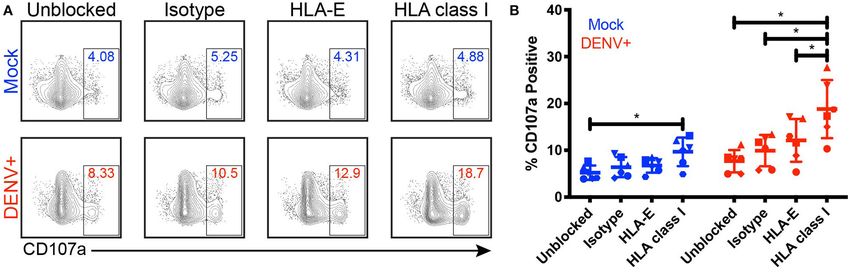
BRIEF RESEARCH REPORT
                                                                                                                                                  published: 23 July 2019
                                                                                                                                          doi: 10.3389/fcimb.2019.00268

                                              HLA Upregulation During Dengue
                                              Virus Infection Suppresses the
                                              Natural Killer Cell Response
                                              Julia L. McKechnie 1† , Davis Beltrán 2,3,4† , Arcelys Pitti 2 , Lisseth Saenz 2 , Ana B. Araúz 5 ,
                                              Rosemary Vergara 6 , Eva Harris 7 , Lewis L. Lanier 8 , Catherine A. Blish 1,6* and
                                              Sandra López-Vergès 2,3*
                                              1
                                               Program in Immunology, Stanford University School of Medicine, Stanford, CA, United States, 2 Department of Research in
                                              Virology and Biotechnology, Gorgas Memorial Institute for Health Studies, Panama City, Panama, 3 Institute for Scientific
                                              Research and Technology Services (INDICASAT-AIP), Panama City, Panama, 4 Department of Biotechnology, Acharya
                                              Nagarjuna University, Guntur, India, 5 Hospital Santo Tomas, Panama City, Panama, 6 Department of Medicine, Stanford
                                              University School of Medicine, Stanford, CA, United States, 7 Division of Infectious Diseases and Vaccinology, School of
                                              Public Health, University of California, Berkeley, Berkeley, CA, United States, 8 Department of Microbiology and Immunology
                                              and the Parker Institute for Cancer Immunotherapy, University of California, San Francisco, San Francisco, CA, United States
                           Edited by:
            Stephen Noel Waggoner,
Cincinnati Children’s Hospital Medical        Dengue virus (DENV) is the most prevalent mosquito-borne virus in the world and a major
                 Center, United States        cause of morbidity in the tropics and subtropics. Upregulation of HLA class I molecules
                       Reviewed by:           has long been considered a feature of DENV infection, yet this has not been evaluated
                       Anuja Mathew,
           University of Rhode Island,
                                              in the setting of natural infection. Natural killer (NK) cells, an innate immune cell subset
                         United States        critical for mounting an early response to viral infection, are inhibited by self HLA class
                 Salim Iqbal Khakoo,
                                              I, suggesting that upregulation of HLA class I during DENV infection could dampen the
                          University of
       Southampton, United Kingdom            NK cell response. Here we addressed whether upregulation of HLA class I molecules
                    *Correspondence:          occurs during in vivo DENV infection and, if so, whether this suppresses the NK cell
                      Catherine A. Blish      response. We found that HLA class I expression was indeed upregulated during acute
                    cblish@stanford.edu
                  Sandra López-Vergès
                                              DENV infection across multiple cell lineages in vivo. To better understand the role of
                 slopez@gorgas.gob.pa         HLA class I upregulation, we infected primary human monocytes, a major target of
     † These   authors have contributed
                                              DENV infection, in vitro. Upregulation of total HLA class I is dependent on active viral
                    equally to this work      replication and is mediated in part by cytokines and other soluble factors induced by
                                              infection, while upregulation of HLA-E occurs in the presence of replication-incompetent
                    Specialty section:
                                              virus. Importantly, blocking DENV-infected monocytes with a pan-HLA class I Fab
          This article was submitted to
        Microbes and Innate Immunity,         nearly doubles the frequency of degranulating NK cells, while blocking HLA-E does not
                 a section of the journal     significantly improve the NK cell response. These findings demonstrate that upregulation
      Frontiers in Cellular and Infection
                            Microbiology      of HLA class I during DENV infection suppresses the NK cell response, potentially
               Received: 20 May 2019
                                              contributing to disease pathogenesis.
               Accepted: 10 July 2019
                                              Keywords: dengue virus, anti-viral response, human leukocyte antigen class I, HLA-E, natural killer cells,
               Published: 23 July 2019
                                              monocytes
                               Citation:
      McKechnie JL, Beltrán D, Pitti A,
        Saenz L, Araúz AB, Vergara R,         INTRODUCTION
      Harris E, Lanier LL, Blish CA and
            López-Vergès S (2019) HLA
                                              Dengue virus (DENV) is a positive-strand RNA virus of which there are four serotypes
    Upregulation During Dengue Virus
Infection Suppresses the Natural Killer
                                              (DENV-1 to DENV-4). The virus is transmitted between humans by its vector, Aedes
                         Cell Response.       mosquitoes. Each year, an estimated 390 million people are infected with DENV (Bhatt et al.,
   Front. Cell. Infect. Microbiol. 9:268.     2013). While most DENV infections are not life-threatening, severe infections can result
       doi: 10.3389/fcimb.2019.00268          in hemorrhage, plasma leakage, shock, organ failure, and death (Kyle and Harris, 2008).

Frontiers in Cellular and Infection Microbiology | www.frontiersin.org               1                                                  July 2019 | Volume 9 | Article 268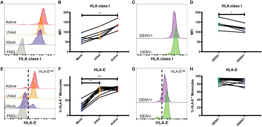
McKechnie et al.                                                                                               HLA Upregulation During DENV Infection

The incidence of dengue is rapidly rising (World Health                      I and HLA-E. We then used in vitro DENV-infected primary
Organization, 2012), increasing the need for a better                        monocytes to determine mediators of HLA class I upregulation.
understanding of how the human immune system responds to                     Finally, we co-cultured primary NK cells with autologous,
DENV infection. There is significant interest in elucidating the             DENV-infected monocytes in the presence of HLA class I
role of natural killer (NK) cells during DENV infection. NK                  blocking Fabs to determine the impact of HLA class I expression
cells are innate lymphoid cells that play a key role during the              on the NK cell response.
early stages of viral infection. Previous studies have shown that
NK cells are activated in vivo during DENV infection (Azeredo,
2006; Petitdemange et al., 2016) and that activated NK cells may             MATERIALS AND METHODS
be an indicator of a positive prognosis (Azeredo, 2006). NK                  DENV Patients and Ethical Statement
cell activation in response to virally infected cells is dependent           Adult DENV patients with
McKechnie et al. HLA Upregulation During DENV Infection
Human sHLA-E ELISA Testing Quantification of Cytokine Production by
Human sera from 6 DENV confirmed patients and 31 healthy Luminex
donors were diluted 1:10 and assayed with the Human The concentrations of cytokines in conditioned supernatants
MHCE/HLA-E ELISA Kit (Biomatik) per manufacturer’s from the aforementioned DENV-infected primary monocyte
instructions. Optical densities were used to calculate cultures were assessed in duplicate using a multiplex cytokine
concentration (ng/mL) with a 4 parametric logistic regression assay by Luminex per the manufacturer’s instructions.
analysis using GraphPad Prism 7.
NK Cell Degranulation Assay
Monocytes were infected with DENV-2 at an MOI of 2 and
Monocyte and NK Cell Preparation incubated for 24 h. After incubation, monocytes were left
Monocytes were isolated from PBMCs by negative selection using unblocked, blocked with an anti-pan human HLA class I Fab
a human Pan Monocyte Isolation Kit (Miltenyi). Autologous NK (generated from DX17, BD Bioscience), with an anti-human
cells were isolated by negative selection using a human NK Cell HLA-E Fab (generated from 3D12, BioLegend), or with isotype-
Isolation Kit (Miltenyi) and cultured in complete RPMI-1640 matched control mouse IgG1 Fab (generated from MG1-45,
media with 300 IU/mL of IL-2 (R&D Systems) for 22 h. BioLegend) all at 7.3 µg/mL for 30 min before adding autologous,
IL-2-activated NK cells at a 1:5 effector to target (E:T) ratio.
Fabs were produced using mouse IgG1 Fab F(ab)2 Kits (Thermo
DENV Infection of Primary Monocytes and Scientific) and verified by gel electrophoresis and Coomassie
Analysis of HLA Expression Blue staining. During the 4 h co-culture, cells were incubated
Aedes albopictus C6/36 cells were infected with DENV- with brefeldin A (eBioscience), monensin (eBioscience), and
2 laboratory strain 429557 (NR-12216). Supernatants APC-H7-conjugated anti-CD107a (H4A3, BD Bioscience) per
were harvested on day 7 or 8 post-infection, filtered, and manufacturer’s instructions. Cells were stained with PerCP-
ultracentrifuged on a D-sorbitol cushion at 59,439 RCF at 4◦ C Cy5.5-conjugated anti-CD3, FITC-conjugated anti-CD7, Alexa
for 3 h. Virus was titrated using a Vero cell focus-forming Fluor 700-conjugated anti-CD16 (3G8, BioLegend), PE-Cy7-
assay (Bayless et al., 2016). Concentrated virus was stored at conjugated anti-CD56 (HCD56, BioLegend), and Zombie Aqua
−80◦ C. Virus was UV-inactivated at 500 µJ × 100 on ice in Fixable Viability dye, then analyzed using a CytekTM Aurora
flat-bottom 96-well plates using a Stratagene UV Stratalinker. analyzer and FlowJo R 10.2.
Virus inactivation was verified by focus-forming assays.
All experiments were repeated with multiple virus batches. Statistical Analysis
Monocytes were mock-infected, exposed to UV-inactivated A Friedman test, followed by Dunn’s multiple comparisons test
DENV-2, or infected with active DENV-2 at a multiplicity was used to determine significant differences between paired data
of infection (MOI) of 2 for 2 h in infection media (RPMI- with three conditions. A paired Wilcoxon signed-rank test was
1640 media with 2% FBS, 1% penicillin/streptomycin, 1% used to determine significant differences between DENV– and
L-glutamine, and 20 mM HEPES). After 2 h, cells were washed, DENV+ cells. A Friedman test with FDR correction followed
resuspended in 24 h infection media (infection media without by a one-tailed Wilcoxon matched-pairs signed-rank test with
HEPES), and incubated at 37◦ C, 5% CO2 for 22 or 46 h. a holm correction was used to analyze the Luminex data. Fab
Cells were stained with FITC-conjugated anti-CD3 (UCHT1, blocking data was analyzed using a Friedman test followed by
BioLegend), FITC-conjugated anti-CD7 (CD7-6B7, BioLegend), paired Wilcoxon signed-rank tests. All statistical analysis was
PE-Cy7-conjugated anti-HLA-E (3D12, BioLegend), PE- done using GraphPad Prism 8, R version 3.4.2, R version 3.6.0,
conjugated anti-pan HLA class I (W6/32, BioLegend), flavivirus and the compare_means function in the open source ggpubr
group antigen (4G2, Novus Biologicals) conjugated to Alexa R package.
FluorTM 647 using Alexa Fluor 647 Antibody Labeling Kit (Life
Technologies), and LIVE/DEADTM Fixable Yellow Dead Cell RESULTS
Stain Kit (Life Technologies) before analysis on a MACSQuant
Analyzer and FlowJo R 10.2. We evaluated HLA class I expression on PBMCs from
a Panamanian cohort of 8 qRT-PCR confirmed, DENV-2-
infected adults within 5 days of symptom onset and 31
Supernatant Swap Assay healthy Panamanian adult controls (Supplementary Table 1).
Conditioned supernatants from aforementioned DENV-infected The expression patterns of HLA class I across cell subsets
primary monocyte cultures were UV-inactivated as previously were visualized with viSNE. This algorithm separated immune
described. They were then used to culture primary monocytes cell subsets into clusters based on expression of key lineage
from the same donors from which the supernatants were markers; manual gating confirmed cluster identity (Figure 1A
collected. After a 24 h incubation, monocytes were stained and Supplementary Figure 1A). Analysis based on protein
with APC-conjugated anti-CD3, APC-conjugated anti-CD7, PE- expression revealed marked upregulation of total HLA class
conjugated anti-pan HLA class I, and Zombie Aqua Fixable I and HLA-E in DENV-infected adults compared to healthy
Viability dye (BioLegend), then analyzed using a CytekTM Aurora controls across multiple immune cell subsets (Figure 1B and
analyzer and FlowJo R 10.2. Supplementary Figure 1B). As HLA-E can also be shed as
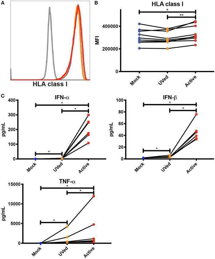
Frontiers in Cellular and Infection Microbiology | www.frontiersin.org 3 July 2019 | Volume 9 | Article 268McKechnie et al. HLA Upregulation During DENV Infection soluble HLA-E (sHLA-E), which has been implicated as a and 48 (Figures 2C,D) hpi, respectively, than infected potential viral mechanism of NK cell evasion (Shwetank monocytes in the same culture. This suggests HLA class I et al., 2013, 2014), we performed an sHLA-E ELISA. There upregulation is primarily restricted to infected cells with a was no significant difference in the concentration of sHLA- modest impact on uninfected cells, likely due to changes E between the DENV-infected adults and healthy controls in the cytokine milieu. Alternatively, bystander cells in our (Supplementary Figure 2). Together, these findings indicate that assay may have been infected at a level below our limit upregulation of HLA class I occurs on the cell surface of multiple of detection. immune cell subsets during acute in vivo DENV infection. Expression of HLA-E was also altered during DENV Interestingly, viSNE visualization revealed that upregulation infection. At 24 and 48 hpi, exposure to UV-inactivated of total HLA class I and HLA-E was not uniform across all DENV or infection with active DENV resulted in a majority monocytes. Instead, there were clear HLA class Ihigh and of monocytes becoming HLA-Ehigh (Supplementary Figure 5E HLA-Ehigh expressing monocytes. We gated on these cells and Figure 2E). Similarly, at 48 hpi, the percentage of HLA- (Figure 1C and Supplementary Figure 3A) and used an Ehigh monocytes in the UVed and active DENV cultures unbiased generalized linear mixed model (GLMM) (Seiler et al., was 2.8- and 3-fold higher, respectively, compared to mock- 2019) to identify associated markers. The GLMM identified infected monocytes (Figure 2F). The increase of HLA-Ehigh 10 markers (CD14, HLA-DR, HLA-E, LFA-3, CCR2, CD95, monocytes was 3-fold for both virus conditions compared CD48, ICAM-1, CD163, and Nectin-1) whose expression was to mock at 24 hpi (Supplementary Figure 5F). At both time associated with HLA class Ihigh monocytes (Figure 1D) and points, bystander monocytes in the active DENV cultures 5 markers (HLA class I, CD11b, ULBP-1,2,5,6, CD163, and had a bimodal distribution of HLA-Elow and HLA-Ehigh MICA/B) whose expression was associated with HLA-Ehigh cells, while infected cells were all HLA-Ehigh (Figure 2G and monocytes (Supplementary Figure 3B). We then verified Supplementary Figure 5G). Infected monocytes displayed a 1.3- these markers by comparing the expression level of each fold increase in the percentage of HLA-Ehigh monocytes at 24 hpi marker in HLAhigh expressing monocytes to the expression compared to bystander monocytes (Supplementary Figure 5H), level in HLAlow expressing monocytes from DENV-infected but at 48 hpi there was no longer a significant difference adults (Supplementary Figures 3C, 4A). Finally, we gated (Figure 2H). These results suggest that the response to viral down to monocytes expressing all 10 or all 5 markers in proteins, rather than viral replication, is the main driver of DENV-infected adults (Supplementary Figures 3D, 4B), and HLA-E upregulation. found that the 10 marker subset and the 5 marker subset Considering that bystander monocytes expressed higher levels expressed HLA class I and HLA-E, respectively, at significantly of HLA class I than DENV-infected monocytes, we wanted to higher levels than total monocytes from the same donor test whether secreted factors, such as cytokines, viral proteins, (Figures 1E,F and Supplementary Figures 3E,F) verifying and other molecules produced during active DENV infection that combinatorial gating using the aforementioned markers were sufficient to upregulate HLA class I. To this end, we UV- accurately identifies HLA class Ihigh and HLA-Ehigh monocytes. treated conditioned supernatants from previous 48 h cultures of Using the same gating scheme, we gated on the 10 and 5 primary monocytes that were mock-infected, exposed to UV- marker monocyte subsets in healthy controls and found inactivated DENV, or infected with active DENV. We then that HLA class I expression by the 10 marker subset and isolated uninfected monocytes from the same donors from HLA-E expression by the 5 marker subset were both 1.6- which the supernatants were collected and cultured them in fold higher in DENV-infected adults compared to healthy the conditioned supernatants for 24 h. Supernatants collected controls (Supplementary Figures 3G, 4C). These results from active, DENV-infected cultures led to a significant, if indicate that upregulation of HLA class I and HLA-E by modest, 1.1-fold increase in HLA class I expression compared monocytes during in vivo DENV infection only occurs on to monocytes cultured in supernatants collected from the mock- specific monocyte subsets. infected and UV-inactivated DENV cultures (Figures 3A,B). To better understand the effects of HLA class I upregulation, This increase in HLA class I expression shows that soluble factors we modeled DENV infection in vitro using primary monocytes secreted during active DENV infection could be contributing to isolated from healthy donors. Further, to understand how HLA class I upregulation. In order to investigate the potential viral replication vs. the presence of viral proteins alters role of cytokines in mediating this increase in HLA class I HLA class I expression, we compared the effects of “active,” expression, we used Luminex to determine the concentration replication-competent DENV, with that of UV-inactivated of cytokines present in the conditioned supernatants. We virus incapable of viral replication. At 24 h post-infection found that the concentrations of IFN-α, IFN-β, and TNF-α (hpi), HLA class I expression did not significantly differ were 759.2-, 26-, and 271.7-fold higher, respectively, in the between active DENV, UV-inactivated DENV, or mock- active condition compared to mock and 59.9-, 12.3-, and 2.8- infected conditions (Supplementary Figures 5A,B). By 48 fold higher, respectively, in the active condition compared to hpi, HLA class I expression was 2.1-fold higher in monocytes the UVed condition (Figure 3C). These findings suggest that infected with active DENV compared to mock (Figures 2A,B). upregulation of HLA class I during active DENV infection is Interestingly, in the active virus cultures, the uninfected largely driven by viral replication, and is likely mediated to bystander monocytes had a modest 1.1- and 1.2-fold higher some extent by cytokines. It is important to note that viral HLA class I expression at 24 (Supplementary Figures 5C,D) RNA, proteins, and particles present in the supernatant of Frontiers in Cellular and Infection Microbiology | www.frontiersin.org 4 July 2019 | Volume 9 | Article 268
McKechnie et al. HLA Upregulation During DENV Infection
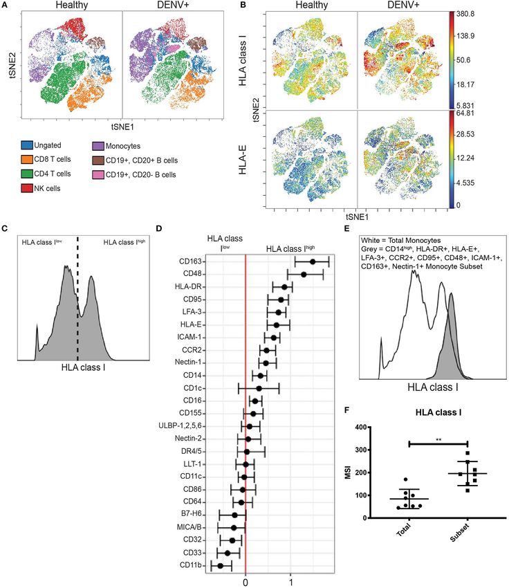
FIGURE 1 | HLA class I upregulation occurs during in vivo DENV infection. (A) Visualization of immune cell subsets in PBMCs from acute Panamanian DENV patients
and healthy Panamanian controls using viSNE. The plots represent pooled data from n = 8 DENV patients and n = 31 controls. To assure equal donor representation,
4,375 events were used from each DENV patient and 1,129 events were used from each healthy control, resulting in 34,999 pooled events to generate both the
DENV+ and healthy control viSNEs. Color key demonstrates major cell populations as determined by gating overlaid upon the viSNE visualization, demonstrating
clusters of major cell subsets. (B) viSNE visualization of total HLA class I and HLA-E expression in whole PBMCs from DENV patients and healthy controls, generated
as in (A). (C) Representative histogram from a DENV patient illustrating HLA class Ihigh and HLA class Ilow expressing monocytes gated on for generalized linear
(Continued)
Frontiers in Cellular and Infection Microbiology | www.frontiersin.org 5 July 2019 | Volume 9 | Article 268McKechnie et al. HLA Upregulation During DENV Infection
FIGURE 1 | mixed model (GLMM) analysis. (D) GLMM analysis of markers associated with HLA class Ihigh and HLA class Ilow expressing monocytes. (E)
Representative histogram from a DENV patient showing increased HLA class I expression by the monocyte subset gated on using the 10 markers identified in (D)
(CD163, CD48, HLA-DR, CD95, LFA-3, HLA-E, ICAM-1, CCR2, Nectin-1, and CD14) compared to total monocytes from the same donor. (F) Summary data from all
eight DENV patients. Wilcoxon signed-rank test **P < 0.01.
FIGURE 2 | Primary monocytes upregulate HLA class I and HLA-E during in vitro DENV infection. Primary monocytes isolated from whole PBMCs from healthy blood
bank donors were mock-infected (blue), exposed to UV-inactivated DENV (orange), or infected with active DENV (red) for 48 h. Representative histograms of HLA
class I (A) and HLA-E (E) expression in total monocytes cultured in the respective conditions. Fluorescence minus one (FMO) shown in gray. HLA class I MFI in total
monocytes (B) as well as bystander (DENV–) and infected (DENV+) monocytes (D). Representative histograms of HLA class I (C) and HLA-E (G) expression in
bystander monocytes (DENV–, green) and infected monocytes (DENV+, purple). Percentage of HLA-Ehigh total monocytes (F) as well as bystander and infected
monocytes (H). Two independent experiments measuring HLA class I were performed with 6 donors. The average for each donor is represented in the graphs. n = 12
for HLA-E experiments. Friedman test followed by Dunn’s multiple comparisons test was used to analyze total monocytes. Wilcoxon signed-rank test was used to
analyze DENV– vs. DENV+ monocytes. *P < 0.05, **P < 0.01, ***P < 0.001.
active infection cultures could also contribute to HLA class nearly double the frequency of CD107a+ NK cells compared
I upregulation. to DENV-infected monocytes blocked with the isotype-matched
Given that NK cell activation is dampened by the expression control Fab, increasing from 9.9 to 18.8%. Additionally, HLA
of self-HLA class I on potential target cells, we investigated the class I-blocking of DENV-infected monocytes resulted in a 6.7%
impact of DENV-mediated HLA class I upregulation on NK increase in CD107a+ NK cells compared to blocking HLA-
cell degranulation in response to DENV-infected cells. We co- E. Blocking HLA class I on DENV-infected monocytes also
cultured DENV-infected primary monocytes with autologous nearly doubled the frequency of CD107a+ NK cells compared to
primary NK cells in the presence of an isotype-matched control blocking HLA class I on mock-infected cells, increasing from 9.7
Fab, an anti-pan HLA class I blocking Fab, or an anti-HLA-E to 18.8% (P = 0.0312). For all of the blocking conditions, except
blocking Fab for 4 h and measured the percentage of CD107a+ HLA-E, the DENV co-cultures had a statistically significant
NK cells as a marker of degranulation and killing activity increase in CD107a+ NK cells compared to the mock-infected
(Figures 4A,B). Fabs were used instead of whole IgG to avoid co-cultures. Thus, these data demonstrate that HLA class I
killing via ADCC following binding of the anti-HLA antibodies upregulation can dampen the NK cell response to DENV-
to the target cells. Blocking HLA class I on mock-infected infected cells.
monocytes led to a 9.7% frequency of CD107a+ NK cells, a 4.5%
increase from 5.2% in the unblocked, mock-infected monocyte DISCUSSION
condition. Because NK cells can become activated when they
are unable to bind self-HLA class I, this result demonstrates Roughly one-third of the world’s population is at risk of
that the Fabs were effectively blocking their targeted proteins. acquiring DENV, making it critically important that we elucidate
Blocking HLA class I on DENV-infected monocytes resulted in immune factors that contribute both to disease protection and
Frontiers in Cellular and Infection Microbiology | www.frontiersin.org 6 July 2019 | Volume 9 | Article 268McKechnie et al. HLA Upregulation During DENV Infection FIGURE 3 | Soluble factors secreted during active DENV infection upregulate HLA class I expression. Conditioned supernatants from experiments shown in Figure 2 were UV-treated and used to culture primary monocytes from healthy blood bank donors (n = 9). After a 24 h incubation, expression of total HLA class I was analyzed by flow cytometry. Histograms from a single representative donor (A) as well as summary data from all 9 donors (B) are shown. Friedman test followed by Dunn’s multiple comparisons test, *P < 0.05, **P < 0.01. (C) Cytokine concentrations in conditioned supernatants from experiments shown in Figure 2 were analyzed by Luminex. Values shown are the average of two reads for each sample (n = 6). Friedman test with FDR correction followed by a one-tailed Wilcoxon matched-pairs signed-rank test with holm correction, *P < 0.05. pathogenesis. Mechanisms by which DENV evades the innate natural infection or infection of undifferentiated primary human immune response by inhibiting the production and signaling immune cells. Here, we show natural DENV infection leads of type I IFNs, as well as other aspects of the cellular antiviral to increased expression of HLA class I and HLA-E in adult response, have been well-reported (Morrison et al., 2012; Green patients. We also found that soluble factors produced during et al., 2014). Similarly, pathways that might promote DENV active infection contributed to HLA class I upregulation. Finally, escape from NK cell recognition have been proposed, including blocking HLA class I on DENV-infected monocytes enhanced the a potential role for the upregulation of HLA class I molecules by ability of NK cells to degranulate in response to DENV-infected DENV-infected cells to inhibit the NK cell response by binding cells. Together, these findings show HLA class I upregulation inhibitory KIRs or CD94/NKG2A (Beltrán and López-Vergès, during active DENV infection suppresses NK cell degranulation. 2014; Petitdemange et al., 2014; Mathew, 2018). However, these HLA class I upregulation during flavivirus infection has been data have primarily arisen from in vitro infection of mouse or previously described and attributed to various mechanisms such human cell lines, rather than more physiologic systems such as as NFκB activation (Kesson and King, 2001), increased transport Frontiers in Cellular and Infection Microbiology | www.frontiersin.org 7 July 2019 | Volume 9 | Article 268
McKechnie et al.                                                                                                               HLA Upregulation During DENV Infection

 FIGURE 4 | Blocking HLA class I improves NK cell responses to DENV-infected cells. Primary NK cells and monocytes were isolated from whole PBMCs from healthy
 blood bank donors (n = 6). NK cells were activated for 22 h with IL-2. Monocytes were mock-infected (blue) or infected with active DENV (red) at an MOI of 2 for 24 h.
 Prior to co-culture with autologous NK cells, monocytes were blocked for 30 min with an isotype-matched control Fab, an anti-HLA-E blocking Fab, or an anti-pan
 HLA class I blocking Fab. Monocytes and NK cells were co-cultured for 4 h before NK cell expression of CD107a was evaluated by flow cytometry. Flow cytometry
 plots from a single representative donor (A) as well as summary data from all 6 donors (B) are shown. Friedman test followed by paired Wilcoxon signed-rank tests,
 *P < 0.05.

of peptides into the endoplasmic reticulum for HLA loading                             the presentation of a capsid peptide which prevents HLA-
(Momburg et al., 2001), and the presence of IFN-β (Glasner                             E engagement with CD94/NKG2A (Nattermann et al., 2005;
et al., 2017). We show replication-competent virus was required                        Davis et al., 2016). No such mechanisms for modulating HLA-
for significant HLA class I upregulation, but that HLA class                           E expression and its affinity for the CD94/NKG2 receptors
I expression was highest in uninfected bystander monocytes.                            have been reported for DENV. The NetMHCpan 4.0 server
Further, supernatants collected from active DENV cultures were                         predicts several DENV-2 peptides with strong and weak binding
able to upregulate HLA class I on uninfected monocytes and                             to HLA-E∗ 01:01, making it possible that DENV peptides
contained higher concentrations of IFN-α, IFN-β, and TNF-α                             modulate NKG2A/C binding. Together, these results suggest
compared to supernatants from mock-infected or UV-inactivated                          HLA-E upregulation is mediated by both virus-dependent and
DENV cultures indicating HLA class I upregulation is mediated                          virus-independent mechanisms, and could influence NK cell
at least in part by soluble factors. These findings pose a potential                   recognition through NKG2A/C.
mechanism for HLA class I upregulation in which soluble factors                           Here we extend prior studies demonstrating that HLA class I
secreted by DENV-infected cells induce increased HLA class                             upregulation during flavivirus infection in cell lines can inhibit
I expression on all cells in an effort to promote cytotoxic                            NK cell activation (Lobigs et al., 1996; Momburg et al., 2001;
T lymphocyte responses. However, the DENV-infected cells                               Hershkovitz et al., 2008; Glasner et al., 2017; Drews et al.,
express lower levels than the bystander cells because DENV may                         2018). For the first time, we used patient samples and more
encode proteins that interfere with processes driving HLA class I                      physiologically relevant in vitro infection and co-culture systems
upregulation to escape the T cell response (Ye et al., 2013; Green                     with undifferentiated primary human immune cells. By directly
et al., 2014; Guzman and Harris, 2015; Glasner et al., 2017).                          blocking NK cell binding to HLA class I using Fabs, we found
    Interestingly, upregulation of HLA-E likely involves different                     HLA class I expression on DENV-infected cells significantly
mechanisms than upregulation of other HLA class I molecules.                           dampens NK cell degranulation. This provides the first direct
We observed a significant increase in HLA-E expression in                              evidence that upregulation of HLA class I is responsible for
response to active DENV as well as UV-inactivated DENV.                                inhibition of NK cell responses to flavivirus-infected cells. We
This suggests that direct sensing of viral products by innate                          did observe some non-specific increase in NK cell degranulation
immune receptors and the resulting cytokines, rather than                              in the presence of the isotype-matched control antibody, but
pathways induced during viral replication, may be the primary                          this effect was dwarfed by the increase in NK cell degranulation
contributors to HLA-E upregulation. However, DENV-infected                             in response to DENV-infected cells in the presence of HLA
monocytes expressed the highest levels of HLA-E, implying                              class I blocking Fabs. These findings indicate that HLA class I
DENV itself is also mediating HLA-E upregulation. Intriguingly,                        expression dampens the magnitude of the NK cell response.
cytomegalovirus proteins can modulate the surface expression                              Previous studies using cytokine-activated endothelial cells
of HLA-E by encoding HLA leader sequence mimics with                                   showed that blocking surface HLA-E and sHLA-E increased NK
reduced binding affinity to the CD94/NKG2 receptors (Heatley                           cell killing, illustrating the importance of HLA-E expression as
et al., 2013). Similarly, human immunodeficiency virus-1 has                           a potential NK escape mechanism in some vascular diseases
been found to upregulate HLA-E expression, resulting in                                (Coupel et al., 2007). However, similar to Drews et al. we did not

Frontiers in Cellular and Infection Microbiology | www.frontiersin.org             8                                                   July 2019 | Volume 9 | Article 268
McKechnie et al. HLA Upregulation During DENV Infection
observe a significant modulating effect of HLA-E expression on DATA AVAILABILITY
NK cell activity against DENV-infected cells (Drews et al., 2018).
HLA-E binds to both inhibitory NK cell receptor CD94/NKG2A Requests to access the dataset should be directed to Catherine
and activating receptor CD94/NKG2C (Braud et al., 1998; Valés- Blish at cblish@stanford.edu.
Gómez et al., 1999; Kaiser et al., 2005). Notably, HLA-E binds
to CD94/NKG2A with higher affinity (Valés-Gómez et al., 1999; ETHICS STATEMENT
Kaiser et al., 2005). The fact that NK cell binding to HLA-E can
result in both activating and inhibitory signaling with a dominant The studies involving human participants were reviewed and
advantage toward inhibitory signaling could explain our results approved by the IRB of Hospital del Niño (CBIHN-M-0634),
and those of Drews et al. HLA-E’s greater affinity toward then confirmed by the committees of ICGES, CSS, Santo Tomas
CD94/NKG2A also suggests that increased HLA-E expression on Hospital, and Stanford University. Written informed consent
DENV-infected cells might be part of the viral escape strategy. to participate in this study was provided by the participants or
Specifically blocking NKG2A or NKG2C in NK cell-infected participants’ legal guardian/next of kin.
cell co-cultures could identify the role of inhibitory signaling
through NKG2A vs. activating signaling through NKG2C in AUTHOR CONTRIBUTIONS
DENV recognition.
In contrast to Drews et al. and Shwetank et al. who observed JM, DB, CB, and SL-V contributed to the conceptualization,
an increase in sHLA-E in the supernatants of DENV-infected formal analysis, investigation, data curation, and the preparation
HMEC-1 cells and Japanese encephalitis virus-infected human of the writing of the original draft. JM, DB, AP, LS, AA,
brain microvascular endothelial cells, respectively, we saw no and RV contributed to the methodology. DB, EH, LL,
significant increase in sHLA-E in the serum of DENV patients CB, and SL-V contributed to the resources. EH and LL
compared to healthy controls (Shwetank et al., 2013; Drews contributed to the reviewing and editing of the manuscript.
et al., 2018). This suggests that sHLA-E shedding is not increased CB and SL-V contributed to the supervision and project
at the systemic level during in vivo DENV infection and is administration. DB, CB, and SL-V contributed to the
consequently unlikely to contribute strongly to suppression of the funding acquisition.
NK cell response.
This study has limitations, the most significant of which is FUNDING
the small sample size of DENV-infected adults in our cohort.
Despite the modest numbers, the conclusions we drew from This work was supported by NIH R21AI135287 and
these in vivo data were clear and supported by our in vitro R21AI130523 to CB, grants 9044.51 from the Ministry of
experiments using primary human immune cells. The second Economy and Finance of Panama and 71-2012-4-CAP11-
limitation is that we were unable to clearly determine the role 003 from SENACYT to SL-V, National Science Foundation
of HLA-E on the NK cell response to DENV infection given Graduate Research Fellowship DGE-1656518 to JM and NIH
its binding to both inhibitory CD94/NKG2A and activating training grant T31-AI-07290 (PI Olivia Martinez). DB (Ph.D.
CD94/NKG2C receptors. student) and SL-V are members of the Sistema Nacional
To our knowledge, ours is the first study showing de Investigación (SNI) of SENACYT, Panama. CB is the
upregulation of HLA class I molecules in acute dengue Tashia and John Morgridge Faculty Scholar in Pediatric
patient samples, suggesting different drivers of HLA-E Translational Medicine from the Stanford Maternal Child
upregulation vs. upregulation of other HLA class I proteins Health Research Institute and an Investigator of the Chan
during DENV infection, and showing enhanced primary NK Zuckerberg Biohub.
cell degranulation upon blocking HLA class I on primary
DENV-infected monocytes. Our in vivo HLA class I expression ACKNOWLEDGMENTS
data need to be confirmed with additional DENV cohorts at
different time points of the disease, spanning all serotypes We thank all health institutions, patients, and their families,
and degrees of disease severity, and including pediatric Luis Bonilla from the Blood Bank of Santo Tomas Hospital for
patients. This will be vital to determining what temporal, providing blood components, and the Stanford Human Immune
viral, and age-related factors affect HLA class I upregulation, Monitoring Center. We thank Dr. Taia Wang for critical reading
as well as whether there is a correlation between disease of the manuscript and Dr. Anne-Maud Ferreira for statistical
severity and HLA class I expression. Future experiments analysis of the Luminex data.
are also required to determine what factors and pathways
mediate HLA class I upregulation in monocytes and how SUPPLEMENTARY MATERIAL
HLA class I expression is modulated in other immune cell
subsets. Overall, this study furthers our understanding of the The Supplementary Material for this article can be found
impacts of DENV infection on innate immune cells and their online at: https://www.frontiersin.org/articles/10.3389/fcimb.
intercellular interactions. 2019.00268/full#supplementary-material
Frontiers in Cellular and Infection Microbiology | www.frontiersin.org 9 July 2019 | Volume 9 | Article 268McKechnie et al. HLA Upregulation During DENV Infection
REFERENCES on NF-kappa B activation. J. Infect. Dis. 184, 947–954. doi: 10.1086/3
23603
Azeredo, E. L. (2006). NK cells, displaying early activation, cytotoxicity and Kyle, J. L., and Harris, E. (2008). Global spread and persistence of dengue. Annu.
adhesion molecules, are associated with mild dengue disease. 143, 345–356. Rev. Microbiol. 62, 71–92. doi: 10.1146/annurev.micro.62.081307.163005
doi: 10.1111/j.1365-2249.2006.02996.x Laoprasopwattana, K., Libraty, D. H., Endy, T. P., Nisalak, A.,
Bayless, N. L., Greenberg, R. S., Swigut, T., Wysocka, J., and Blish, C. A. Chunsuttiwat, S., Ennis, F. A., et al. (2007). Antibody-dependent
(2016). Zika virus infection induces cranial neural crest cells to produce cellular cytotoxity mediated by plasma obtained before secondary
cytokines at levels detrimental for neurogenesis. Cell Host Microbe 20, 423–428. Dengue virus infections: potential involvement in early control
doi: 10.1016/j.chom.2016.09.006 of viral replication. J. Infect. Dis. 195, 1108–1116. doi: 10.1086/5
Beltrán, D., and López-Vergès, S. (2014). NK cells during dengue disease and 12860
their recognition of Dengue virus-infected cells. Front. Immunol. 5:192. Libraty, D. H., Pichyangkul, S., Ajariyakhajorn, C., Endy, T. P., and Ennis, F.
doi: 10.3389/fimmu.2014.00192 A. (2001). Human dendritic cells are activated by Dengue virus infection:
Bhatt, S., Gething, P. W., Brady, O. J., Messina, J. P., Farlow, A. W., Moyes, C. enhancement by gamma interferon and implications for disease pathogenesis.
L., et al. (2013). The global distribution and burden of dengue. Nature 496, J. Virol. 75, 3501–3508. doi: 10.1128/JVI.75.8.3501-3508.2001
504–507. doi: 10.1038/nature12060 Lobigs, M., Blanden, R. V., and Müllbacher, A. (1996). Flavivirus-induced
Braud, V. M., Allan, D. S., O’Callaghan, C. A., Söderström, K., D’Andrea, A., Ogg, up-regulation of MHC class I antigens; implications for the induction
G. S., et al. (1998). HLA-E binds to natural killer cell receptors CD94/NKG2A, of CD8+ T-cell-mediated autoimmunity. Immunol. Rev. 152, 5–19.
B and C. Nature 391, 795–799. doi: 10.1038/35869 doi: 10.1111/j.1600-065X.1996.tb00908.x
Cheng, Y., King, N. J. C., and Kesson, A. M. (2004). Major histocompatibility Mathew, A. (2018). Defining the role of NK cells during Dengue virus infection.
complex class I (MHC-I) induction by West Nile virus: involvement of 2 Immunology 154, 557–562. doi: 10.1111/imm.12928
signaling pathways in MHC-I up-regulation. J. Infect. Dis. 189, 658–668. Mei, H. E., Leipold, M. D., Schulz, A. R., Chester, C., and Maecker, H. T. (2015).
doi: 10.1086/381501 Barcoding of live human peripheral blood mononuclear cells for multiplexed
Costa, V. V., Ye, W., Chen, Q., Teixeira, M. M., Preiser, P., Ooi, E. E., et al. (2017). mass cytometry. J. Immunol. 194, 2022–2031. doi: 10.4049/jimmunol.1402661
Dengue virus-infected dendritic cells, but not monocytes, activate natural killer Momburg, F., Müllbacher, A., and Lobigs, M. (2001). Modulation of Transporter
cells through a contact-dependent mechanism involving adhesion molecules. associated with Antigen Processing (TAP) -mediated peptide import
MBio 8:e00741–17. doi: 10.1128/mBio.00741-17 into the endoplasmic reticulum by flavivirus infection. J. Virol. 75,
Coupel, S., Moreau, A., Hamidou, M., Horejsi, V., Soulillou, J.-P., and 5663–5671.
Charreau, B. (2007). Expression and release of soluble HLA-E is an Morrison, J., Aguirre, S., and Fernandez-Sesma, A. (2012). Innate immunity
immunoregulatory feature of endothelial cell activation. Blood 109, 2806–2814. evasion by Dengue virus. Viruses 4, 397–413. doi: 10.3390/v4030397
doi: 10.1182/blood-2006-06-030213 Nattermann, J., Nischalke, H. D., Hofmeister, V., Kupfer, B., Ahlenstiel, G.,
Davis, Z. B., Cogswell, A., Scott, H., Mertsching, A., Boucau, J., Wambua, Feldmann, G., et al. (2005). HIV-1 infection leads to increased HLA-E
D., et al. (2016). A conserved HIV-1-derived peptide presented expression resulting in impaired function of natural killer cells. Antivir. Ther.
by HLA-E renders infected T-cells highly susceptible to attack 10, 95–107.
by NKG2A/CD94-bearing natural killer cells. PLoS Pathog. 12:e10 Nightingale, Z. D., Patkar, C., and Rothman, A. L. (2008). Viral
05421. doi: 10.1371/journal.ppat.1005421 replication and paracrine effects result in distinct, functional
Drews, E., Adam, A., Htoo, P., Townsley, E., and Mathew, A. (2018). Upregulation responses of dendritic cells following infection with dengue
of HLA-E by dengue and not Zika viruses. Clin. Transl. Immunol. 7:e1039. 2 virus. J. Leukoc. Biol. 84, 1028–1038. doi: 10.1189/jlb.02
doi: 10.1002/cti2.1039 08105
Durbin, A. P., Vargas, M. J., Wanionek, K., Hammond, S. N., Gordon, Petitdemange, C., Wauquier, N., Devilliers, H., Yssel, H., Mombo,
A., Rocha, C., et al. (2008). Phenotyping of peripheral blood I., Caron, M., et al. (2016). Longitudinal analysis of natural
mononuclear cells during acute dengue illness demonstrates infection killer cells in Dengue virus-infected patients in comparison to
and increased activation of monocytes in severe cases compared to chikungunya and chikungunya/Dengue virus-infected patients.
classic dengue fever. Virology 376, 429–435. doi: 10.1016/j.virol.2008. PLoS Negl. Trop. Dis. 10:e0004499. doi: 10.1371/journal.pntd.00
03.028 04499
Glasner, A., Oiknine-Djian, E., Weisblum, Y., Diab, M., Panet, Petitdemange, C., Wauquier, N., Rey, J., Hervier, B., Leroy, E., and Vieillard, V.
A., Wolf, D. G., et al. (2017). Zika virus escapes NK cell (2014). Control of acute Dengue virus infection by natural killer cells. Front.
detection by upregulating major histocompatibility complex Immunol. 5:209. doi: 10.3389/fimmu.2014.00209
class I molecules. J. Virol. 91:e00785-17. doi: 10.1128/JVI.00 Seiler, C., Kronstad, L. M., Simpson, L. J., Le Gars, M., Vendrame, E., Blish, C.
785-17 A., et al. (2019). Uncertainty Quantification in Multivariate Mixed Models for
Green, A. M., Beatty, P. R., Hadjilaou, A., and Harris, E. (2014). Innate immunity Mass Cytometry Data. arXiv [stat.AP]. Available online at: http://arxiv.org/abs/
to Dengue virus infection and subversion of antiviral responses. J. Mol. Biol. 1903.07976.
426, 1148–1160. doi: 10.1016/j.jmb.2013.11.023 Shwetank., Date, O. S., Carbone, E., and Manjunath, R. (2014). Inhibition
Guzman, M. G., and Harris, E. (2015). Dengue. Lancet 385, 453–465. of ERK and proliferation in NK cell lines by soluble HLA-E released
doi: 10.1016/S0140-6736(14)60572-9 from Japanese encephalitis virus infected cells. Immunol. Lett. 162, 94–100.
Heatley, S. L., Pietra, G., Lin, J., Widjaja, J. M. L., Harpur, C. M., Lester, S., doi: 10.1016/j.imlet.2014.07.010
et al. (2013). Polymorphism in human cytomegalovirus UL40 impacts on Shwetank., Date, O. S., Kim, K. S., and Manjunath, R. (2013). Infection of human
recognition of human leukocyte antigen-E (HLA-E) by natural killer cells. J. endothelial cells by Japanese encephalitis virus: increased expression and release
Biol. Chem. 288, 8679–8690. doi: 10.1074/jbc.M112.409672 of soluble HLA-E. PLoS ONE 8:e79197. doi: 10.1371/journal.pone.0079197
Hershkovitz, O., Zilka, A., Bar-Ilan, A., Abutbul, S., Davidson, A., Mazzon, M., et al. Sun, P., Morrison, B. J., Beckett, C. G., Liang, Z., Nagabhushana, N., Li, A.,
(2008). Dengue virus replicon expressing the nonstructural proteins suffices to et al. (2017). NK cell degranulation as a marker for measuring antibody-
enhance membrane expression of HLA class I and inhibit lysis by human NK dependent cytotoxicity in neutralizing and non-neutralizing human sera from
cells. J. Virol. 82, 7666–7676. doi: 10.1128/JVI.02274-07 dengue patients. J. Immunol. Methods 441, 24–30. doi: 10.1016/j.jim.2016.
Kaiser, B. K., Barahmand-Pour, F., Paulsene, W., Medley, S., Geraghty, D. E., and 11.005
Strong, R. K. (2005). Interactions between NKG2x immunoreceptors and HLA- Sun, P., Williams, M., Nagabhushana, N., Jani, V., Defang, G., and Morrison, B.
E ligands display overlapping affinities and thermodynamics. J. Immunol. 174, J. (2019). NK cells activated through antibody-dependent cell cytotoxicity
2878–2884. doi: 10.4049/jimmunol.174.5.2878 and armed with degranulation/IFN-γ production suppress antibody-
Kesson, A. M., and King, N. J. C. (2001). Transcriptional regulation of major dependent enhancement of dengue viral infection. Sci. Rep. 9:1109.
histocompatibility complex class I by flavivirus West Nile is dependent doi: 10.1038/s41598-018-36972-2
Frontiers in Cellular and Infection Microbiology | www.frontiersin.org 10 July 2019 | Volume 9 | Article 268McKechnie et al. HLA Upregulation During DENV Infection Valés-Gómez, M., Reyburn, H. T., Erskine, R. A., López-Botet, M., and Conflict of Interest Statement: The authors declare that the research was Strominger, J. L. (1999). Kinetics and peptide dependency of the binding conducted in the absence of any commercial or financial relationships that could of the inhibitory NK receptor CD94/NKG2-A and the activating receptor be construed as a potential conflict of interest. CD94/NKG2-C to HLA-E. EMBO J. 18, 4250–4260. doi: 10.1093/emboj/18. 15.4250 Copyright © 2019 McKechnie, Beltrán, Pitti, Saenz, Araúz, Vergara, Harris, Lanier, World Health Organization (2012). Global Strategy for Dengue Prevention and Blish and López-Vergès. This is an open-access article distributed under the terms Control 2012-2020. Available online at: https://apps.who.int/iris/bitstream/ of the Creative Commons Attribution License (CC BY). The use, distribution or handle/10665/75303/9789241504034_eng.pdf reproduction in other forums is permitted, provided the original author(s) and the Ye, J., Zhu, B., Fu, Z. F., Chen, H., and Cao, S. (2013). Immune evasion copyright owner(s) are credited and that the original publication in this journal strategies of flaviviruses. Vaccine 31, 461–471. doi: 10.1016/j.vaccine.2012. is cited, in accordance with accepted academic practice. No use, distribution or 11.015 reproduction is permitted which does not comply with these terms. Frontiers in Cellular and Infection Microbiology | www.frontiersin.org 11 July 2019 | Volume 9 | Article 268
You can also read