Translating genetic and functional data into clinical practice: a series of 223 families with myotonia
←
→
Page content transcription
If your browser does not render page correctly, please read the page content below
https://doi.org/10.1093/brain/awab344                                                                                      BRAIN 2022: 00; 1–14           | 1

Translating genetic and functional data into
clinical practice: a series of 223 families

                                                                                                                                                                Downloaded from https://academic.oup.com/brain/advance-article/doi/10.1093/brain/awab344/6371181 by guest on 11 March 2022
with myotonia
Karen Suetterlin,1,2 Emma Matthews,1,3 Richa Sud,4 Samuel McCall,4 Doreen Fialho,1,5
James Burge,1,5 Dipa Jayaseelan,1 Andrea Haworth,4 Mary G. Sweeney,4
Dimitri M. Kullmann,6 Stephanie Schorge,6,7 Michael G. Hanna1 and Roope Männikkö1

High-throughput DNA sequencing is increasingly employed to diagnose single gene neurological and neuromuscu-
lar disorders. Large volumes of data present new challenges in data interpretation and its useful translation into
clinical and genetic counselling for families. Even when a plausible gene is identified with confidence, interpret-
ation of the clinical significance and inheritance pattern of variants can be challenging. We report our approach to
evaluating variants in the skeletal muscle chloride channel ClC-1 identified in 223 probands with myotonia conge-
nita as an example of these challenges. Sequencing of CLCN1, the gene that encodes CLC-1, is central to the diag-
nosis of myotonia congenita. However, interpreting the pathogenicity and inheritance pattern of novel variants is
notoriously difficult as both dominant and recessive mutations are reported throughout the channel sequence,
ClC-1 structure-function is poorly understood and significant intra- and interfamilial variability in phenotype is
reported.
Heterologous expression systems to study functional consequences of CIC-1 variants are widely reported to aid
the assessment of pathogenicity and inheritance pattern. However, heterogeneity of reported analyses does not
allow for the systematic correlation of available functional and genetic data. We report the systematic evaluation
of 95 CIC-1 variants in 223 probands, the largest reported patient cohort, in which we apply standardized function-
al analyses and correlate this with clinical assessment and inheritance pattern. Such correlation is important to
determine whether functional data improves the accuracy of variant interpretation and likely mode of inherit-
ance.
Our data provide an evidence-based approach that functional characterization of ClC-1 variants improves clinical
interpretation of their pathogenicity and inheritance pattern, and serve as reference for 34 previously unreported
and 28 previously uncharacterized CLCN1 variants. In addition, we identify novel pathogenic mechanisms and
find that variants that alter voltage dependence of activation cluster in the first half of the transmembrane
domains and variants that yield no currents cluster in the second half of the transmembrane domain. None of the
variants in the intracellular domains were associated with dominant functional features or dominant inheritance
pattern of myotonia congenita.
Our data help provide an initial estimate of the anticipated inheritance pattern based on the location of a novel
variant and shows that systematic functional characterization can significantly refine the assessment of risk of an
associated inheritance pattern and consequently the clinical and genetic counselling.

Received May 24, 2021. Revised July 13, 2021. Accepted August 05, 2021. Advance access publication September 16, 2021
C The Author(s) (2021). Published by Oxford University Press on behalf of the Guarantors of Brain.
V
This is an Open Access article distributed under the terms of the Creative Commons Attribution License (https://creativecommons.org/licenses/by/4.0/), which
permits unrestricted reuse, distribution, and reproduction in any medium, provided the original work is properly cited.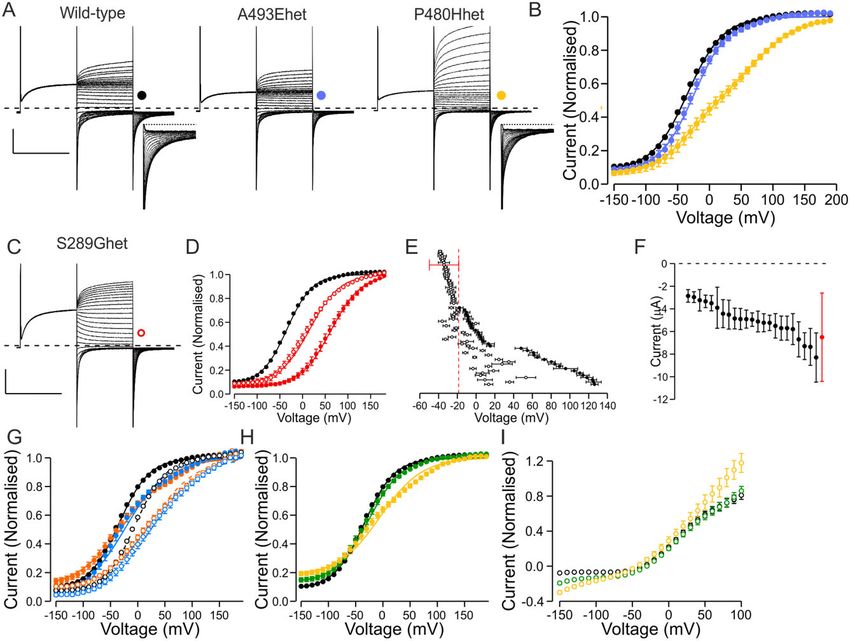
2   | BRAIN 2022: 00; 2–14                                                                                                 K. Suetterlin et al.

1   MRC International Centre for Genomic Medicine in Neuromuscular Diseases, Department of Neuromuscular
    Disease, UCL Queen Square Institute of Neurology, London, UK
2   AGE Research Group, NIHR Newcastle Biomedical Research Centre, Newcastle-upon-Tyne Hospitals NHS
    Foundation Trust and Newcastle University, Newcastle-upon-Tyne, UK
3   Atkinson Morley Neuromuscular Centre, Department of Neurology, St Georges University Hospitals NHS
    Foundation Trust, London, UK
4   Neurogenetics Unit, UCL Queen Square Institute of Neurology, London, UK
5   Department of Clinical Neurophysiology, King’s College Hospital, London, UK
6   Department of Clinical and Experimental Epilepsy, UCL Queen Square Institute of Neurology, London, UK

                                                                                                                                                    Downloaded from https://academic.oup.com/brain/advance-article/doi/10.1093/brain/awab344/6371181 by guest on 11 March 2022
7   Department of Pharmacology, UCL School of Pharmacy, London, UK

Correspondence to: Roope Männikkö
MRC International Centre for Genomic Medicine in Neuromuscular Diseases
Department of Neuromuscular disease, UCL Queen Square Institute of Neurology, London, UK
E-mail: r.mannikko@ucl.ac.uk

Keywords: skeletal muscle channelopathy; chloride channel; myotonia congenita; ClC-1; CLCN1

Introduction                                                              typically associated with dominant inheritance of myotonia con-
                                                                          genita4,17,18 while variants without dominant negative effects are
Myotonia congenita is the most common skeletal muscle channel-            associated with recessive inheritance.19–21
opathy.1 It is caused by a reduction in the repolarizing chloride             To enable accurate use of functional data in a clinical diagnostic
current, resulting in an increase in the excitability of the muscle       setting a correlation between distinct functional and clinical features
membrane,2 leading to a delay in terminating muscle contraction           needs to be established. This is currently complicated by phenotypic
following voluntary activity. This manifests clinically as stiffness      variability but also by heterogeneity in the methodology of the acqui-
and rigidity of affected muscles. Myotonia congenita has dominant         sition of genetic, clinical and functional data that often do not allow
and recessive forms, both caused by mutations in CLCN1 that re-           direct comparison of distinct functional features with the inheritance
sult in reduced function of the encoded skeletal muscle chloride          data between studies. In addition, some variants are found in both
channel ClC-1.3,4 Sequencing of CLCN1 has become integral to con-         dominant and recessive pedigrees, suggesting that the correlation
firming myotonia congenita diagnosis following clinical assess-           between functional features and the inheritance pattern is not linear.
ment. However, it is important that myotonia is not erroneously           Finally, in some cases the functional data do not match the predicted
attributed to an identified CLCN1 variant as myotonia can also be         pathogenicity and inheritance data.22–24 Thus, an evidence-based
caused by myotonic dystrophy (DM), a multisystem and potential-           guide for assessment of pathogenicity and inheritance pattern based
ly lethal disorder, and by gain-of-function mutations of the skel-        on specific functional features of ClC-1 variants is needed for pur-
etal muscle sodium channel Nav1.4 (encoded by SCN4A).                     poseful clinical and genetic counselling.
Mutations in distinct myotonia-associated genes (CLCN1, SCN4A,                We use functional expression to inform the diagnosis and gen-
DMPK, CNBP) can co-occur in a patient and modify the presentation         etic counselling in patients with myotonia congenita. Here we re-
compared to a patient carrying a single gene mutation.5–11                port analysis of the correlation of the functional properties of 95
     ClC-1 is a homodimer.12,13 Each subunit contains its own chloride-   distinct missense variants with the reported inheritance pattern of
selective pore and is composed of 18 intramembrane a-helices (con-        223 probands in a diagnostic service setting and assess the impli-
ventionally numbered A to R) organized in two topologically related       cations for the use of functional data to improve genetic counsel-
repeats with opposite membrane orientations, and an intracellular         ling in myotonia congenita.
domain with two cystathionine beta-synthase (CBS) repeats. The
chloride-selective pores can be gated individually or concurrently, in
processes known as fast and slow gating, respectively. Both gates are     Materials and methods
opened by membrane depolarization. ClC-1 voltage sensitivity arises
                                                                          Ethics
from the interaction of the channel with chloride ions.14–16
     Interpretation of the clinical significance of a ClC-1 variant can   The study was conducted as part of a service evaluation of the
be difficult as myotonia congenita exhibits intra- and interfamilial      NHS England Highly Specialised Muscle Channelopathy Service at
variability in the phenotype, severity and penetrance. In addition,       the National Hospital for Neurology and Neurosurgery. No proce-
it is currently difficult to accurately predict pathogenicity or inher-   dures were performed outside of routine clinical care. The oocytes
itance of a novel variant based purely on the amino acid change           were recovered from Xenopus laevis toads in accordance with the
and its location in the ClC-1 channel.                                    Animals (Scientific Procedures) Act 1986.
     Functional assessment of mutations associated with myotonia
congenita has revealed that they either reduce functional expres-
sion or shift the voltage dependence of channel activation towards
                                                                          Genetics
depolarized voltages, thereby reducing the chloride current at            The diagnostic molecular genetics laboratory at the National
physiological voltages. A mutant subunit can show dominant                Hospital for Neurology and Neurosurgery is the UK national centre
negative effects on coexpression with wild-type subunits, typically       for myotonia congenita genetic testing. Since 2007 this consists of
by shifting the voltage dependence of activation of the heterodi-         sequencing all 23 exons of CLCN1 plus flanking intronic regions, or
meric channel to depolarized voltages.17,18 Variants that show            targeted sequencing of specific exons for relatives of individuals in
dominant negative effects in functional expression analysis are           whom variants have already been identified. For samples processed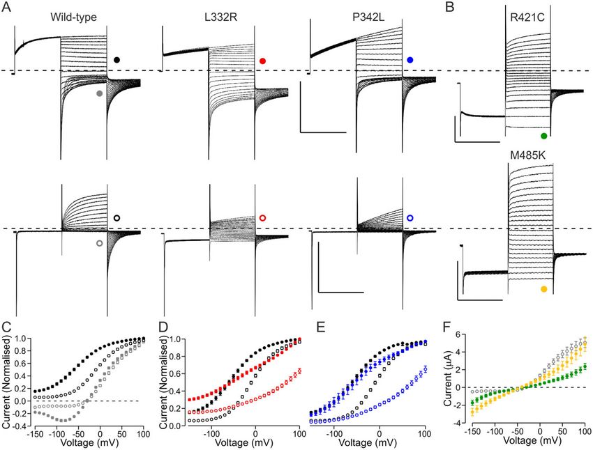
ClC-1 functional data in diagnosis of MC                                                                        BRAIN 2022: 00; 3–14         | 3

before 2007, it was routine for initial sequencing to be of mutation     wild-type or mutant mRNA. The heterozygous condition is simulated
hotspots only. At least the proband subsequently underwent full          by injecting a 1:1 mixture of wild-type and mutant mRNA. The
sequencing of all CLCN1 exons apart from the few circumstances           oocytes were incubated in modified Barth’s solution (in mM): NaCl
where it was not possible to obtain DNA to do so. In cases where no      88, KCl 1, MgSO4 1.68, HEPES 10, Ca(NO3)2 0.47, NaHCO3 2.4, CaCl2 0.41,
mutations are found, a single recessive mutation is identified or a      supplemented with penicillin and streptomycin routinely for 30–72 h
homozygous mutation is identified, Multiplex Ligation-dependent          at 15 C before electrophysiological recordings. Each variant was
Probe Amplification is performed to assess for exon deletions or         studied in more than one batch of oocytes.
duplications. Whenever possible, the inheritance and allelic distribu-
tion of the variants is investigated by sequencing CLCN1 in family       Electrophysiology
members. Sequencing of SCN4A (full gene or hotspots), DM1 and
DM2 retest was triggered after a ClC-1 variant was identified if the     Two-electrode voltage clamp experiments were performed using

                                                                                                                                                     Downloaded from https://academic.oup.com/brain/advance-article/doi/10.1093/brain/awab344/6371181 by guest on 11 March 2022
phenotype was atypical or it was uncertain if the CLCN1 variant could    GeneClamp 500B, DigiData 1200 or 1550 Interface and Clampex soft-
account for dominant myotonia congenita. In some samples SCN4A           ware (all Axon Instruments) at room temperature in ND96 extracellu-
and CLCN1 were screened in parallel.                                     lar media (in mM): NaCl 96, KCl 2, MgCl2 1, HEPES 5, CaCl2 1.8, pH 7.4.
    DNA was extracted from blood using standard methods.                 Recording electrodes were filled with 3 M KCl and had a tip resistance
Bidirectional direct DNA sequencing was performed using a Big            51 MX. Data were filtered at 1 kHz and sampled at 5 kHz.
Dye Terminator sequencing kit [Applied Biosystems (ABI)] and a               From a holding voltage of –80 mV, an activating pre-pulse step
3730 automated DNA sequencer (ABI). DNA sequences were ana-              to + 60 mV for 250 ms was applied before test voltage steps ranging
lysed using v.2.5 SeqScape Analysis software (ABI). All 23 CLCN1         from –150 to + 190 mV in 10 mV increments for 250 ms, followed by
exons are compared to larger databases including 1000 genomes,           a tail voltage step to –100 mV. For most cells the same protocol was
dbSNP, ExAC and Exome Variant Server.                                    also applied with a holding voltage of –40 mV as well as an add-
                                                                         itional protocol where the holding voltage was –80 mV and the
                                                                         pre-pulse step taken to –140 mV. These protocols are referred to as
Clinical and genetic assessment of clinical                              Vh = –40mV and Vpp = –140 mV, respectively.
inheritance pattern
The CLCN1 variants in each pedigree were assigned as dominant,           Data analysis
recessive or sporadic, based on available demographic, clinical,
                                                                         Data analysis and presentation were prepared using Clampfit (Axon
electrophysiological and genetic data that were collected from re-
                                                                         instruments), Origin (OriginLab) and Excel (Microsoft) software.
ferral forms and/or clinic notes (Tables 1, 2 and Supplementary
                                                                            The tail currents were routinely measured 4 ms after the test
Tables 1–3). Dominant variants were single variants sufficient to
                                                                         pulse. The current–voltage relationship was fitted with the
cause the myotonia congenita phenotype and associated with par-
                                                                         Boltzmann equation:
ent to child transmission. The inheritance pattern of some domin-
ant variants was specified ‘with variable penetrance’ (i) when the
                                                                                       IðVÞ ¼ Imax =f1 þ exp ½ðV1=2  VÞ = Vc Þ þ Cg          (1)
proband’s parents self-reported as asymptomatic but one was
found to have the variant and clinical or electrographic myotonia
                                                                         where Imax is the amplitude of the fit, C the offset current, V1/2 the
on examination; or (ii) in families with no known history of con-
                                                                         voltage at which the current is (Imax + C) / 2 and Vc the slope factor.
sanguinity, parents self-reported as asymptomatic and a nephew,
                                                                         We fixed the value of C in the fitting process to the baseline cur-
niece, aunt, uncle or half-sibling was reported to be affected.
                                                                         rent level at the most hyperpolarized voltages. In some cases, two
Recessive variants were found in homozygosis or compound het-
                                                                         Boltzmann equations were required to fit the data:
erozygosis and associated with asymptomatic parents. Sporadic
variants were found in isolation in probands with asymptomatic
parents and no other family history of myotonia congenita.                         IðVÞ ¼ C þ Imax  ðA=f1 þ exp ½ðV1=2ð1Þ  VÞ=Vc1 Þg
                                                                                          þ ð1  AÞ=f1 þ exp ½ðV1=2ð2Þ  VÞ=Vc2 gg            (2)
    As expected at service level, the phenotyping and genotyping
data for family members was not always complete. In
Supplementary Tables 1–3 we specify whether the assessment of            with C and Imax as before while A, V1/2(1), Vc1 and (1 – A), V1/2(2), Vc2
inheritance pattern was confirmed by segregation of the variant          are the fraction, the voltage of half-maximal activation and the
with the clinical symptoms or if it was based on reports. When a         slope factor of the first and second Boltzmann component, re-
family history was not available or was insufficient to determine        spectively. The time constant of activation was assessed by fitting
the inheritance pattern, the variant is listed as unknown (Tables 1,     a two-component exponential equation to data following settling
2 and Supplementary Tables 1–3). Some variants were classified as        of capacitive transients, and the weighted average of time con-
‘uncertain pathogenicity’ as specified in the ’Results’ section.         stants is presented.
                                                                            Cells without a clear component of activation that could be
                                                                         described with a Boltzmann equation and with tail current ampli-
Molecular biology                                                        tude 51 mA following a step to + 80 mV were considered devoid of
The mutations were introduced into wild-type CLCN1 cDNA by               functional ClC-1 channel expression.
Quikchange site-directed mutagenesis (Agilent).18 Successful mu-            Data are presented as mean ± standard error of the mean (SEM)
tagenesis was confirmed by sequencing the entire insert. The             unless otherwise stated. To assess if clustering of the variants
mRNA was transcribed from MluI linearized vector using                   across the functional and structural groups was significant, we
mMessageMachine SP6 kit (Ambion).                                        used a two-tailed Fisher’s test.

Xenopus laevis oocytes                                                   Data availability
                                                           25
Oocytes were extracted from adult female Xenopus laevis, isolated        Functional data are available on reasonable request to the corre-
after incubation with 2 mg/ml Collagenase Type A (Roche) in OR-2 (in     sponding author. The clinical and genetic data are not publicly
mM): NaCl 82.5, KCl 2, MgCl2 1, HEPES and injected with 2.5 ng of        available.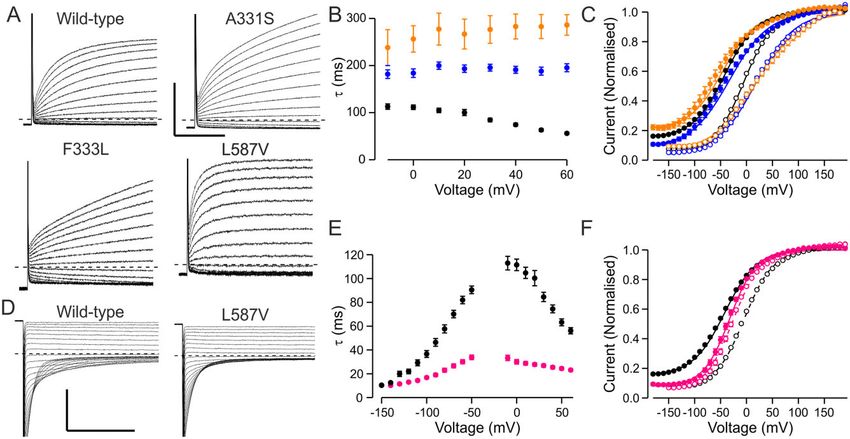
4 | BRAIN 2022: 00; 4–14 K. Suetterlin et al.
Table 1 Inheritance patterns of variants with wild-type-like or recessive functional features
Variant Functional Clinical/Genetic Sum Location
Dominant Sporadic Recessive Uncertain Unknown
p.His29Pro Wild-type-like – – 1 – – 1 IC
p.Ser70Leu Wild-type-like – – 1 – – 1 IC
p.Arg105Cys Wild-type-like – – – 1 – 1 IC
p.Leu106Val* Wild-type-like – – – 1 – 1 IC
p.Gln154Arg Wild-type-like – – – 1 – 1 TM1
p.Gly222Ser Wild-type-like – – – 1 – 1 TM1
Downloaded from https://academic.oup.com/brain/advance-article/doi/10.1093/brain/awab344/6371181 by guest on 11 March 2022
p.Val327Ile Wild-type-like – – – – 2 2 TM1
p.Ala331Ser* Wild-type-like – – 1 – – 1 TM1
p.Phe333Leu* Wild-type-like – – 1 – – 1 TM1
p.Ala402Val Wild-type-like – 1 1 – – 2 TM2
p.Pro408Ala Wild-type-like – – 2 1 3 6 TM2
p.Val456Ile* Wild-type-like – – – – 1 1 TM2
p.Ala493Thr* Wild-type-like – – – – 1 1 TM2
p.Phe494Leu* Wild-type-like – – – 1 – 1 TM2
p.Leu587Val* Wild-type-like – – 2 – – 2 IC
p.Gly594Val* Wild-type-like – – – – 1 1 IC
p.Arg611His* Wild-type-like – – – 1 1 2 IC
p.Met646Thr* Wild-type-like – – 1 – – 1 IC
p.His664Pro* Wild-type-like – – – 1 – 1 IC
p.Arg669Cys Wild-type-like – 1 – – – 1 IC
p.Gly688Arg* Wild-type-like – – – – 1 1 IC
p.Pro744Thr Wild-type-like – – – 1 – 1 IC
p.Thr837Ile* Wild-type-like – – – 1 – 1 IC
p.Val851Met Wild-type-like – – 2 – –. 2 IC
p.Gly898Arg* Wild-type-like – – – 1 – 1 IC
Total Wild-type-like - 2 12 11 10 35
p.Phe167Leu Recessivea – – 7 4 2 13 TM1
p.Gly190Arg Recessiveb – – 3 1 – 4 TM1
p.Leu198Val Recessivea – – – 1 – 1 TM1
p.Ala221Glu* Recessiveb – – 1 – – 1 TM1
p.Gly233Ser Recessiveb – – 1 – – 1 TM1
p.Val273Met Recessivea – – – – 1 1 TM1
p.Gly276Ser Recessivea – – 1 – – 1 TM1
p.Cys277Arg Recessiveb – – 1 – – 1 TM1
p.Gly285Val* Recessiveb 1 – – – – 1 TM1
p.Glu291Lys Recessiveb – – 1 – – 1 TM1
p.Arg317Leu Recessivea – – 1 – – 1 TM1
p.Ala320Val Recessivea – – 1 – – 1 TM1
p.Arg338Gln Recessivea – – 1 – 1 2 TM1
p.Gly355Arg Recessiveb – – 1 – – 1 TM2
p.His369Pro Recessiveb – – – – 1 1 TM2
p.Val397Asp* Recessiveb – – – – 1 1 TM2
p.Phe413Cys Recessiveb – – – – 1 1 TM2
p.Glu422Lys Recessiveb – – – 1 – 1 TM2
p.Trp433Arg Recessiveb – – – – 1 1 TM2
p.Phe463Ile* Recessiveb – – 1 – – 1 TM2
p.Gly483Ser* Recessivea – – 1 – – 1 TM2
p.Met485Val Recessiveb – – 6 4 1 11 TM2
p.Ala493Glu Recessiveb – – 1 – – 1 TM2
p.Glu500Lys* Recessiveb – – 1 – – 1 TM2
p.Pro521Thr* Recessiveb – – 1 – – 1 TM2
p.Ala529Val* Recessiveb – – – – 1 1 TM2
p.Glu548Lys Recessiveb – – – – 1 1 TM2
p.Thr550Met Recessiveb – – – 1 – 1 TM2
p.Pro558Ser Recessiveb – – 2 – – 2 TM2
p.Met560Thr Recessivea – – – – 1 1 TM2
p.Ala566Thr Recessiveb – – 2 – – 2 TM2
p.Gln583Arg* Recessivea – – 1 – 1 2 TM2
p.Val640Phe* Recessiveb – – 1 – – 1 IC
p.Asp822Asn* Recessivea – – 1 – – 1 IC
p.Pro883Thr Recessivea – – 1 – 2 3 IC
Total Recessive 1 - 38 12 15 66
Asterisks following variant name indicate variants not reported in the literature previously. Functional = classification according to functional feature; Clinical/Genetic =
number of pedigrees with distinct inheritance patterns of clinical symptoms. Location = if the variant is found in intracellular domain (IC: residues 1–110, 586–988), first (TM1:
residues 111–344) or second (TM2: residues 345–585) of the transmembrane repeats. Data for the variants that were not missense are presented in Supplementary Table 3.
a,b
For recessive variants it is specified if the variant in homomeric condition expresses currents with shifted voltage dependence of activationa or reduced current amplitudeb.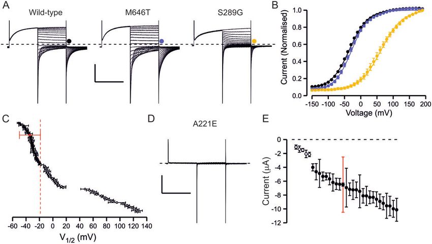
ClC-1 functional data in diagnosis of MC                                                                       BRAIN 2022: 00; 5–14         | 5

Results                                                                  than 1.5  SD cut-off (Supplementary Table 4). In the absence of
                                                                         loss-of-function effects this variant was not considered
Genetic and clinical overview of the cohort                              pathogenic.
                                                                             The current amplitude of wild-type channels was also variable
Our cohort comprised 223 probands referred for genetic testing for
                                                                         (–6.3 ± 0.2 mA, SD = 3.6 mA, range –0.5 to –30 mA). Twenty-five variants
myotonia congenita in whom CLCN1 variants were identified. A
                                                                         did not show any chloride currents (Supplementary Table 4) and for
total of 115 distinct mutations were identified in the cohort, 95 of
                                                                         five variants with wild-type-like voltage dependence of activation
which were missense while 20 were non-missense (truncating, fra-
                                                                         (A221E, H369P, V397D, F413C, E422K) and one variant with shifted
meshifting, intronic, duplications, deletions or silent). The non-
                                                                         voltage dependence of activation (W303R) many of the oocytes did
missense variants were included in the cohort only if they were
                                                                         not show currents (Fig. 1 and Supplementary Table 4) and the mean
compound heterozygous with missense mutations.
                                                                         amplitude of the currents from those oocytes that yielded currents

                                                                                                                                                    Downloaded from https://academic.oup.com/brain/advance-article/doi/10.1093/brain/awab344/6371181 by guest on 11 March 2022
    A single heterozygous variant was found in 116, homozygous
                                                                         was less than the mean + SD for oocytes expressing wild-type chan-
variant in 27 (one with two homozygous variants), compound het-
                                                                         nels (Fig. 1E). Although not displaying complete loss-of-function
erozygous variants in 75 and more than two variants in five pro-
                                                                         these variants were considered pathogenic by reducing functional
bands (Supplementary Table 1). In total, 309 variants
                                                                         expression of the channel.
(Supplementary Tables 1–3), of which 263 were missense
                                                                             Most (91/95) of the channel variants could be described as hav-
(Supplementary Table 2), were found in the cohort and were
                                                                         ing wild-type-like features (25 variants), no or reduced chloride
assigned an inheritance pattern. For the missense variants this
                                                                         currents (31 variants) or chloride currents with shifted voltage de-
was either dominant (81), sporadic (21) or recessive (74)
                                                                         pendence of activation (35 variants) (Fig. 1 and Supplementary
(Supplementary Table 2). For a further 54 missense variants, family
                                                                         Table 4).
history was unavailable or insufficient to determine the inherit-
ance pattern. For 33 missense variants the assessment of patho-
genicity or inheritance was complicated by co-allelic CLCN1              Functional properties of extraordinary ClC-1
variants, the presence of variants in other myotonia-associated          variants
genes, by the variant not segregating with myotonia congenita            Four variants showed properties that could not be described by
symptoms, or by reported dominant inheritance in compound het-           reduced current amplitude or shifted voltage dependence of acti-
erozygous probands where it could not be determined which of             vation alone.
the variants was associated with dominant inheritance. These var-            Two variants (L332R and P342L) showed depolarization-acti-
iants were assigned ‘with uncertain pathogenicity’. For 50 of the        vated currents but it was difficult to describe the voltage depend-
missense variants in the cohort with recessive or dominant inher-        ence of activation with a Boltzmann equation. In addition, the tail
itance patterns, the assessment was based on both clinical and           current amplitude declined when studied with pre-pulses to
genetic segregation data and for 105 variants this was based on          hyperpolarized voltages (VPP = –140 mV) but increased when using
clinical symptoms only (Supplementary Table 2). Of the 33 pedi-          a more depolarized holding voltage (Vh = –40mV) (Fig. 2A, D and E).
grees with dominant inheritance and confirmed segregation, six           The voltage dependence of activation of wild-type channels too is
met our criteria for variable penetrance (Supplementary Table 2).        clearly dependent on the voltage protocol (Fig. 2A and C). To com-
    Seventy-eight of the variants were found in a single pedigree        pare wild-type, L332R and P342L behaviours, the currents for each
only (64 missense variants) (Supplementary Tables 1–3). The most         cell were normalized to the tail current amplitude following a volt-
common variant, G230E, was identified in 40 pedigrees. Eight var-        age step to + 100 mV using the Vh = –40 mV protocol. In response to
iants were found associated with more than one inheritance pat-          a voltage step to 0 mV using the VPP = –140 mV protocol the nor-
tern (dominant, sporadic, recessive), of which three (G285E, F307S       malized wild-type-channel activity was 58%, but only 31% for
and A313T) were associated with both recessive and dominant in-          L332R and 24% for P342L channels, demonstrating reduced activity
heritance. The only variant with genetic segregation data available      for mutant channels at physiological voltages (Fig. 2D and E).
to confirm association with both dominant and recessive inherit-             Two variants (M485K and R421C) showed currents at hyperpo-
ance was G285E. Variable penetrance has been reported previously         larized voltages (Fig. 2B and F). M485K channels also showed de-
for G285E.18,19,26                                                       polarization-activated currents but with a voltage dependence
    To our knowledge, 34 of the missense variants have not been          of activation that was shifted to depolarized voltages
previously reported as associated with myotonia congenita and a          (Supplementary Table 4). Oocytes expressing R421C channels
further 28 have been reported but not functionally characterized.        showed small depolarization-activated currents (mean tail current
                                                                         amplitude –1.1 ± 0.1 mA) with shifted voltage dependence of activa-
Functional assessment and classification of ClC-1                        tion (Supplementary Table 4). The current amplitude at –120 mV
                                                                         was –1.8 ± 0.3 mA for M485K cells, –0.9 ± 0.2 mA for R421C channels
variants
                                                                         but –0.4 ± 0.05 mA for wild-type cells (Vpp = –140 mV protocol).
The effect of the CLCN1 missense variants on ClC-1 channel func-
tion was tested in the Xenopus laevis oocyte expression system for
                                                                         Assessment of dominant negative effect of ClC-1
the 95 missense variants. The V1/2 for wild-type channels was –
34.2 ± 0.6 mV (n = 308) (Fig. 1 and Supplementary Table 4). As the V1/   variants
2 of cells expressing wild-type channels was variable [standard de-      To simulate the heterozygous condition of the patient and to as-
viation (SD) = 10.4 mV, range –68.6 to –7.8 mV], we assigned a cut-      sess the inheritance pattern of the variant, mRNA encoding mu-
off voltage (V1/2 for the wild-type channel ±1.5  SD = –18.6 mV) to     tant and wild-type subunits were co-injected into oocytes (Fig. 3).
decide whether the V1/2 was wild-type-like or pathogenic (Fig. 1C).      We indicate the simulated heterozygous condition by adding the
Using this cut-off voltage, the well-known pathogenic variant            suffix ‘het’ to the variant name, e.g. F167Lhet.
F167L with a modest positive shift in the voltage dependence of ac-         Twenty-three of the 31 variants that showed no or reduced
tivation27 (V1/2 = –17.7 mV; Supplementary Table 4) was classified       currents when expressed alone showed currents with wild-
as pathogenic while variants with V1/2 negative to that were not.        type-like voltage dependence of activation in simulated hetero-
One variant, H664P, activated at more hyperpolarized voltages            zygous conditions (Fig. 3E and Supplementary Table 4). The
6 | BRAIN 2022: 00; 6–14 K. Suetterlin et al.
Table 2 Inheritance patterns of variants with dominant or extraordinary functional features
Variant Functional Clinical/Genetic Sum Location
Dominant Sporadic Recessive Uncertain Unknown
p.Met128Ile Dominant 2 – – – – 2 TM1
p.Cys179Tyr* Dominant 1 – – – – 1 TM1
p.Ser183Pro Dominant 1 – – – – 1 TM1
p.Gly190Ser Dominant – – 3 – – 3 TM1
p.Leu198Pro Dominant – – – – 1 1 TM1
p.Gly200Glu* Dominant – – 1 – – 1 TM1
Downloaded from https://academic.oup.com/brain/advance-article/doi/10.1093/brain/awab344/6371181 by guest on 11 March 2022
p.Ala218Val Dominant 1 – – – – 1 TM1
p.Gly230Glu Dominant 29 5 – 1 5 40 TM1
p.Pro234Thr* Dominant – – 1 – – 1 TM1
p.Pro234Leu* Dominant – – – 1 – 1 TM1
p.Thr268Met Dominant 1 – – – 2 3 TM1
p.Cys271Arg Dominant 1 – – – – 1 TM1
p.Gly276Asp Dominant 2 – – – – 2 TM1
p.Gly285Glu Dominant 5 3 9 4 5 26 TM1
p.Ser289Gly* Dominant 1 – – – – 1 TM1
p.Ser289Asn Dominant 1 1 – – – 2 TM1
p.Ser289Ile* Dominant – – 1 – – 1 TM1
p.Phe297Ser Dominant 7 – – – – 7 TM1
p.Val299Leu Dominant – – 2 – – 2 TM1
p.Trp303Arg Dominant 12 2 – – 2 16 TM1
p.Phe306Leu Dominant 2 1 – – 2 5 TM1
p.Phe307Ser Dominant 1 2 4 – 3 10 TM1
p.Ala313Thr Dominant 9 1 1 – 1 12 TM1
p.Ala313Val Dominant 2 – – 2 1 5 TM1
p.Val321Leu Dominant – 1 – – – 1 TM1
p.Thr328Ile Dominant 1 – – – 4 5 TM1
p.Pro480His Dominant – – – 2 – 2 TM2
p.Pro480Ser Dominant 1 – – – 1 2 TM2
p.Gly523Asp Dominant – 1 – – – 1 TM2
p.Val536Ile Dominant – – – – 1 1 TM2
p.Gly551Asp Dominant – – – – 1 1 TM2
Total Dominant 80 17 22 10 29 158
p.Leu332Arg* Other – 1 – – – 1 TM1
p.Pro342Leu* Other – 1 – – – 1 TM1
p.Arg421Cys Other – – 1 – – 1 TM2
p.Met485Lys* Other – – 1 – – 1 TM2
Total All 81 21 74 33 54 263
Asterisks following variant name indicate variants not reported in the literature previously. Functional = classification according to functional feature; Clinical/Genetic =
number of pedigrees with distinct inheritance patterns of clinical symptoms. Location = if the variant is found in intracellular domain (IC: residues 1–110, 586–988), first (TM1:
residues 111–344) or second (TM2: residues 345–585) of the transmembrane repeats. ‘Other’ indicates that the functional features could not be classified as wild-type-like, dom-
inant or recessive. Data in row ‘All’ includes data from Tables 1 and 2.
current amplitude in simulated heterozygous condition was not amplitude is significantly reduced at physiological voltages. For 12
reduced much below 50% of current amplitude from oocytes variants, the voltage dependence in simulated heterozygous con-
expressing wild-type channels (Fig. 3F). ditions was wild-type-like.
Eight variants with no or reduced currents as homomers pro- Thus, in total 35 variants that showed pathogenic changes in
duced currents with altered voltage sensitivity of activation on homomeric condition did not show dominant negative effects on
coexpression (Fig. 3B and Supplementary Table 4). The voltage de- channel function in simulated heterozygous condition suggesting
pendence of the heterozygous channels was better fit with recessive inheritance, while 31 variants showed a shifted voltage
Boltzmann equation with two rather than with one component. dependence of activation, suggesting dominant inheritance.
The component that activated at more hyperpolarized voltages The voltage dependence of activation of P342Lhet and L332Rhet
had a V1/2 similar to that of wild-type channels (Supplementary channels could be fitted with a Boltzmann equation and was wild-
Table 4), consistent with these currents being produced by a mixed type-like (Fig. 3 and Supplementary Table 4). However, when using
population of wild-type homomers and wild-type/mutant dimers. the Vpp = –140 mV protocol the voltage dependence of activation
For all variants with a shift in the voltage dependence of activa- was shifted about + 30 mV compared to wild-type, suggesting that
tion, the V1/2 of the simulated heterozygous channel was less posi- the simulated heterozygous form retains an increased sensitivity
tive than that of the homomeric mutant channel, except for the to voltage protocols (Fig. 3). The current amplitude at hyperpolar-
F297S variant that showed a greater shift in the V1/2 in simulated ized voltages relative to the amplitude at depolarized voltages was
heterozygous than in homomeric form (Fig. 3E and Supplementary larger for M485Khet and R421Chet channels compared to wild-
Table 4). For 23 variants, the V1/2 was positive to the voltage set as type channels (Fig. 3). The voltage dependence of the depolariza-
a cut-off for assigning pathogenicity in homomeric conditions. tion-activated current was shifted for M485Khet and wild-type-like
This suggests that in heterozygous conditions the current for R421Chet channels (Supplementary Table 4).ClC-1 functional data in diagnosis of MC BRAIN 2022: 00; 7–14 | 7
Downloaded from https://academic.oup.com/brain/advance-article/doi/10.1093/brain/awab344/6371181 by guest on 11 March 2022
Figure 1 Overview of functional properties of ClC-1 variants. (A) Representative traces of wild-type channel, a variant with wild-type-like functional
features (M646T) and a variant with shifted voltage dependence of activation (S289G). From holding voltage of –80 mV, 250 ms pre-pulse step to
+ 60 mV, 250 ms test voltage steps from –150 mV to + 190 mV in 10-mV increments (only traces in response to pulses up to + 60 mV are shown) and a
tail voltage step to –100 mV were applied. Scale bars: = 250 ms (x), 3 mA (y). (B) Voltage dependence of activation of wild-type (black), M646T (blue) and
S289G (yellow) channels. Current at the beginning of tail voltage step was normalized to peak amplitude of the Boltzmann fit for each cell and the
mean ± SEM normalized current data are shown. Solid lines show fit of the Boltzmann equation to mean data. (C) Mean V1/2 ± SEM is shown for each
variant. If the V1/2 of the variant was depolarized relative to the cut-off voltage (wild-type ± 1.5 SD red dashed vertical line) the variant was consid-
ered pathogenic. If the V1/2 was to the left of the cut-off it was considered wild-type-like. (D) Representative current traces of a variant (A221E) with
minimal ClC-1 currents. Scale bars as in A. (E) Mean ± SEM tail current amplitude of variants with wild-type-like voltage dependence. Red bars show
SD of wild-type current amplitude data. While most variants showed wild-type-like current amplitude, for five variants many cells did not express
currents and when the currents were detectable the mean amplitude was outside the limits of wild-type ± SD. These variants were considered patho-
genic due to reduced expression. Numbers are shown in Supplementary Table 4.
Correlation of functional properties with inheritance the association with myotonia congenita was uncertain. All pro-
patterns of clinical symptoms bands with unknown inheritance pattern and all but one of the
probands with uncertain association with myotonia congenita car-
We next analysed how the distinct functional features of the 95
ried compound heterozygous CLCN1 variants. Only two variants
missense variants (‘Functional’ column Tables 1 and 2 and
with recessive functional features were found as a lone mutation.
Supplementary Table 4) correlates with the inheritance pattern
G285V was found in heterozygosis in a dominant pedigree with a
assigned by analysing the clinical and genetic features of the 223
parent carrying the variant and displaying mild myotonia. DM1,
probands (‘Clinical/Genetic’ column in Tables 1, 2 and
DM2 and SCN4A mutations were excluded in the proband. In an-
Supplementary Table 2). To ensure equal weight for each variant,
other pedigree, the association of a lone F167L variant with myo-
the variants that were associated with more than one inheritance
tonia congenita was uncertain as it was identified together with a
category were assigned a fractional value based on the frequency
known pathogenic SCN4A variant.
they appeared in the distinct categories (‘Clinical/Genetic’ column in
The non-missense variants were compound heterozygous with
Tables 1 and 2). For example, A313T was found in 12 probands: nine
dominant, one sporadic and one recessive pedigree, and in one pedi- missense variants as per inclusion criteria. Consistently, when the
gree the inheritance was unknown. It was assigned with 0.75 domin- inheritance pattern of clinical symptoms could be determined it
ant, 0.083 sporadic, 0.083 recessive and 0.083 unknown inheritance. was always recessive (Supplementary Table 3). Pathogenicity of
Wild-type-like functional features suggest the variant is not some of the non-missense variants included in this cohort
associated with myotonia congenita. Accordingly, the pathogen- remains to be determined.
icity was uncertain for 39% of these variants and for a further 24% For missense variants with dominant functional features the
the data were insufficient to confirm the inheritance pattern. For inheritance pattern was dominant for 41%, sporadic for 11%, reces-
only 37% of these variants was clinical data sufficient to assess the sive for 19%, unknown for 21% and uncertain for 8% of variants.
inheritance pattern: two probands (6%) were sporadic carrying a For 8/10 pedigrees where the association with myotonia congenita
lone heterozygous variant with asymptomatic parents and the was uncertain the inheritance was reported dominant but the pro-
remaining variants (31%) were found in recessive pedigrees. band carried two CLCN1 variants and it could not be confirmed
For variants with recessive functional features the family his- which variant was dominant. Two other uncertain cases include
tory was available to confirm recessive inheritance for 58%. For probands where a heterozygous A313V or G285E variant occurred
28% the inheritance pattern could not be confirmed and for 11% together with a known pathogenic SCN4A variant.8 | BRAIN 2022: 00; 8–14 K. Suetterlin et al.
Downloaded from https://academic.oup.com/brain/advance-article/doi/10.1093/brain/awab344/6371181 by guest on 11 March 2022
Figure 2 Functional properties of variants where the dysfunction could not be described by analysis of V1/2 or current amplitude only. (A)
Representative current traces of wild-type, L332R and P342L channels. Voltage protocol was as in Fig. 1A except that the holding voltage was –40 mV
(Vh = –40 mV) (top row) or as in Fig. 1A but the pre-pulse step was to –140 mV (VPP = –140 mV) (bottom row). Scale bars = 250 ms (x), 5 mA (y). (B)
Representative current traces of R421C and M485K channels. Voltage protocol was as in Fig. 1A except that the pre-pulse step was to –140 mV. (A and
B) Traces in response to test pulses up to + 60 mV are shown. Scale bars = 250 ms (x), 1 mA (R421C) or 3 mA (M485K) (y). (C) Data for wild-type channels
are shown in response to test voltages (grey) or to tail voltage (black) using the Vh = –40 mV protocol (solid symbols) and VPP = –140 mV protocol (open
symbols). Data for each cell are normalized to current in response to (grey) or immediately after (black) a voltage step to + 100 mV. Only a subset of
wild-type cells was analysed (n = 18). (D and E) Data for L332R (red) and P342L (blue) are shown for Vh = –40 mV (solid) and VPP = –140 mV (open) proto-
cols. Data are normalized as in C. L332R n = 13 and P342L n = 11. Wild-type data as in C are shown in black. (F) Mean current at the end of the test pulse
is plotted against the test voltage for R421C (green) (n = 19) and M485K (n = 9) channels. Wild-type data for cells in C are shown in grey.
Two of the variants with a mild positive shift in V1/2 in the functional features (80%) than variants with wild-type-like (41%,
simulated heterozygous condition compared to the cut-off voltage, P 5 0.01) or dominant (24%, P 5 0.001) functional features. The per-
A313T and P480S (Supplementary Table 4), were identified mainly centage of variants associated with a dominant inheritance pat-
in dominant pedigrees (75 and 50%, respectively). In contrast, all tern of clinical symptoms was significantly higher for variants
variants with shifted voltage dependence of activation in the with dominant functional features (52%) than variants with wild-
homomeric condition but wild-type-like voltage dependence in type-like (0%, P 5 0.001) or recessive (4%, P 5 0.001) functional fea-
the simulated heterozygous condition were identified in recessive tures. Sporadic variants showed dominant or wild-type-like but
pedigrees. This suggests that the cut-off V1/2 value was useful in not recessive functional features.
discerning variants associated with recessive inheritance from Variants with prominent loss-of-function following a hyperpo-
variants with a risk of dominant inheritance. larizing pre-pulse (L332R, P342L) were found in pedigrees with
For statistical analysis of the correlation of functional and in- sporadic inheritance, suggesting that a single variant may be suffi-
heritance data, we excluded the variants with insufficient infor- cient to cause clinical symptoms. The M485K variant with hyper-
mation to determine the inheritance pattern (Fig. 4). The polarization-activated currents and shifted voltage dependence of
percentage of variants with uncertain association with myotonia depolarization-activated currents in the simulated heterozygous
congenita was significantly higher for variants with wild-type-like condition was found in a large pedigree with recessive inheritance.
functional features (51%) than variants with recessive (16%, The inheritance pattern for R421C with hyperpolarization-acti-
P 5 0.05) or dominant (10%, P 5 0.01) functional features. The per- vated currents and wild-type-like depolarization-activated cur-
centage of variants with recessive inheritance pattern of clinical rents in the simulated heterozygous condition was also recessive,
symptoms was significantly higher for variants with recessive with an asymptomatic heterozygous carrier among the parents.ClC-1 functional data in diagnosis of MC BRAIN 2022: 00; 9–14 | 9
Downloaded from https://academic.oup.com/brain/advance-article/doi/10.1093/brain/awab344/6371181 by guest on 11 March 2022
Figure 3 Properties of simulated heterozygous mutant channels. (A) Representative current traces of wild-type channels and mutant channels that
displayed no currents in the homomeric condition. In simulated heterozygous condition these either showed currents with wild-type-like (A493Ehet)
or shifted (P480Hhet) voltage dependence of activation. Inserts show a zoom in on the first 90 ms of the tail current to illustrate two distinct compo-
nents of activation for P480Hhet channels. Scale bars = 250 ms (x), 3 mA (y). (B) Voltage dependence of activation of wild-type, A493Ehet and P480Hhet
channels. The data were normalized as in Fig. 1B except for P480Hhet where tail currents in individual cells were normalized to peak amplitude of a
double Boltzmann equation. (C) Representative current traces of a variant with shifted voltage dependence of activation in homomeric condition
(S289G) co-expressed with wild-type subunits. Scale bars = 250 ms (x), 3 mA (y). (D) Voltage dependence of activation of wild-type (black), S289G (solid
red) and S289Ghet (open red) channels. (E) V1/2 of simulated heterozygous channels. Mean wild-type data ± 1.5 SD cut-off voltage is shown in red,
data in homomeric conditions with solid symbols and in simulated heterozygous conditions with open symbols. For variants with no, or reduced cur-
rents, in homomeric conditions only data in simulated heterozygous condition are shown. Variants with V1/2 left of the cut-off voltage were consid-
ered recessive, while variants right of the cut-off voltage were considered dominant. Data for variants for which two-component Boltzmann
equation was used to describe the voltage dependence are not included. (F) Current amplitude of simulated heterozygous variants that in homomeric
condition showed no or reduced currents and in heterozygous conditions showed wild-type-like voltage dependence. None of the variants sup-
pressed current amplitude to below 50% of wild-type current amplitude, consistent with an absence of dominant negative effects on wild-type sub-
unit function. (G) Voltage dependence of activation of L332Rhet (orange) and P342Lhet (blue) channels using the Vh = –40 mV (solid symbols) or VPP = –
140 mV (open symbols) protocols. While the V1/2 was wild-type-like using the Vh = –40 mV protocol, this was shifted when using the VPP = –140 mV
protocol [V1/2(wild-type) = –4.4 ± 0.4 mV, n = 271, V1/2(L332Rhet) = 27.0 ± 4.1 mV, n = 8, V1/2(P342L) = 28.4 ± 4.9 mV, n = 6]. (H) Voltage dependence of acti-
vation of R421Chet (green) and M485Khet (yellow) channels. (I) Voltage dependence of currents at the end of the test voltage pulse for R421Chet
(green) and M485Khet (yellow) measured using the VPP = –140 mV protocol. Data were normalized to peak tail current amplitude of the same cell
measured using the standard protocol. The normalized current data indicate increased currents at hyperpolarized voltages for the simulated hetero-
zygous mutant channels compared to wild-type channels.
F297S, the only variant with V1/2 more positive in the simulated myotonia congenita. Accordingly, for many of these variants the
heterozygous than in the homomeric condition, was only found in association with myotonia was uncertain, but in several pedigrees
dominant pedigrees. the clinical and genetic data support an association with myotonia
congenita. We investigated whether functional features other than
current amplitude or voltage dependence of activation could indi-
Alternative pathogenic mechanisms of variants with
cate alternative pathogenic mechanisms.
wild-type-like voltage dependence of activation Two variants, A331S and F333L, showed a wild-type-like volt-
More than a quarter of the studied variants show wild-type-like age dependence of activation following a depolarizing pre-pulse
expression levels and voltage dependence of activation, despite (Supplementary Table 4). However, both showed clearly reduced
being found in patients with clinical features suggestive of rates of activation (Fig. 5A and B). Consequently, following a10 | BRAIN 2022: 00; 10–14 K. Suetterlin et al.
excluded when a variant is functionally wild-type-like based on
voltage dependence and current amplitude alone. Other loss-of-
function features may contribute, such as alterations in rates of
channel activation or deactivation (Fig. 5) or splicing, as shown for
V327Lvariant.28 Another variant c.1222A4G (P408A) creates an AG
dinucleotide that activates a cryptic acceptor site and potentially
alters splicing of CLCN1 mRNA (Human Splicing Finder). Potential
splicing defects could be assessed by studying patient CLCN1
mRNA or with the minigene assay.29 It is also possible that some
variants with wild-type-like functional features but classified as
Downloaded from https://academic.oup.com/brain/advance-article/doi/10.1093/brain/awab344/6371181 by guest on 11 March 2022
recessive (H29P and S70V) are in fact innocuous as they were com-
Figure 4 Correlation of the functional properties with the inheritance pound heterozygous with variants with dominant functional fea-
pattern of clinical symptoms. Bar graph of inheritance patterns of all tures (P234T and F307S, respectively). The classification as
pedigrees for missense variants with wild-type-like (WT-like), recessive recessive may be a result of variable penetrance of the dominant
and dominant functional features. Dominant inheritance pattern is
variants, as shown previously for F307S variant. Finally, it is pos-
indicated in red, sporadic inheritance in yellow and recessive inherit-
ance in blue. Variants with uncertain association with clinical symp- sible that the pathogenic mechanism involves disruption of
toms are shown in grey. Variants with unknown inheritance pattern muscle-specific modulation of the channel that cannot be detected
are excluded from the figure. in the Xenopus oocyte expression system. ClC-1 modulation
involves intracellular domains30,31 where many of the variants
hyperpolarizing pre-pulse (Vpp = –140 mV), the voltage dependence with wild-type-like functional features are located (Table 1, see
of activation was shifted about + 25 mV compared to wild-type below).
channels (Fig. 5C). A331S was found in a proband with P480fs When a variant with wild-type-like functional feature was
frameshift mutation with asymptomatic parents while F333L was associated with myotonia congenita the inheritance pattern was
found in homozygosis in a pedigree where the parents were con- never dominant (Fig. 4). However, two variants with wild-type-like
firmed heterozygous asymptomatic carriers. These data indicate functional features (A402V and R669C) were found as a lone muta-
that slow activation may contribute to myotonia congenita with tion in sporadic pedigrees with asymptomatic parents. It remains
recessive inheritance. to be determined whether and how these variants with no detect-
One variant with wild-type-like voltage dependence of activa- able pathogenicity in functional assay are the sole cause of myo-
tion, L587V, showed an accelerated rate of activation and closing tonia in these probands.
(Fig. 5A, D and E). The variant was found in homozygosis in two Taken together, our data indicate that variants with wild-type-
pedigrees with confirmed asymptomatic parents, suggesting that like functional properties carry a significant risk of uncertain asso-
the variant contributes to recessive myotonia congenita. ciation with myotonia congenita and consequently alternative
Accelerated closing is a loss-of-function feature that results in genetic causes of myotonia, e.g. SCN4A mutations and myotonic
reduced increment in current amplitude at physiological voltages dystrophy should be excluded as a priority, particularly when a
when switching from hyperpolarizing to depolarizing pre-pulse wild-type-like variant is found in isolation.
conditions. For wild-type channels the mean open probability at –
60 mV increases from 0.17 to 0.47 (276%) when switching from
Variants with recessive functional features
hyperpolarizing (Vpp = –140mV protocol) to depolarizing (Vh = –
40 mV protocol) pre-pulse condition. Respective values for L587V Variants with recessive functional features were mainly
are 0.24 (Vpp = –140 mV) and 0.31 (Vh = –40 mV) [29% increase, associated with recessive myotonia congenita (Fig. 4). In keeping
P 5 0.001 for both protocols (Mann–Whitney test)]. These data indi- with this, all probands with functionally recessive variants for
cate that a lack of increase in ClC-1 channel activity at physiologic- whom the clinical inheritance pattern was unknown had com-
al voltage following electrical activation of the muscle may pound heterozygous variants. Only one variant (G285V) was found
contribute towards myotonia. in isolation in a pedigree with dominant family history. The mech-
anism of dominant inheritance of this variant remains to be
determined.
Discussion These data suggest that variants with recessive functional
Our functional data for 95 ClC-1 missense variants identified in properties should be reported as recessive and that finding these
223 myotonic probands provide strong support for an evidence- variants as the sole heterozygous variant should trigger investiga-
based guide for the use of functional data in the diagnosis of myo- tions into alternative mechanisms of myotonia.
tonia congenita. Thirty-four of these ClC-1 variants were novel
and 28 had not previously been functionally characterized, some Variants with dominant functional features
of which showed new pathogenic mechanisms that have implica-
Fifty-two per cent of variants with dominant functional features
tions for understanding the function of the CLC-1 channel. We
were found in pedigrees with dominant inheritance of clinical
also identified clustering of variants with distinct functional fea-
symptoms (Fig. 4B), 14% in heterozygous probands with sporadic
tures on the ClC-1 structure.
inheritance of myotonia congenita and a further 10% in dominant
pedigrees although classified with ‘uncertain association with
Variants with wild-type-like functional features myotonia congenita’ as they were compound heterozygous with
Consistent with the lack of detrimental effects in functional ana- another CLCN1 variant and segregation data were not available to
lysis, variants with wild-type-like functional features are signifi- confirm which variant was associated with dominant inheritance
cantly more likely to have uncertain association with myotonia or with a known pathogenic SCN4A variant. It is likely that the
congenita compared to variants with pathogenic functional fea- CLCN1 variants modify the presentation of SCN4A variants in these
tures. However, an association with myotonia congenita is not probands.8–11ClC-1 functional data in diagnosis of MC BRAIN 2022: 00; 11–14 | 11
Downloaded from https://academic.oup.com/brain/advance-article/doi/10.1093/brain/awab344/6371181 by guest on 11 March 2022
Figure 5 Alternative pathogenic mechanisms. (A) Representative current traces showing time course of activation of wild-type, A331S, F333L and
L587V. Holding voltage was –80 mV, responses steps to voltages between –60 and + 60 mV are shown. (A and D) Scale bars = 50 ms (x), 5 mA (y). (B and
C) Time constant (B) and voltage dependence (C) of activation for wild-type (black), A331S (blue) and F333L (orange) channel. Solid symbols show data
for Vh = –40 mV protocol, open symbols for VPP = –140 mV protocol. (D) Representative current traces showing time course of deactivation of wild-type
and L587V channels. Holding voltage was –80 mV, traces show the time course of closure following pre-pulse to + 60 mV for voltage range + 50 to –
150 mV. (E and F). Time constants of activation and deactivation (E) and voltage dependence of activation (F) for wild-type (black) and L587V (pink)
channels. Solid symbols show data for the Vh = –40 mV protocol, open symbols for the VPP = –140 mV protocol.
The clinical inheritance of 24% of the functionally dominant and a single variant was intracellular. Finally, wild-type-like var-
variants was recessive. Although for most of these variants the iants were predominantly found in intracellular termini (60%, 15/
shift in the voltage dependence of activation was modest, S289I, 25), while five variants were found in TM1 and TM2 each. This dis-
a variant with one of the largest shifts was found in a recessive tinct, predominant distribution of variants with dominant, reces-
pedigree with a reportedly asymptomatic parent. Dominant in- sive without shift in voltage dependence of activation as
heritance had variable penetrance in 7% of variants, including homomers and wild-type-like functional features in TM1, TM2 and
the most common dominant variant G230E that was found in two intracellular domains, respectively, was significant (Fig. 6B, C) and
apparently unaffected individuals. In our cohort variants with helps guide preliminary predictions on functional features and
dominant functional features had greatly increased risk of dom- consequently on pathogenicity and inheritance pattern of novel
inant inheritance compared to those with recessive or wild-type- variants. However, functional characterization is still necessary as
like functional features. However, it is possible that none of the different substitutions of a single residue can have drastically dif-
variants with dominant functional features has full clinical ferent effects (e.g. M485V and M485K). Nevertheless, it is notable
penetrance. that in our cohort none of the variants found in the intracellular
domains were associated with dominant inheritance or functional
features. Therefore, in patients with a lone intracellular ClC-1 vari-
Structure-function considerations
ant, alternative mechanisms of myotonia should be excluded as a
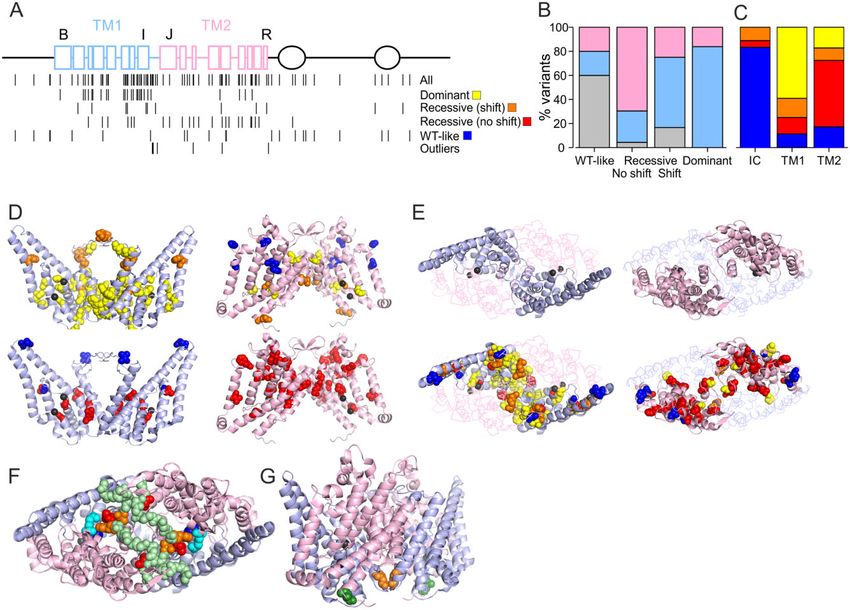
When plotted on the ClC-1 primary structure a disproportionately priority.
high number of variants (48%) were located in the first of the TM1 is mainly located towards the intracellular end of the
roughly identical halves of the transmembrane domain (TM1) of intramembrane domain where the chloride ions are found in the
ClC-1 subunit (helices B-I and the IJ-linker, residues 111–344) but selectivity filter pathway (Fig. 6D).32 The voltage dependence of
only 19% of variants were found in the intracellular N and C ter- ClC-1 channel activation is sensitive to Cl– concentration and
mini (1–110, 586–988) (Tables 1, 2 and Fig. 6). Seventy-nine per cent arises from interactions with the channel and the ion.14–16 It is
(37/47) of the variants that shifted the voltage dependence of acti- thus likely that the dominant variants localized towards the intra-
vation in homomeric or simulated heterozygous condition, includ- cellular side shift the voltage dependence of activation by altering
ing the variants with extraordinary functional properties, are these interactions. The TM1 also forms most of the subunit inter-
located in TM1, while a further eight (17%) were on the second half face (Fig. 6E), suggesting that mutations in one subunit can affect
of transmembrane domain (TM2) (residues 345–585) and two var- the chloride conducting pathway in the neighbouring subunit
iants (4%) were intracellular (Fig. 6A–C). Of the variants with through this interface. F297S, the only variant with voltage de-
shifted voltage dependence of activation in simulated heterozy- pendence of activation showing a larger shift in simulated hetero-
gous condition 84% (26/31) were found on TM1 and the remaining zygous than in the homomeric condition, affects a residue located
16% on TM2. In contrast, 16/23 (70%) of the variants with recessive at the subunit interface with the main chain of the two F297 resi-
functional features without shift in voltage dependence of activa- dues in close contact (Fig. 6G). Functional data indicate that a sym-
tion in homomeric condition were found on TM2, six in TM1 (26%) metric F297S channel shows a smaller shift in voltage dependenceYou can also read