Top Ophthalmology Tips for the Practicing Veterinarian
←
→
Page content transcription
If your browser does not render page correctly, please read the page content below
Top Ophthalmology Tips for the Practicing Veterinarian

                                     By Martin Coster, DVM, MS, DACVO
                                     angell.org/eyes
                                     ophthalmology@angell.org
                                     617-522-7282
                                     January 2021

                                     This article shares some tips and tricks for performing successful eye exams.

1.   It’s all in the holder
     The key to performing a decent eye exam is to have the animal restrained appropriately, which may take one or
     two (and sometimes even three) assistants. The pet’s eyes should be at the examiner’s eye level, which means in
     most cases the pet should be on an exam table, with the examiner seated at a good height to make eye-to-eye
     contact without sacrificing ergonomics. An adjusting stool and/or table will therefore be needed. The pet’s head
     should be restrained with one hand under the chin and one at the back of the skull. This means a second holder
     may be needed to control the body if the patient will not stay still. A third assistant may be needed to open the
     eyelids or control front legs. Pre-visit sedation or anti-anxiety medications to facilitate a less-stressed, less
     fearful visit can be very helpful to performing a complete eye exam.1,2

2. Darkness is key
   The ideal examination room is one in which external light sources (windows, doors, and even light seeping in
   around doors) can be completely blocked out. An easily-accessible dimmer switch on the room lights can help
   greatly to allow the eye exam to be performed at varying levels of light intensity. A red light can be used to
   provide working illumination. This reduces incidental light reflections on the surface of the eye, which
   maximizes the examiner’s ability to look into the eye with direct illumination.

 Angell Animal Medical Center • 350 S. Huntington Ave., Boston, MA 02130 • 617-522-7282 • fax: 617-989-1635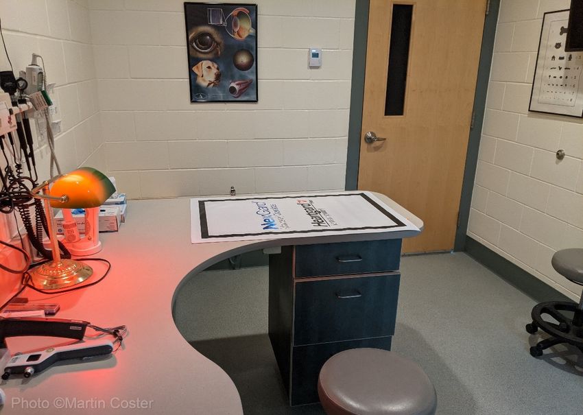
Figure 1: Ophthalmology examination room; note blacked-out window on the door, red light, and
adjustable stools. In this setup, the main room lighting is controlled by a dimmer switch easily accessible
under the examination table.
3. Don’t forget the basic tests
Performing Schirmer tear test, fluorescein stain, and intraocular pressure are essential tests in order to make a
diagnosis. Dry eye in particular is often overlooked for lack of a tear test, the normal value of which is
15mm/min or greater, in the absence of clinical signs. Intraocular pressure should be interpreted in light of other
exam findings. For example, although a normal pressure of approximately 15mmHg is expected, this may be
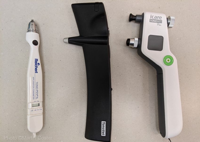
considered glaucomatous in a case of severe uveitis. As a variety of Icare® TONOVETs have become
increasingly popular due to ease of use compared with the Tono-Pen®, knowing the normal values for your
instrument is important. For example, the TONOVET Plus may read around 4 to 5 mmHg higher than the
standard TONOVET, although the TONOVET Plus may be providing a truer pressure measurement.3
Angell Animal Medical Center • 350 S. Huntington Ave., Boston, MA 02130 • 617-522-7282 • fax: 617-989-1635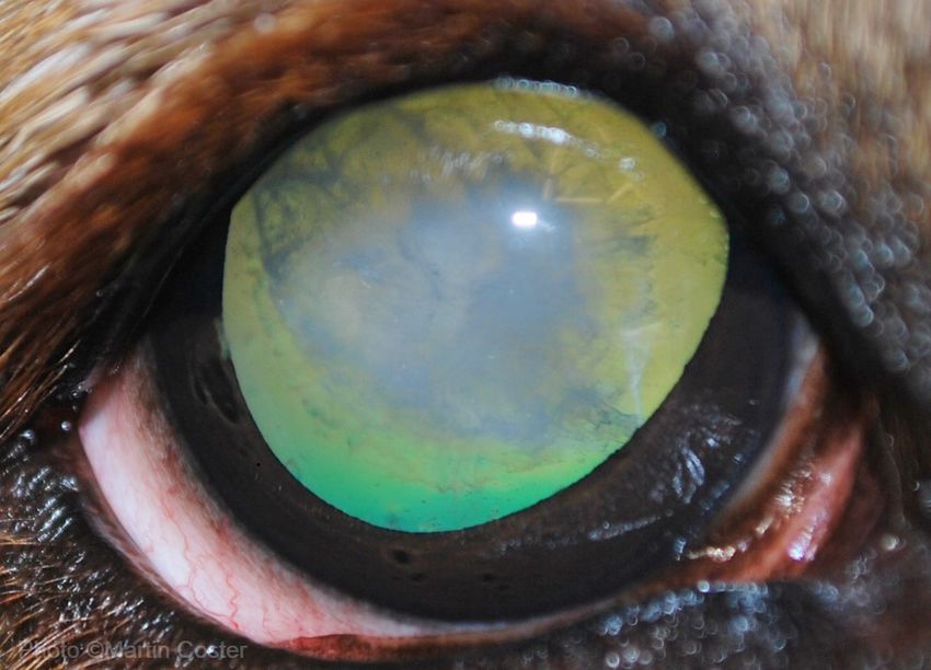
Figure 2: Three generations of tonometers; the Reichert Tono-Pen® XL; the TONOVET®; the Icare®
        TONOVET Plus. The Schiotz tonometer is not pictured as it has been consigned to our time capsule.

4.   Follow an ordered exam
     A thorough eye exam should have an ordered approach that is consistent every time. This maximizes the
     chance that every structure will be looked at and thus all conditions can be diagnosed. An “outside-in”
     approach is most common, assessing global vision and pupillary light reflex assessments, facial conformation
     and orbit, eyelids including third eyelid, conjunctiva, cornea, anterior chamber, iris, lens, vitreous, and retinal
     exam. Most practices do not have a slitlamp biomicroscope as used by ophthalmologists. Therefore, the
     magnification provided by a headloupe is very helpful, and having a well-charged bright transilluminator can
     make a huge difference over a dimmer light source. An ophthalmoscope can be used with its slitbeam for
     further anterior chamber assessment. Fundic examination is best performed with an indirect lens (20 or 28
     diopter) and bright transilluminator.

5.   Prescribe out when necessary
     Many veterinarians are unable to carry numerous ophthalmic medications in their pharmacy. Fortunately, most
     ophthalmic drugs are also used in humans, and thus do not all need to be stocked in-house. This may sound
     like a simple tip, but all too often we see referral patients in whom a correct diagnosis was made but
     appropriate medications were not prescribed due to “lack” of availability.

 Angell Animal Medical Center • 350 S. Huntington Ave., Boston, MA 02130 • 617-522-7282 • fax: 617-989-1635
6.   Drugs that penetrate into the eye
     Not all topical drugs penetrate through an intact corneal epithelium. For antibiotics, choices are limited to
     fluoroquinolones (e.g. ofloxacin and ciprofloxacin) or chloramphenicol. Non-steroidal anti-inflammatory drugs
     (NSAIDs, e.g. diclofenac, flurbiprofen, and ketorolac) and of course all glaucoma drops penetrate into the eye,
     but only certain steroid salts will penetrate. These are prednisolone acetate and the dexamethasone found in
     neo-poly-dexamethasone; in this latter case, solution may penetrate better than ointment. Antibiotics, NSAIDs,
     and steroids will not penetrate much deeper than the aqueous humor, and so posterior segment disease must be
     treated with systemic medications.

7.   Neo-Poly-Hydrocortisone is rarely used by this author
     This steroid does not penetrate into the eye so cannot treat uveitis, and is rarely strong enough for ocular
     surface inflammation. Its use is thus quite limited. Allergic inflammation might respond but may do better with
     specific antihistamine eye drops such as patanol or ketotifen.

8.   Never underestimate pain
     Clients frequently report their pet is comfortable, while also reporting the animal squinting at home. A careful
     history can elucidate other symptoms of pain, such as reduced appetite or altered behavior, or rubbing at the
     eye or face. Whether treating corneal ulcers, conjunctivitis, glaucoma, or uveitis, pain control should not be
     overlooked. Topical anesthesia (e.g. proparacaine) is inappropriate except for diagnostics, due to potential
     epithelial toxicity. Systemic pain control with an oral NSAID, gabapentin, fentanyl patch, and/or
     injectable/oral opioids should be considered as indicated.

9.   Sclerosis vs. Cataract
     One of the hardest determinations to make is whether an eye has cataract versus lenticular a.k.a. nuclear
     sclerosis (complicated even further when both are present). Generally, nuclear sclerosis should not occur in an
     animal younger than 7-8 years of age, and should be bilaterally symmetrical when present. Iatrogenic dilation
     with tropicamide or dark room examination with a relatively dim (so as not to elicit the pupillary light reflex)
     direct light source may be needed to visualize the perimeter of the lens. Dense nuclear sclerosis can obscure
     the fundus and behave like a cataract, but generally the fundus should be easily visualized even through
     sclerosis. The outer cortex should be clear with sclerosis. True cataract can be bilaterally symmetrical, but is
     rarely homogenous throughout the lens, and obscures fundic examination.

 Angell Animal Medical Center • 350 S. Huntington Ave., Boston, MA 02130 • 617-522-7282 • fax: 617-989-1635
Figure 3: Nuclear sclerosis a.k.a. lenticular sclerosis, in the left eye of a dog; note the pearlescent central
      (nuclear) opacity. This opacity should be in symmetry with the other eye. This eye is iatrogenically dilated
      with tropicamide.

Angell Animal Medical Center • 350 S. Huntington Ave., Boston, MA 02130 • 617-522-7282 • fax: 617-989-1635
Figure 4: Nuclear cataract, in the right eye of a dog. Note the dense, heterogeneous white opacity
obscuring the central view of the yellow/green tapetal reflection.
10. Tips for a fundic exam
The retinal exam is the hardest part of an eye exam and is thus often unfortunately overlooked or skipped
entirely. Iatrogenic dilation with tropicamide (assuming intraocular pressure is normal and glaucoma or lens
luxation are not suspected) is extremely important for a full retinal exam; however the time limitations of
private practice, and owner constraints on time, can often preclude this. In these instances, dark room
examination using a slightly dimmer light source and a 28 diopter lens (as opposed to a 20 diopter lens used
for dilated exams) can provide visualization through a mid-range pupil. Once again, it is essential to have a
trained assistant or two, to restrain the patient and hold open the eyelids as needed. Standing arms distance
away from the eye, bring a transilluminator up close to the patient’s eye, obtain a tapetal reflection and then
bring the light source back to the level of your own eye, still directed at the fundus. At this point, bring an
indirect lens into alignment with the light and patient’s eye, close enough to the eye that you can observe the
iris and pupil. Once the pupil is centered in the lens, bring the lens closer to you until it is about a finger’s
length away from the eye (stabilize your hand by extending a finger to touch the side of the face or bridge of
the nose). If you lose the view of the fundus, or to alter position, swing the lens away, re-gain the tapetal
reflection, and swing the lens back into place.
Angell Animal Medical Center • 350 S. Huntington Ave., Boston, MA 02130 • 617-522-7282 • fax: 617-989-1635Footnotes & References
1. In an abstract presented at the 49th Annual Scientific Meeting Of The American College Of Veterinary
Ophthalmologists, Minneapolis, Minnesota, Sept 26‐29, 2018, titled “Effects of oral trazodone on canine
tear production and intraocular pressure” Pelych et al concluded that “administration of 5 or 9 mg/kg of
trazodone does not significantly affect tear production or IOP in dogs within 6 hours”.
2. If used for pre-visit sedation, gabapentin might lower intraocular pressure in the dog, but not a clinically
significant amount; however its effect on glaucoma have not been studied: Shukla AK, Pinard CL, Flynn
BL, Bauman CA. Effects of orally administered gabapentin, tramadol, and meloxicam on ocular variables
in healthy dogs. Am J Vet Res. 2020 Dec;81(12):973-984. doi: 10.2460/ajvr.81.12.973. PMID: 33251843.
3. Minella AL, Kiland JA, Gloe S, McLellan GJ. Validation and comparison of four handheld tonometers in
normal ex vivo canine eyes. Vet Ophthalmol. 2020 Jun 1. doi: 10.1111/vop.12780. Epub ahead of print.
PMID: 32478941.
Angell Animal Medical Center • 350 S. Huntington Ave., Boston, MA 02130 • 617-522-7282 • fax: 617-989-1635You can also read