Therapeutic Antibodies Against Shiga Toxins: Trends and Perspectives - Frontiers
←
→
Page content transcription
If your browser does not render page correctly, please read the page content below
REVIEW
                                                                                                                                                   published: 10 February 2022
                                                                                                                                              doi: 10.3389/fcimb.2022.825856

                                             Therapeutic Antibodies Against Shiga
                                             Toxins: Trends and Perspectives
                                             Izabella de Macedo Henrique 1, Flavia Sacerdoti 2, Raissa Lozzardo Ferreira 1,
                                             Camila Henrique 1, Maria Marta Amaral 2, Roxane Maria Fontes Piazza 1* and Daniela Luz 1*
                                             1 Laboratório de Bacteriologia, Instituto Butantan, São Paulo, Brazil, 2 Laboratorio de Fisiopatogenia, Instituto de Fisiologı´a y

                                             Biofı´sica Bernardo Houssay (IFIBIO Houssay-CONICET), Departamento de Fisiologı´a, Facultad de Medicina, Universidad de
                                             Buenos Aires, Buenos Aires, Argentina

                                             Shiga toxins (Stx) are AB5-type toxins, composed of five B subunits which bind to Gb3
                                             host cell receptors and an active A subunit, whose action on the ribosome leads to protein
                            Edited by:
                                             synthesis suppression. The two Stx types (Stx1 and Stx2) and their subtypes can be
                      Jorge Goldstein,
 Consejo Nacional de Investigaciones         produced by Shiga toxin-producing Escherichia coli strains and some Shigella spp. These
   Cientı´ficas y Técnicas (CONICET),        bacteria colonize the colon and induce diarrhea that may progress to hemorrhagic colitis
                             Argentina
                                             and in the most severe cases, to hemolytic uremic syndrome, which could lead to death.
                        Reviewed by:
                      Nora Lı´a Padola,
                                             Since the use of antibiotics in these infections is a topic of great controversy, the treatment
         National University of Central      remains supportive and there are no specific therapies to ameliorate the course.
               Buenos Aires, Argentina
                                             Therefore, there is an open window for Stx neutralization employing antibodies, which
                          Emily Mallick,
              Fluid-Screen Cambridge,        are versatile molecules. Indeed, polyclonal, monoclonal, and recombinant antibodies have
                         United States       been raised and tested in vitro and in vivo assays, showing differences in their neutralizing
                       Pablo Chacana,
      Instituto Nacional de Tecnologı´a
                                             ability against deleterious effects of Stx. These molecules are in different phases of
               Agropecuaria, Argentina       development for which we decide to present herein an updated report of these
                   *Correspondence:          antibody molecules, their source, advantages, and disadvantages of the promising
          Roxane Maria Fontes Piazza
                                             ones, as well as the challenges faced until reaching their applicability.
       roxane.piazza@butantan.gov.br
                          Daniela Luz        Keywords: Shiga toxin-producing E. coli, Stx toxins, antibodies, therapy, trends
          daniela.luz@butantan.gov.br

                   Specialty section:
        This article was submitted to        1 INTRODUCTION
    Molecular Bacterial Pathogenesis,
               a section of the journal      Shiga toxins (Stxs) are potent cytotoxic proteins that can be produced and secreted by Shigella
              Frontiers in Cellular and      dysenteriae 1 and by some serogroups of Escherichia coli (called Stx1 in E. coli), which also can
                Infection Microbiology       produce a second type of Stx, called Stx2, antigenically distinct of Stx/Stx1, but with the same action
       Received: 30 November 2021            mode (Melton-Celsa and O'Brien, 2014). Shiga toxin-producing Escherichia coli (STEC) is a family
         Accepted: 11 January 2022           of bacteria that share the possibility to secrete Stx. STEC are foodborne pathogens that may colonize
        Published: 10 February 2022          and damage the human colon, where they secrete Stx that gain access to the bloodstream and
                                 Citation:   damage different target organs: mainly kidney and brain. Indeed, STEC infection may develop
Henrique IM, Sacerdoti F, Ferreira RL,       hemolytic uremic syndrome (HUS) because of Stx in the target organs.
 Henrique C, Amaral MM, Piazza RMF
                                                 Currently, there are no protective measures or therapies against Stx intoxication, and the
         and Luz D (2022) Therapeutic
     Antibodies Against Shiga Toxins:
                                             treatment is solely supportive and includes rehydration therapy, and, where necessary, dialysis. The
               Trends and Perspectives.      neutralization of Stx before the appearance of HUS severe symptoms is one of the promising
Front. Cell. Infect. Microbiol. 12:825856.   approach; therefore, this review summarizes one of the most studied neutralization molecules,
       doi: 10.3389/fcimb.2022.825856        the antibodies.

Frontiers in Cellular and Infection Microbiology | www.frontiersin.org                 1                                         February 2022 | Volume 12 | Article 825856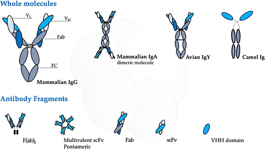
Henrique et al.                                                                                                          Antibodies Against Shiga Toxins

    Antibodies are key molecules for therapeutic proposal. Its                  described that each Stx variant differs in pathogenicity. While
modular structure enables it to be engineered to have tags for                  Stx1a strains are associated with hospitalization and bloody
purification and immunoprecipitation, conjugation sites to                       diarrhea, Stx1c and Stx1d are less often associated and not
improve chemical space, or mutagenesis to map the CDRs,                         enough information is available about the clinical significance
which allow affinity improvement (Gitlin, 1966; Tiller and                       of Stx1d (Kumar et al., 2012; EFSA BIOHAZ Panel et al., 2020).
Tessier, 2015; Basu et al., 2019). As recombinant molecules,                    On the other hand, Stx2a and Stx2c are clinically more related
they can be produced by several distinct hosts, such as                         with severe cases of HUS, and additionally, it is generally
mammalian, insect, yeast, and bacterial systems, the last one                   described that Stx2a expressing STEC strains develop more
requires low-cost production media and equipment, besides                       severe cases of HUS with a higher risk of encephalopathy
being able to synthesize and express practically unlimited                      (Orth et al., 2007). Differently, Stx2d and Stx2e are associated
amounts of antibodies to almost any antigen, therefore, an                      with milder or asymptomatic infections (Friedrich et al., 2002;
interesting molecule used either by industry or academia,                       Orth et al., 2007). Stx2e strains are not often found in STEC
ranging from bench to large-scale production (Robinson                          infections associated with human disease, and Stx2f strains have
et al., 2015).                                                                  recently been isolated from patients with HUS (Friesema et al.,
    The main aim of this review is to provide an update regarding               2015; De Rauw et al., 2018). Stx2g subtype is also rarely
antibodies raised towards Stx to prevent their toxic effects, by                associated with human illness and not usually associated with
contextualizing the intoxication problem and how antibodies can                 severe illness (Prager et al., 2011).
be used as a therapeutic approach to solve it.                                      The pathology associated with the Stx toxicity starts with Stx-
                                                                                producing bacterial infection. Figure 1 summarizes the disease
1.1 Shiga Toxins and Their Toxic                                                course and main outcomes of Stx intoxication, which begins with
Outcomes                                                                        diarrhea that can be self-limited. In some cases, it may evolve to
Stxs are the main virulence factor of STEC and are responsible                  bloody diarrhea, hemorrhagic colitis (HC), and, in
for developing HUS. Stx is encoded in the late region of the                    approximately 15% of the infections (Tarr et al., 2005), develop
genome of lambdoid prophages integrated into the bacterial                      HUS characterized by thrombocytopenia, microangiopathic
chromosome (O'Brien et al., 1983) and is optimally expressed                    hemolytic anemia, and acute kidney injury (Repetto et al., 2013).
after the induction of the lytic cycle. In this regard, Stx phages                  Development of HUS occurs approximately 7 days after the
constitute an important lateral gene transfer mechanism that                    onset of gastrointestinal symptoms and 4 days after the onset of
may contribute to the emergence of new STEC strains (Schmidt,                   bloody diarrhea (Bitzan, 2009). Intervention strategies for
2001; Bielaszewska et al., 2014). An example of this gene transfer              blocking Stx, as primary therapy for directly preventing HUS
occurred in the outbreak in Europe in 2011 where an                             development, may be applied in this period between STEC
enteroaggregative E. coli strain acquired the Stx phage, and this               infection and before the appearance of HUS symptoms.
newly STEC strain affected mainly adults (Borgatta et al., 2012).                   The clinical features of cytotoxic effects of Stx are determined
    Stx can be classified in antigenically different types and subtypes:         by the damage to endothelial cells of small vessels mainly
Stx (from Shigella sp.), Stx1 (Stx1a, Stx1c, Stx1d, Stx1e), and Stx2            localized in the colon, kidney, and central nervous system
(Stx2a, Stx2c, Stx2d, Stx2dact, Stx2e, Stx2f, Stx2g, Stx2h, Stx2i, Stx2j,       (CNS) (Richardson et al., 1988); however, several other organs
Stx2k—recently reported but have yet to be broadly accepted—and                 can be affected, such as pancreas and liver, and consequently,
Stx2l). Stx subtypes differ in their amino acid sequence, and an                endothelial damage can be widespread in the microvasculature
analysis of the Stx protein sequences showed that Stx1, Stx1c, and              (Luna et al., 2021). Nevertheless, not all cells undergo cell death
Stx1d have 93%–100% homology and Stx2a to Stx2g have also high                  upon binding and uptake of Stx. Toxin binding to platelets,
homology (93%–100%), except for Stx2 and Stx2f (69%) (Golshani                  leukocytes, and erythrocytes can lead to their activation without
et al., 2016). Among the several Stx subtypes, the prototype toxin for          inducing cytotoxicity (Karpman and Ståhl, 2014). During
each group is now designated Stx1a or Stx2a (Melton-Celsa and                   activation, cells release microvesicles (MVs) and it has been
O'Brien, 2014).                                                                 described that Stx may be released within MVs (MVs-Stx) from
    All Stx types and their subtypes are AB5 toxins characterized               blood cells during HUS (Ge et al., 2012; Arvidsson et al., 2015;
by the presence of a one active A domain (~32 kDa) that blocks                  Ståhl et al., 2015). So, these MVs-Stx can transport Stx into the
cell protein synthesis by cleavage of ribosomal RNA, and a                      kidney, evading the immune system, and contributing to kidney
pentameric binding domain B (~7.7 kDa each), with close                         failure in HUS patients (Ståhl et al., 2015).
affinity to the glycosphingolipid globotriaosylceramide (Gb3,                        The kidney is seriously affected by Stx because of the presence
CD77) and, to a lesser extent, globotetraosylceramide (Gb4)                     of specially Stx-sensitive cells that express high amounts of Gb3
(Legros et al., 2018), which are found in a variety of human                    receptor as microvascular endothelial cells that express 50-fold
cells, such as glomerular and brain endothelial cells. Receptor-                higher Gb3 levels than macrovascular endothelial cells (Obrig
mediated internalization of the toxin results in the inhibition of              et al., 1993; Obrig, 2010). Moreover, because of the high volume
protein synthesis, ribotoxic stress that finally leads to apoptosis              of blood flow and filtration rate, the possibility of Stx interaction
(Tesh et al., 1993; Melton-Celsa and O'Brien, 2014).                            with cells of renal microvasculature and the filtration barrier
    Not all Stx subtypes have been associated with severe illness               increase (Obrig, 2010). The thrombotic microangiopathy lesion
(Beutin et al., 2004; Hofer et al., 2012). In this regard, it has been          is the typical injury caused by Stx in the kidney because of the

Frontiers in Cellular and Infection Microbiology | www.frontiersin.org      2                                February 2022 | Volume 12 | Article 825856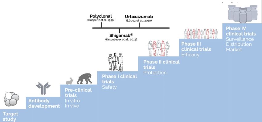
Henrique et al.                                                                                                                    Antibodies Against Shiga Toxins

 FIGURE 1 | Clinical course and main outcomes of Stx-producing bacterial infection. Modified from (Bruyand et al., 2018).

direct action of Stx on glomerular endothelial cells that consists                   detected inside neurons that upregulated the Gb3 receptor
of thickening of arterioles and capillaries, swelling and                            (Tironi-Farinati et al., 2010).
detachment of endothelial cells from the basement membrane,
and platelet thrombi that obstruct the microcirculation (Zoja                        1.2 Stx-Producing Bacteria
et al., 2010). Inflammation also can contribute to endothelial                        STEC and its subgroup enterohemorrhagic E. coli (EHEC) are
damage. In this sense, Stx induces the release of pro-                               the major causes of gastroenteritis and competent Shiga-toxin
inflammatory cytokines, leukocyte recruitment, platelet                               producers. Worldwide, around 2.8 million people were infected
aggregation, and fibrin deposition. All these events lead to                          per year, and over 250,000 illnesses occurred in the USA due to
partial or complete vessel occlusion by microthrombi and the                         STEC, and although several serotypes are harmful, one-third
consequent microangiopathic hemolytic anemia (Exeni et al.,                          originate from the O157:H7 serotype (Bird, 2019).
2018). The kidney injury is expressed as different degrees of renal                      In Latin America, the southern countries are the most
failure (Repetto, 2005; Repetto et al., 2013), and the endothelial                   affected, especially Argentina. There, HUS is endemic, and
dysfunction is essential to the development of microvascular                         although Argentina has the greater incidence rates in the entire
lesions in HUS (Morigi et al., 1995; Ruggenenti et al., 2001).                       world, the correlation of infection rates between countries is
    The main effects of Stx on the endothelial cells are                             problematic since each country has unique testing parameters
intracellular edema and a decrease of cell viability by apoptosis                    that make this evaluation difficult; a preoccupation due to the
and necrosis (Amaral et al., 2013). Likewise, Stx also causes                        outbreaks in Argentina also relays the relevant role of the nation
damages to renal epithelial cells by the inhibition of protein                       in the bovine meat exportation (Torres et al., 2018; Torti et al.,
synthesis, apoptosis, and necrosis, showing the direct effect of                     2021). For that reason, meaningful research in this field is done
this toxin on the renal tubules (Karpman et al., 1998; Kaneko                        in Latin America (Torres et al., 2018).
et al., 2001; Creydt et al., 2006).                                                      Majority of STEC infection occurs through fecal-oral
    Moreover, there is evidence that a direct effect of Stx is also on               contamination, contaminated water, and food consumption,
the central neural system (Obata et al., 2008). It was                               such as undercooked meat (below 71°C), unpasteurized food,
demonstrated that Stx2 has a direct action in the brain of rats                      contaminated vegetables, and person-to-person contact
since it produces damage in neurons, astrocytes,                                     (Alconcher et al., 2020). Cattle are the principal reservoirs for
oligodendrocytes, and endothelial cells (Goldstein et al., 2007).                    STEC, and the bacteria can survive for months in soil, water, or
Furthermore, Stx2 breaks the blood-brain barrier (BBB) and                           organic material (Sapountzis et al., 2020).
damages cells that modulate motor functions (Pinto et al., 2017).                        Upon ingestion, STEC/EHEC resides in the intestinal tract
Stx2 may act through Gb3 neuronal receptors, and this toxin was                      and adheres to the gut epithelium of the distal ileum and colon.

Frontiers in Cellular and Infection Microbiology | www.frontiersin.org           3                                     February 2022 | Volume 12 | Article 825856
Henrique et al. Antibodies Against Shiga Toxins
Fimbriae promote an initial binding, which, in EHEC infections, antigen-binding site (Fab), which determines the molecule
is followed by the effector protein (Esp proteins) injection by a specificity (Nilvebrant and Sidhu, 2018) (Figure 2). The Fab
type III secretion system (T3SS) (Donnenberg and Kaper, 1992; fragment contains three hypervariable regions called
Garmendia et al., 2005; Gaytá n et al., 2016). After injection of the complementary domain regions (CDR) responsible for
translocated intimin receptor (Tir) into the host cell plasma hypervariability and specificity against different antigens.
membrane, it interacts with the bacterial outer membrane The versatility and specificity of these molecules attract
protein intimin, triggering the intimate attachment to the host significant attention for their use as therapeutic tools. Indeed,
cell and the effacement of the brush border microvilli, initiating the global market for monoclonal antibodies is projecting a
actin polymerization and subsequent formation of attaching and compound annual growth rate (CAGR) of 5.3% between the
effacing (A/E) lesions. The genes encoding Tir, intimin, and the years 2020 and 2026 and its prospect to reach the value of 11.77
T3SS are localized on the chromosomal locus of enterocyte billion dollars in 2026 (Kaplon and Reichert, 2021). There are
effacement (LEE) pathogenicity island present only on EHEC numerous motivations to be interested in antibodies. From the
strains. LEE-negative STEC strains may also produce severe business view, engineered antibodies represent potential
disease since other adhesins may be involved in adhesion/ solutions to challenges facing the industry, including the
colonization of this subset of bacteria to enterocytes shortage of innovative candidates in the pipeline and low
(McWilliams and Torres, 2014) and from the unusual HUS- approval success rates for new therapeutics.
inducing E. coli strain EAEC of serotype O104:H4 bearing stx2 The comparatively high approval success rates for
gene, which was responsible for the major outbreak in Germany monoclonal antibodies (mAbs), for example, are probably one
and parts of Europe in 2011 (Bielaszewska et al., 2011; Mellmann reason for the growing interest in the development of these
et al., 2011). therapeutics. As described by Hay et al. (2014), biologics such as
Shigellosis is bacillary dysentery caused by Shigella; these mAbs have a probability of approval of near 1 in 4 compared
bacteria, at first, attack epithelial cells of the large intestine, with that of new molecular entities, which is nearly 1 in 8.
and then the infection reaches nearby cells. Transmission can Basically, mAbs are granted marketing approvals at twice the rate
happen by the fecal-oral route, also due to contaminated food of small molecule drugs (Kaplon and Reichert, 2019). On the
and water, or through vectors like flies. Shigella as STEC has the other hand, from the medical science perspective, innovative
incredible characteristic of causing an illness with a low infection protein engineering allows for the design of antibody molecules
dose of only 100 organisms. This characteristic makes Shigella a with decreased immunogenicity, enhanced effector functions,
danger to human health and a significant food safety concern and improved pharmacokinetic properties (Reichert, 2009).
since this pathogen has been responsible for so many epidemics To generate antibodies for therapeutic application, there are
(Lampel et al., 2018). three main approaches: (i) by animal immunization to generate
There are four serogroups; S. dysenteriae with 12 serotypes; polyclonal antibodies (pAbs); (ii) by lymphocyte immortalization
S. flexneri with 6 serotypes; S. boydii with 18 serotypes; and creating single clones secreting specific mAbs; and (iii) by DNA
S. sonnei with 1 serotype, also known as A, B, C, and D, recombinant technology and heterologous expression system
respectively (Hale et al., 1996). creating diverse recombinant (rAbs) antibody formats for
The serogroups A and B are the promoters of endemic and different goals (Cosson and Hartley, 2016).
epidemic shigellosis (respectively) in developing countries, with Following, we provide a review of the antibodies raised against
high transmission rates and significant cases of fatality rates. Stx and their different presentation (Figure 2) to update the efforts
Only A can cause infection; C and D serogroups are mild, towards the search for an effective neutralizing molecule against
causing watery or bloody diarrhea (Williams and Berkley, the most severe Stx intoxication symptoms.
2018). However, in general, Shigella dysenteriae type 1 strains
are the most reported to be associated with Stx toxin production 2.1 Antibodies Against Shiga Toxin Activity
(Mark Taylor, 2008; Williams and Berkley, 2018). Before becoming a biopharmaceutical, any therapeutic molecule,
from vaccine candidates to antibodies must get through several
testing stages to ensure that the final compound is safe and
2 ANTIBODIES AS THERAPEUTIC TOOLS efficient for human administration. These steps include
establishing the pharmacology and biochemistry of the
Antibodies are ubiquitous molecules, and because of their ability molecule of interest through various in vitro and in vivo tests
to highly recognize and bind to an antigen, besides mediating the to assess its safety (Tamimi and Ellis, 2009). Figure 3
interaction with other cells and molecules of the immune system, summarizes these steps and presents the most advanced
they have been used in a wide range of biotechnological antibodies raised against Stx, which reached clinical trials.
approaches (Basu et al., 2019). Moreover, Table 1 summarizes all the anti-Stx antibodies cited
Although the mammalian immune system can produce five from basic polyclonal until innovative recombinant antibodies,
classes of immunoglobulin, the IgG is the most used for antibody their format, and stage of development.
research. It has a “Y”-shape structure consisting of two light and It is noteworthy that, once a biopharmaceutical is approved, it
two heavy chains that form a conserved crystallizable fragment must be manufactured by the standards of purity and stability, as
(FC), responsible for effector functions of the antibody, and an per regulatory agencies (Mohs and Greig, 2017), as well as being
Frontiers in Cellular and Infection Microbiology | www.frontiersin.org 4 February 2022 | Volume 12 | Article 825856Henrique et al. Antibodies Against Shiga Toxins
FIGURE 2 | Schematic representation of the different antibody source, structure, and arrangement presented at this review. The figure highlights the domains of the
mammalian IgG, the most used antibody molecule.
evaluated for its postmarketing safety, with the monitoring of Below, we describe in detail all approaches used to develop
possible adverse events (Tamimi and Ellis, 2009). anti-Stx antibodies.
Herein, in this review, we intend to discuss antibodies against
Stx and their trends and perspectives; for that, we employed as 2.2 Polyclonal Antibodies
selection criteria the publications in PubMed between January 1, The polyclonal antibody obtainment starts with the animal
1989 and October 1, 2021, with predilection given to renowned immunization, which in general is either mice or rabbits and
papers and review published in the last 10 years or so, with the consists of a purified antibody mixture from the immunized
tool “MeSH” using the terms “hemolytic uremic syndrome,” animal serum. The term polyclonal is the mixture of antibodies
“thrombotic microangiopathy,” “Shiga toxin,” “Shiga-like that comes from different lymphocyte clones and responds
toxins,” “verotoxin,” and “antibodies” in combination with the against different epitopes of the target toxin (Cosson and
term “pathophysiology,” “causes,” “therapy,” and “treatment.” Hartley, 2016). pAbs have been used for neutralizing the
We restricted our search to English publications. We first toxicity of bacterial toxins since the golden age of microbiology
focused on review articles to provide more detail. Selected (circa 1850), initially for diphtheria and tetanus, which was
reports from the past 10 years but did not exclude important granted a Nobel prize to Emil von Behring in 1901 (Froude
and highly cited older publications. For greater elucidation of et al., 2011). Regarding Stx toxins, it was not different; indeed
these data, it searched the reference lists of selected articles many groups started obtaining pAbs as soon as the first
identified by this search strategy and selected those relevant to description of Stx intoxication was published (O'Brien et al.,
the main topic. In the last decade, some broad and relevant 1983). Several polyclonal strategies were attempted towards Stx,
reviews have been published; we highlighted (Keir et al., 2012; such as the use of immunized animal serum, bovine colostrum,
Page and Liles, 2013; Hall et al., 2017; Torres et al., 2018; and enriched IgY from avian eggs.
Kakoullis et al., 2019; Joseph et al., 2020; Lingwood, 2020; First, we highlight the work developed by O’Brien’s group,
Mühlen and Dersch, 2020; Hwang et al., 2021) those that which has contributed to the knowledge of Shiga toxin since the
covered different aspects of STEC such as epidemiology, 1980s. The Stx2-producing E. coli O157:H7 strain 86-24 was used
diagnosis, serotypes, and HUS from pathogenicity to to produce anti-Stx2 polyclonal rabbit antisera (Kokai-Kun et al.,
therapeutic options. Therefore, our decision was to focus on 2000). At rodent infection model, this rabbit anti-Stx2 pAb was
the therapeutic strategies using antibodies for Stx blockage. administered in a single dose intraperitoneally. To determine the
Frontiers in Cellular and Infection Microbiology | www.frontiersin.org 5 February 2022 | Volume 12 | Article 825856Henrique et al. Antibodies Against Shiga Toxins
FIGURE 3 | Development steps of biopharmaceuticals until human use approval and the most advanced antibodies raised against Stx. The discovery of a new drug
as therapeutic antibodies starts with the target study followed by the antibody development (choosing of which antibody, type, format, avidity, and affinity) and
achieving the functional test in vitro and in vivo (using different animal models, starting with mice and rabbits until the most closely related to humans as apes),
reaching the challenge assay in an animal model, which is part of the preclinical trials. Once the molecule shows relevant ability to neutralize the effect of the target,
human clinical trials could be performed. The clinical trials are generally divided into four phases: The first one enrolls dozens of volunteers, and the main endpoint is
to test the safety of the molecule. By phase II, the molecule is tested in hundreds of volunteers to attest protection towards the toxin effect. At the last phase before
regulatory agency approval, controlled randomized double-blind assays are performed in millions of volunteers to test the molecule efficacy. Once passed at those
clinical trials, the therapeutic molecule can be approved by the regulatory agencies to be administered in humans, which is considered phase IV, in which the
surveillance of its long-term administration is going to take place (Tamimi and Ellis, 2009). The polyclonal developed by Huppertz (Huppertz et al., 1999) and
Shigamab® (Desoubeaux et al., 2013) tested safety as a phase I clinical trial. On the other hand, urtoxazumab tested safety and pharmacokinetic, which could be
considered a clinical trial 1/2 (Ló pez et al., 2010).
efficacy, fecal pellets were collected at different times (Huppertz et al., 1999). On the other hand, some studies
postinfection and tested for toxin presence. From days 3–5 showed effectivity of colostrum containing anti-Shiga antibody
postchallenge, it was observed that the antiserum diminished administration to protect animals from death (Funatogawa et al.,
the E. coli O157:H7 burden and extended the animal survival. 2002; Seita et al., 2013). Similarly, colostrum IgG against Shiga
However, by the seventh day, the level was similar to that of the toxin and bovine lactoferrin completely prevented lethality of
control group (Mohawk et al., 2010). E. coli O157:H7 in a weaned mouse model (Albanese et al., 2018).
Another important study was done by Takeda and his In addition, an early study in children showed that bovine
coworkers in characterizing Shiga toxin and investigating the colostrum is well tolerated, reduced the frequency of loose
prominent therapies to neutralize it. Initially, human serum anti- stools, and eliminated bacterial infection (Huppertz et al.,
Stx1 and anti-Stx2 from HUS patients and those commercially 1999). In this line of thought, spray-dried and reconstituted
available were tested against both toxins. Only one tested serum hyperimmune bovine colostrum against Stx2 preserved the
showed a neutralizing ability against all Stx1 (125 pg/ml); on the protective capacity against E. coli O157:H7 pathogenicity in
other hand, none of the tested serum neutralize Stx2 (Takeda vitro and in vivo models. In this regard, the hyperimmune
et al., 1993; Adachi et al., 1998). Furthermore, the same group bovine colostrum against Stx has been proposed as a
raised an antibody anti-Stx2 in rabbits, which showed in vivo preventive tool for STEC/EHEC infection control in bovine
protection against neurotoxicity caused by Stx2 when applied and humans (Garimano et al., 2021).
intrathecally within 2 h (Fujii et al., 1998). Egg yolk antibodies (IgY) can be obtained noninvasively by
In a different approach, immunoglobulin-rich bovine immunizing avians with specific antigens. IgY is a typical low
colostrum preparation containing a high titer of anti-Stx1 and molecular weight egg yolk antibody of birds, reptiles,
anti-Stx 2 was tested in patients; however, the treated patient did amphibians, and lungfish (Figure 2) (Parma et al., 2011). It
not show a significant difference compared with the placebo- has interesting advantages such as obtaining better yields, higher
treated patient; also, HUS development or other possible antibody titers, and lower costs than IgG generation from the
infection complications were not analyzed by study subjects plasma of mammals (Svendsen et al., 1995; Sriram and
Frontiers in Cellular and Infection Microbiology | www.frontiersin.org 6 February 2022 | Volume 12 | Article 825856Frontiers in Cellular and Infection Microbiology | www.frontiersin.org
Henrique et al.
TABLE 1 | List of antibodies included on this paper and their development stage.
Reference Name Target Molecular format Source Development
stage
Mohawk et al. (2010); Kokai-Kun et al. (2000) Anti-Stx2 antiserum Stx2 Polyclonal Rabbit serum Preclinical
Takeda et al. (1993) Stx1; Stx2 Polyclonal HUS patient and In vitro
commercial serum
Fujii et al. (1998) Stx2 Polyclonal Rabbit serum In vivo
Huppertz et al. (1999) Stx1; Stx2 Polyclonal Immunoglobulin-rich Clinical trials
bovine colostrum
Funatogawa et al. (2002); Seita et al. (2013) Stx1; Stx2 Polyclonal Colostrum IgG In vivo
Garimano et al. (2021) Stx2 Polyclonal Hyperimmune bovine In vivo
colostrum
Parma et al. (2011) (Tironi-Farinati et al., 2010) Stx2B; Stx2 Polyclonal IgY Egg yolks In vivo
Feng et al. (2013) Stx2e Polyclonal IgY Egg yolks Preclinical
Fathi et al. (2020) Stx1; Stx2 Polyclonal IgY Egg yolks Preclinical
Padhye et al. (1989) 3C10a; 4c9b; 5E1b; 6Flb; 9c9a; 10D11a; 10D12b; 10F4b; 1C5a; Stx1A/B; Stx2A/B Monoclonal IgG/IgM Hybridoma In vitro
1E1b; 2D5b; 3E4b; 4F10b; 8H10b
Nakao et al. (1999) VTm1.1 Stx2 B Monoclonal IgG Hybridoma In vitro
Nakao et al. (2002) 5-5B; 6-5C; 13-3E; 13-5C;18-6D Stx1 B Monoclonal IgG Hybridoma In vitro
Ma et al. (2008) 5F3 and 5C11, 1A4 and 1A5 Stx2 A; Stx2 B Monoclonal IgG Hybridoma In vivo
Rocha et al. (2012) 3E2 and 2E11 Stx1; Stx2 Monoclonal IgG Hybridoma In vitro
He et al. (2013); Russo et al. (2014); Skinner et al. (2015) Stx2-1; Stx2-2; Stx2-4; Stx2-5; Stx2-6 Stx2 Monoclonal IgG Hybridoma Preclinical
Ma et al. (2008) 5EF Stx2 Monoclonal scFv Recombinant In vivo
Strockbine et al. (1985); (Bitzan et al., 2009); Desoubeaux caStx1 and caStx2 (Shigamabs®) Stx1 B; Stx2 A Monoclonal IgG1 Chimeric Clinical trials
et al. (2013); Tzipori et al. (2004)
7
Kimura et al. (2002); Yamagami et al. (2001); Moxley et al. Urtoxazumb (TMA-15) Stx2 b Monoclonal IgG Humanized Clinical trials
(2017); Ló pez et al. (2010)
Mukherjee et al. (2002a); Sheoran et al. (2003) 1G3; 2F10; 3E9; 4H9; 5C12; 5H8c; 6G3c Stx2-a Monoclonal IgG Humanized In vivo
Mukherjee et al. (2002b); Jeong et al. (2010) 2D9b; 5A4a; 10F4a; 15G2a; 15G9b Stx1-b Monoclonal IgM/IgG Humanized In vivo
Iwata et al. (2014) Stx1 B Monoclonal IgG/IgA Recombinant In vitro
Inoue et al. (2004) 5-5b rec Stx1 Monoclonal Fab Recombinant phage In vitro
display
Luz et al. (2015); Luz et al. (2018) C11 Stx1; Stx2-b Monoclonal scFv/Fab Recombinant phage In vivo
display
Luz et al. (2021) Fab F8:Stx2 Stx2 Monoclonal Fab Recombinant phage In vitro
display
Tremblay et al. (2013) Stx1; Stx2 Monoclonal VHH Recombinant In vivo
Mejıá s et al. (2016) Stx2b Monoclonal VHH Recombinant In vivo
February 2022 | Volume 12 | Article 825856
a
IgG.
b
IgM.
c
Stx2-b.
Antibodies Against Shiga ToxinsHenrique et al. Antibodies Against Shiga Toxins
Yogeeswaran, 1999). In this scenario, Parma and colleagues further analyzed as an anti-Stx2 promising blocker; it will be
(Parma et al., 2011) used this know-how to produce anti-Stx2 discussed below in the Recombinant Antibodies section of this
IgY obtained from egg yolks of laying hens immunized with a review (Kimura et al., 2002). The same group also developed five
recombinant Stx2B subunit. The anti-Stx2 IgY was able to different anti-Stx1 mAbs with affinity to subunit B, with the
recognize Stx2B and Stx2 under denatured conditions, as well ability to neutralize Stx1 cytotoxicity in vitro (Nakao et al., 2002).
as block the biological activity of Stx2 in Vero cells and protect Similarly, Ma et al. (2008) developed four novel mAbs anti-Stx2
mice from the Stx2 toxicity after antibody-toxin preincubation (2 for Stx2A, and 2 for Stx2B), all of which have shown strong
(Parma et al., 2011). Similarly, Fathi and his coworkers (Fathi neutralization activities in vitro and in vivo. As the VTm1.1
et al., 2020) obtained IgY polyclonal antibodies from eggs after antibody, the mAbs raised by Ma and colleagues were also
chicken immunization with EHEC O157:H7 supernatant transformed into scFv molecules and will be further
(containing Stx1a and Stx2a). Mice challenge injected with discussed below.
5LD50 of Stx showed that the concentration of 2 mg/mice IgY Our group also developed and described mAbs against Stx
was able to reach 100% survival rate, while the entire control toxins. The mAb anti-Stx1 (3E2) and anti-Stx2 (2E11) were
group died after 4 days. The raised antibody was able to generated using Stx1a and Stx2a toxoids as antigens. These mAbs
neutralize Stx effects after preincubation, which suggests that it showed neutralizing ability against either purified toxins or
would be a promising prophylactic candidate. In 2013, an different Stx subtypes produced by STEC isolates (Rocha
interesting paper from Feng and collaborators (Feng et al., et al., 2012).
2013) discussed the use of IgY to neutralize Stx2e, known for He and colleagues (He et al., 2013) tested the ability of
causing porcine edema disease. In vitro and in vivo tests different mAbs, namely, Stx2-1, Stx2-2, Stx2-4, Stx2-5, and
demonstrated neutralizing capacity upon Stx2e, but high Stx2-6 to neutralize Stx2 activity in Vero cells; only mAb Stx2-
antibody titers were needed to achieve that goal (Feng 5 showed a significant neutralization activity in the cell-based
et al., 2013). assays. These mAbs were further characterized by Cheng and
Even though such polyclonal antibodies showed promising coworkers (Cheng et al., 2013), who tested the mAbs ability to
neutralization abilities in vitro and in vivo, none of them got protect mice from death. Challenge assays were performed by
through further than the preclinical stage at the testing different doses of mAbs individually or combined. In
biopharmaceutical path, which we believe is because its animal contrast to the Vero cell toxin neutralization assays, mAbs Stx2-1
origin, which could trigger anti-antibody effect, inactivates the (anti-Stx2 Subunit A) and Stx2-2 (anti-Stx2) completely
therapeutic antibody before neutralizing the toxin activity protected mice from death with only 5 µg/mouse of mAbs.
(Figure 3). Moreover, the use of animal serum is a limiting MAb Stx2-5 (anti-Stx2 subunit B) provided the highest level of
process since it is limited by the size of the immunized animal. protection, showing full protection at 1 µg/mouse (Cheng et al.,
The discovery of lymphocyte immortalization to obtain 2013; He et al., 2013).
hybridomas, which could grow in cell culture unlimitedly as Russo and colleagues (Russo et al., 2014) analyzed several
well as secrete specific antibodies, was a breakpoint in parameters of mice infected with Stx2a and evaluated the
antibody research. neutralizing ability of the mAb 11E10. Besides protection from
death, the mAb also prevented kidney damage, which is a
2.3 Monoclonal Antibodies promising feature, since HUS especially affects these organs
Following the polyclonal antibody generation, Köhler and (Russo et al., 2014).
Milstein (Köhler and Milstein, 1975) discovered a method to Although promising, either polyclonal approaches or the
immortalize mouse antibody-producing cells by fusing target- hybridoma development results in molecules with an animal
specific lymphocytes with myeloma cells. This was a landmark in origin, and as therapeutic tool, the administration of these
the development of antibodies; with such remarkable molecules could lead to a human antimurine antibody
consequences, they were awarded with the Nobel Prize for (HAMA) response, which may trigger several side effects
Physiology and Medicine in 1984. The antibodies obtained besides the inactivation of the antibody effectivity.
from this approach are specific to a unique epitope, giving In order to overcome this problem and make it possible for
high specificity to the developed molecule. the discovered antibody to reach the human use stages, the
The first work to describe the generation of mAbs against Stx1 researchers used the DNA recombinant technologies to improve
and Stx2 was performed by Padhye et al. (1989); however, only the mAbs, whether by chimerization, humanization (by using
the Stx1 mAb prevented the death of mice exposed to Stx1 transgenic animals which express the human IgG molecule, for
(Padhye et al., 1989). Following this, other groups also generate example), or raising new rAbs at different formats, classes, or
mAbs against Shiga toxins. sources to obtain antibodies with lower immunogenicity.
The mAb VTm1.1 was raised against Stx2 subunit B (epitope
Ser30, Ser53, Glu56, Gln65, Asn68, and Asp69) from Escherichia 2.4 Recombinant Antibodies
coli O157:H7 (Nakao et al., 1999). The VTm1.1 mAb was able to rAbs have numerous practical advantages over animal-derived
neutralize the cytotoxic activity of Stx2 and subtypes derived molecules, such as control of antibody selection, format,
from STEC isolates from patients but not those derived from production system, and storage. Indeed, in some approaches,
animals (Nakao et al., 1999). The VTm1.1 molecule, later called rAbs can be obtained without animal immunization, so they can
TMA-15, after humanization and heterologous expression, was be selected in a less biased manner. Moreover, they can be
Frontiers in Cellular and Infection Microbiology | www.frontiersin.org 8 February 2022 | Volume 12 | Article 825856Henrique et al. Antibodies Against Shiga Toxins
designed to have a unique specificity or multiple ones, in intravenous, double-blind, placebo-controlled doses tested in
different formats (Figure 2), besides being able to be produced healthy adults or pediatric patients with a confirmed STEC
unlimitedly and be stored safely (Cosson and Hartley, 2016). infection (Ló pez et al., 2010).
Thus, some groups worked to transform their pAbs and mAbs Another approach to obtain whole human IgG by hybridoma
into these veritable molecules. Even though the rAbs format technology is by transgenic animal immunization (HumAb). To
possibility is wider than presented here, we highlight only the do that, the animals are genetically modified to produce human
strategies used against Shiga toxins. immunoglobulin (Ig) heavy and light chain loci, in general, the
Indeed, Ma et al. (2008) also tested their anti-Stx1 and anti- mouse is the animal used. Mukherjee and colleagues (Mukherjee
Stx2 mAbs and used them to obtain scFv molecules, which are et al., 2002a; Mukherjee et al., 2002b) developed a panel of
smaller and easier to obtain from bacterial system. Their scFv hybridomas against both types of Shiga toxins. A total of 37 of
were capable of neutralizing Stx toxicity, suggesting that there specific anti Stx2 and 13 against Stx1 humanized antibodies
would be promising candidates against Shiga toxin-producing (HumAbs) were isolated and tested. These HumAbs were able to
bacterial infection. Similarly, Luz et al. (2015) obtained scFv anti- prolong the survival of mice in an Stx1 toxicosis model
Stx2 from a murine hybridoma previously characterized by (Mukherjee et al., 2002a) and higher survival of gnotobiotic
Rocha et al. (2012). The anti-Stx2 scFv also showed piglets was observed when treated 48 h after challenge with an
neutralizing ability; however, because of its murine origin, in Stx2a-producing STEC strain (Sheoran et al., 2003).
this work, the scFv was directed to function as a diagnostic tool. Interestingly, only 5C12 (anti-Stx2) protected piglets infected
The problem of immunogenicity towards murine therapeutic either with Stx1- or Stx2-producing strains (Jeong et al., 2010).
antibodies led the researchers to search for other strategies. A different strategy was performed by Iwata et al. (2014), by
developing an IgG/IgA hybrid against the B subunit of Stx1. The
2.4.1 Chimeric Antibodies or Humanized original molecules have a murine origin and were engineered to
Antibodies—IgG Format be expressed in Chinese hamster ovary cells (CHO-K1) as
The chimeric murine-human monoclonal antibodies (chi-Abs) monomeric or dimeric formats. Both hybrid formats showed
were developed by Strockbine et al. (1985), comprising the the ability to abolish the Stx cytotoxic effect on Vero cells;
variable regions of the murine Stx1- (B-subunit) or Stx2- however, the dimeric hybrid was 10-fold more efficient than
neutralizing (A-subunit) antibodies 13C4 and 11E10 fused to monomeric, suggesting that the binding site tetravalent
the light chain of human IgG1; the chi-Abs were named caStx1 characteristic may contribute to this neutralization efficacy.
and caStx2 (Shigamabs®), which showed ability to neutralize
Stxs in mice (Strockbine et al., 1985; Bitzan et al., 2009). In 2.4.2 Antibody Selection by Phage Display—Fab and
addition, they were well tolerated in healthy human volunteers scFv Formats
when given as a single dose either separately or in combination In 2018, George P. Smith and Sir Gregory P. Winter, the
(Dowling et al., 2005; Bitzan et al., 2009). Moreover, phase 2 researchers responsible for the development of the phage
clinical trial was performed enrolling STEC-infected children, display technique were awarded with the Nobel Prize in
showing the safety of its administration (Desoubeaux et al., Chemistry. George Smith first described the use of phage
2013). Unfortunately, the efficacies of hybrid antibodies were display in 1985 and the proof of concept for phage-displayed
often found to be lower compared with the murine parent peptide libraries in 1990. Also in 1990, Sir Gregory Winter and
antibodies (Tzipori et al., 2004). However, the major concern his colleagues reported the expression of a functional and
regarding chimeric mAbs is that they still retain murine IgG correctly folded antibody fragment in filamentous phages.
elements that could trigger HAMA effect. Since then, this technology has been used for antibody
To overcome this possibility, some groups rely on humanized research and development by organizations located around the
antibodies, which combine the murine antibody complementary world, resulting in more than 80 antibodies in clinical trials for
regions with a human framework and constant regions. The different diseases (Kaplon and Reichert, 2019).
mAb VTm1.1 developed by Nakao et al. (1999) used this The pioneer group to obtain anti-Stx antibody fragments by
approach to produce TMA-15 (urtoxazumab) in cells Sp2/O- this technology was Inoue and colleagues (Inoue et al., 2004).
Ag14 (Yamagami et al., 2001; Kimura et al., 2002). The They generated anti-Stx1 Fab neutralizing antibodies fragments
humanized TMA-15 was tested in postinfection experiments to selected by phage display library prepared from anti-Stx1
prevent Stx2 binding to the B subunit, protecting mice from a hybridoma isolated genes (Nakao et al., 2002). These Fab were
lethal challenge with STEC when given within 24 h of infection, produced in a bacterial system, which differs from regular mAbs
as well as able to reduce brain lesions and death in a gnotobiotic or whole IgG rAbs, consisting in a low-cost process (Luz
piglet model (Yamagami et al., 2001; Kimura et al., 2002; Moxley et al., 2015).
et al., 2017). Indeed, the postinfection administration approach is Likewise, our group generated two anti-Stx2 Fab fragments by
relevant since, in the clinic, the treatment is done after symptoms phage display using the human synthetic antibody library F and
appear (Yamagami et al., 2001). This is one of a few anti-Stx were expressed in bacterial systems (Persson et al., 2013; Luz
antibodies to reach clinical trials phase I (Figure 3). The TMA- et al., 2015; Luz et al., 2021). These Fab are fully human, which
15 (urtoxazumab) was found to be well-tolerated and safe after diminishes the possibility of antigenic reaction against them. The
being tested by intravenous application in a single randomized, FabF8:Stx2 showed specificity only for Stx2 and protected
Frontiers in Cellular and Infection Microbiology | www.frontiersin.org 9 February 2022 | Volume 12 | Article 825856Henrique et al. Antibodies Against Shiga Toxins
human glomerular endothelial cells (HGEC) against Stx2 3 TRENDS
cytotoxicity (up to 83%), morphological alterations (90%), and
apoptosis (93%). These protections were observed preincubating Antibodies are versatile molecules, and their pharmaceutical
and to a lesser extent coincubating the toxin with FabF8:Stx2. In generation can range from polyclonal plasma from infected
addition, this molecule was able to neutralize the cytotoxic effects animals, monoclonal antibody-secreting hybridoma, to
of toxins secreted by Shiga toxin-producing E. coli strains recombinant antibody fragments. Indeed, innovative recombinant
harboring different stx gene subtypes (Luz et al., 2021). On the DNA technologies have enhanced the murine mAb clinical efficacy
other hand, FabC11:Stx2 showed affinity to the B subunit and led to regulatory approvals for immunoglobulin and monovalent
(YTKYNDDTFT and GKIEFSKYNEDDTF epitopes) and antibody fragment molecules. Certainly, these molecules have been
cross-reacted with Stx1. It also protected both HGEC and the focus of the strengths of the global biopharmaceutical industry in
human proximal tubular epithelial cells (HK-2) against the order to convey innovative antibody therapeutics to patients for
cytotoxicity and morphological alterations induced by Stx2. several diseases, mainly cancer, some immunological disorders, and
This protection, more prominent in HK-2 cells, has a dose- recently for SARS-Cov-2. However, this scenario is not the same for
dependent behavior and occurred either pre- or coincubating the toxins produced by bacteria in general, in which we may include
toxin with the antibody (Luz et al., 2018). Moreover, FabC11: antibodies against Stx.
Stx2 protected mice from death and kidney damage when The worst outcomes of Stx infections are HC and HUS, and for
administered after preincubation (Luz et al., 2018). that, the use of antibiotics is a debating issue, while some antibiotics
Another rAbs format were also described against Stx, such as such as beta-lactams and trimethoprim/sulfamethoxazole may be
the scFv anti-Stx1 and anti-Stx2 developed by Neri et al. (2011). detrimental, others appear to be safe and can prevent the
The antibody fragments were selected from a human naive development of HUS. Importantly, fosfomycin appears to be the
library AIM-5 (Morino et al., 2001) against both toxins, but antibiotic with the most positive results from clinical studies and
just the scFv anti-Stx1 was able to neutralize the toxin; however, may be able to prevent HUS development, especially if administered
it was the first monomeric antibody described showing a within the first 2 or 3 days from diarrhea onset. Likewise,
neutralizing ability towards Stx 1 toxicity. fluoroquinolones have also shown positive outcomes in clinical
studies, despite demonstrating unfavorable results in in vitro
2.4.3 Nanobodies—VHH Format studies. Other agents, such as colistin, gentamycin, and rifamycin,
The VHH format is one of the possible single-domain antibodies have shown promising results in in vitro studies and require further
(nanobodies), which are small antigen-binding fragments evaluation (Kakoullis et al., 2019). An ideal STEC infection
generated from heavy chains only present in camelid antibiotic therapy should kill or inhibit the bacteria without
antibodies, which do not express light chains (Figure 2). inducing Stx expression at any concentration. In this sense, in
Unlike mouse heavy variable chain, the VHH are in general combination with neutralizing antibodies to Stx1 and Stx2, the
soluble and stable for in vitro production (Holliger and Hudson, tigecycline-antibody treatment fully protected Vero cells from Stx
2005). To overcome or at least reduce the immunogenicity toxicity, even when the STEC bacteria and the Vero cells were
problem related to animal origin, these fragments are cultured together (Skinner et al., 2015). However, there is still a need
e ng i ne e r ed w it h t h e m u t a t io n t h a t m i ni m i ze s th e for preventive early therapy of STEC infections to avoid HUS
immunogenic and hydrophobic residues followed by a development. In this regard, antibodies are an excellent and
presentation in display technique, such as the phage display versatile approach.
previously discussed here. Most of the antibodies raised either polyclonal, or
One group used this approach to obtain anti-Stx nanobodies. monoclonal, or recombinant presented in this review rely on
Tremblay and his coworkers (Tremblay et al., 2013) raised anti- bench studies, mainly in vitro, and some were tested in mice and
Stx1 and anti-Stx2 VHH and created VHH-based heterodimers piglets showing that they mainly differ in their protective efficacy
as a toxin-neutralizing agent. This single toxin-neutralizing agent and/or their specificity to Stx subtypes, but they are promising
consists in a double-tagged VHH heterotrimer (one Stx1-specific tools. It’s worth mentioning two mAbs: urtoxazumab (TMA-15)
VHH, one Stx2-specific VHH, and one Stx1/Stx2 cross-specific and Shigamabs® (caStx1 and caStx2) which were tested in
VHH). Their engineered antibody-based strategy was effective in humans. TMA-15 was found to be well-tolerated and safe in
preventing all Stx1- and Stx2-related symptoms when healthy adults or pediatric patients in intravenous applications
coadministered with an effector antibody and opened new (Ló pez et al., 2010). The dose-related safety was not noted and
therapeutic approaches to managing the disease (Tremblay anti-urtoxazumab antibodies were not detected, suggesting low
et al., 2013). immunogenicity, but further investigation is needed of
Likewise, a trivalent VHH molecule (two copies of anti-Stx2B urtoxazumab to assure security. Shigamabs® was evaluated in
VHH and one anti-seroalbumin VHH) was raised by Mejı́as et al. forty-five children with macroscopic bloody diarrhea for less
(2016), which has a higher half-life and showed high therapeutic than 36 h, and STEC-positive stools were randomized into three
activity against Stx2 toxicity in three different mouse models groups receiving either 1 or 3 mg/kg of each antibody or placebo.
(single i.v. Stx2 lethal dose, several i.v. incremental Stx2 doses, In general, the adverse events were mild and transient and
and intragastric STEC infection), suggesting being a promising equally distributed between groups. Three serious adverse
option to treating STEC infections to prevent or ameliorate HUS events, including two HUS cases, were reported, and all were
outcome (Mejı́as et al., 2016). considered unrelated to the drug study. Moreover, one patient
Frontiers in Cellular and Infection Microbiology | www.frontiersin.org 10 February 2022 | Volume 12 | Article 825856Henrique et al. Antibodies Against Shiga Toxins
developed an asymptomatic immune response against caStx2. In summary, there are still many challenges to overcome in
Shigamabs® thus appeared safe and well-tolerated in STEC- order to reach the desirable anti-Stx neutralizing molecules;
infected children (Desoubeaux et al., 2013). The small sample however, one thing is indisputable, antibodies are the closest
size made it difficult to infer any trends in efficacy in this study. known molecules to a perfect weapon against these
However, different clinical trial phases must still confirm their powerful toxins.
efficacy. As described for vaccine trials, therapy employing
antibodies will face the same problems: while phase II clinical
trials can be carried out, the availability of patients with STEC
infections for phase III trials is limited, besides, the time of
AUTHOR CONTRIBUTIONS
presentation at the physician or in hospital will most likely be Conceptualization: IMH, RMFP, and DL. Data curation: IMH,
after the onset of bloody diarrhea or late stages of watery RLF, and CH. Writing—original draft preparation: IMH, FS, CH,
diarrhea, making an early intervention difficult (Mühlen and RLF, MMA, RMFP, and DL. Writing—review and editing:
Dersch, 2020). RMFP and DL. Supervision: DL and RMFP. All authors have
read and agreed to the published version of the manuscript. All
authors listed have made a substantial, direct, and intellectual
4 PERSPECTIVES contribution to the work and approved it for publication.
Based upon the in vivo experiments so far presented, passive
antibody transfer is a viable therapeutic option for STEC
infection, since Stx seems to be delivered at a continual low FUNDING
dose. However, the knowledge about the time when the Stx
enters the bloodstream and the Stx levels in the blood and This work was supported by São Paulo Research Foundation
infected tissues is scarce. Therefore, a very critical point for (FAPESP-2015/17178-2 and FAPESP-2018/13895-0 to RP and
that with Stx antibodies that has to be considered for successful 2013/03160-9 and 2019/24276-1 to DL) and by the “National
therapy is the time point and dosage of antibody administration: Agency for Promotion of Science and Technology” (grant
infected patients might be protected against the development of number ANPCYT-PICT 2017-0617 to MA), the “University of
HUS when the antibodies are given shortly after the onset of Buenos Aires” (grant number UBACYT-20020170200154BA to
diarrhea. Also, for these small molecules such Fab, there is a MA), and the “National Scientific and Technical Research
necessity to increase its half-life by conjugating them to carriers. Council” (grant number CONICET: PUE 0041 to MA). IH, a
Another point to be considered is even though there are some master student is a CAPES-recipient fellow (CAPES-PROEX
animal models which mimic some STEC infection 88.887.509.845/2020-00; RLF, an undergraduate student was a
characteristics, in general, mice and piglets do not develop recipient of a fellowship from FAPESP (2018/24659-5) and
either bloody diarrhea or HUS as humans, making it hard to Fundaç ão Butantan, and currently, she is a recipient of a
prove the results indicating that protective effect of Stx-specific fellowship from the Brazilian National Council (PIBIC-CNPq).
antibodies cannot easily be transferred to humans. Furthermore, CH, a PhD student is a FAPESP-recipient fellow (2017/17213-8).
as Shiga toxin-producing bacterial infections occur as outbreaks, RP received a fellowship from the National Council of Scientific
it is difficult to enroll volunteers for clinical trials. and Technological Development (CNPq 303969/2017-2).
Hemolysis and Release of Complement-Coated Red Blood Cell-Derived
REFERENCES Microvesicles in Hemolytic Uremic Syndrome. J. Immunol. 194 (5), 2309–
Adachi, E., Yoshino, K., Kimura, T., Matsumoto, Y., and Takeda, T. (1998). Anti- 2318. doi: 10.4049/jimmunol.1402470
Verotoxin-Neutralizing Antibody in Intravenous Gammaglobulin Basu, K., Green, E. M., Cheng, Y., and Craik, C. S. (2019). Why Recombinant
Preparations. Kansenshogaku Zasshi 72 (8), 808–812. doi: 10.11150/ Antibodies - Benefits and Applications. Curr. Opin. Biotechnol. 60, 153–158.
kansenshogakuzasshi1970.72.808 doi: 10.1016/j.copbio.2019.01.012
Albanese, A., Sacerdoti, F., Seyahian, E. A., Amaral, M. M., Fiorentino, G., Beutin, L., Krause, G., Zimmermann, S., Kaulfuss, S., and Gleier, K. (2004).
Fernandez Brando, R., et al. (2018). Immunization of Pregnant Cows With Characterization of Shiga Toxin-Producing Escherichia Coli Strains Isolated
Shiga Toxin-2 Induces High Levels of Specific Colostral Antibodies and From Human Patients in Germany Over a 3-Year Period. J. Clin. Microbiol. 42
Lactoferrin Able to Neutralize E. Coli O157:H7 Pathogenicity. Vaccine 36 (3), 1099–1108. doi: 10.1128/JCM.42.3.1099-1108.2004
(13), 1728–1735. doi: 10.1016/j.vaccine.2018.02.060 Bielaszewska, M., Mellmann, A., Zhang, W., Köck, R., Fruth, A., Bauwens, A., et al.
Alconcher, L. F., Rivas, M., Lucarelli, L. I., Galavotti, J., and Rizzo, M. (2020). Shiga (2011). Characterisation of the Escherichia Coli Strain Associated With an Outbreak
Toxin-Producing Escherichia Coli in Household Members of Children With of Haemolytic Uraemic Syndrome in Germany, 2011: A Microbiological Study.
Hemolytic Uremic Syndrome. Eur. J. Clin. Microbiol. Infect. Dis. 39 (3), 427– Lancet Infect. Dis. 11 (9), 671–676. doi: 10.1016/S1473-3099(11)70165-7
432. doi: 10.1007/s10096-019-03738-1 Bielaszewska, M., Schiller, R., Lammers, L., Bauwens, A., Fruth, A., Middendorf,
Amaral, M. M., Sacerdoti, F., Jancic, C., Repetto, H. A., Paton, A. W., Paton, J. C., B., et al. (2014). Heteropathogenic Virulence and Phylogeny Reveal Phased
et al. (2013). Action of Shiga Toxin Type-2 and Subtilase Cytotoxin on Human Pathogenic Metamorphosis in Escherichia Coli O2:H6. EMBO Mol. Med. 6 (3),
Microvascular Endothelial Cells. PloS One 8 (7), e70431. doi: 10.1371/ 347–357. doi: 10.1002/emmm.201303133
journal.pone.0070431 Bird, P. (2019). STEC, EHEC, or E. Coli O157? Differentiating Between Targets.
Arvidsson, I., Ståhl, A. L., Hedström, M. M., Kristoffersson, A. C., Rylander, C., Food Saf. Magazine. Available at: https://www.food-safety.com/articles/6260-
Westman, J. S., et al. (2015). Shiga Toxin-Induced Complement-Mediated stec-ehec-or-e-coli-o157-differentiating-between-targets.
Frontiers in Cellular and Infection Microbiology | www.frontiersin.org 11 February 2022 | Volume 12 | Article 825856You can also read