Mitotic progression and dual spindle formation caused by spindle association of de novo-formed microtubule-organizing centers in parthenogenetic ...
←
→
Page content transcription
If your browser does not render page correctly, please read the page content below
GENETICS, 2023, 223(2), iyac178
https://doi.org/10.1093/genetics/iyac178
Advance Access Publication Date: 14 December 2022
Investigation
Mitotic progression and dual spindle formation caused by
spindle association of de novo–formed microtubule-
organizing centers in parthenogenetic embryos of
Drosophila ananassae
Downloaded from https://academic.oup.com/genetics/article/223/2/iyac178/6896485 by guest on 07 September 2023
Kazuyuki Hirai ,1,* Yoshihiro H. Inoue ,2 Muneo Matsuda1
1
Department of Biology, Kyorin University School of Medicine, Mitaka, Tokyo 181-8611, Japan
2
Biomedical Research Center, Kyoto Institute of Technology, Kyoto, Kyoto 606-8585, Japan
*Corresponding author: Email: kahirai@ks.kyorin-u.ac.jp
Abstract
Facultative parthenogenesis occurs in many animal species that typically undergo sexual reproduction. In Drosophila, such development
from unfertilized eggs involves diploidization after completion of meiosis, but the exact mechanism remains unclear. Here we used a
laboratory stock of Drosophila ananassae that has been maintained parthenogenetically to cytologically examine the initial events of
parthenogenesis. Specifically, we determined whether the requirements for centrosomes and diploidization that are essential for devel
opmental success can be overcome. As a primal deviation from sexually reproducing (i.e. sexual) strains of the same species, free asters
emerged from the de novo formation of centrosome-like structures in the cytosol of unfertilized eggs. Those microtubule-organizing
centers had distinct roles in the earliest cycles of parthenogenetic embryos with respect to mitotic progression and arrangement of mi
totic spindles. In the first cycle, an anastral bipolar spindle self-assembled around a haploid set of replicated chromosomes. Participation
of at least one microtubule-organizing center in the spindle was necessary for mitotic progression into anaphase. In particular, the first
mitosis involving a monastral bipolar spindle resulted in haploid daughter nuclei, one of which was associated with a microtubule-organ
izing center whereas the other was not. Remarkably, in the following cycle, biastral and anastral bipolar spindles formed that were fre
quently arranged in tandem by sharing an aster with bidirectional connections at their central poles. We propose that, for diploidization
of haploid nuclei, unfertilized parthenogenetic embryos utilize dual spindles during the second mitosis, as occurs for the first mitosis in
normal fertilized eggs.
Keywords: acentrosomal spindle poles, diploidization, monastral bipolar spindles, parallel microtubule interactions, syncytial nuclear
divisions
Introduction sperm penetration in the uterus (Page and Orr-Weaver 1997;
Parthenogenesis refers to development from an ovum that was Heifetz et al. 2001). Following oviposition by virgin females of
not previously stimulated or penetrated by a sperm. The capacity Drosophila species (Markow 2013), meiosis has been released from
of unfertilized eggs to develop as diploid embryos involves import arrest in metaphase I and proceeds to completion (Doane 1960;
ant alterations to the basic constraints on accidental initiation of Mahowald et al. 1983). In unfertilized eggs, all haploid meiotic pro
parthenogenesis in sexually reproducing species. During the evo ducts including both the presumptive female pronucleus and po
lution of sexual reproduction, parthenogenetic reproduction has lar bodies are normally arrested in a mitotic, or more properly, a
arisen sporadically in certain but diverse taxa including metaphase-like state of the first nuclear cycle, indicating that
Drosophila (Stalker 1954; Suomalainen 1962; Templeton 1983; DNA replication and chromosome condensation take place but
Suomalainen et al. 1987; Normark 2003; Engelstädter 2008; chromosome segregation does not (Gergely et al. 2000; Riparbelli
Neaves and Baumann 2011; Markow 2013; Riparbelli et al. 2017; et al. 2000; Pé rez-Mongiovi et al. 2005; Gartenmann et al. 2020;
Galis and van Alphen 2020; Bell 2021). Vazquez-Pianzola et al. 2022). Furthermore, early embryonic de
In Drosophila, reproduction is essentially sexual, but the process velopment occurs in a syncytium—that is, oocyte meiosis and
of early embryonic development is highly adaptable to partheno early cleavage divisions of fertilized embryos occur without ac
genesis (Templeton 1983; Markow 2013; Riparbelli et al. 2017; companying cytokinesis, resulting in an embryo with all cleavage
Avilé s-Pagan and Orr-Weaver 2018; Lv et al. 2021). Egg activation nuclei and polar bodies within a common cytoplasm (Sullivan
occurs during passage through the oviduct, independently of and Theurkauf 1995; Fogarty et al. 1997; Riparbelli et al. 2000;
Received: September 17, 2022. Accepted: November 22, 2022
© The Author(s) 2022. Published by Oxford University Press on behalf of the Genetics Society of America.
This is an Open Access article distributed under the terms of the Creative Commons Attribution License (https://creativecommons.org/licenses/by/4.0/), which
permits unrestricted reuse, distribution, and reproduction in any medium, provided the original work is properly cited.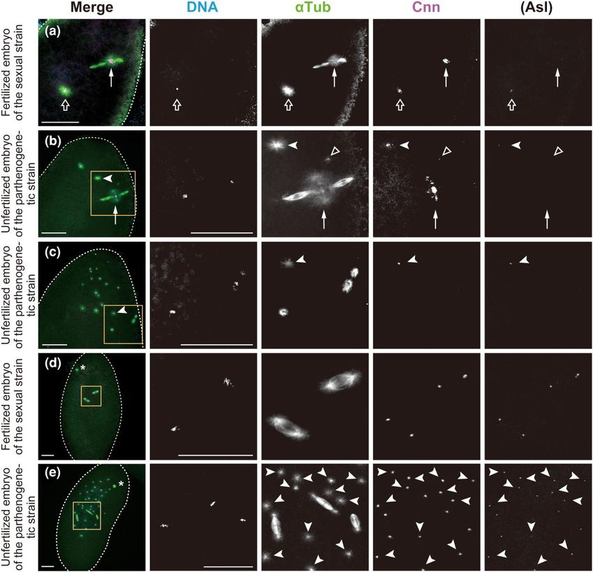
2 | GENETICS, 2023, Vol. 223, No. 2
Fischer et al. 2004; Pé rez-Mongiovi et al. 2005; Deshpande and development is entirely under maternal control through
Telley 2021). In fact, these features are associated with partheno stored mRNA and proteins (Farrell and O’Farrell 2014; Yuan
genesis from unfertilized eggs of some, if not all, Drosophila species et al. 2016).
(Stalker 1954; Sprackling 1960; Carson et al. 1969; Futch 1972; Centrosomes are also essential for parthenogenetic embryo
Carson 1973; Futch 1979; Templeton 1983; Riparbelli and Callaini genesis (Debec et al. 2010; Nabais et al. 2017). In many systems, in
2003; Matsuda and Tobari 2004; Eisman and Kaufman 2007; cluding in Drosophila, the formation of centrosomes de novo has
Riparbelli and Callaini 2008; Chang et al. 2014), as well as gynogen been extensively reported and in some cases mechanistically
esis (Fuyama 1986a, 1986b; Loppin et al. 2005) and androgenesis studied in vitro (Riparbelli et al. 2017; Pereira et al. 2021; Takumi
(Komma and Endow 1995) from fertilized eggs of D. melanogaster. and Kitagawa 2022). De novo formation of centrioles can be
In these examples of unisexual development with only maternal trigged in cases where cells lack all centrioles. Accordingly,
or paternal chromosomes, genetic evidence has been presented MTOCs are self-organized in the cytosol of unfertilized partheno
for patterns of diploidization by fusion between two haploid sets genetic eggs of insects such as the viviparous pea aphid (Riparbelli
Downloaded from https://academic.oup.com/genetics/article/223/2/iyac178/6896485 by guest on 07 September 2023
of chromosomes of meiotic products or cleavage nuclei, but it re et al. 2005), the hymenopteran Nasonia vitripennis (Tram and
mains to be seen how two distinct sets of chromosomes are suc Sullivan 2000), and Drosophila mercatorum (Riparbelli and Callaini
cessfully gathered into close apposition in the syncytium. 2003; Eisman and Kaufman 2007; Riparbelli and Callaini 2008).
Diploidization of haploid parental chromosome complements In unfertilized embryos of parasitic wasps and honeybees, centro
is a special feature of fertilized eggs. Zygotic diploid nuclei somes are produced from oocyte nuclear envelope-derived cyto
are generally formed during the mitotic phase of the first cleavage plasmic organelles with high concentrations of γ-tubulin (Ferree
division in many sexually reproducing animal species (Kawamura et al. 2006). In general, centrioles can be self-assembled without
2001; Loppin and Karr 2005; Lindeman and Pelegri 2012; template centrioles, relying on only the concentration of compo
Reichmann et al. 2018; Rahman et al. 2020; Cavazza et al. 2021; nents and PCM proteins (Pereira et al. 2021). Although new cen
Schneider et al. 2021). For early Drosophila embryos, the trioles generally assemble in the vicinity of pre-existing
microtubule-organizing centers (MTOCs), centrosomes, are es centrioles in most proliferating cells, the centriole pair is not a
sential (Rothwell and Sullivan 1999; Basto et al. 2006; Stevens completely essential component of centrosome formation
et al. 2007; Varmark et al. 2007; Rodrigues-Martins et al. 2008). (Martin and Akhmanova 2018). In syncytial embryos, the primary
In fertilized eggs, two centrosomes are newly constructed function of nucleus-associated centrosomes is to provide astral
from template centrioles contributed by the sperm and mater microtubules for proper nuclear spacing as cleavage divisions pro
nally provided pericentriolar material (PCM) components in ceed (de Saint Phalle and Sullivan 1998; de-Carvalho et al. 2022).
cluding a number of proteins involved in microtubule However, it remains to be seen whether the centrosomes are crit
nucleation, which compensate for the prior elimination of ical for the initiation of parthenogenetic development.
maternal centrosomes during oogenesis (Loppin et al. 2015; In Drosophila species, obligate parthenogenesis is known to oc
Blake-Hedges and Megraw 2019). The lack of centrosomes dur cur only in one species Drosophila mangabeirai, whereas rare facul
ing meiosis, as well as the inhibition of de novo centrosome for tative parthenogenesis, referred to as tychoparthenogenesis, is far
mation in unfertilized eggs, is speculated to be a mechanism to more common at least under laboratory conditions (Templeton
prevent parthenogenesis in Drosophila (Pimenta-Marques et al. 1983). One such species Drosophila ananassae provides unique ex
2016; Yamashita 2018). perimental material with which to study the cytology of partheno
Upon fertilization, the centrosomes are initially associated genetic embryos. Genetic variants that are associated with the
with the male pronucleus located deep within the egg and serve ability to carry out parthenogenesis have been isolated from nat
as the sperm aster, along which the distant female pronucleus ural populations of otherwise sexually reproducing D. ananassae
is transported to close proximity of the male pronucleus in and its closely related species (Futch 1972, 1973; Matsuda and
preparation for the first cleavage. The centrosomes then separ Tobari 2004). A previous study using a self-sustaining partheno
ate and form opposite poles of a mitotic spindle, organizing a genetic strain showed that a causal gene maps to the left arm of
microtubule array between them. Referred to as the gonomeric chromosome 2 (Matsuda and Tobari 2004).
spindle, this first mitotic spindle consists of two halves of the bi Identification of the exact means by which a diploid nucleus
polar microtubule arrays, each encompassing a parental set of forms at the beginning of parthenogenetic development is crucial
replicated chromosomes. These microtubule units arranged in to elucidating the potential reproductive mechanisms. Here we
parallel are linked together at their distal ends, with an aster report a detailed cellular analysis of meiosis and the first two
at the poles. Along the spindle, the haploid complements per cleavage divisions in unfertilized embryos produced by the par
sist in separated groups at the equator, and then the two groups thenogenetic strain of D. ananassae, by comparison with those in
of separated chromatids become intermingled at the spindle unfertilized eggs and fertilized embryos produced by females of
poles, generating the zygotic diploid nuclei each associated two sexual strains of the same species. The present study shows
with a centrosome (Guyénot and Naville 1929; Callaini and that, in parthenogenetic embryos, free MTOCs that are produced
Riparbelli 1996; Komma and Endow 1997; Williams et al. 1997; de novo in the cytosol play a central role in directing the earliest
Tirián et al. 2000; Loppin et al. 2015; Yamaki et al. 2016; cleavage divisions and in delineating mitotic events by which di
Hirai et al. 2018). The nuclei undergo extremely rapid ploidization can be achieved. In the egg, an anastral bipolar spin
syncytial divisions, forming the blastoderm. In addition to the dle arose from self-organization of microtubules around
microtubule-organizing roles, centrosomes serve as hubs for replicated chromosomes of a meiotic product. Progression of the
the integration and coordination of other biological processes first mitosis in the haploid state depended on incorporation of at
(Arquint et al. 2014; Ryniawec and Rogers 2021). In syncytial least one cytoplasmic MTOC into the mitotic spindle. During the
Drosophila embryos, centrosomes attached to spindle poles are second division, parthenogenetic embryos often formed dual
implicated in the spatial control of Cyclin B destruction, regu spindles, in which two separate spindles—each encompassing a
lating the exit from mitosis (Huang and Raff 1999; Wakefield single set of chromosomes—were arranged in tandem by sharing
et al. 2000). It should be noted that the first stages of embryonic an MTOC at the connected spindle poles. Based on our cytological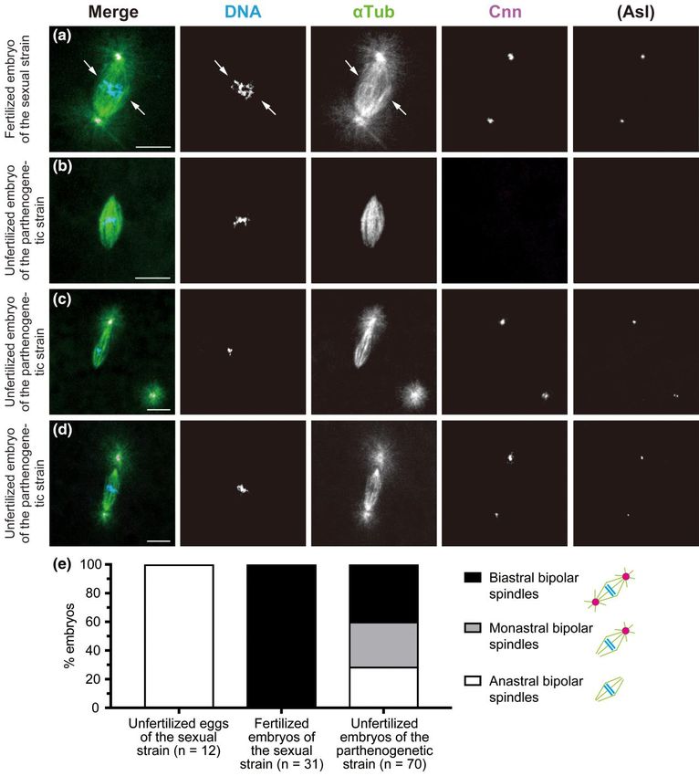
K. Hirai et al. | 3
observations, we propose a model for the steps by which an essen followed by mass matings using 10 females in each vial. After
tial diploid nucleus is generated in parthenogenetic embryos of the establishment of the cross on day 0, parents were transferred
D. ananassae. to new vials on days 2, 4, 6, and 8 and were then discarded on day
10. The offspring were scored no later than day 18 after the fe
males were allowed to lay eggs in each vial. Regular X-bearing
Materials and methods eggs fertilized by X- and Y-bearing sperm yield X/X (+) female
D. ananassae strains and X/Y (y) male offspring, respectively. Two classes of exception
The parthenogenetic stock, designated y-Im, was derived from al ova resulting from X chromosome nondisjunction or loss during
flies caught in Taputimu, American Samoa (Futch 1972, 1973; meiosis are recoverable: diplo-X eggs fertilized by Y-bearing
Matsuda and Tobari 2004). It has been maintained in the labora sperm and nullo-X eggs fertilized by X-bearing sperm, which
tory for decades as a self-sustaining parthenogenetic line. The X give rise to X/X/Y (y) female and 0/X (w) male offspring, respect
chromosome of this strain is marked with yellow (y), a spontan ively. X/X/Y (y) females are phenotypically indistinguishable
Downloaded from https://academic.oup.com/genetics/article/223/2/iyac178/6896485 by guest on 07 September 2023
eous mutation that occurred in the original stock. Gross morph from parthenogenetic y females. Exceptional w males can also
ology of ovaries and eggs appears normal. The obligate sexual arise from abnormal loss of the maternal X chromosome at the
strains used in the present study are AABBg1 and BKK17, which first mitosis of y/w zygotes. Loss of a paternal X chromosome re
originated in Hawaii, USA and Bangkok, Thailand, respectively. sults in X/0 (y) males, which are indistinguishable from regular
Control embryos were essentially obtained from the AABBg1 X/Y (y) males. The frequency of w males among offspring was
stock, unless otherwise specified. Genetically heterozygous (F1 hy calculated as [w males × 100]/[y+ w+ females + y males + w
brid) females were produced as y+ females from the cross between males].
y females of the parthenogenetic strain and wild-type males of the
AABBg1 strain. yAM-203 (abbreviated as y hereafter) and white (w)
strains were used for the X chromosome segregation test. Stock
maintenance and crosses were performed on standard Collection of mature oocytes and embryos,
cornmeal-glucose-yeast-agar medium at 24°C. All D. ananassae immunostaining, and imaging
stocks were provided by Kyorin Fly, part of the National To examine meiotic spindles in mature oocytes, late-stage oocytes
BioResource Project of Ministry of Education, Culture, Sports, were separated from ovaries of 6- to 10-day-old females that were
Science, and Technology, Japan. mated with males of the sexual strain and virgin females of the
parthenogenetic strain and were treated as described (Takeo
Mitotic chromosome preparation et al. 2010). Oocytes fixed with formaldehyde were stained for im
We made chromosome preparations with brains from third instar munofluorescence with primary antibodies rat monoclonal
larvae using the air-dry method (Hirai et al. 2004), and chromo anti-Tubulin (YL1/2, 1:300; Abcam) and rabbit anti-centrosomin
somes were stained with Giemsa. Squash preparations of embry (Cnn; 1:3,000; Lucas and Raff 2007), followed by secondary anti
onic chromosomes were made using embryos collected within 2 h bodies Alexa Fluor 488–conjugated goat anti-rat IgG (1:800;
after deposition as described (Vidwans et al. 2002), and chromo Thermo Fisher Scientific) and Cy3-conjugated AffiniPure goat
somes were visualized by DAPI staining. At least three sets of mi anti-rabbit IgG (1:800; Jackson ImmunoResearch Laboratories),
totic chromosomes in anterior, central, and posterior parts of as well as the DNA dye DAPI.
individual embryos at the syncytial blastoderm stage were exam Virgin or mated females were allowed to lay eggs on grape juice
ined. Karyotypes were analyzable in ∼40% of the embryos in the agar plates supplemented with fresh yeast paste. Embryos were
sexual strain (n = 136), but in only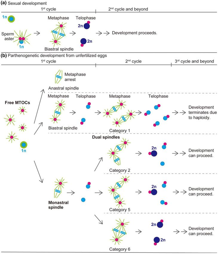
4 | GENETICS, 2023, Vol. 223, No. 2
Fig. 1. Development of parthenogenetic embryos. a) Parthenogenetic females are essentially XX diploids. The chromosome spread from the neuroblast of
Downloaded from https://academic.oup.com/genetics/article/223/2/iyac178/6896485 by guest on 07 September 2023
a female larva shows pairing of four pairs of homologous chromosomes, typical of normal Drosophila somatic cells. The X and fully heterochromatic
fourth chromosomes are indicated. b) An unfertilized egg laid by the sexual strain AABBg1 was fixed with methanol and stained for microtubules
(α-Tubulin, αTub) and DNA (DAPI). The two-color image on black background shows a bipolar array of microtubules self-assembled around condensed
chromosomes of a meiotic product. The chromosome complement is inferred to be haploid from the number of chromosome arms; it is however
ambiguous. c) The percentage of embryos that reached anaphase of the first cleavage or a more advanced stage. Embryos produced by mated females of
the sexual strain and virgin females of the parthenogenetic strain were fixed within the time intervals from 10 min to the indicated time points (20, 30, 60,
120, and 180 min) after deposition and examined. The number of embryos analyzed ranged from 107 to 339 per time interval. Data are represented as the
mean ± 95% confidence interval. d and e) Images of embryonic chromosomes. A diploid complement of chromosomes from the sexual strain (d) and a
haploid complement of chromosomes from the parthenogenetic strain (e). f and g) Nonactivated mature oocytes were formaldehyde fixed, and
stained as in (b). Females of both the sexual (f) and parthenogenetic (g) strains exhibited normal meiotic arrest at metaphase I with anastral bipolar
spindles. Scale bars, 10 μm.
Results and discussion a rate of 4.9% (Table 1). Clearly, the majority of embryos failed
to hatch as larvae (Table 1). The extensive embryonic lethality in
Parthenogenetic reproduction in D. ananassae
dicates that a key process for successful developmental progres
The self-sustaining parthenogenetic strain y-Im of D. ananassae
sion occurs during embryonic stages. It is also worth noting
is capable of virgin reproduction (geographic origin of the strain:
that, despite a complete lack of parthenogenetic capability in fe
Tutuila Island, American Samoa; Futch 1972). Female larvae
males of the sexual strain, parthenogenesis occurred in unfertil
that were examined from the stock were diploid for the
ized eggs laid by virgin females of F1 hybrids (i.e. from matings
X chromosome and three autosomes in neuroblasts, as
between females of the parthenogenetic strain and males of the
expected for this species (Fig. 1a; n = 12; Kaufmann 1936;
sexual strain), albeit in a very small percentage of cases (0.3%,
Marchetti et al. 2022). A large majority of adult individuals
Table 1). This genetic analysis argues that the parthenogenetic
were females, but 0.3% (7/2,627) were males, which were con
trait behaves recessively, and it is consistent with the idea of re
sidered to be sterile X0 diploid individuals that resulted from
pression of parthenogenesis in the sexual strains of the species.
the loss of an X chromosome during early mitosis (Futch
In addition, given the weak effect elicited in F1 hybrids, we cannot
1972). In the context of haploid–diploid life cycle of animal re
rule out the other possibility that increased gene dosage confers
production, we would like to employ the ploidy values that indi
the capacity of parthenogenesis.
cate the number of sets of chromosomes as a multiple of the
To better understand chromosomal inheritance from mother
haploid genome, but not chromatin amount or DNA content
to parthenogenetic offspring, we next examined chromosome
that changes during the cell cycle, in this paper.
segregation during meiosis and maternally driven earliest embry
To determine the developmental stage critical for successful
onic divisions by a conventional, highly sensitive genetic assay. By
parthenogenesis that gives rise to adults, we successively counted
following the transmission of genetically marked X chromosomes
the number of first instar larvae, pupae, and emergent adults that
in a cross, we were able to examine their behavior. Eggs that were
developed from unfertilized eggs laid by females of the strain.
aneuploid for the X chromosome were detectable; upon fertiliza
Unfertilized eggs laid by females developed into viable adults at
tion, those eggs were recoverable with different phenotypes
among offspring. We could not analyze autosomes here. When
Table 1. Development of eggs laid by females of the sexual and y/y females from the parthenogenetic strain were crossed to w/Y
parthenogenetic strains of D. ananassae. males of the sexual strain, both sexual (+ females, y males, and
Developmental Female parent
w males) and parthenogenetic (y females) offspring were pro
stage duced simultaneously (Table 2). w males are diagnostic of X
Mated Virgin Virgin females of Virgin chromosome misbehavior, as they result from exceptional
females females parthenogenetic females nullo-X ova fertilized by X-bearing sperm. The females of the par
of sexual of sexual strain (n = 713) of F1 thenogenetic strain produced w male offspring at a frequency of
strain strain hybrids 0.3%, which was significantly higher than the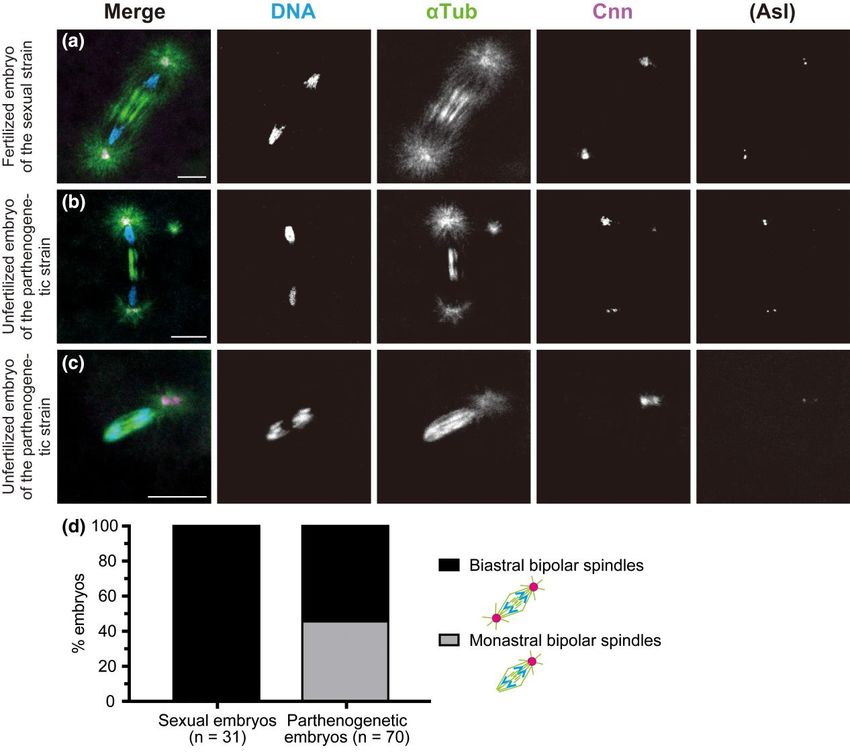
K. Hirai et al. |    5

Table 2. Segregation data of the X chromosome.

                                                         Offspring from y/y ♀ × w/Y ♂                                                       Frequency of w ♂ (%)

Female parent                             +♀                  y♀                  y♂                 w♂                 Total

Sexual strain                           3,503                    0               3,385                 2                6,890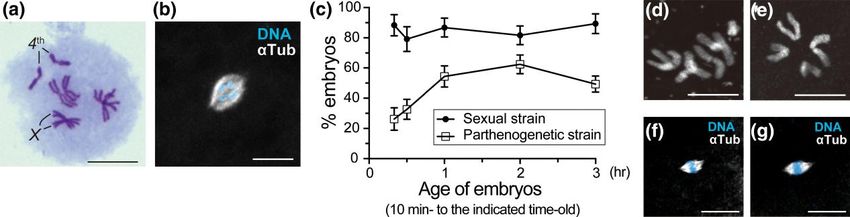
6 | GENETICS, 2023, Vol. 223, No. 2
Downloaded from https://academic.oup.com/genetics/article/223/2/iyac178/6896485 by guest on 07 September 2023
Fig. 2. De novo formation of MTOCs in the cytosol of activated eggs produced by females of the parthenogenetic strain. Laid eggs and embryos were fixed
and treated with antibodies against α-Tubulin (αTub) for microtubules and Cnn and Asl (not overlaid in merged images but only shown independently) for
the PCM, as well as the DNA dye DAPI. In the merged images, the superimposed staining of microtubules and Cnn appears white. a and d) Fertilized
embryos laid by females of the sexual strain AABBg1. b, c, and e) Unfertilized embryos laid by females of the parthenogenetic strain. The edge of each
embryo is outlined with dashed lines for clarity. The boxed regions in the merged images in (b–e) are shown at higher magnification in the adjacent
panels. a) The fertilized oocyte in metaphase II exhibits the meiotic aster located between the central poles of the two tandem spindles of the female
meiotic apparatus (arrow) and the sperm aster in the center of the egg (outlined arrow). The former includes only Cnn but not Asl, whereas the latter
includes both Cnn and Asl. Chromosomes on the spindles are not visible in the processed images. b) The unfertilized egg laid by the parthenogenetic
strain shows two free asters, which are formed from MTOCs that include Cnn and Asl (one of them is indicated by a white arrowhead) at the anterior of
the egg cytosol. A punctiform aster (outlined triangle) is detectable immediately adjacent to the prominent aster at the central poles of the meiotic
spindles (arrow). c) After completion of meiosis, meiotic products are surrounded by arrays of microtubules. One is located centrally in the egg (on the
lower left side of the boxed region), while the others lie near the cortex. Free asters of variable sizes are randomly distributed at the anterior of the egg. The
arrow indicates one of the asters. d) The sexual embryo during the second mitosis shows synchronous mitotic progression with biastral bipolar spindles.
The polar body lies near the cortex (asterisk). e) This parthenogenetic embryo contains a total of three mitotic spindles, in addition to the polar body
(asterisk) and ∼30 free asters that are predominantly distributed in a region approximately one-half of the egg’s length from the anterior end
(arrowheads). One of the bipolar spindles is monastral, whereas the others are biastral. Ploidy level of the chromosome complements is not known. Scale
bars, 50 μm.
A striking difference between eggs laid by females of the sexual both centrosome markers Cnn and Asl (Fig. 2a). In contrast, no as
and parthenogenetic strains was noticed in aster formation in the ters were detected in unfertilized eggs of the sexual strains ar
egg cytosol. In fertilized eggs of the sexual strain, the clearly vis rested in a mitotic state of the first cycle (n = 102 for AABBg1
ible sperm aster formed in the center of the egg (Fig. 2a, outlined eggs, 10 min–3 h after deposition; n = 136 for AABBg1 eggs, 3–6 h
arrow). Next to the sperm nucleus, the radial arrays of microtu after deposition; and n = 52 for BKK17 eggs, 10 min–6 h
bules were nucleated by the MTOCs, which were labelled with after deposition). However, in unfertilized eggs of theK. Hirai et al. | 7
parthenogenetic strain, MTOCs with Cnn and Asl labeling (i.e. sizes of asters on individual biastral spindles (Fig. 3d), suggests
centrosome-like structures) formed particularly at the anterior that free MTOCs produced in the cytosol could coalesce into either
of the earliest embryos, at a short distance from meiotic spindles one or both anastral poles of a bipolar spindle. We occasionally
and nuclei (Fig. 2b, arrowheads). We refer to the MTOCs that form observed earlier association of MTOCs with microtubules that ac
spontaneously in the egg cytosol as “free MTOCs,” and “asters” as cumulated in the vicinity of individual chromosomes, but not the
the microtubules emanating outward from any MTOCs. Those whole set of chromosomes (Supplementary Fig. 2, b and c). The
free MTOCs appeared as early as anaphase I (Supplementary consequence of such an occurrence is not, however, known.
Fig. 2a) and were detected in the majority of embryos laid by the
parthenogenetic strain: 73.3% of eggs in anaphase I–telophase II Mitotic progression occurs with astral, but not
(n = 30; Fig. 2, a and b) and >90% of post-meiotic embryos that anastral, bipolar spindles during the first mitosis
were fixed 10–30 min after deposition (n = 292; Fig. 2, c and e). of parthenogenetic embryos
Nonactivated oocytes in prometaphase I–metaphase I completely To see the effect of MTOCs associated with the first mitotic spindle
Downloaded from https://academic.oup.com/genetics/article/223/2/iyac178/6896485 by guest on 07 September 2023
lacked asters (Fig. 1g, n = 49). In summary, de novo formation of on mitotic progression, we then examined embryos in the subse
free MTOCs occurs after egg activation in the egg cytosol in the quent anaphase and telophase stages. Mitotic progression into
parthenogenetic strain. anaphase is heralded by the synchronous movement of two sets
Centrosomes duplicate precisely once per cell cycle during the of separated chromatids away from the metaphase plate to op
canonical cell cycle (Callaini and Riparbelli 1990; Debec et al. posite spindle poles, where the chromosomes are packaged into
1996), but we noted that the number of free MTOCs varied across new daughter nuclei during telophase. In the control sexual em
individual embryos. To assess de novo formation of MTOCs and bryos, all examined anaphase–telophase spindles of the first mi
their possible overduplication independently of nuclear divisions, tosis were biastral and bipolar (Fig. 4a; n = 31). However, in
we quantified free MTOCs over time in embryos showing no cleav embryos produced by the parthenogenetic strain, anaphase and
age divisions beyond the first metaphase-like state. The number of telophase figures were detectable on bipolar spindles that were
free MTOCs observed was 6.7 ± 10.0, 10.1 ± 12.0, 10.7 ± 7.4, and 5.9 not only biastral (Fig. 4b) but also monastral (Fig. 4c). These em
± 4.9 (mean ± SD) in 10- to 30-min-old (n = 18), 10- to 60-min-old bryos exhibited orderly segregation of chromosomes. No anastral
(n = 17), 10- to 90-min-old (n = 19), and 10- to 150-min-old (n = 15) spindles showed progression into anaphase in embryos from the
embryos, respectively. An increase in the number of MTOCs over parthenogenetic strain (Fig. 4d; n = 70), despite their presence dur
time was not evident, suggesting a transient production of ing the prior metaphase (Fig. 3, b and e). It should be emphasized
MTOCs but not their self-propagation in the cytosol. that the presence of one MTOC at a bipolar spindle is both neces
sary and sufficient to confer mitotic progression.
Free MTOCs can be incorporated into the spindle The ratio of monastral to biastral spindles observed during
for the first mitosis of parthenogenetic embryos anaphase–telophase (biastral, 54.3%; monastral, 45.7%) mirrors
Following the completion of meiosis and the pronuclear stage, that during the prior stages (biastral, 56.0%; monastral, 44.0%;
syncytial embryonic development begins with a series of rapid nu n = 50 from Fig. 3e). This suggests that, during the first mitosis of par
clear divisions. To investigate the influence of free MTOCs par thenogenetic embryos, astral bipolar spindles do indeed segregate
ticularly at the earliest stage of parthenogenetic development, the chromosomes, regardless of whether they are biastral or monas
we thoroughly examined the first mitosis using a collection of em tral. In contrast, mitosis in the parthenogenetic strain embryos termi
bryos fixed within 30 min after deposition. This developmental nated in a metaphase-like state without transition to anaphase with
stage was assigned by the presence of a single mitotic spindle in in bipolar spindles devoid of any associated MTOCs. Such a mitotic ar
dividual embryos. In the control (sexually developing) embryos, a rest with anastral bipolar spindles is reminiscent of that which nor
biastral bipolar spindle was assembled around parental sets of mally occurs in laid unfertilized eggs of sexual strains (Fig. 1b).
chromosomes in the interior of the embryo (Fig. 3a; n = 12). Obviously, with MTOCs that have been produced de novo in the cyto
Typical of the first mitosis of the Drosophila zygote, the metaphase sol of eggs from the parthenogenetic strain, engagement of at least
spindle appeared to be composed of two units of microtubule ar one of the free MTOCs in the first mitotic spindle is required to exit
rays each encompassing a haploid set of replicated chromosomes. from the metaphase state. More specifically, in parthenogenetic em
In unfertilized embryos of the parthenogenetic strain, meiotic pro bryos, the MTOC is not required for organizing a metaphase spindle
ducts were surrounded by microtubules, at least immediately around replicated chromosomes of a meiotic product, but associ
after deposition (Fig. 2c). The embryos then self-assembled a bipo ation of a focused pole of the self-assembled bipolar spindle with
lar spindle around the seemingly haploid set of chromosomes po an MTOC is crucial for the initiation of anaphase.
sitioned in the interior, apart from polar bodies lying near the This notion is plausible because a similar mitotic arrest pheno
cortex (Fig. 3, b–d). Clearly, the spindles formed in parthenogenetic type has been reported in Drosophila embryos defective in centro
embryos were smaller (Fig. 3, b–d) relative to those formed around some localization at spindle poles. Mutations in D. melanogaster
parental sets of chromosomes in sexual embryos (Fig. 3a), indica Cyclin B3 (Yuan and O’Farrell 2015; Garrido et al. 2020) and Elys
tive of the former’s haploidy (Komma and Endow 1995, 1997; Brent (Hirai et al. 2018) exhibit maternal-effect lethality during a
et al. 2000). Nonetheless, the predicted ploidy level is ambiguous. metaphase-like state of the first mitosis, accompanied by an anas
Although the spindles similarly self-assembled around a mei tral spindle that results from centrosome detachment from the
otic product located centrally in unfertilized eggs of the sexual spindle poles. Moreover, in fertilized eggs that are defective in pro
strain, they were invariably anastral (Fig. 1b); this was not always nuclear migration, an anastral spindle is assembled around repli
the case in parthenogenetic embryos. Allocation of asters asso cated chromosomes of the female pronucleus, at a distance from
ciated with a bipolar spindle of the first mitosis differed substan the male pronucleus, where mitotic progression is halted in a
tially between individuals (n = 70); the spindles were anastral metaphase-like state (Llamazares et al. 1999; Gergely et al. 2000;
(28.5%; Fig. 3b), monastral (31.4%; Fig. 3c), or biastral (40.0%; Riparbelli et al. 2000; Dix and Raff 2007; Varmark et al. 2007;
Fig. 3d). The presence of monastral spindles containing one astral Vazquez-Pianzola et al. 2022). We suggest that the MTOCs at the
and one anastral pole (Fig. 3c), as well as a clear difference in the spindle poles regulate the mechanism of the spindle assembly8 | GENETICS, 2023, Vol. 223, No. 2
Downloaded from https://academic.oup.com/genetics/article/223/2/iyac178/6896485 by guest on 07 September 2023
Fig. 3. The first mitosis in sexual and parthenogenetic embryos. Laid embryos were fixed and treated with antibodies against α-Tubulin (αTub) for
microtubules and Cnn and Asl (not overlaid in merged images but only shown independently) for the PCM, as well as the DNA dye DAPI. In the merged
images, the superimposed staining of microtubules and Cnn appears white. a) In the fertilized egg produced by the sexual strain AABBg1, the first mitotic
spindle is bipolar and biastral around two groups of replicated chromosomes derived from the ovum and sperm (arrows). b–d) In unfertilized embryos
produced by the parthenogenetic strain, a set of replicated chromosomes, which is considered to be haploid, aligns at the equator during the first mitosis.
Those spindles are smaller relative to the spindle in (a). Based on the number of asters associated with a bipolar spindle, spindles fall into three classes: (b)
an anastral bipolar spindle that formed in an embryo containing four free asters in the cytosol (not visible in this image), (c) a monastral bipolar spindle
and a neighboring free aster, and (d) a biastral bipolar spindle that formed in an embryo containing six free asters (not visible in this image). The asters at
the opposite spindle poles vary markedly in size. e) The proportion of different forms of the first mitotic spindle. Scale bars, 10 μm.
checkpoint. In D. melanogaster syncytial embryos, chromosome Dual spindle formation during the second mitosis
segregation is triggered by cohesin cleavage and downregulation of parthenogenetic embryos by arrangement of
of Cdk1 (Oliveira et al. 2010). Thus, it may be that, in parthenogen two spindles in tandem
etic embryos of D. ananassae, an MTOC located at one of the poles We next wished to know what mechanism controls the second mi
of the first mitotic spindle can promote anaphase onset, setting totic cycle with the daughter nuclei produced by the first cleavage
the key biochemical events in motion within the entire spindle. division of parthenogenetic embryos. In the control (sexuallyK. Hirai et al. | 9
Downloaded from https://academic.oup.com/genetics/article/223/2/iyac178/6896485 by guest on 07 September 2023
Fig. 4. Chromosome segregation occurs on astral, but not anastral, bipolar spindles during the first mitosis of parthenogenetic embryos. Embryos laid by
females of the parthenogenetic strain were fixed and treated with antibodies against α-Tubulin (αTub) for microtubules and Cnn and Asl (not overlaid in
merged images but only shown independently) for the PCM, as well as the DNA dye DAPI. In the merged images, the superimposed staining of
microtubules and Cnn appears white. a) During late anaphase of the first mitosis in a sexually developing embryo of the sexual strain AABBg1,
chromosomes of maternal and paternal origin are gathered into a single mass as separated chromatids synchronously migrate toward the poles of the
biastral bipolar spindle. b and c) Mitotic progression during the first mitosis of parthenogenetic embryos. The spindles are smaller relative to those
formed in the sexual embryo (a). b) Late anaphase on the biastral bipolar spindle of the first mitosis in unfertilized embryos of the parthenogenetic strain.
The upper chromosome set looks brighter than the lower one because of the difference in depth resulting from the tilted axis of the division. This is true
for the microtubule-organizing centers of the nuclei and asters. One of the six free asters in the cytosol is shown in the upper right region of the image.
c) Early anaphase on the monastral bipolar spindle of the first mitosis in unfertilized embryos produced by females of the parthenogenetic strain. The
embryo contains one free aster (not visible in this image). d) Distribution of different forms of bipolar spindles during anaphase–telophase of the first
mitosis. Note that no anastral spindle showed mitotic progression into anaphase. Scale bars, 10 μm.
developing) embryos, after DNA replication, progression of second frequency is in excellent agreement with that of the biastral bipo
mitosis was always synchronous and occurred with two inde lar spindles noted during the prior first anaphase–telophase
pendent, biastral bipolar spindles. The individual spindles were stages (54.3%; Fig. 4d), lending support for the apparently normal
clearly separated but at a relatively close distance in the syncyt nuclear and centrosomal cycles. We thus infer that most, if not
ium (Fig. 2d; n = 26). In parthenogenetic embryos, however, mis all, category 1 embryos in the second cycle result from the first
cellaneous configurations of spindles were present around two mitosis with a biastral bipolar spindle. Those embryos seem to
sets of chromosomes that could be classified into six categories undergo syncytial cleavage divisions in the haploid state
(Fig. 5; n = 59). It should be noted first that no parthenogenetic em (Supplementary Fig. 1b), resulting in lethality before hutching.
bryos contained two anastral spindles, which is consistent with The remaining five categories of mitotic configurations, which
the halting of mitotic progression on anastral bipolar spindles were specific to parthenogenetic embryos, are presumed to have
during the first mitosis, as mentioned above. About half of the em arisen from a first mitosis that was carried out on monastral bipo
bryos displayed an apparently normal pair of biastral bipolar spin lar spindles (Fig. 4c). Of the resultant daughter nuclei, which were
dles during the second mitosis (category 1, 50.8%; Fig. 5a). This considered to be haploid, one was associated with an MTOC and10 | GENETICS, 2023, Vol. 223, No. 2
Downloaded from https://academic.oup.com/genetics/article/223/2/iyac178/6896485 by guest on 07 September 2023
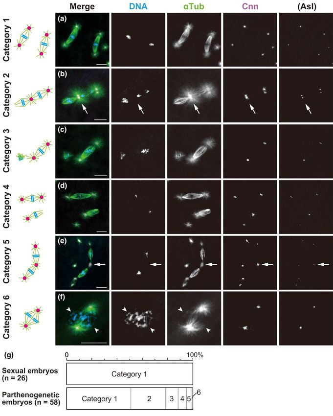
Fig. 5. Dual spindles are formed by sharing an aster at the central poles during the second mitosis of parthenogenetic embryos. Laid embryos were fixed
and treated with antibodies against α-Tubulin (αTub) for microtubules and Cnn and Asl (not overlaid in merged images but only shown independently) for
the PCM, as well as the DNA dye DAPI. In the merged images, the superimposed staining of microtubules and Cnn appears white. Based on the
configurations of their mitotic figures, embryos undergoing the second mitosis were assigned to the following six categories, listed in decreasing order of
frequency. a) Category 1: two independent biastral spindles form. b) Category 2: two spindles form which are tandemly arranged by sharing an aster at the
central pole (arrow). For the dual spindles, one of the distal poles is anastral whereas the other is astral. c) Category 3: a biastral spindle forms around one
set of chromosomes and short arrays of microtubules form around the other set of chromosomes. d) Category 4: two independent spindles form that are
biastral and monastral. For the monastral spindle, the astral pole appears more tapered than the anastral pole. e) Category 5: dual spindles form that are
similar to those of category 2, but they differ only in that both distal poles of the dual spindle are astral in category 5. One of the distal poles (in the lower
left) is associated with asters in pairs, whereas the other distal pole and the central poles (arrow) are associated with individual asters. f) Category 6: two
units of bipolar arrays of microtubules form around the adjoining sets of chromosomes. The units are closely apposed, sharing asters at the spindle poles.
The arrowheads mark the neighboring two sets of chromosomes on the spindle. g) The graph quantifies the six mitotic configuration categories formed
during the second cleavage division. Scale bars, 10 μm.K. Hirai et al. | 11
the other was not. Among the configurations of two spindles that rather exerted an impact on development during the first cleavage
were seen only in parthenogenetic embryos, a pair of tandemly ar division. Despite the disparate architecture of these MTOCs lo
ranged spindles was most common (category 2, 27.1% and cat cated at the central poles of meiotic and mitotic spindles, they
egory 5, 3.4%; Fig. 5, b and e, respectively). Those distinctive may share the common property of arranging two spindles in tan
dual spindles consisted of two bipolar spindles that were con dem. This is remarkable given that, during anaphase II, separated
nected at their central poles by sharing a single MTOC (Fig. 5, ar chromatids migrate to the central poles from discrete metaphase
rows), whereas the distal poles were kept completely apart. We plates of the spindles and then the non-daughter nuclei come into
assume that those dual spindles resulted from the coupling of close apposition (Riparbelli and Callaini 1996; Endow and Komma
neighboring biastral and anastral spindles through a shared 1997).
MTOC during the second mitosis, as discussed below. In rare in
stances, a bipolar spindle formed around two distinct chromo Possible microtubule interactions between
some sets that were juxtaposed to each other (category 6, 1.7%; anastral spindle poles and astral microtubules
emanating from separate MTOCs
Downloaded from https://academic.oup.com/genetics/article/223/2/iyac178/6896485 by guest on 07 September 2023
Fig. 5f). It is not known whether this configuration results from un
ion of two independent bipolar spindles, or assembly of an In post-meiotic parthenogenetic embryos, microtubules nucleate
all-inclusive bipolar spindle. It should be noted, however, that in the vicinity of a meiotic product and become organized into a
the spindle configuration of category 6 observed in parthenogen bipolar array, depending on centrosome-independent mechan
etic embryos bears a close resemblance to the normal first mitotic isms (Duncan and Wakefield 2011; Petry 2016). However, mitotic
spindle in fertilized eggs of the sexual strain, which is composed of progression into anaphase was permitted only with astral bipolar
two units of microtubules around parental haploid sets of chro spindles, which formed quite frequently during the first mitosis of
mosomes (Fig. 3a). parthenogenetic embryos (>70%; Fig. 3e). We thus infer that a high
In category 3 embryos, a biastral bipolar spindle assembled affinity of interaction between the aster emanating from a free
around one of the two chromosome sets, and the other chromo MTOC and an anastral mitotic spindle might have resulted in
some set located nearby was surrounded by apparently short mi such a conjoined structure, but this has not been demonstrated
crotubules displaying random polarity with no aster association directly. It has been shown that a significant proportion of spindle
(10.2%; Fig. 5c). This situation probably reflected an early frame microtubules are disconnected from the centrosome within a nor
in the temporal sequence of the concurrent assembly of two dif mal astral spindle (Merdes and Cleveland 1997; Chavali et al. 2015;
ferent types of spindles. The involvement of MTOCs may have fa Martin and Akhmanova 2018). In D. melanogaster, the microtubule-
cilitated the rapid constitution of bipolar spindles, whereas based processive motor dynein is required to maintain the attach
anastral bipolar spindle assembly, which depends solely on ment of centrosomes to mitotic spindle poles in early embryos
microtubule self-organization around chromosomes, may be a (Robinson et al. 1999) and Schneider 2 cultured cells (Maiato
slower process, as has been shown in D. melanogaster somatic cells et al. 2004; Goshima et al. 2005; Morales-Mulia and Scholey 2005).
(Basto et al. 2006; Lecland et al. 2013; Poulton et al. 2014). According to a model for pole focusing of astral spindles
It was curious that we failed to detect embryos with biastral (Goshima et al. 2005), temporal cross-linking between kinetochore
and anastral bipolar spindles during the second mitosis. Instead, fibers and astral microtubules by the non-clarlet disjunctional
as category 4 embryos showed, biastral and monastral bipolar protein facilitates recruitment of dynein for minus end–directed
spindles were distributed independently (Fig. 5d). It is thus highly transport of kinetochore fibers along astral microtubules
likely that anastral spindle poles strongly attract a nearby aster. toward the centrosome, forming parallel filament overlapping.
One of the poles of an anastral spindle may often be caught by as Attachment of astral microtubules to separate spindle microtu
tral microtubules that emanate from the neighboring biastral bules might be an integral part of astral spindle formation during
spindle, resulting in the dual spindle in category 2 (27.1%; the first mitosis of parthenogenetic embryos of D. ananassae. It
Fig. 5b), and, less frequently, by aster emanating from an adjacent would be an intriguing possibility that this process depends on
free MTOC, resulting in a monastral spindle (category 4, 6.8%; parallel microtubule interactions involving dynein. The concur
Fig. 5d). This prediction was supported by the presence of dual rent presence of meiotic products surrounded by microtubules
spindles associated with asters at both distal poles, which oc and free MTOCs nucleating asters in a limited area of partheno
curred infrequently (category 5, 3.4%; Fig. 5e). The form might re genetic embryos (Fig. 2c) could have increased the accessibility
sult from both binding of a resident MTOC of the neighboring of astral microtubules to the self-assembled anastral spindle. It
biastral spindle to one of the poles of the originally anastral spin may be that initial microtubule interaction occurs most frequent
dle and addition of a free MTOC to another anastral pole. Taken ly during early steps of anastral spindle assembly with dispersed
together, our observations illustrate the plasticity of the arrange spindle microtubule minus ends (Supplementary Fig. 2b).
ment of two spindles in the syncytium and reveal the role of asters In parthenogenetic embryos, the first mitosis on a monastral
as an “inter-spindle linker.” bipolar spindle was reflected in the construction of one astral
In the distinctive configurations of spindles represented by cat and one anastral spindle in the near distance during the second
egories 2 and 5, we were observing true second mitotic spindles ra cycle, each encompassing replicated chromosomes of one of the
ther than meiosis II spindles of structural similarity. Meiotic daughter nuclei. Again, as discussed above for the first mitosis,
spindles arrayed in tandem lack asters at both distal poles and it is tempting to speculate about a dominant influence of
contain a non-centrosomal aster only between the central poles MTOCs on dual spindle formation. Presumably, astral microtu
(Fig. 2, a and b; Riparbelli and Callaini 1996; Endow and Komma bules emanating from MTOCs, which are located in the cytoplasm
1998; Wilson and Borisy 1998; Riparbelli and Callaini 2005). or attached to a bipolar biastral spindle, interact with microtubule
Instead, the asters on the dual spindles in parthenogenetic em minus ends of a forming or completed anastral spindle.
bryos were attached to both central and distal poles, and, more Association of MTOCs to anastral spindle poles may depend on
over, the mitotic asters grew out from MTOCs with Asl labeling the orientation of the divisions and distribution of free MTOCs
(Fig. 5, b and e). Thus, free MTOCs produced de novo in the egg in the syncytium. Perhaps dual spindles, which are arranged in
cytosol did not gain immediate access to meiotic spindles, but tandem with a shared MTOC (Fig. 5, b, e, and f), result from12 | GENETICS, 2023, Vol. 223, No. 2
interaction between astral microtubules emanating from a spin haploid sets of separated chromatids at both poles of the first mi
dle pole-located MTOC and microtubule minus ends of coexisting totic spindle (Figs. 4a and 6a). Such a fusion between non-
anastral spindle. Likewise, asters of free MTOCs could interact daughter nuclei could also occur late during the second mitosis
with the remaining anastral pole of dual spindles (Fig. 5e), or ei of parthenogenetic embryos, at the connected poles of dual spin
ther one (Fig. 5d) or both (Fig. 5a) of the poles of an independent dles (Figs. 5, b, e, f and 6b). Importantly, those resulting diploid
anastral spindle. During normal development of D. melanogaster nuclei in parthenogenetic embryos are associated with an
early embryos, astral microtubules of biastral bipolar spindles MTOC, ensuring subsequent proliferation as nuclear cycles con
act to prevent abnormal fusion of spindles and collision of cleav tinue. The remaining haploid sets of chromosomes that might
age nuclei (Baker et al. 1993). A recent report shows that, in telo have migrated to the distal poles may fall out or be outcompeted
phase of syncytial nuclear divisions, distance between by the diploid lineage thereafter in the syncytium. The probable
non-daughter nuclei is maintained by an aster-mediated repul diploidization mechanism involving fusion of haploid cleavage
sion, where antiparallel microtubule interactions are involved in nuclei can account for the complete genetic homozygosity of par
Downloaded from https://academic.oup.com/genetics/article/223/2/iyac178/6896485 by guest on 07 September 2023
a mechanical link between astral microtubules extending from thenogenetic individuals of this species (Futch 1972, 1973;
neighboring centrosomes of two separate spindles (Deshpande Matsuda and Tobari 2004).
et al. 2022). In contrast, particularly when centrosomes are defect Overall, in D. ananassae, de novo formation of free MTOCs in the
ive in early embryos, abnormal connections between poles of dis egg cytosol primarily distinguished embryos produced by females
tinct spindles located in proximity are often caused by sharing an of the parthenogenetic strain from those of the sexual strains. The
MTOC, resulting in a configuration referred to as trains or rosettes behavior of the asters formed from MTOCs apparently holds the
(Megraw et al. 1999; Vaizel-Ohayon and Schejter 1999; key to the execution of parthenogenesis. Although the participa
Pé rez-Mongiovi et al. 2005; Archambault et al. 2007; Zhang and tion of such MTOCs in a bipolar spindle that self-assembles
Megraw 2007). around a haploid set of chromosomes leads to mitotic progres
sion, it is considered critical that no more than one MTOC is in
Possible routes of diploidization for haploid eggs volved during the first mitosis so that dual spindles form during
during parthenogenetic development in the subsequent (i.e. second) mitosis (Fig. 6b). We thus suggest
D. ananassae that parthenogenesis with a low probability of success in D. ana
Once dual spindles formed in parthenogenetic embryos, the nassae is primarily attributable to the variability in the number
structure was steadily maintained and was the means by which of MTOCs and their initial distribution within the egg.
chromosome segregation occurred (Fig. 5b). Although the conse Realization of a monastral mitotic spindle for the first mitosis
quence of the second mitosis with dual spindles and the subse probably depends on interactions between anastral spindle poles
quent development were of great interest to us in the present and asters nucleated by free MTOCs. It is just conceivable that
study, this analysis was hampered, largely because of the diffi successful editing of parthenogenetic embryos so as to reduce
culty in identifying corresponding embryos in fixed samples. In the number of de novo–formed MTOCs to about one and setting
fact, during the second mitosis, asynchronous mitotic progression it next to the meiotic product farthest from the cortex could en
with two spindles, one in metaphase and the other in anaphase or hance the probability of monastral spindle formation during the
telophase, was evident in 16.9% of the embryos (n = 2/30 in cat first mitosis and, eventually, the efficiency of parthenogenetic
egory 1, n = 6/16 in category 2, n = 1/6 in category 3, and n = 1/4 in success.
category 4). As expected, we observed embryos carrying odd- Unlike the process in D. ananassae, parthenogenesis of unfertil
numbered spindles (e.g. three spindles in an embryo as shown ized embryos of D. mercatorum occurs via an anastral bipolar spin
in Fig. 2e). Moreover, as nuclear cycles continued in parthenogen dle that is self-assembled during the first haploid mitosis and
etic embryos, aberrant mitotic figures became apparent. Small functions in an orderly way, resulting in the production of daugh
thin bipolar arrays of microtubules associated with a subset of ter nuclei lacking MTOCs (Riparbelli and Callaini 2003, 2008;
chromosomes were arranged radially around one MTOC (on the Eisman and Kaufman 2007). Embryos of Sciara coprophila undergo
left side of Supplementary Fig. 1c). Also present were tripolar spin a similar process (de Saint Phalle and Sullivan 1998). Thus, the
dles (on the center right side of Supplementary Fig. 1c) and irregu role of centrosomes and the regulation of mitotic progression dur
lar microtubule arrays around dispersed chromatin involving ing syncytial cleavage divisions can differ between species.
supernumerary MTOCs (Supplementary Fig. 1d). Besides the for Nevertheless, regarding the diploidization mechanism of par
mation of wholly haploid embryos (Supplementary Fig. 1b), such thenogenetic embryos, centrosomes are assumed to be involved
defects in spindle assembly would also be responsible for many in capturing two of the haploid cleavage nuclei distributed in early
of the observed mortality of parthenogenetic embryos (Table 1). embryos of D. mercatorum (Eisman and Kaufman 2007), as we ob
Although achieving diploidy during parthenogenesis in served in the second mitosis of D. ananassae embryos. These find
Drosophila is generally considered to involve fusion of haploid nu ings thus underscore the important role of centrosomes in
clei (Templeton 1983), we do not know exactly how it occurs in coordinating diploidization of haploid nuclei at the beginning of
parthenogenetic embryos of D. ananassae. However, the observa sexual, as well as parthenogenetic, development.
tions presented here lead us to hypothesize that the tandemly Unfertilized eggs of D. melanogaster, which are arrested in a mi
aligned configuration of dual spindles could explain a mechanistic totic state, normally lack maternal PCM and centrioles and also
basis for moving haploid sets of chromosomes for fusion. Figure 6 the ability to form them de novo. However, numerous free
shows a model for diploidization in parthenogenetic embryos in MTOCs can be produced by overexpression of five different cen
comparison with that in sexual embryos. It is intriguing that, re trosomal proteins (Spindle assembly abnormal-4, Spindle assem
garding the mechanism of diploidization, dual spindles that bly abnormal-6, Sak kinase/Polo-like kinase 4, Anastral spindle 2,
form during the second mitosis of parthenogenetic embryos are and Asl) individually in the female germline (Peel et al. 2007;
essentially analogous to the first mitotic spindle of fertilized Rodrigues-Martins et al. 2007; Stevens et al. 2010) or by a
eggs in sexual strains with two parental sets of chromosomes. In dominant-negative allele of the Dhc64C cytoplasmic dynein heavy
fertilized eggs, diploidization occurs by the gathering of two chain gene (Belecz et al. 2001), and maternal centrosomes can beYou can also read