Neurobiological Alterations in Females With PTSD: A Systematic Review - Frontiers
←
→
Page content transcription
If your browser does not render page correctly, please read the page content below
SYSTEMATIC REVIEW
                                                                                                                                                   published: 13 June 2022
                                                                                                                                           doi: 10.3389/fpsyt.2022.862476

                                                Neurobiological Alterations in
                                                Females With PTSD: A Systematic
                                                Review
                                                Elizabeth Eder-Moreau 1 , Xi Zhu 1,2 , Chana T. Fisch 1 , Maja Bergman 1 , Yuval Neria 1,2* and
                                                Liat Helpman 3,4
                                                1
                                                 New York State Psychiatric Institute, Columbia University Irving Medical Center, New York, NY, United States, 2 Department
                                                of Psychiatry, Columbia University Irving Medical Center, New York, NY, United States, 3 Department of Counseling and
                                                Human Development, Faculty of Education, University of Haifa, Haifa, Israel, 4 Psychiatric Research Unit, Tel Aviv Medical
                                                Center, Tel Aviv, Israel

                                                Most females experience at least one traumatic event in their lives, but not all develop
                                                PTSD. Despite considerable research, our understanding of the key factors that
                                                constitute risk for PTSD among females is limited. Previous research has largely focused
                                                on sex differences, neglecting within group comparisons, thereby obviating differences
                                                between females who do and do not develop PTSD following exposure to trauma.
                                                In this systematic review, we conducted a search for the extent of existing research
                                                utilizing magnetic resonance imaging (MRI) to examine neurobiological differences among
                                                females of all ages, with and without PTSD. Only studies of females who met full
                              Edited by:        diagnostic criteria for PTSD were included. Fifty-six studies were selected and reviewed.
         Alessandra Maria Passarotti,
       University of Illinois at Chicago,
                                                We synthesized here findings from structural MRI (sMRI), functional MRI (fMRI), diffusion
                           United States        tensor imaging (DTI), and resting state functional connectivity (rs-FC MRI) studies,
                      Reviewed by:              comparing females with and without PTSD. A range of biopsychosocial constructs that
          Jennifer Strafford Stevens,
                                                may leave females vulnerable to PTSD were discussed. First, the ways timing and type
      Emory University, United States
                   Nathaniel Harnett,           of exposure to trauma may impact PTSD risk were discussed. Second, the key role
      McLean Hospital, United States            that cognitive and behavioral mechanisms may play in PTSD was described, including
                  *Correspondence:              rumination, and deficient fear extinction. Third, the role of specific symptom patterns and
                          Yuval Neria
      yuval.neria@nyspi.columbia.edu
                                                common comorbidities in female-specific PTSD was described, as well as sex-specific
                                                implications on treatment and parenting outcomes. We concluded by identifying areas
                    Specialty section:          for future research, to address the need to better understand developmental aspects
          This article was submitted to
                      Psychopathology,
                                                of brain alterations, the differential impact of trauma types and timing, the putative role
                a section of the journal        of neuroendocrine system in neurobiology of PTSD among females, and the impact of
                 Frontiers in Psychiatry        social and cultural factors on neurobiology in females with PTSD.
          Received: 26 January 2022
            Accepted: 20 April 2022             Keywords: PTSD, females, sex differences, neuroimaging, MRI
           Published: 13 June 2022

                             Citation:
     Eder-Moreau E, Zhu X, Fisch CT,
                                                INTRODUCTION
  Bergman M, Neria Y and Helpman L
  (2022) Neurobiological Alterations in         Nearly 90% of adults in the U.S. report lifetime exposure to at least one potentially traumatizing
    Females With PTSD: A Systematic             event, while only 8.3% go on to develop posttraumatic stress disorder (1). Females report slightly
 Review. Front. Psychiatry 13:862476.           lower rates of overall trauma exposure as compared to males, but are twice as likely to meet
     doi: 10.3389/fpsyt.2022.862476             diagnostic criteria for PTSD (2). Females’ proclivity to develop PTSD more frequently than

Frontiers in Psychiatry | www.frontiersin.org                                          1                                            June 2022 | Volume 13 | Article 862476
Eder-Moreau et al. Neurobiological Alterations in Females: PTSD
males may be partially explained by greater exposure to specific females with PTSD. Seligowski et al. (19) similarly make
types of trauma relative to males (e.g., intimate partner violence). important contributions to the current body of literature,
However, such an explanation is too glib and fails to consider highlighting differences between neural correlates of PTSD
relevant biological (sex) and psychosocial (gender) substrates of in males and females while also emphasizing the need for
PTSD in females that could provide further clarification as to why greater understanding of differences among females. A review of
females develop PTSD at higher rates than males, and why some neuroimaging findings specific to trauma exposed female samples
females develop PTSD while others do not. with and without PTSD would complement this literature, and
Over the past decade, scholars have begun to examine elucidate future directions for both between and within group
neurobiological markers of PTSD more closely. A growing comparisons of females with PTSD.
body of evidence reflects differential neurological and clinical The goal of the present review is therefore to examine and
manifestations of trauma exposure according to sex [e.g., (3)]. summarize the extant body of research relevant to within-
Females appear to exhibit specific symptom patterns that vary group neuroimaging exploration of females with a diagnosis of
according to trauma type, timing, and duration (4). For example, PTSD. We hope to clarify neurobiological symptoms of PTSD
adult female victims of severe abuse, particularly sexual abuse, in females, their prognosis for recovery, and areas for future
may display higher rates of dissociation than victims of other research. We refer to the female sex throughout the review
types of trauma (5, 6). Varying rates of PTSD among females have in order to accurately portray the literature and associated
also been related to the neuroendocrine system, wherein estradiol findings for a number of reasons. First, sex, defined as a
regulation through the hypothalamus has been identified as a dichotomous variable stipulated at birth by biology and genetics,
protective factor against developing PTSD following a traumatic encompasses the underlying neural mechanisms of PTSD and
event (7–9). Thus, the likelihood that females develop PTSD the neuroendocrine system. Conversely, gender is a nonbinary,
following trauma exposure may partially dependent on the phase socially defined construct that influences concepts of femininity
of the menstrual cycle at the time of exposure. The extent of and masculinity (19). While gender undoubtedly influences
a female’s risk of experiencing symptoms of PTSD may also be PTSD outcomes, there is ample evidence that the influence of
associated with learned coping styles influenced by social and such psychosocial factors on the development of PTSD may vary
cultural factors in the environment. by sex (19). Additionally, in many of the studies summarized
Psychosocial and cultural factors influencing symptom participants’ gender identity is not consistently reported in the
severity in PTSD in females pertain to gender socialization, literature, term “female” may be a more accurate description of
social support, and cultural definitions of self and femininity, the relevant literature. While we use the term female and mainly
and ascription to traditional gender norms (10–13). Researchers refer to biological differences between PTSD and trauma exposed
have long posited that females process emotional information controls in the aftermath of exposure to trauma, it is important
differently from males and that there is significant within- to note that these differences are not necessarily innate only and
group variability that may be attributed to the aforementioned may interact with environmental factors.
constructs (14, 15). For example, females who ruminate
more demonstrate less cognitive flexibility and are thus
more vulnerable to negative psychological outcomes, including METHODS
exacerbated PTSD symptoms (15, 16). Such coping styles may
be learned as part of cultural socialization to gender, wherein Database searches in PubMed, PsychInfo, and EBSCOHost
socialized masculinity is associated with more problem-oriented were conducted to find peer-reviewed scholarly works exploring
coping styles and femininity with emotional coping (15). Some, neural mechanisms and brain morphology related to PTSD in
such as rumination, have been associated with poor psychological females, elucidated by neural imaging through MRI. We did
outcomes and distinct neural correlates (17). Culture may also not limit the timeframe of publications to allow for breadth
influence the adaptation of personality traits associated with poor in results. Search criteria included female samples of who had
neurological and psychological outcomes, such as neuroticism, undergone MRI scans related to PTSD. Search terms included
found to have its own neural correlates (16). In sum, it is likely “neuroimaging,” “sex differences,” “PTSD,” “women,” “females,”
that females’ distinct risk for PTSD and subsequent severity “MRI” and “fMRI.” The initial search returned 87 articles, the
is comprised of social, environmental, and biological factors. abstracts of which were subsequently screened to determine
Despite the evidence that such covariates might cause substantial whether they met inclusion and exclusion criteria. Only studies
within group variability, few authors have reviewed existing that included MRI scans (sMRI, fMRI, rs-FC) of female samples
neuroimaging literature regarding the difference between trauma who met the full diagnostic criteria for PTSD were included.
exposed females who do an do not develop PTSD. No animal studies met inclusion criteria. Studies including male
Literature reviews examining the role of biological sex in samples were included as long as there were within-group
PTSD highlight the influence of the human and animal female analyses among females. Relevant articles were imported into
neuroendocrine system on PTSD symptoms, fear responses, Covidence, a software system designed to assist in systematic
and extinction [e.g., (8, 18, 19)]. They report mixed findings literature reviews, and submitted to other reviewers for their
in the literature, noting that varied impacts of estradiol and opinion. Of the initial articles, 24 were deemed irrelevant, leaving
progesterone on fear extinction and PTSD symptoms underscore a total of 63 articles for full-text review. Following the review of
the need for a greater understanding of differences among the full-text, ten additional studies were removed because they
Frontiers in Psychiatry | www.frontiersin.org 2 June 2022 | Volume 13 | Article 862476Eder-Moreau et al. Neurobiological Alterations in Females: PTSD
did not meet the inclusion criteria. Subsequent to the initial somatosensory network sizes in resting state fMRI tasks, which
search, a complementary manual search was conducted to ensure vary according to symptom severity (59, 68). We interpret these
that the authors captured the full breadth of literature on PTSD in findings of the protraction of deficits in the FPN, together with
females. An additional search was conducted using the keywords reduced GMV in the amygdala and visual cortex, to suggest
“pediatric PTSD,” “MRI,” and “females.” This search returned 18 that female adult survivors of CSA may struggle with aspects
articles, of which only three were eligible for review according to of executive functioning, including visual memory, attention
inclusion criteria. As such, a total of 56 studies were included, as shifting, and learning.
shown in Table 1. The severity of these deficits is likely influenced by the
timing of CSA. For example, the literature has established that
sexual abuse between 10 and 11 years is likely to damage the
RESULTS amygdala (59). Other critical periods have been established for
the hippocampus (3–5 years), corpus callosum (9–10 years), and
In this literature review, we present putative factors impacting
the prefrontal cortex (14–16 years) (4). The ways that these
neurobiological and clinical presentations of PTSD in females.
neurobiological presentations manifest as clinical symptoms
We review the role of key external determinants including
of PTSD will be discussed further in a following section of
trauma timing and trauma type, intrapersonal considerations
the review.
and neural correlates that place females particularly at risk of
Additional literature about childhood maltreatment and
developing PTSD, and conclude with the ways that these may
abuse pertains to samples of adolescents and adult females
culminate in the clinical presentations of PTSD in females. We
with mixed types and histories of physical, emotional, and
then address the impacts PTSD has on both mothers’ abilities
sexual abuse in childhood, which we will refer to as childhood
to relate to their children, the neural substrates of mothers with
maltreatment. There is expanding evidence to support the
PTSD, and the subsequent effects that has on their children.
notion that female adolescent brain development is affected by
Finally, we end with a discussion of treatment outcomes among
childhood maltreatment. For example, in adolescent victims of
females with PTSD.
mixed types of childhood abuse, brain volumes are compromised
in the insula, splenium, hippocampal, and frontal lobe suggesting
The Role of Trauma Characteristics in an increased rate of myelination in comparison with healthy
Neurobiological Presentation controls (67). This may lead to alterations in executive
Trauma Timing functioning and processing of self-referential information (5, 30).
Trauma that occurs early in life severely influences brain The deviation from normative development in the splenium,
development (70). The extent and location of the impact is otherwise one of the fastest-developing parts of the corpus
modulated by the timing, type, and duration of the traumatic callosum in normative samples between 4 and 18, can debilitate
exposure during childhood (4, 68, 71). Because male and female visual focus and the modulation of visual information in the
brains mature differently due to sex differences endogenous to thalamus (76). In all, these varied pathways of neurological
the neuroendocrine system, it is important to understand the growth due to childhood abuse have the potential to impact
effects of childhood trauma on the brain of females. One of development across the lifespan.
the types of childhood maltreatment studied most frequently in As females develop into adulthood, these deficits appear
the PTSD literature en masse is childhood sexual abuse (CSA) to continue throughout the brain, manifesting as overall
(66, 72). Surprisingly, there were few neuroimaging studies that reduced GMV and cortical thickness (CT) in some areas
met the inclusion criteria for our review. This section of the (i.e., the amygdala, visual cortex, midcingulate cortex,
review presents the immediate and the long-term neurobiological subiculum and presubiculum; see Figure 1) and increased
impacts of CSA in females with PTSD documented in the CT in others (i.e., FPN) (5, 59, 63, 64, 69, 72, 74). Resting state
neuroimaging literature. functional connectivity appears interrupted between the visual,
Neurodevelopmental outcomes of female victims of CSA with frontoparietal, and default mode networks (59). Similarly, adult
PTSD appear to differ from those of healthy controls. Areas females with a history of childhood abuse show more activity
of activation in the brain associated with CSA can be seen in the amygdala in response to fearful stimuli than healthy
in Figure 1. In adolescence, female victims of CSA showed controls, which may suggest challenges in top-down regulation
compromised BOLD activity using reinforcement learning fMRI (49). In some female survivors of childhood maltreatment, such
tasks showed compromised activity and increased cortical neurobiological profiles may result in challenges with executive
thickness within the frontoparietal network (FPN) (68, 69, 73). functioning, self-referential processing, social relationships, and
As survivors develop into adulthood, such impacts to the FPN emotional regulation [e.g., (5, 57, 67)].
may translate into compromised neurological and cognitive As with CSA, the nature and extent of the neurobiological
functioning (63, 74). and subsequent psychological impact of childhood maltreatment
Adult female victims of CSA that occurred before the age appears to be associated with its severity and duration (59, 67,
of twelve may exhibit decreased gray matter volume (GMV) in 69, 72–74). For example, dissociation severity has been negatively
the visual cortex and right basolateral and cortical amygdala, correlated with GMV in the ventral attention network (VAN)
in addition to smaller splenium than healthy controls (63, 75). (59). The duration of childhood maltreatment and CSA and
Similarly, they continue to exhibit alterations in the FPN and corresponding severity of neurological alterations in males and
Frontiers in Psychiatry | www.frontiersin.org 3 June 2022 | Volume 13 | Article 862476Eder-Moreau et al. Neurobiological Alterations in Females: PTSD
TABLE 1 | List of studies included.
References Sample size Sample mean Trauma type Trauma timing Imaging type
age and SD
when available
Adult samples
Aupperle et al. (20) 14 40.07 (SD = 7.44) IPV Adulthood fMRI: Cued anticipation task
Aupperle et al. (21) 22 34.60 (SD = 9.40) IPV Adulthood fMRI: Stop/signal Task
Berman et al. (22) 62 25.21 Sexual assault Adulthood MRI, fMRI- rsFC
Berman et al. (23) 62 25.21 Sexual assault Adulthood MRI
Brown et al., (24) 70 32 Interpersonal violence Adulthood fMRI: Implicit emotional
interference/conflict task
Brown et al. (16) 61 31.05 Interpersonal violence Adulthood fMRI: Emotion interference/conflict
matching task, rsFC
Buchholz et al. (17) 39 31.33 (SD = 9.38) Interpersonal violence Adulthood fMRI: Emotion interference task
Cisler et al. (25) 16 33.8 (SD = 10.8) Interpersonal violence Adulthood fMRI: trauma memory recall task
Cisler et al., (26) 40 33.26 Interpersonal violence Adulthood fMRI: trauma memory recall task
Cisler et al., (25) 16 33.8 (SD = 10.8) Interpersonal violence Adulthood fMRI: trauma memory recall task
Crombie et al. (27) 121 33.15 Interpersonal violence Adulthood MRI
Felmingham et al., (28) 86 Not indicated Mixed (accidents and Adulthood fMRI: fear perception task
interpersonal violence)
Fonzo et al. (29) 24 Not indicated IPV Adulthood fMRI: emotional face-matching task, rsFC
Fonzo et al. (30) 33 Not indicated IPV Adulthood fMRI: emotional face processing task,
rsFC
Graziano et al., (31) 21 31.9 (11.04) Mixed interpersonal violence Adulthood DTI
Graziano et al., (32) 78 31.45 Mixed interpersonal violence Adulthood DTI
Jovanovic et al. (33) 41 39.2 Mixed, specific type not Not indicated fMRI: Go/No go task
indicated
Landre et al. (34) 34 24.8 Sexual abuse Adulthood MRI
Moser et al. (35) 35 33.49 Interpersonal violence Adulthood fMRI: separation task
Moser et al. (36) 48 33.6 (5.4) Interpersonal violence Adulthood fMRI: separation task
Neumeister et al. (37) 36 26.47 Interpersonal violence Adulthood fMRI: trauma-related picture processing
task
Neumeister et al. (38) 38 26.84 Interpersonal violence Adulthood fMRI: trauma related word-processing task
New et al. (39) 42 36.3 Sexual abuse Adulthood fMRI: explicit emotion regulation task
Philippi et al. (40) 71 31.93 (9.39) Interpersonal violence Adulthood fMRI: Self-related impact coding task,
rsFC
Privratsky et al., (41) 65 33.7 (9) Not indicated Not indicated fMRI: fear conditioning and extinction task
Ross et al. (42) 29 31.17 Interpersonal violence Adulthood fMRI: reinforcement learning task
Sartin-Tarm et al. (43) 43 30.8 (8.2) Interpersonal violence Adulthood fMRI: fear conditioning and extinction task
Satterthwaite et al. (44) 105 31.22 IPV Adulthood fMRI: rsFC
Schechter et al. (45) 45 26.18 Interpersonal violence Adulthood fMRI: maternal separation task
Schechter et al. (46) 59 34.2 (5.7) Interpersonal violence Adulthood fMRI: maternal separation task
Simmons et al. (47) 30 35.73 IPV Adulthood fMRI: cued anticipation task
Simmons et al. (48) 30 34.58 IPV Adulthood fMRI: cued anticipation task
Stevens et al. (49) 40 38.4 Mixed Adulthood fMRI: emotion regulation fear and neutral
face processing task
Strigo et al., (50) 38 35.62 IPV Adulthood fMRI: experimental pain paradigm task
Vatheuer et al. (51) 99 29.38 Not specified Not specified MRI
Weaver et al., (52) 19 Interpersonal violence Adulthood fMRI: emotional conflict task
Weaver et al., (53) 61 32.34 Interpersonal violence Adulthood fMRI: reward-punishment contingency
task
Bluhm et al., (54) 32 38.53 Childhood maltreatment Childhood fMRI: Free thinking task
Chalavi et al., (55) 61 42 Childhood maltreatment, Childhood MRI
Adult IPV
Chalavi et al., (5) 65 39.48 Childhood maltreatment Childhood MRI
(Continued)
Frontiers in Psychiatry | www.frontiersin.org 4 June 2022 | Volume 13 | Article 862476Eder-Moreau et al. Neurobiological Alterations in Females: PTSD
TABLE 1 | Continued
References Sample size Sample mean Trauma type Trauma timing Imaging type
age and SD
when available
Frewen et al. (56) 30 37.22 (7.00) Childhood maltreatment, Childhood fMRI: emotional imagery/numbing task
interpersonal violence
Frewen et al. (57) 44 30.86 Childhood maltreatment Childhood fMRI: visual-verbal self-other referential
processing task
Kitayama et al., (58) 18 37.3 Childhood maltreatment Childhood MRI
Lebois et al. (59) 65 34.37 (12.2) Childhood maltreatment Childhood fMRI: interference, masked faces, and rest
tasks
Ludascher et al. (60) 25 28.38 Childhood maltreatment Childhood fMRI: Dissociation-script task
Sierk et al. (61) 42 40.12 Childhood maltreatment Childhood fMRI Diffusion MRI
Steuwe et al. (62) 32 32.06 (12.03) Childhood maltreatment Childhood fMRI: eye contact task
Steuwe et al., (62) 32 32.06 Childhood maltreatment Childhood fMRI: eye contact task
Tomoda et al. (63) 37 19.75 Childhood sexual abuse Childhood MRI
Veer et al. (64) 24 27.5 Childhood maltreatment childhood MRI
Child/adolescent
samples
Cisler et al., (65) 34 13 Childhood maltreatment Childhood/ fMRI: implicit threat processing
Adolescence
DeBellis and Keshavan 183 11.72 Childhood maltreatment Childhood/ MRI
(66) Adolescence
Klabunde et al. (67) 59 13.9 Interpersonal violence Childhood MRI
Letkiewicz et al. (68) 60 15.3 Interpersonal violence Adolescence fMRI: reinforcement learning task
Ross et al., (69)** 253 14.7 (1.9) Childhood maltreatment Adolescence MRI
fMRI, functional Magnetic Resonance Imaging; MRI, Magnetic Resonance Imaging; IPV, Intimate Partner Violence; DTI, Diffusion Tensor Imaging.
** Includedadolescents and adults.
females is also likely a combination of biological and psychosocial childhood abuse overlap with those specifically examining CSA.
considerations. For example, if a child feels safe disclosing abuse Type of abuse notwithstanding, the impact of the abuse on brain
to a caregiver then the continuance of the abuse might be less. development is directly correlated with the age at which the
In such cases, the child’s social environment would therefore abuse occurred, its severity, and duration (4, 66). The literature
have facilitated a lesser duration of abuse, perhaps leading to also highlights the distinct severity of the neurobiological impact
improved neurobiological outcomes (77). This again underscores of abuse prior to the age of 12 [e.g., (63)], thereby providing
the probable relationship between environment and biology in support for critical periods of neurological development in
the development of PTSD and potential neurological impact females. The extent of the modifications in neurobiological
(77). Further neuroimaging research is needed to examine the presentations resulting from abuse is likely also a function of
specific interaction of nonbiological and biological factors on environmental and social factors.
neurobiological outcomes of childhood maltreatment and sexual
abuse in females. Trauma Type
Females are more vulnerable to interpersonal violence than
Trauma Timing Summary males (78, 79). Broadly, interpersonal violence is emotional,
Female survivors of CSA and other forms of childhood physical, or sexual violence by a perpetrator who may or may
maltreatment have distinct neurobiological profiles when not be an intimate partner. The literature describes two types of
compared to healthy controls, with modified aging in various interpersonal violence that are more commonly directed toward
parts of the brain associated with emotion regulation, executive females: intimate partner violence (IPV) and sexual assault.
functioning, and learning. Specifically, the literature accentuates IPV is defined as physical, sexual, or emotional violence at the
differences in the connectivity and cortical thickness of FPNs hands of a romantic partner or significant other (78, 80). In the
between healthy controls and survivors of sexual abuse. literature, sexual assault is also examined without specification
Concurrently, it indicates varying outcomes in GMV in the as to whether the perpetrator is an intimate partner. Other
visual cortex, frontal lobe, corpus callosum, and the VAN in studies investigate interpersonal violence using samples that
survivors of other types of childhood maltreatment. Because combine types of interpersonal violence (e.g., physical, sexual,
much of the literature on childhood maltreatment works with emotional abuse). Therefore, a neural typology of specific types
samples who have suffered different types of abuse, it is likely of interpersonal violence is difficult to ascertain. We will explore
that some of the results in the literature pertaining to general the evidence for existing patterns in the following sections.
Frontiers in Psychiatry | www.frontiersin.org 5 June 2022 | Volume 13 | Article 862476Eder-Moreau et al. Neurobiological Alterations in Females: PTSD
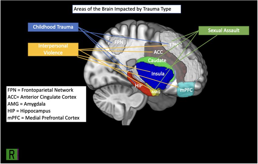
FIGURE 1 | Brain regions implicated in IPV, childhood trauma and sexual assault.
Intimate Partner Violence deviations from normative abilities to focus and integrate
Adult females are more likely than males to experience and contextual information into emotional responses (35). Some
report being victims of intimate partner violence in their female survivors of IPV-PTSD exhibit disorganized connectivity
lifetime than males (78), yet only eight of the neuroimaging between the frontal and limbic systems, resulting in sustained
studies reviewed specifically address neural correlates of IPV. periods of heightened arousal that may cause them to misdirect
Although scarce, existing literature suggests that female survivors and fixate their attention on nonthreatening stimuli (82).
of IPV with PTSD exhibit neurobiological changes relative to Taken together, these deficits may leave females with PTSD
healthy controls. Aberrations from healthy controls manifest vulnerable to further neural dysregulation in the face of future
in various ways across the frontoparietal, default, and salience trauma. Clinical presentations of PTSD in female survivors
networks, and translate into difficulty with executive functioning, of PTSD may also be impacted by social and environmental
emotional regulation, and hypervigilance [e.g., (29, 81)]. variables, such as lack of perceived social support and cultural
Within-group comparisons suggest divergence among females messages about blame in intimate partner violence (10, 81).
with IPV-PTSD from healthy controls in activity in the amygdala, The magnitude of the effect of these and other environmental
anterior cingulate cortex, insula, dorsolateral prefrontal cortex, and social covariates on neurobiological presentations in female
and medial prefrontal cortex (21, 29, 82). In general, it appears survivors of IPV remains to be explored in the neuroimaging
that some female survivors of IPV may struggle to accurately literature examined for the present review.
appraise situations as threatening or not and, in turn, to adjust
neural responses to task demand (29, 82). Challenges with top- Sexual Assault
down regulation are reflected in trends toward increased activity Adult females are more vulnerable to SA than males (79),
in the medial and dorsolateral PFC, anterior ACC, and insula making the neurobiological profiles of female survivors of SA
evaluating social and emotional stimuli in comparison to healthy with PTSD of particular interest. Of the studies reviewed, five
controls (21, 29, 35, 82–84). Conversely, a proclivity toward directly examined survivors of sexual assault. Similar to IPV
hypoactivation of the frontal and parietal lobes when neural survivors, the female samples of sexual assault presented with
activation is required suggest further challenges modulating varying alterations of GMV, activity and functional connectivity
arousal, perhaps leading to avoidance and numbness often in the brain areas frequently implicated in PTSD (e.g., the
reported in PTSD (29, 50, 82). In some females, such issues insula, amygdala, right caudate, prefrontal cortex, and medial
may be further compounded by hyperactivity in the anterior occipital cortex). The composite of the neural correlates found in
cingulate cortex and dorsolateral prefrontal cortex, suggesting the literature suggest hyperactivity in the insula and prefrontal
Frontiers in Psychiatry | www.frontiersin.org 6 June 2022 | Volume 13 | Article 862476Eder-Moreau et al. Neurobiological Alterations in Females: PTSD
cortex, in addition to aberrant GMV in the amygdala, right Other Trauma Types
caudate, and insula in female survivors of SA as compared There is a gap in the neuroimaging literature surrounding the
to healthy controls (22, 23). Similar to IPV survivors, the neural correlates of PTSD in females related to types of trauma
female samples of SA survivors also presented with changing other than those described above (i.e., medical trauma, natural
degrees of activity and resting-state functional connectivity disasters, or other). Our literature search rendered only three
(rsFC) in the brain areas frequently implicated in PTSD (e.g., neuroimaging studies examining the neural correlates of PTSD
the insula, amygdala, right caudate, and medial occipital cortex) stemming from other types of trauma in females meet our
(22, 23, 34). Hypoactivation in the prefrontal cortex provides inclusion criteria. One examines the neural correlates of PTSD
further support for challenges with emotional regulation and among breast cancer survivors, finding no significant differences
the executive functioning required for attention shifting (39). in amygdala and hippocampal brain volume as compared to
Collectively, female survivors of SA who have PTSD may face healthy controls (87). One did not clearly specify the types
greater difficulties in the realms of information processing related of trauma that the females in their samples had experienced,
to self and others, hyperarousal, and executive functioning than and another mixed trauma from accidents with interpersonal
healthy controls. As a result, they may be more susceptible to violence. The results of these studies are discussed in later
PTSD symptoms of greater severity if faced with subsequent sections in which we identify neural correlates of specific
traumatic events. symptom clusters of PTSD in females.
Given the paucity of studies specifically examining the neural
correlates of IPV-PTSD due to sexual assault in female samples, it Trauma Type Summary
is difficult to determine how females with IPV and sexual assault Although it is difficult to determine the association between the
might be distinct in their neurobiological presentation. Results type of interpersonal violence females incur and neurobiological
suggest similar presentations wherein there may be compromised deficits in functioning, the literature appears to establish that
ability to manage arousal in the amygdala and insula, thereby interpersonal violence impacts females’ brains (see Figure 1).
engendering deficits in automatic emotional processing. Such More specifically, it seems to cause weakened regulation of
alterations may necessitate greater activation of the prefrontal arousal by impacting GMV in the amygdala and insula, while
cortex, perhaps reflecting the brain’s attempt to overcompensate creating a deficit in rsFC between the insula, amygdala, and
for a lack of control in the amygdala and insula. Relative prefrontal cortex that weakens executive, cognitive, and social
to healthy controls, differences in social processing are fairly functioning. What remains unknown is the extent to which this
common in female survivors of sexual abuse, perhaps due to impact is influenced by individual differences within samples,
smaller volumes of the right caudate. There is also research to such as variability among subjects’ trauma load, symptom
suggest that there are sociocultural elements that may predispose severity. Further, study designs are largely cross-sectional
females to sexual assault, such as living in poverty and prior and sample sizes are generally small, thereby reducing the
history of abuse (85). Additional research is needed to determine generalizability of the results found. Finally, as we highlighted in
whether there is a difference in neural underpinnings of sexual each of the sections above, the extent of the interaction between
assault and IPV, and the ways that society and culture may trauma exposure and sociocultural factors on neurobiological
influence outcomes and neurobiological symptoms of PTSD in presentations in females with and without PTSD remains largely
female survivors of sexual assault and IPV. unexplored by neuroimaging research.
Mixed Samples of Interpersonal Violence Underlying Purported Mechanisms
Remaining studies addressing interpersonal violence in females In addition to differential susceptibility to specific types
combine types of interpersonal violence, diluting the impact of of gendered trauma, there are individual determinants that
each type of interpersonal violence on the female brain. It is influence neurobiological presentations in females with PTSD.
therefore difficult to establish a unique neurobiological profile In the present section, we will examine some of these, including
for female survivors of interpersonal violence with PTSD based the neuroendocrine system, coping mechanisms, and behavioral
on extant literature. However, results echo those discussed in mechanisms of PTSD in females.
prior sections, with deviations from healthy controls in GMV in
addition to variations in subcortical activity and rsFC. Different The Neuroendocrine System
findings from those already reported provide insight into the The impact of the neuroendocrine system on fear circuitry in the
role of the corpus callosum in the brain, specifically the genu brain is well-documented among healthy females. Specifically,
(31). Others have found increased right caudate volumes, perhaps due to the high amount of estrogen and progesterone receptors
leading to difficulty managing social expectations and threat in the amygdala and hippocampus, these areas are particularly
appraisal (26, 27). Increased GMV in this region of the brain have susceptible to differential functioning and volume throughout the
been associated with other forms of psychopathology, including female menstrual cycle (8). These fluctuations can manifest as
psychosis (86), further emphasizing the degree of compromised more frequent and severe symptoms of reexperiencing intrusive
functioning in females with PTSD. Because most are studies memories, negative alterations in mood (e.g., increased anxiety
examining fMRIs related to specific symptoms of PTSD, they and depression), and difficulty with memory and executive
will be reviewed in further detail when we examine clinical functioning. For example, extinction recall is negatively impacted
presentations of PTSD and their neural correlates. during the early luteal phase of the menstrual cycle (8, 88).
Frontiers in Psychiatry | www.frontiersin.org 7 June 2022 | Volume 13 | Article 862476Eder-Moreau et al. Neurobiological Alterations in Females: PTSD
Despite the abundant evidence that the neuroendocrine (90), and neuroticism, a personality trait related to general
system influences individual PTSD symptoms in female humans psychopathology at large (15, 16).
and rodents, only one study met our inclusion criteria of a
diagnosis of PTSD. Sartin-Tarm et al. (43) examined the impact Rumination
of estradiol on fear habituation in females with and without Researchers have found that females ruminate more than men
PTSD and found that estradiol was positively correlated with and that rumination, an emotion-focused coping style defined
activation in the anterior cingulate cortex and dorsomedial as self-referential thought focused on negative outcomes, is
prefrontal cortex during fear habituation responses. Low levels a transdiagnostic consideration in psychopathology (15). The
of estradiol negatively predicted the ability to habituate to tendency to ruminate has been correlated with sociocultural
fear extinction among those with PTSD. These results further considerations such as identification with femininity, as well
stress the importance of the timing of the traumatic event as biological sex factors like estradiol levels (12, 91). As the
on neurological susceptibility to trauma responses and the other constructs reviewed, rumination and its impact on PTSD
development of PTSD. symptoms is therefore likely the product of nature and nurture.
Extant literature characterizes rumination as the function of
Emotion Regulation and Gendered Coping potential deficits with executive functioning, including memory
Mechanisms (92), attentional shift (16), emotional processing, and coping
Challenges with emotional regulation among females with PTSD (15). These correlate with hyperconnectivity between the right
reflect changes in several regions of the brain, presumably amygdala and the mPFC, PCC, precuneus, and the orbital
impacted by posttraumatic stress. The neuroimaging literature cortex, reflecting the need to make a greater cognitive effort
posits that females’ implicit and explicit emotional regulation to shift attention sets (21). Conjointly, such weaknesses may
is affected by altered neural connectivity. Broadly, it suggests cause those who ruminate to fixate on negatively-valenced
hypoactivity in the vmPFC and hyperactivity in the amygdala emotional information, thereby leading to exacerbated outcomes
(49, 89). This hyperactivity may result in excess physiological and among females with PTSD. For example, rumination in females
emotional arousal in females with PTSD and, in turn, lead to with PTSD has been positively correlated with the frequency
deficits in executive functioning required for emotion regulation. and intensity of reexperiencing symptoms, corresponding to
This is particularly true when females diagnosed with PTSD are increased GMV in the left isthmus cingulate (40, 90, 93).
faced with tasks of increasing cognitive demand or emotional
load (21, 39, 52, 89). Neuroticism
Emotion regulation is also affected by the positive or negative Much like rumination, neuroticism, a personality construct
evaluation of stimuli and its relevance to the initial traumatic defined as one’s proclivity toward experiencing negative
event. Among females with PTSD, more threatening stimuli emotional states, is associated with poor psychological outcomes
are associated with increased activation in the hippocampus (16, 94). Those who endorse higher levels of neuroticism may
and amygdala and decreased connectivity between the insula, experience high levels of anxiety, have difficulty coping with
the dACC, and the amygdala relative to healthy controls (29, challenging situations, and display poorer emotion regulation
53). Challenges with emotional regulation are also evident in than others 16). Females with PTSD who display more neurotic
bottom-up regulation wherein females show hypoactivity of the tendencies experience PTSD symptoms of greater severity
vmPFC in response to positive stimuli (56). Taken together, it (95), making it important to understand the neural correlates
appears that females with PTSD exhibit distinct modifications of neuroticism.
in neurobiology that result in greater emotional dysregulation Like rumination, neuroticism interferes with the ability
relative to healthy controls, often as a function of cognitive to objectively evaluate environmental stimuli, due to an
load. These neurobiological alterations may interfere with the overactivation of brain regions associated with fear, self-
executive functioning required for emotion regulation (21, referential processing, and value judgments needed to make
33). Overall, these patterns in the literature reflect a neural accurate decisions. Neuroticism is positively correlated with
hypoactivity when faced with positive events, and hyperactivity hyperactivation in the amygdala, right PFC, dmPFC, and
and an attentional bias when faced with negative events. It is parahippocampus (16). Together, rumination and neuroticism
likely that the negative attention bias, together with the gendered leave females with PTSD more vulnerable to reexperiencing,
coping mechanisms previously discussed (i.e., rumination and negative alterations in mood, difficulty with memory, and
neuroticism) contribute to the other symptoms of hyperarousal accurate fear-based learning.
and create a unique profile of arousal in females with PTSD.
Some of the challenges with emotional regulation discussed Behavioral Mechanisms
may be related to socialization to more gendered coping Behavioral mechanisms of PTSD involve fear-based learning,
styles and, in turn, impact neurobiological presentations in such as extinction, value expectation, and inhibition. Fear
females with PTSD. Certain emotion-focused coping styles extinction is an important part of PTSD symptom reduction
and personality traits may leave females more vulnerable and recovery and is therefore a large part of APA-recommended
to developing psychopathology when faced with stressors or PTSD treatments like exposure therapy (96, 97). Fear extinction
traumatic events (15). Perhaps two of those most documented requires the ability to accurately assess potentially fearful future
are rumination, relevant to the PTSD symptom of reexperiencing events, to inhibit fear responses when they arise and decrease
Frontiers in Psychiatry | www.frontiersin.org 8 June 2022 | Volume 13 | Article 862476Eder-Moreau et al. Neurobiological Alterations in Females: PTSD
arousal, and the eventual ability to learn from experiences (98). In addition, the literature reflects patterns of hyperactivity
The neuroimaging literature suggests that females diagnosed with in the amygdala, insula, dmPFC, and striatum with deficits
PTSD present with deficits in the neural underpinnings of each of in connectivity between the amygdala and the vmPFC. The
these processes, contributing to rigid thinking and learning styles mechanisms reviewed may leave females more vulnerable to
which thereby hinder their recovery (21). worsening trauma symptoms and unique presentations of
Accurate assessment of future events requires application of PTSD and comorbid disorders. In the following section, we
prior knowledge and experiences to future situations and their examine the literature surrounding neural correlates of clinical
possible outcomes. The literature suggests that many females presentations of PTSD in females, including symptom types,
with PTSD may struggle to integrate prior experiences into their severity, and comorbidity.
expectations and responses to situations and tasks at hand, in
threatening and nonthreatening situations (42). Because females Presentation
with PTSD may have difficulty incorporating lessons learned into The DSM-5 classifies PTSD according to criteria across four
decision making and problem solving, they may resort more to symptom clusters: reexperiencing, or experiencing unwanted
trial and error in problem solving tasks (68). This may be due to memories of the traumatic event; hyperarousal, evidenced by
a propensity to prediction errors, combined with an attentional symptoms such as being startled easily or hypervigilance;
bias in the brain toward negative information that is consistent avoidance of stimuli that are reminders of the traumatic event;
with inaccurate value expectations (26, 48). Females with PTSD and increased negative affect across several domains, including
are more neurologically reactive to anticipated negative events irritability, anhedonia, and feelings of alienation from others
and less so to anticipated positive events than healthy controls, (100). As previously noted, these symptoms often differ in
thereby further complicating the formation of expectations (47). severity and duration, often according to the type and timing of
Neuroimaging results suggest that these outcomes are associated trauma. For females, clinical and neurobiological presentations
with deficits in the FPN, and hypoconnectivity in the ACC and may also be a function of the phase of their menstrual cycle in
dmPFC (26, 68). which the trauma occurs, together with age (8). In the present
Inhibition is also an important part of reversing fear-based section we will review the different types of clinical presentations
learning, involving the ability to monitor one’s own mental and that arise in females with PTSD, according to symptom cluster.
physiological state and to purposefully react to the same (21, 99).
The literature reviewed reflects the notion that females with Reexperiencing
PTSD may struggle with inhibition, such that they may find A hallmark of PTSD is reexperiencing the traumatic event
it difficult to regulate arousal in response to external stimuli. through intrusive thoughts, memories, or nightmares, which may
These challenges are reflected in hyperactivation of the insula, be triggered by direct or indirect reminders of the traumatic
dmPFC, the striatum, the amygdala, and hypoactivation of the event (100). The neuroimaging literature on females with PTSD
vmPFC (21, 33, 48). Such abnormalities may result in difficulties examines reexperiencing through fMRI imaging of subjects
with cognitive control, attention shifting, and processing of being exposed to direct or indirect reminders of the traumatic
self-referential information, all processes required to inhibit experience [e.g., (35)], or through the correlation of self-report
arousal (47, 99). Such deviations from normative social learning, of reexperiencing with GMV and rsFC [e.g., (27)]. In general,
expectation formation, and inhibition may contribute to the the evidence suggests that females who indicate higher rates
documented mixed outcomes of exposure therapy in among of reexperiencing symptoms exhibit altered neural correlates
females with PTSD. associated with visual memory and inhibition, and increased
Successful extinction has been negatively correlated with activity between the limbic and default networks.
activation in the vmPFC and bilateral amygdala (96). However, Among females with PTSD, reexperiencing is associated
females with PTSD display the opposite neurological profile with reduced CT in the left inferior and mid temporal gyrus
when engaging in extinction learning tasks. Another important and increased GMV in the lingual gyrus (22, 27). Diffusion
factor that is not often considered by the neuroimaging literature Tensor Imaging (DTI) similarly suggests compromised white
is the impact of individual estradiol levels on extinction learning, matter volumes (WMV) of the postcentral gyrus in the corpus
frequently overlooked by extant neuroimaging evidence (43). callosum. The alterations of WMV in the corpus callosum may
Although previously discussed, it is important to highlight it here suggest deficits in the processing and communication of visual
as a factor that may make females more vulnerable to varying information between hemispheres (21, 31). The increased GMV
results in behavioral treatments of PTSD. of the lingual gyrus, also associated with inhibition and top-
down regulation, may lead to difficulties in preventing memories
Underlying Mechanisms Summary from surging to conscious awareness. This may lead females with
Females have distinct coping styles and ways of regulating PTSD to feel flooded by visual memories of their traumatic event,
their emotions, corresponding to personality (e.g., neuroticism), thereby increasing overall arousal (101).
cognition, (e.g., rumination), and behavior (e.g., fear processing). Distress associated with reminders of traumatic events is
These are each associated with a specific neural presentation reflected in an increased rsFC between the right and left
and may be linked to both gender and sex. Although each amygdala, the right inferior frontal gyrus, the right hippocampus,
has specific neural correlates, they appear to reflect a common and the visual and dorsomedial cortex (27, 37, 38, 102). It
pattern of decreased connectivity between the insula and dmPFC. is concurrently associated with hypoconnectivity between the
Frontiers in Psychiatry | www.frontiersin.org 9 June 2022 | Volume 13 | Article 862476Eder-Moreau et al. Neurobiological Alterations in Females: PTSD
lingual cluster and the fusiform cortex, responsible for the reduced CT in the occipital gyri, as well as the left lateral fissure
voluntary and involuntary processing of trauma memories (22). and right posterior cingulate (27). The posterior cingulate cortex
The combination of hyper- and hypoactivity in these areas of has been found to mediate information between emotions and
the brain may reflect the brain’s altered ability to assimilate memory (104), while the lateral fissure and occipital gyri have
traumatic memories and prevent hyperarousal when confronted been associated with visual and working memory. The reduction
with reminders of the same. in CT may suggest a decreased capacity to process emotional
memories and uncertainty about the safety of their surroundings
Hyperarousal due to prediction errors. In turn, individuals with PTSD may
Hyperarousal among individuals with PTSD manifests as develop avoidance as a coping skill to escape overwhelming
irritability, impulsivity, hypervigilance, difficulty sleeping, feelings that may be associated with their inability to accurately
inability to concentrate, and exaggerated startle responses (102). process emotional and environmental stimuli.
These symptoms may indicate deficits in emotion regulation,
often described in PTSD as part of a “feedback loop” that leads to Specifiers: PTSD With Depersonalization or
further arousal, thereby creating challenges with concentration Derealization
and reliable evaluation of environmental stimuli (101). The most recent diagnostic classification for PTSD includes
Hypervigilance can generally be defined as a sense of constant a specifier for a dissociative subtype (PTSD-D) to identify
awareness of one’s surroundings, even in situations one knows the presence of persistent symptoms of depersonalization or
to be safe. Surprisingly, we found the neural correlates of derealization. DSM-5 defines depersonalization as “persistent or
hypervigilance to be largely understudied in females with PTSD. recurrent experiences of feeling detached from, and as if one
However, the little evidence that exists describes a pattern of were an outside observer of, one’s mental processes or body,” and
hyperconnectivity between the amygdala, insula, ACC and the derealization as “persistent or recurrent experiences of unreality
superior colliculus and locus cereleus in females with PTSD of surroundings” (105). PTSD-D can therefore be summarized
(102). These areas of the brain, responsible for visual and auditory as a presentation of PTSD in which there is a disconnection, or
processing and arousal, suggest overactivation which, together dissociation, from one’s lived experience.
with the negative attention bias previously discussed, may lead to Extant neuroimaging literature comparing females with
appraisals of otherwise neutral stimuli as threatening (21, 102). PTSD-D to healthy controls suggests that dissociative
experiences relate to hyperactivity in the somatomotor network
Negative Affect (i.e., the right superior temporal gyrus, involved in auditory
Negative affect and changes in mood are often reflected in processing) and frontoparietal lobes, specifically in the left
self-blame, exaggerated negative perceptions of self and others, inferior, precentral, and medial gyri (59, 60). Higher rates of
anhedonia, and detachment or estrangement from others (102). dissociation severity are negatively related to activation in the
Extant literature reports that many females with PTSD have an parahippocampal gyrus, associated with memory formation and
overwhelmingly negative self-image coupled with tremendous spatial location of objects, and hypoactivity in the ventromedial
guilt relative to those without PTSD. They may find themselves prefrontal cortex (59, 60, 106). Accordingly, the neurobiological
debilitated in their ability to modify such negative cognitive profile of females with PTSD-D suggests difficulties with
distortions, due to rigidity in thinking, which has been connected emotion regulation, likely caused by a sense of sensory and
to poor psychological outcomes (48, 57). Neural correlates reflect emotional overwhelm. Altered activity in the vmPFC may
a greater preoccupation with the self than others as well as a suggest deficits in amygdala regulation (89), leading to challenges
negative attention bias toward their own qualities, relative to in downregulating excessive arousal, and hypervigilance among
healthy controls. This attention bias may manifest as a tendency some females with PTSD. This presentation is heightened by
to accept more negative qualities about themselves, while also deficits in synchronization between key brain regions including
endorsing more positive qualities about others. Examinations of the amygdala, hippocampus, thalamus, and the brain stem (61),
neural activity suggest greater activity of the visual cortex when proposed to be linked to the experience of dissociation.
imagining the self as opposed to others, in addition to greater
activation in the mPFC when considering negative qualities about Presentation Summary
themselves (57). The correlates of self-blame mirror those of Females with PTSD present with altered GMV and WMV in
reexperiencing, exhibiting reduced GMV in the lingual gyrus, areas of the brain responsible for emotion and visual processing.
such that self-blame may involve an aspect of reexperiencing These deviations from volumes in healthy controls correspond
among females with PTSD (21, 57). Cumulatively, these results to hypoactivity of brain structures involved in top-down and
may suggest difficulty regulating emotions in relation to the self bottom-up emotion regulation (i.e., the vmPFC) and executive
and negative cognitive distortions about the self, with favorable functioning (i.e., the dmPFC), in addition to hyperactivity
biases toward others. of areas related to arousal (i.e., the amygdala) and sensory
processing (i.e., the right superior temporal gyri).
Avoidance
Among males and females, avoidance is associated with Comorbidities
heightened global PTSD symptom severity (103). Females with A review of clinical presentations of PTSD in females would
PTSD who engage in avoidance with greater frequency show be incomplete without a discussion of the literature related to
Frontiers in Psychiatry | www.frontiersin.org 10 June 2022 | Volume 13 | Article 862476Eder-Moreau et al. Neurobiological Alterations in Females: PTSD
comorbidities. As many as 83% of males and females diagnosed associated with reduced hippocampal GMV with concurrent
with PTSD present with a comorbid disorder, and female increased GMV in the right putamen, pallidum, and striatum as
gender has been identified as a risk factor for the same (107, a function of the severity of dissociative symptoms (5, 55). It is
108). As such, it is important to review the neuroimaging unclear from the current body of literature whether these neural
literature regarding specific comorbidities that occur with PTSD. correlates also correspond to those with PTSD-D.
PTSD rarely occurs by itself, and it is frequently associated
with comorbidities such as Major Depressive Disorder (MDD), Summary of Comorbidities
Dissociative Identity Disorder (DID), Borderline Personality In general, there is a lack of information surrounding
Disorder (BPD), and Substance Abuse Disorder (55, 109, 110). comorbidities with PTSD and associated neural correlates
Past literature has studied neural correlates of each of the in female samples that precludes conclusions about specific
aforementioned comorbidities with PTSD in females except neurobiological profiles of each. Perhaps one of the most
Substance Abuse Disorder. challenging components of studying such comorbidities is
that PTSD symptoms are shared among so many psychiatric
PTSD and Major Depressive Disorder disorders, such that determining specific neural correlates is
Reports on the incidence of PTSD-MDD vary, but the literature particularly challenging (44). It is perhaps this overlap in
reports it as one of the most commonly occurring comorbidities symptoms and variety of presentations that lead to differing
among patient populations with PTSD. Recent statistics show outcomes and prognoses among females with PTSD.
that up to 89% of males and females with PTSD present
with PTSD-MDD (108–110). Despite the high prevalence of Sex Related Outcomes of PTSD
depression among PTSD populations, there is little neuroimaging We turn now to an examination of the outcomes of PTSD
research that specifically investigates the neural correlates of among females, looking at some of the ways that trauma further
PTSD-MDD in females. The only study we found that met the impacts them specifically. We begin by reviewing literature on
inclusion criteria for this review implicated a hypoconnectivity the ways that PTSD impacts mothering and corresponding neural
between the amygdala and the frontal and temporal lobes correlates and end with a review of treatment outcomes.
in PTSD-MDD (44). These results replicate others found in
literature that examined PTSD, perhaps because so many studies Parenting Outcomes: Mothers With PTSD and Their
contain samples with mixed presentations of PTSD in females, Children
including PTSD-MDD [e.g., (49)]. Despite these findings, there is Children of mothers with PTSD are more likely to experience
still not enough information to clearly establish a neurobiological traumatic events than children of healthy mothers (111). The
profile for PTSD-MDD. literature documents one possible explanation as lower levels of
attunement and maternal sensitivity among mothers with PTSD,
PTSD and Borderline Personality Disorder particularly if a mother endured severe childhood trauma [e.g.,
A comparison of the symptoms of BPD and PTSD in the (45, 112)]. Parenting requires the ability to mentalize (i.e., take
DSM-5 reveal an overlap of symptoms. Patients of BPD and another’s perspective by identifying their emotions), empathize,
PTSD both present with difficulty with emotion regulation; an and respond sensitively to a child’s needs (36). As we have
unstable sense of self, marked by negative self-image; dissociative documented, PTSD may result in emotional dysregulation and
symptoms; and a proclivity toward impulsivity or behaviors a proclivity toward interpreting neutral social information as
that might incur self-harm (100). As such, one might expect threatening, resulting in a state of persistent hyperarousal. In
that the literature would reflect similarly impacted areas in mothers, it is likely that such constant hyperarousal complicates
the brain, both in GMV and rsFC, perhaps on a larger scale their ability to respond sensitively to their young.
due to the presence of a dual diagnosis. However, the study Evidence for heightened arousal when faced with stress in
that met the inclusion criteria for the present review actually relation to their young can be found in decreased activation
found no significant differences between GMV of the amygdala, of the vmPFC among mothers with PTSD relative to healthy
vmPFC, or bilateral ACC when comparing females with BPD controls (36, 46). Altered emotion regulation may cause mothers
and PTSD to those with PTSD alone (51). The dearth of with PTSD to respond differently to separation from their
neuroimaging literature surrounding BPD and comorbid PTSD, children than healthy controls, manifesting as heightened arousal
en masse with these results that suggest insignificant differences and increased activity in the dmPFC (36). Thus, mothers with
between neurobiological profiles, suggests the need for further PTSD may have more difficulty engaging in parental reflective
investigation of this comorbidity. functioning relative to healthy controls, thereby complicating the
formation of a secure attachment with their children.
PTSD and Dissociative Identity Disorder Children of mothers who suffered childhood abuse exhibit
PTSD-DID has been examined largely as it pertains to differences patterns that mirror their mothers’ emotion regulation,
in morphology between females afflicted with the disorder evidenced by decreased vmPFC activity. Relative to children of
and healthy controls. Results are again insufficient in volume healthy controls, the literature points to a higher probability that
to reveal a specific neurobiological profile of PTSD-DID, but children of mothers with PTSD endure a traumatic event or
portray interesting findings surrounding subcortical GMV in develop psychopathology, perhaps related to emotion regulation
females with PTSD-DID. More specifically, PTSD-DID has been and arousal (46, 111). Together, the neuroimaging literature
Frontiers in Psychiatry | www.frontiersin.org 11 June 2022 | Volume 13 | Article 862476You can also read