The roles and diagnostic value of miRNA-1246 in the serum of patients with intracranial aneurysms
←
→
Page content transcription
If your browser does not render page correctly, please read the page content below
Translational Neuroscience 2022; 13: 172–180
Research Article
Haijie Jiang, Yansheng Ding, Lili Wu, Chunyan Jiang, Chengdong Wang*
The roles and diagnostic value of miRNA-1246 in
the serum of patients with intracranial aneurysms
https://doi.org/10.1515/tnsci-2022-0227 Keywords: intracranial aneurysm, microRNAs, diagnosis,
received April 01, 2022; accepted June 8, 2022 bioinformatics
Abstract
Background ‒ Inflammatory response is one of the impor-
tant factors affecting the formation of intracranial aneurysm.
miR-1246 is involved in the regulation of several inflamma- 1 Introduction
tory diseases; however, its expression levels and the
mechanism of action in intracranial aneurysms remain Intracranial aneurysm is one of the most dangerous vas-
further investigated. cular diseases of the brain. Its sudden rupture of intra-
Methods ‒ Bioinformatics was used to analyze the levels cranial aneurysms is the main cause of non-traumatic
of micro-RNAs (miRNAs) in the serum of intracranial subarachnoid hemorrhage, which can result in severe
aneurysm patients as well as in the intracranial aneurysm damage to the central nervous system as well as to sev-
tissues downloaded from the GEO RNA-seq database. Blood eral other organs. Because most intracranial aneurysms
samples were collected pre-operatively from patients with are asymptomatic before rupture, early detection of intra-
intracranial aneurysms as well as from healthy volunteers, cranial aneurysms can be effective for monitoring their
and miRNA-1246 expression levels were detected using progression and taking preventive measures. Digital sub-
quantitative reverse transcriptase polymerase chain reac- traction angiography (DSA) is currently the best approach
tion. Meanwhile, the diagnostic value of miR-1246 for intra- for diagnosing intracranial aneurysms, but DSA is not sui-
cranial aneurysm was explored using the receiver operating table for early screening and treatment of intracranial
characteristic (ROC) curve. aneurysms since it is invasive and does not show the distal
Principle findings and results ‒ Serum levels of miR-1246 vessels of the occlusion, and in addition, it is costly and
were elevated in intracranial aneurysm patients. Bioinfor- potentially risky to visualize the location of the vascular
matics studies revealed that the target genes of miR-1246, lesion by injecting iodine contrast. Recently, the detection
TP53, glycogen synthetase kinase (GSK), and transcription of micro-RNA (miRNA) in circulating immune cells has
factor YY1 may play important roles in the development of been used as a means to screen for diagnostic markers of
intracranial aneurysms. miR-1246 is involved in inflamma- vascular diseases such as intracranial aneurysms, abdom-
tory response, lipid, and atherosclerotic signaling pathways. inal aortic aneurysms, and stroke. miRNAs are known to
Conclusions and significance ‒ High level of miR-1246 inhibit protein translation by regulating the 3′ untrans-
is found in the serum of patients with intracranial aneur- lated region of target gene mRNAs and are involved
ysms and may serve as a diagnostic or/and treatment in important biological processes such as apoptosis and
marker for intracranial aneurysms. metabolism in the organism [1]. Several miRNAs have been
previously identified to be associated with the develop-
ment and progression of intracranial aneurysms and can
have an impact on the processes of angiogenesis and
* Corresponding author: Chengdong Wang, Clinical Laboratory,
Weifang People’s Hospital, Diagnosis by Clinical Examination, extracellular matrix degradation and on the biological pro-
Weifang, China, e-mail: shenjingneierke@126.com cesses such as inflammatory responses and apoptosis of
Haijie Jiang: Department of Medical Laboratory, Weifang Medical vascular smooth muscle cells (VSMC) through various
University, Molecular Biological Diagnosis of Cerebrovascular pathways. The formation and development of intracranial
Disease, Weifang, China
aneurysms are the results of endothelial dysfunction and
Yansheng Ding, Chunyan Jiang: Clinical laboratory,
Weifang People's Hospital, Weifang, China
conversion of the VSMC phenotype to a pro-inflammatory
Lili Wu: Neurology department, Weifang People's Hospital, Weifang, phenotype. miR-409-3p is a widely studied miRNA in
China inflammatory diseases, and it triggers the inflammatory
Open Access. © 2022 Haijie Jiang et al., published by De Gruyter. This work is licensed under the Creative Commons Attribution 4.0
International License.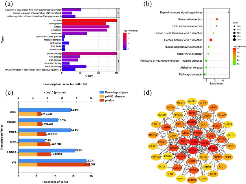
Diagnosis of intracranial aneurysms 173
response mainly by targeting Nr4a2 to activate the NF-κB the Medical Ethics Committee of the Weifang People’s
pathway [2]. Hospital.
In this study, we used second-generation gene sequen-
cing to screen for differentially expressed miRNAs asso- Informed consent: Informed consent has been obtained
ciated with inflammation in the serum of patients with from all individuals included in this study.
intracranial aneurysms. qRT-PCR was used to detect the
serum levels of miRNAs in patients with intracranial aneur-
ysms and healthy individuals. Bioinformatics was used to 2.2 Screening and bioinformatics analysis
analyze the differentially expressed miRNAs and to explore of target miRNAs
the significance of inflammatory response in intracranial
aneurysms and the diagnostic value of inflammation- There were two main data sources in this study including
related miRNAs in intracranial aneurysms. (i) the second-generation gene sequencing (RNA-seq)
data for the blood levels of miRNAs in patients with intra-
cranial aneurysms; (ii) the data of differentially expressed
miRNAs in tissues of patients with intracranial aneurysms
2 Materials and methods were obtained and screened from GEO database. Second-
generation gene sequencing to determine the levels of
2.1 Study subjects and sample collection miRNA in the serum of the patients with intracranial
aneurysms was partially conducted by researchers from
Patients who attended the Neurosurgery Department at BGI Genomics Co., Ltd. The GSE46336 and GSE66239
the Weifang People’s Hospital in the Shandong Province data sets were downloaded from the GEO database of the
from September 2020 to December 2021 were diagnosed National Center for Biotechnology Information (NCBI) and
with intracranial aneurysm and selected for the study, contained 10 intracranial aneurysm samples and 13 control
while healthy individuals in the Health Management samples (middle cerebral artery tissue and superficial
Center were selected as healthy controls. The inclusion temporal artery tissue). GSE46336 and GSE66239 expres-
criteria for the group of healthy individuals were the fol- sion profiles were analyzed using GEO2R tool, and the
lowing: (i) matched to patients in terms of age, gender, screening criteria were P < 0.05, the absolute value of
and other aspects of medical history; and (ii) healthy log2FC > 1.5. After the screening, differentially expressed
individuals who did not have inflammation-related dis- microRNAs (DEMs) were identified from the overlap using
eases, cerebrovascular-related diseases, history of various Venn diagrams, and those with differential expression in
types of tumors, and immune diseases. The inclusion cri- both tissues and serum and consistent expression were con-
teria for the group of patients with intracranial aneurysms sidered as differentially expressed miRNAs. The miRNet data-
were the following: (i) patients presenting with sudden base (https://www.mirnet.ca/), a comprehensive miRNAs
onset of headache, ptosis, dizziness or diplopia, and diag- target gene database, was used to predict the target genes
nosed by DSA; and (ii) patients with various inflammatory- of the screened differentially expressed miRNAs in the
related diseases, cerebrovascular-related diseases, history aneurysm tissues of intracranial aneurysm patients, and
of various tumors, and immune diseases other than intra- the target genes of the screened highly expressed specific
cranial aneurysms were excluded. miRNAs were transcribed factor annotation by FunRich
Three (3) mL of venous blood was collected from the (v3.3.1) software. Transcription factors that met P < 0. 05
patient’s pre-operative elbow vein (fasting for more than were used as entry factors for further analysis. gene onto-
8 h), and blood from healthy controls was also collected logy (GO) enrichment analysis and kyoto encyclopedia of
at the same time. The blood samples were immediately genes and genomes (KEGG) signaling pathway prediction
centrifuged at 3,500 rpm for 5 min after collection. The were performed using the DAVID database (https://david.
serum was collected in two 1.5 mL RNAase-free centrifuge ncifcrf.gov/) for the screened target genes with high expres-
tubes, sealed and numbered, and immediately stored in a sion specificity for DEMs. The criterion for screening was
−80°C freezer. P < 0.05. The obtained DEMs of target genes were uploaded
to a string database (https://string-db.org/) to predict the
Ethical approval: The research related to human use has relationship between the target genes and then visualized
been complied with all the relevant national regulations, using Cytoscape software (https://cytoscape.org/) to find
institutional policies and in accordance with the tenets the top five genes by various computational methods
of the Helsinki Declaration, and has been approved by including the top five. The core target genes located in174 Haijie Jiang et al.
the top 50 were also calculated using the degree of con- includes the following: 10 μL of 2× Transtart Tip Green qPCR
nectivity search. CytoHubba sorts the nodes by several Super mix, 6 μL of five-fold diluted cDNA template, 2 μL of
topological algorithms based on the characteristics of forward primer, and 2 μL of reverse primer. The qPCR was
the pre-loaded protein protein interaction (PPI) network. performed under the following conditions: 95°C for 30 s
(1 cycle), 95°C for 5 s, and 60°C for 30 s (45 cycles). For
spiking, two replicate wells were added for each cDNA.
2.3 Determine miRNA levels in serum After waiting for the amplification reaction to be completed,
the relative quantification method (with cel-miR-39 as a
The miRNA was extracted using the Easy Pure miRNA kit. reference) was used to determine the miRNA level in the
200 μL of serum was mixed with 1 mL lysis buffer for 30 s patient’s serum. The formula was used for calculation as
and then incubated at room temperature for 5 min before 2−ΔCt (ΔCt = Cttarget miRNA − Ctcel-miR-39). The Ct values of
adding nematode outer reference solution, and the solu- each target miRNA and cel-miR-39-3p for each patient
tion was mixed thoroughly and left for 10 min. Chloroform were used as the average of the Ct values of the two repli-
(200 μL) was then added to the serum and mixed well, cate wells. The reagents used in this experiment were
and the mixture was centrifuged at 10,000×g for 15 min obtained from the Beijing TransGen Biotech Co., Ltd.
at 4°C; the upper aqueous phase was aspirated to another
RNAase-free centrifuge tube. The volume of the aqueous
phase was measured accurately, and one-third volume of 2.4 Statistical methods
anhydrous ethanol was added to the aqueous phase and
mixed well. The obtained solution was transferred to the SPSS 26.0 software as well as GraphPad Prism 9 was used
RNA Spin Column which was centrifuged at 12,000×g for to analyze the data. The normality of the data was tested
30 s at room temperature and the effluent was retained. using the Shapiro–Wilk test. Normally distributed data
The effluent was transferred to a clean 2-mL RNAase-free were expressed as mean ± standard deviation (Mean ± SD),
centrifuge tube, and 1.25 times the volume of effluent was and non-normally distributed data were expressed as
mixed with anhydrous ethanol. The obtained solution was median and quartiles M (P25 and P75). The non-parametric
transferred to a miRNA Spin Column and centrifuged at Mann–Whitney U test was used to analyze whether there
12,000×g for 30 s at room temperature, and then, the were differences in the data distribution of serum miR-1246
effluent was discarded (the volume of solution is larger levels in the intracranial aneurysm group and healthy con-
than the volume of the miRNA Spin Column; this step is trol group. ROC curves were used to assess the diagnostic
repeated until the entire solution is added). Wash buffer value of miRNA in the serum of patients with intracranial
(500 μL) was added to the miRNA Spin Column, centrifuged aneurysms. The difference was considered statistically sig-
at room temperature at 12,000×g for 30 s, and then, the nificant by statistical analysis P < 0.05.
effluent was discarded. The procedure was repeated once.
miRNA Spin Column was centrifuged at room temperature
at 12,000×g for 2 min in order to completely remove the 3 Results
residual ethanol from it. The miRNA Spin Column was
placed into another 1.5 mL RNAase-free centrifuge tube,
and 35 μL of RNAase-free water was added from the center 3.1 Clinical profile of patients with
of the miRNA Spin Colum, and then, it was allowed to stand intracranial aneurysm
at room temperature for 1 min. The miRNA Spin Column
was centrifuged for 1 min at 12,000×g at room temperature A total of 58 patients with intracranial aneurysms were
to elute the miRNA. The tubes were sealed and numbered, included in this study, including 7 cases of multiple intra-
and the extracted miRNA was directly reverse-transcribed to cranial aneurysms and 51 cases of single intracranial
cDNA or stored in an −80°C freezer. The miRNA was used as aneurysms. There were 23 male patients and 35 female
a template for reverse transcription to synthesize cDNA. The patients in the intracranial aneurysm group. A total of 44
reactions include the following: 10 μL of 2× Transcript healthy individuals were included in the study. There
miRNA reaction mix, 9 μL of extracted miRNA template, were 23 male patients and 21 female patients in the
and 1 μL of Transcript miRNA RT enzyme mix. The reverse healthy group. The difference in the composition ratio
transcription reaction was performed at 37°C for 1 h and between male and female patients was not statistically
then heated to 85°C for 5 s temperature. The quantitative significant between the intracranial aneurysm group and
reverse transcriptase-polymerase chain reaction (qRT-PCR) the healthy control group (P = 0.130). The average age ofDiagnosis of intracranial aneurysms 175
Table 1: Clinical information of the intracranial aneurysm patient group and healthy control group
Intracranial aneurysm group (n = 58) Health group (n = 44) P-Value
Gender Male (n = 23) Male (n = 23) 0.205b
Female (n = 35) Female (n = 21)
Age 55.02 ± 9.03 55.30 ± 10.03 0.883a
Multiple intracranial aneurysms Yes (n = 7)
No (n = 51)
Note: a – t-test; b – chi-square test.
the intracranial aneurysm group was 55.02 ± 9.03 years, including YY1, ARID3A, DLX3, RORA, HOXD8, and LHX3
and the age of the healthy control group was 55.30 ± 10.03 years. (Figure 2c). Functional annotation and signaling pathway
The difference in age between the intracranial aneurysm prediction were performed for 319 target genes of DEMs.
group and the healthy control group was not statistically The results reveal that the relevant target genes were
significant (P = 0.574 (Table 1)). mainly focused on the pathways such as inflammatory
response-related pathways, lipid and atherosclerotic sig-
naling pathways, Alzheimer’s disease, and neurodegen-
3.2 Screening differentially expressed erative disease pathways (Figure 2a and b, Tables 3 and 4).
Subsequently, the top five key genes were each taken using
miRNAs
different calculation methods of CytoHubba, and the five key
genes with the highest number of occurrences in different
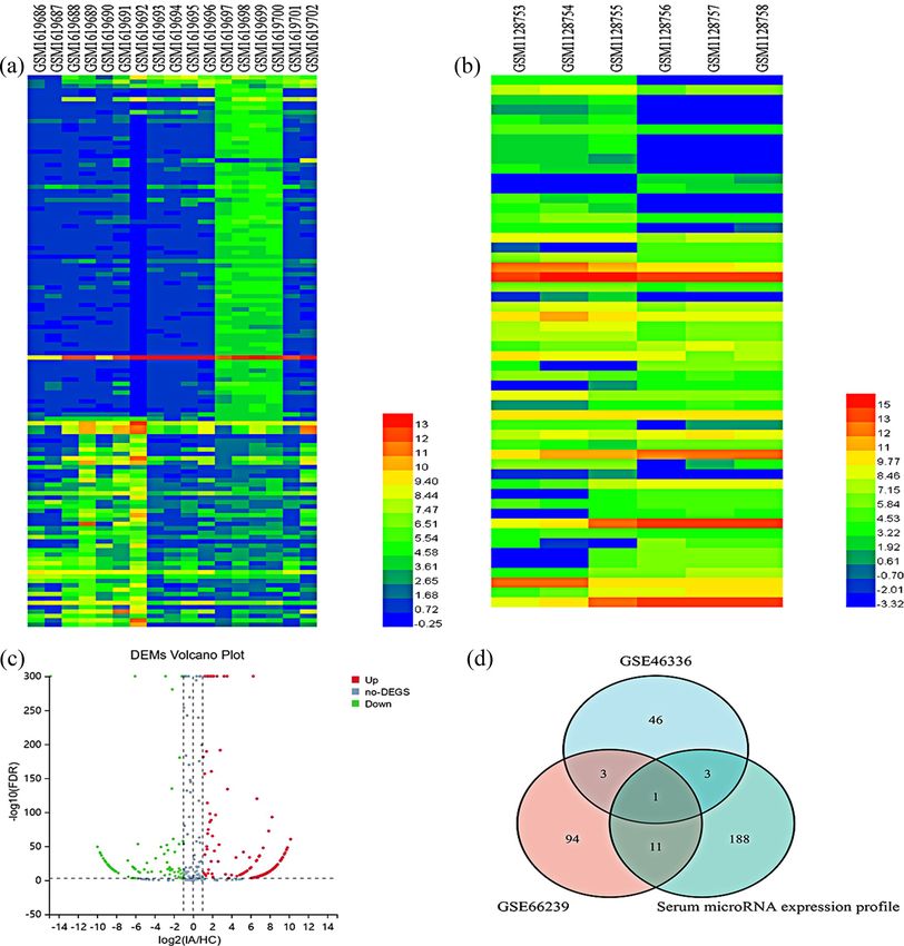
A total of 109 differentially expressed miRNAs were screened
methods were used as the final key genes, namely, TP53,
and identified from the GSE66239 dataset, among which
GSK3B, XPO1, UBE2I, and DHX15 (Table 2 and Figure 2d).
35 differentially expressed miRNAs were upregulated and
74 differentially expressed miRNAs were downregulated
(Figure 1a); 53 differentially expressed miRNAs were screened
and identified from the GSE46336, among which 31 differen- 3.4 Serum expression levels of miR-1246 in
tially expressed miRNAs were upregulated and 22 differen- intracranial aneurysm group and healthy
tially expressed miRNAs were downregulated (Figure 1b). control group
The miRNAs whose expression showed a consistent status
in both datasets include hsa-miR-1246, hsa-miR-127-3p, The expression levels of miRNA-1246 in serum of healthy
hsa-miR-409-3p, and hsa-miR-654-3p, among which hsa- control group and aneurysm group were determined using
miR-409-3p and hsa-miR-1246 showed a consistent high qRT-PCR. The miR-1246 expression level was 0.985 (0.434,
expression status with log2FC values greater than 2 in both 2.815) in the intracranial aneurysm group and 0.116 (0.058,
datasets. By gene sequencing as well as online software 0.209) in the control group, respectively. Compared with the
analysis, 203 differentially expressed miRNAs were screened control group, the miR-1246 level was increased in the
and identified, including 138 miRNAs with upregulated serum of intracranial aneurysm group, and the difference
expression levels and 65 miRNAs with downregulated was statistically significant (P < 0.001) (Figure 3a). The AUC
expression levels (Figure 1c). Based on the above results, value of serum miR-1246 for the diagnosis of intracranial
it was found that miRNA-1246 showed upregulation of aneurysm was 0.909 (Figure 3b), and the sensitivity of miR-
expression levels in both tissues and serum (Figure 1d), 1246 for the diagnosis of intracranial aneurysm was 79.55%,
bioinformatics analysis, and study were performed. and the specificity was 89.66%, both with statistically sig-
nificant differences (P < 0.001).
3.3 Bioinformatic analysis of miRNA-1246
A total of 319 target genes regulated by miRNA-1246 were 4 Discussion
predicted by miRNet database (https://www.mirnet.ca/).
By performing transcription factor analysis on the target Currently, many studies have found that miR-1246 plays
genes of differentially expressed miRNAs, the six tran- an important role in inflammation-related diseases. miR-
scription factors with the largest differences were found, 1246 provides a new avenue for the diagnosis and176 Haijie Jiang et al. Figure 1: Differentially expressed miRNAs from the screening. (a) Heat map of GSE66239 differentially expressed miRNAs; (b) heat map of GSE46336 differentially expressed miRNAs; (c) volcano plot of differentially expressed miRNAs; and (d) Venn diagram of differentially expressed miRNAs in serum and tissue. treatment of intracranial aneurysms. Alexander described inflammatory response. Bai et al. and colleagues estab- miR-1246 as a “key enhancer” of pro-inflammatory responses lished a rat disease model, stimulated human umbilical in mesenchymal stem cells (MSC) or stromal cells [3]. It was vein endothelial cells (HUVEC) using VEGF in order to found that miR-1246 is highly expressed in MSCs or stromal induce corneal neovascular cells in vitro, and performed cells and that miR-1246 regulates the inflammatory response RNA immunoprecipitation (RIP) [4]. These experiments by directly targeting PRKAR1A and PPP2CB, subunits of PKA revealed that miR-1246 in a rat model of corneal neovas- and PP2A, and mediates the secretion of CCL2 and CCL5 cularization was suppressed by lncRNA MIAT expression, via activating NF-κB pathway, by which involves the and it was capable of regulating cell proliferation and
Diagnosis of intracranial aneurysms 177
Figure 2: Bioinformatic analysis of miRNA-1246 target genes. (a) GO analysis; (b) KEGG analysis; (c) transcription factor enrichment
analysis; and (d) PPI analysis.
migration of HUVEC. It has even been found that miR-1246 MCP-1 expression, and restoring collagen biosynthesis [7].
is capable of acting on NF-κB signaling pathway and regu- Lai et al. found that in a rat aneurysm model, injection
lating M2 macrophage polarization through NF-κB pathway of the rats with APC-siRNA further activated the NF-κB
[5], which confirms Hasan’s study [6]. Hasan and colleagues signaling pathway and upregulated expression of MCP-1,
found that after intracranial aneurysm rupture, there is TNF-α, IL-1, IL-6 MMP-2, and MMP-9, as well as enhanced
a change in the ratio of macrophage type M1 to type M2 the levels of p65 phosphorylation [8]. The above findings
[6]. NF-κB enhances the high expression of pro-infla- fully demonstrate that NF-κB pathway is involved in
mmatory molecules such as vascular cell adhesion the development of intracranial aneurysms via regulating
molecule-1 (VCAM-1), monocyte chemoattractant protien-1 inflammatory response. Furthermore, miR-1246 may be
(MCP-1), inducible NO synthase, and matrix metalloprotei-
nases (MMPs) by aneurysmal endothelial cells and macro-
phages at the transcriptional and translational levels. These Table 2: Top five key genes identified by different calculation
inflammatory mediators promote inflammatory cells, espe- methods
cially macrophages, to infiltrate the vessel wall and further
degrade vascular structural proteins. Further studies found Method MCC Degree DMNC MNC EPC
that not only shrinkage of the aneurysm but also an increase Key genes TP53 TP53 KLC1 TP53 TP53
in the thickness of the aneurysm wall was achieved by DHX15 XPO1 TRIP11 XPO1 XPO1
blocking NF-κB and MCP-1 activities at the first month after KHDRBS1 GSK3B KIF15 UBE2I PPP2CA
GSK3B UBE2I MED14 GSK3B UBE2I
the formation of the experimental aneurysm. This may be
HNRNPU DHX15 CAPZA1 PPP2CA GSK3B
achieved by reducing macrophage infiltration, decreasing178 Haijie Jiang et al.
Table 3: KEGG target gene enrichment analysis
Pathway Genes Fold enrichment
Herpes simplex virus 1 infection ZNF320, ZNF460, ZNF283, ZNF480, ZNF160, PTPN11, ZNF12, PIK3CB, 2.52
TAPBP, ZNF717, ZNF627, ZNF85, ZNF548, BCL2, BAX, ZNF557, ZNF841,
ZNF763, TP53, ZNF267
Pathways of neurodegeneration – multiple GSK3B, XBP1, DCTN4, AXIN2, KLC1, SDHB, PPP3CA, CASP7, PSMC6, 1.97
diseases BCL2, DVL3, BAX, SLC25A5, CALM2, ACTR10
Pathways in cancer NOTCH2, GSK3B, PIK3CB, AXIN2, RASGRP3, CASP7, CDK6, CCNE1, 1.76
MSH3, PIM1, BCL2, DVL3, BAX, CALM2, TP53
Salmonella infection CYFIP1, RAB5B, DCTN4, PIK3CB, KLC1, CASP7, ARPC2, BCL2, BAX, TAB3, 3.26
ACBD3, ACTR10, RAB7A
Human papillomavirus infection NOTCH2, ATP6V1A, PPP2CA, GSK3B, CDK6, PPP2R1B, CCNE1, DVL3, BAX, 2.26
AXIN2, PIK3CB, TP53
Alzheimer’s disease GSK3B, PPP3CA, XBP1, CASP7, PSMC6, DVL3, AXIN2, PIK3CB, SLC25A5, 1.95
KLC1, CALM2, SDHB
Lipid and atherosclerosis GSK3B, PPP3CA, XBP1, CASP7, BCL2, NFATC3, BAX, PIK3CB, SOD2, 3.19
CALM2, TP53
MicroRNAs in cancer NOTCH2, MARCKS, UBE2I, CDK6, CCNE1, RDX, CL2, PIM1, PIK3CB, 2.21
TP53, TP63
Human T-cell leukemia virus 1 infection KAT2B, PPP3CA, XPO1, CCNE1, CANX, NFATC3, BAX, PIK3CB, 2.81
SLC25A5, TP53
Thyroid hormone signaling pathway NOTCH2, KAT2B, GSK3B, MED1, MED14, MED13, PIK3CB, MED4, TP53 4.64
also involved in the formation and rupture of intracranial analyzed two genotypes of TP53 and concluded that dif-
aneurysms as a result of targeting and regulating the ferent genotypes of TP53 are capable of playing different
NF-κB pathway and triggering an inflammatory response. roles in intracranial aneurysms. miR-34b/crs4938723 TT
Two important target genes, TP53 and GSK3B, were type has a higher incidence of intracranial aneurysms.
identified in this study. TP53 has been shown to be involved TP53 is capable of mediating the inflammatory response
in the development of several cancers as a tumor sup- involved in the formation of intracranial aneurysms [9].
pressor, and TP53 was also found to have an important GSK3B is not only one of the major regulators of the inflam-
role in inflammation and inflammatory diseases. Li et al. matory response, but also a serine/threonine protein kinase
Table 4: Gene ontology analysis
Term Count P value Group
−6
Regulation of transcription from RNA polymerase II promoter 52 2.41 × 10 BP
Positive regulation of transcription, DNA-templated 27 1.17 × 10⁻⁵ BP
Positive regulation of transcription from RNA polymerase II 37 7.31 × 10⁻⁵ BP
Nucleus 159 4.66 × 10⁻¹⁷ CC
Nucleoplasm 120 5.59 × 10⁻¹⁶ CC
Cytosol 130 4.58 × 10⁻⁹ CC
Cytoplasm 124 1.59 × 10⁻⁷ CC
Membrane 69 1.30 × 10⁻⁶ CC
Cytoplasmic stress granule 8 3.17 × 10⁻⁴ CC
Mediator complex 6 4.25 × 10⁻⁴ CC
Centrosome 20 6.50 × 10⁻⁴ CC
PML body 8 1.19 × 10⁻³ CC
Kinetochore 9 1.26 × 10⁻³ CC
Protein binding 245 1.50 × 10⁻⁹ MF
RNA binding 50 3.70 × 10⁻⁷ MF
DNA binding 44 1.76 × 10⁻⁶ MF
Chromatin binding 21 4.76 × 10⁻⁵ MF
Metal ion binding 67 8.06 × 10⁻⁵ MF
RNA polymerase II transcription factor activity, sequence-s 37 5.56 × 10⁻⁴ MFDiagnosis of intracranial aneurysms 179
(a) hsa-miR-1246 (b) hsa-miR-1246
potential diagnostic marker and therapeutic target for
intracranial aneurysms.
25 *** 1.0
Relative expression
20 0.8 Funding information: The present work was supported by
Sensitivity
15 0.6 Shandong Province Natural Science Gene Surface Project
10 0.4 (ZR2020MH379) and Weifang City Science and Technology
AUC=0.909
Development Plan Project (2020YX056).
5 0.2
0 0.0
IA HC 0.0 0.2 0.4 0.6 0.8 1.0
Conflict of interest: Authors state no conflict of interest.
1 - Specificity
Data availability statement: All data generated or ana-
Figure 3: Expression level and ROC diagnostic curve of serum lyzed during this study are included in this published
miRNA-1246. (a) Expression level of serum miRNA-1246, (b) ROC article and its supplementary information files.
diagnostic curve of serum miRNA-1246
signaling molecule that is widely expressed in many cell References
types [10]. The GSK3B/β-linked protein signaling pathway
has been reported to promote the proliferation and migration [1] Huang L, Li X, Chen Z, Liu Y, Zhang X. Identification of
of vascular smooth muscle cells. Higher phosphorylated inflammation associated circulating long noncoding RNAs and
GSK3B in atheroma leads to β-linked protein activation, genes in intracranial aneurysm rupture-induced subarachnoid
and β-linked protein activation is involved in the resting hemorrhage. Mol Med Rep. 2020;22(6):4541–50.
transformation of vascular smooth muscle cells. This pro- doi: 10.3892/mmr.2020.1154.
[2] Hu L, Si L, Dai X. Exosomal miR-409-3p secreted from activated
cess has also been shown to be involved in arterial aging
mast cells promotes microglial migration, activation and
[11]. Analysis of transcription factors revealed that YY1 neuroinflammation by targeting Nr4a2 to activate the NF-κB
may be associated with the development of intracranial pathway. J Neuroinflammation. 2021;18(1):68.
aneurysms. YY1 is a member of the Polycomb histone [3] Bott A, Erdem N, Lerrer S, Hotz-Wagenblatt A, Breunig C,
family, which acts as a typical multifunctional zinc finger Abnaof K, et al. miRNA-1246 induces pro-inflammatory
responses in mesenchymal stem/stromal cells by
transcription factor that can activate or repress biological
regulating PKA and PP2A. Oncotarget.
processes such as gene transcription [12]. Zhou et al. found 2017;8(27):43897–914.
that YY1 plays a role in the endothelial inflammatory pro- [4] Bai Y, Wang W, Sun G, Zhang M, Dong J. Curcumin inhibits
cess by forming a functional complex through the NF-κB angiogenesis by up-regulation of microRNA-1275 and
pathway and exerting transcriptional regulation on the microRNA-1246: a promising therapy for treatment of corneal
inflammatory genes IL6 and IL8. The miRNAs, target neovascularization. Cell Prolif. 2016;49(6):751–62.
[5] Qian M, Wang S, Guo X, Wang J, Zhang Z, Qiu W, et al.
genes, and their transcription factors in the above study
Hypoxic glioma-derived exosomes deliver microRNA-1246
are comparable with our predicted target genes, sug- to induce M2 macrophage polarization by targeting TERF2IP
gesting that these target genes may be the key genes in via the STAT3 and NF-κB pathways. Oncogene.
the regulatory pathways associated with the development 2020;39(2):428–42.
and inflammation of the intracranial aneurysms [13]. [6] Hasan D, Hashimoto T, Kung D, Macdonald Rl, Winn HR,
Heistad D. Upregulation of cyclooxygenase-2 (COX-2) and
microsomal prostaglandin E2 synthase-1 (mPGES-1) in wall of
ruptured human cerebral aneurysms: preliminary results.
5 Conclusion [7]
Stroke. 2012;43:1964–7.
Shimizu K, Kushamae M, Mizutani T, Aoki T. Intracranial
aneurysm as a macrophage-mediated inflammatory disease.
In conclusion, a number of studies, including ours, sug- Neurol Med Chir (Tokyo). 2019;59(4):126–32.
gest that inflammation is one of the most important fac- [8] Lai Xl, Deng Zf, Zhu Xg, Chen Zh. Apc gene suppresses intra-
tors in the development and rupture of aneurysm. Our cranial aneurysm formation and rupture through inhibiting the
study has shown that miR-1246 is highly expressed in NF-κB signaling pathway mediated inflammatory response.
Biosci Rep. 2019;39(3)1–22.
patients with intracranial aneurysms and that our bioin-
[9] Li L, Sima X, Bai P, Zhang L, Sun H, Liang W, et al. Interactions
formatic analysis indicates an important role of miR-1246 of miR-34b/c and TP53 polymorphisms on the risk of intra-
in regulating the inflammatory pathways, lipid and ather- cranial aneurysm. Clin Develop Immunol.
osclerotic signaling pathways. miR-1246 may serve as a 2014;2012(1):567586.180 Haijie Jiang et al.
[10] Liu WL, Chiang FT, Kao JT. GSK3 modulation in acute lung injury, [12] Zhang C, Zhu X, Hua Y, Zhao Q, Wang K, Zhen L, et al. YY1
myocarditis and polycystic kidney disease-related aneurysm. mediates TGF-β1-induced EMT and pro-fibrogenesis in alveolar
Biochim Biophys Acta Mol Cell Res. 2020;1867(11):118798. epithelial cells. Respir Res. 2019;20(1):249.
[11] Meng J, Liu HL, Ma D. Upregulation of aurora kinase A pro- [13] Zhou Y, Wang M, Zhang J, Xu P, Wang H. MicroRNA-29a-3p
motes vascular smooth muscle cell proliferation and migration regulates abdominal aortic aneurysm development and pro-
by activating the GSK-3β/β-catenin pathway in aortic-dis- gression via direct interaction with PTEN. J Cell Physiol.
secting aneurysms. Life Sci. 2020;262:118491. 2020;235(12):9414–942.You can also read