The Role of Lipids in CRAC Channel Function - MDPI

Page created by Dean Mann
 
CONTINUE READING
The Role of Lipids in CRAC Channel Function - MDPI
biomolecules

Review
The Role of Lipids in CRAC Channel Function
Lena Maltan            , Ana-Marija Andova and Isabella Derler *

                                          Institute of Biophysics, JKU Life Science Center, Johannes Kepler University Linz, AT-4020 Linz, Austria;
                                          lena.maltan@jku.at (L.M.); amandova7@gmail.com (A.-M.A.)
                                          * Correspondence: isabella.derler@jku.at

                                          Abstract: The composition and dynamics of the lipid membrane define the physical properties of
                                          the bilayer and consequently affect the function of the incorporated membrane transporters, which
                                          also applies for the prominent Ca2+ release-activated Ca2+ ion channel (CRAC). This channel is
                                          activated by receptor-induced Ca2+ store depletion of the endoplasmic reticulum (ER) and consists
                                          of two transmembrane proteins, STIM1 and Orai1. STIM1 is anchored in the ER membrane and
                                          senses changes in the ER luminal Ca2+ concentration. Orai1 is the Ca2+ -selective, pore-forming
                                          CRAC channel component located in the plasma membrane (PM). Ca2+ store-depletion of the ER
                                          triggers activation of STIM1 proteins, which subsequently leads to a conformational change and
                                          oligomerization of STIM1 and its coupling to as well as activation of Orai1 channels at the ER-PM
                                          contact sites. Although STIM1 and Orai1 are sufficient for CRAC channel activation, their efficient
                                          activation and deactivation is fine-tuned by a variety of lipids and lipid- and/or ER-PM junction-
                                          dependent accessory proteins. The underlying mechanisms for lipid-mediated CRAC channel
                                          modulation as well as the still open questions, are presented in this review.

                                          Keywords: CRAC channel; STIM1; Orai1; protein-lipid interactions; modulatory proteins; ER-PM
                                          junctions; lipids
         
         
Citation: Maltan, L.; Andova, A.-M.;
Derler, I. The Role of Lipids in CRAC     1. Introduction
Channel Function. Biomolecules 2022,
                                                Biomembranes play an important role in maintaining healthy cell function. They
12, 352. https://doi.org/10.3390/
                                          separate the interior from the exterior of the cell and form various compartments within
biom12030352
                                          the cytosol that perform a variety of cellular functions. Cell membranes are semipermeable,
Academic Editors: Groschner Klaus         act as a diffusion barrier, and maintain a concentration gradient between different cell
and Romanin Christoph                     compartments. They are further involved in the regulated exchange of substances and
Received: 27 January 2022
                                          control various cellular and intracellular processes like cell movement and subcellular
Accepted: 20 February 2022
                                          signal transmission. The cell membrane is composed of two layers, namely the inner and
Published: 23 February 2022
                                          outer leaflets which are made up of a variety of amphiphilic lipids. The hydrophobic
                                          tails of the lipids in both leaflets face each other while the hydrophilic headgroups meet
Publisher’s Note: MDPI stays neutral
                                          the aqueous surroundings [1–5]. The lipid bilayer has an overall thickness of ~4 nm, is
with regard to jurisdictional claims in
                                          fluid, and contains proteins arranged in a mosaic pattern described by the fluid mosaic
published maps and institutional affil-
                                          model [6–8].
iations.
                                                Membrane lipids are categorized into glycerophospholipids, sphingolipids, and
                                          sterols [4,9]. The lipids of the first two groups are bilayer forming lipids. Although
                                          their overall structure, consisting of a hydrophilic headgroup linked to a hydrophobic tail,
Copyright: © 2022 by the authors.
                                          is similar, they may differ in fatty acids (chain length, presence of saturated state, and
Licensee MDPI, Basel, Switzerland.        hydroxylation) and/or headgroup substituents, which leads to the existence of more than
This article is an open access article    1000 different lipid species [5,9]. Sterols are structurally different from the other two groups.
distributed under the terms and           They are composed of four aromatic rings, derived from sterane, and a short aliphatic chain,
conditions of the Creative Commons        which makes them very rigid. It is noteworthy that sterols occur in mammals typically as
Attribution (CC BY) license (https://     cholesterol [4,10–12].
creativecommons.org/licenses/by/                Given the diversity of lipids and the composition of lipid bilayer, it is hardly surprising
4.0/).                                    that they influence not only the membrane properties, but also the structure and function

Biomolecules 2022, 12, 352. https://doi.org/10.3390/biom12030352                                          https://www.mdpi.com/journal/biomolecules
The Role of Lipids in CRAC Channel Function - MDPI
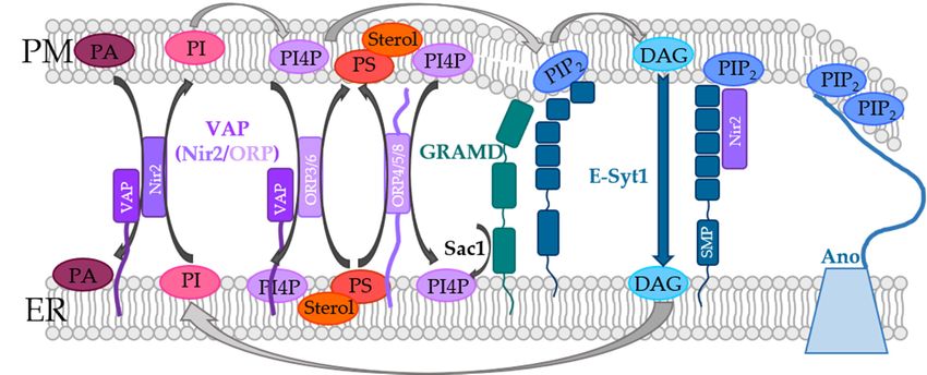
Biomolecules 2022, 12, 352                                                                                            2 of 37

                             of membrane proteins. Direct interactions between proteins and lipids or nonspecific
                             changes in the physicochemical properties of the membrane, including curvature, fluidity,
                             and thickness, can influence protein dynamics. In the case of lipid–protein interplay, a
                             distinction can be made between annular and non-annular binding sites. Annular binding
                             sites show direct interactions between lipids and membrane proteins, such as lipids located
                             in the first transmembrane ring surrounding the protein. Non-annular lipids are located, for
                             example, in crevices or at the interface of protein subunits [13–16]. Additionally, evidence
                             is accumulating that membrane proteins are localized in micro- or nanodomains enriched
                             in sphingolipids and cholesterol. These lipids facilitate the association of many signaling
                             molecules, serve as a sorting platform for signal transduction proteins, and as a source of
                             efficient signal transduction [17–19].
                                   In regard to ion channels, it has been documented for decades that they can sense
                             changes in the compositions of the lipid bilayer [20]. In particular, the so-called mechanosen-
                             sitive channels are sensitive to changes in membrane pressure and the resulting change
                             in membrane curvature and thickness. Examples of these types of ion channels include
                             the bacterial large-conductance mechanosensitive channel (MscL) [21], piezo channels [22],
                             and the potassium-selective TRAAK channel [23]. More recent reports concentrated on
                             the role of signaling lipids, such as phosphatidylinositol-4,5-bisphosphate (PI(4,5)P2, short:
                             PIP2 ) or diacylglycerol (DAG) in the direct modulation of ion channels. A prime example
                             of ion channel gating by direct lipid binding is the inward rectifying K+ (Kir) channel. The
                             direct binding of PIP2 to the transmembrane (TM) region 1 and 2 of all four subunits can
                             trigger a conformational change and subsequently open the channel [24]. Moreover, some
                             voltage-gated K+ (Kv) ion channels can function in a PIP2 dependent manner. Furthermore,
                             a variety of ion channels (e.g., inwardly-rectifying K+ channels, voltage-gated K+ chan-
                             nels, Ca2+ sensitive K+ channels, voltage-gated Na+ channels, N-type voltage-gated Ca2+
                             channels and volume-regulated anion channels) are sensitive to changes in the cholesterol
                             content of membranes [25–28]. Further examples of lipid-regulated ion channels include
                             several members of the transient receptor potential (TRP) channels that can interact with
                             different lipids at different channel positions (e.g., TRPV1/5/6 and TRPM2/8 interact with
                             PIP2 [29–34] whereas TRPC3/6 are activated by DAG binding [35–37]). Lipids also affect
                             channel activity by modulating their trafficking towards and insertion into the plasma
                             membrane [38,39].
                                   Pathological changes in lipid levels can either enhance or impair channel function,
                             which may also play a crucial role in disease progression. In patients with hypercholes-
                             terolemia, the development of atherosclerosis or impaired vasodilatation has been as-
                             sociated with dysfunction or suppression of Kir or Kv channels, probably due to their
                             modulation by cholesterol [25,26,40]. Altered availability of PIP2 or mutations that alter
                             the function of PIP2 -dependent channels have also been reported to adversely affect blood
                             flow control and vascular function [41]. However, the effects of lipids on ion channels
                             are highly variable and there are few examples of pathologies in which lipids and ion
                             channels are co-regulated [25,26,40,41]. Hence, a detailed mechanistic understanding of the
                             co-regulation of lipids and ion channels is required. This review focuses on the so-called
                             Ca2+ release-activated Ca2+ (CRAC) ion channel, a prominent Ca2+ entry pathway into
                             the cell. It consists of two components, one anchored in the endoplasmic reticulum (ER)
                             membrane and the second in the plasma membrane (PM). Before describing these two key
                             proteins of the CRAC channel in detail, the next section discusses the lipid composition
                             and dynamics of the ER and the PM in the following.

                             2. Lipid Compositions and Dynamics in the Membranes of the ER and the Cell
                                  Interestingly, the lipid composition of the ER membrane and the PM differ quite
                             substantially. Even though the ER is the main place in the cell for lipid synthesis, it is
                             mainly composed of phospholipids and a low amount of cholesterol. Its membrane is
                             loosely packed, allowing transport of synthesized lipids and proteins. In contrast, in the
                             PM, sphingolipids and cholesterol are abundant, which are responsible for its increased
The Role of Lipids in CRAC Channel Function - MDPI
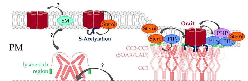
Biomolecules 2022, 12, x FOR PEER REVIEW                                                                                                     3 of 39

Biomolecules 2022, 12, 352
                                 packed, allowing transport of synthesized lipids and proteins. In contrast, in the 3PM,                        of 37

                                 sphingolipids and cholesterol are abundant, which are responsible for its increased stiff-
                                 ness and mechanical resistance [9,42]. Moreover, the lipids in the two leaflets in the ER
                                  stiffness and
                                 membrane           mechanicalcells
                                               of eukaryotic       resistance     [9,42]. Moreover,
                                                                        are symmetrically                 theas
                                                                                                distributed    lipids   in the
                                                                                                                  opposed        twoPM,
                                                                                                                             to the    leaflets
                                                                                                                                            where in
                                  the ERall
                                 almost     membrane
                                              major lipids of exhibit
                                                              eukaryotic      cells are symmetrically
                                                                        an asymmetric                        distributed
                                                                                            distribution [9,43,44].    Whileas the
                                                                                                                                opposed      to the
                                                                                                                                    outer leaflet
                                 isPM,  where almost
                                    primarily    composed  all major    lipids exhibit an asymmetric
                                                               of phosphatidylcholine                         distribution [9,43,44].
                                                                                                (PC) and sphingomyelin          (SM), the While
                                                                                                                                              cyto-
                                  the  outer  leaflet is  primarily    composed       of phosphatidylcholine        (PC)
                                 solic leaflet contains particularly phosphatidylethanolamine (PE) and the charged lipids and    sphingomyelin
                                  (SM), the cytosolic leaflet
                                 phosphatidylinositol              contains
                                                             (PI) and           particularly phosphatidylethanolamine
                                                                         phosphatidylserine        (PS). This asymmetry has (PE)    major  and   the
                                                                                                                                             func-
                                  charged    lipids  phosphatidylinositol         (PI)  and   phosphatidylserine       (PS).
                                 tional implications, e.g., on vesicle formation or the development of local transmembrane     This  asymmetry
                                  has major Furthermore,
                                 potentials.   functional implications,         e.g., on vesicle
                                                                 active transporters               formation
                                                                                          particularly          or the
                                                                                                          maintain       development
                                                                                                                      a higher    contentofoflocal
                                                                                                                                                an-
                                  transmembrane
                                 ionic  lipid species potentials.   Furthermore,
                                                         in the inner                  active
                                                                          leaflet, which    is transporters   particularly
                                                                                               of physiological              maintain
                                                                                                                    importance.           a higher
                                                                                                                                    In addition,
                                  content ofstudies
                                 lipidomic     anionic revealed
                                                          lipid species     in the inner
                                                                      asymmetric           leaflet, which
                                                                                       distribution         is ofchain
                                                                                                       of acyl     physiological
                                                                                                                         structure importance.
                                                                                                                                      properties
                                  In  addition,
                                 [43,45,46].      lipidomic    studies     revealed    asymmetric      distribution   of  acyl chain structure
                                  properties   [43,45,46].
                                       Cholesterol    is an important hydrophobic lipid in the hydrophobic layer of the mem-
                                 brane [47], whichiscontrols
                                        Cholesterol        an important      hydrophobicand
                                                                    lipid organization        lipidconformational
                                                                                                     in the hydrophobic      layerofoffatty
                                                                                                                       ordering          the mem-
                                                                                                                                               acid
                                  brane   [47],  which    controls   lipid   organization     and   conformational      ordering
                                 chains [48]. Cholesterol has been proposed to be dynamically distributed in the PM, which          of  fatty   acid
                                  chainsdepends
                                 likely    [48]. Cholesterol    has been proposed
                                                    on the composition,                  to be dynamically
                                                                               concentration                   distributed
                                                                                                 of phospholipids,             in thein
                                                                                                                       and forces     PM,thewhich
                                                                                                                                              lipid
                                  likely  depends    on  the  composition,      concentration     of phospholipids,
                                 bilayer. However, in this respect more detailed studies are still required [49]. All PMand   forces  in  thephos-
                                                                                                                                               lipid
                                  bilayer. However, in this respect more detailed studies are still required [49]. All PM
                                 pholipids and cholesterol can be synthesized in the ER, which are then transported to the
                                  phospholipids and cholesterol can be synthesized in the ER, which are then transported to
                                 membrane, as reviewed by van Meer et al. [9].
                                  the membrane, as reviewed by van Meer et al. [9].
                                       In the PM, and especially at the contact sites between the ER and the PM, lipids are
                                        In the PM, and especially at the contact sites between the ER and the PM, lipids are
                                 subject to dynamics, which are controlled by means of the so-called phosphoinositide cy-
                                  subject to dynamics, which are controlled by means of the so-called phosphoinositide
                                 cle (Figure 1). Although phosphoinositides (PIs) represent only a small fraction of the total
                                  cycle (Figure 1). Although phosphoinositides (PIs) represent only a small fraction of
                                 lipids in the PM, they are essential players in cellular processes such as actin dynamics,
                                  the total lipids in the PM, they are essential players in cellular processes such as actin
                                 transmembrane protein regulation, and signal transduction [50–57]. PIs belong to the
                                  dynamics, transmembrane protein regulation, and signal transduction [50–57]. PIs belong
                                 group of glycerophospholipids that contain inositol and carry different numbers of phos-
                                  to the group of glycerophospholipids that contain inositol and carry different numbers
                                 phate groups at their headgroups [58,59]. Specific kinases and phosphatases can phos-
                                  of phosphate groups at their headgroups [58,59]. Specific kinases and phosphatases can
                                 phorylate the inositol ring at positions 3, 4, and/or 5, generating different subspecies that
                                  phosphorylate the inositol ring at positions 3, 4, and/or 5, generating different subspecies
                                 are
                                  thatphysiologically     relevant
                                        are physiologically           [58–62].
                                                                relevant     [58–62].

                                 Figure
                                  Figure1.1.Lipid
                                              Lipidtransport
                                                    transportandandphosphoinositide
                                                                    phosphoinositide(PI)(PI)cycle
                                                                                             cycleatatthe
                                                                                                        theER-PM
                                                                                                            ER-PMcontact
                                                                                                                     contactsites.
                                                                                                                              sites.PI
                                                                                                                                     PIisisconsecu-
                                                                                                                                            consecu-
                                 tively
                                  tively transformed
                                          transformed at   the PM
                                                       at the  PM into
                                                                   into PI4P
                                                                        PI4PbybyPIP4K
                                                                                 PIP4KandandPIP
                                                                                              PIP2
                                                                                                     by
                                                                                                   2by   PIP5K
                                                                                                       PIP5K   (PIP
                                                                                                              (PIP 2
                                                                                                                       can
                                                                                                                     2can  be
                                                                                                                          be   converted
                                                                                                                             converted     toto PI4P
                                                                                                                                              PI4P  or
                                 or  PI  by  phosphatases).   PM-receptor    stimulation  can  trigger   PLC  mediated     hydrolysis
                                  PI by phosphatases). PM-receptor stimulation can trigger PLC mediated hydrolysis of PIP2 to form       of  PIP 2 to

                                 form DAG and IP3. While DAG can activate PKC or some ion channels, IP3 activates the ER located
                                  DAG and IP3 . While DAG can activate PKC or some ion channels, IP3 activates the ER located IP3 R.
                                 IP3R. Non-vesicular transport mechanisms (see Section 5) can transport DAG to the ER membrane,
                                  Non-vesicular transport mechanisms (see Section 5) can transport DAG to the ER membrane, where
                                 where it is converted to PI. PITPs transport PI back to the plasma membrane. Furthermore, sterols,
                                  it isand
                                 PS,    converted  totransported
                                            PI4P are  PI. PITPs transport
                                                                   betweenPItheback
                                                                                 twotomembranes
                                                                                      the plasma membrane.        Furthermore,
                                                                                                      via non-vesicular   transportsterols,   PS, and
                                                                                                                                      (see Section
                                  PI4P
                                 5).     are transported   between  the  two  membranes    via non-vesicular    transport   (see Section    5).

                                       In the PM, PI species are phosphorylated by phosphatidylinositol-4-kinase (PI4K)
                                  to generate phosphatidylinositol-4-phosphate (PI4P). Consequently, PI(4,5)P2 or short
                                  PIP2 is produced by phosphorylation of phosphatidylinositol-4-phosphate-5-kinase
The Role of Lipids in CRAC Channel Function - MDPI
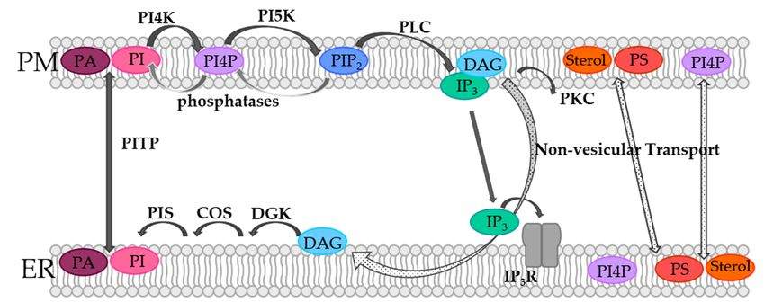
Biomolecules 2022, 12, 352                                                                                           4 of 37

                             (PIP5K) [60,61,63,64]. It is worth noting that specific counterpart phosphatases, as for in-
                             stance the inositol polyphosphate 5-phosphatases (IPP-5Ptases) or the PIP2 -4 phosphatases
                             (PIP2 -4Ptases), dephosphorylate PIP2 to PI4P and PI5P, respectively [65]. PIP2 can be further
                             phosphorylated to PIP3 or reconverted by corresponding other kinases or phosphatases,
                             respectively. Within the PI cycle, PIP2 is then hydrolyzed by phospholipase C (PLC) to
                             diacylglycerol (DAG) and inositol-1,4,5-trisphosphate (IP3 ) (Figure 1). Following this, DAG
                             activates protein kinase C (PKC), while IP3 binds its corresponding receptor in the ER
                             membrane and activates Ca2+ release (Figure 1). When the ER membrane and the PM
                             are in close contact, DAG is transported via non-vesicular transport from the PM to the
                             ER membrane. Subsequently, DAG is sequentially converted by the diacylglycerol kinase
                             (DGK) to phosphatic acid (PA), by CDP-diacylglycerol synthase (CDS) to CDP-DG and
                             by the PI synthase (PIS) to PI. Finally, PI is transferred from the ER membrane to the PM
                             via non-vesicular phosphatidylinositol transfer protein (PITP), which closes the circle and
                             makes further rounds in the PI cycle possible (Figure 1). Furthermore, PA can be converted
                             in the PM from DAG, which is then transported by PITPs to the ER membrane, where
                             it is converted back to PI and transported back to the PM [60,61,63,64]. In this way, the
                             spatiotemporal modulation of certain phosphoinositide lipid species at the ER-PM junction
                             is critical for the control of Ca2+ signaling [13], as specifically outlined in detail for the
                             prominent Ca2+ release-activated Ca2+ (CRAC) ion channel in the following sections.

                             3. CRAC Channels
                                   As mentioned earlier, the CRAC ion channel consists of Orai, a hexameric plasma
                             membrane protein that forms the pore unit [66,67], and the stromal interaction molecule
                             (STIM), which protrudes from the ER membrane. While the Orai family consists of Orai1,
                             Orai2, and Orai3, the STIM proteins include two members STIM1 and STIM2. Orai iso-
                             forms can form heteromeric channels and both STIM1 and STIM2 can interact with Orai1.
                             Therefore, physiological CRAC channels are highly diverse [68–72]. In this review we focus
                             on STIM1 and Orai1, as lipid-mediated regulation has so far been reported exclusively for
                             those two proteins. However, given the structural homology of Orai and STIM isoforms, it
                             cannot be precluded that they interact with lipids.
                                   Upon store-depletion STIM1 can interact cytosolically with Orai1 after its activa-
                             tion [73,74] (Figure 2). Their interplay is therefore only given when the two membranes are
                             in close proximity with each other.
                                   STIM1 exists in the form of a dimer in the quiescent state and senses the luminal Ca2+
                             concentration in the ER via its N-terminal EF-hand [75–77]. This Ca2+ sensing region is
                             connected via a TM domain with the C-terminus, which is involved in the control of the
                             inactive state in the resting state and in the oligomerization of STIM1 and coupling to
                             Orai1 in the activated state. The C-terminus exhibits a folded configuration in the resting
                             STIM1 state in which CC2 and CC3 resemble the capital letter “R” and form an antiparallel
                             V-shape in the dimeric state [78]. In this quiescent state, this V-shape is upside down
                             and faces the ER membrane. A drop in the Ca2+ concentration leads to loss of the bound
                             Ca2+ and subsequently to a huge conformational change of STIM1 [79–82]. This change
                             starts with a slight unfolding of the ER luminal EF-hand and the sterile alpha motif (SAM)
                             domain [76,77]. Following this, the opening signal is transduced via the TM domain to
                             the three coiled-coil domains (CC1-3). In the activated state, CC1 is first zipped together,
                             causing the CC2/CC3-V-shape to flip upward facing now the plasma membrane [83,84].
                             Here, the STIM1 apex (aa 390–410) together with a region N-terminal to the apex (aa
                             370–380; [85]) is now able to interact with Orai1, with the Orai1 C-terminus acting as the
                             main binding site (Figure 2).
The Role of Lipids in CRAC Channel Function - MDPI
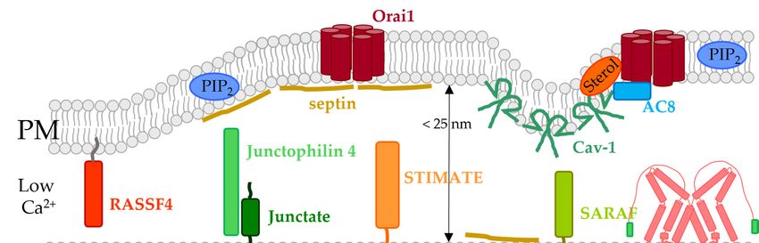
Biomolecules 2022, 12, x FOR PEER REVIEW                                                                                                                  5 of 39
Biomolecules 2022, 12, 352                                                                                                                                 5 of 37

                                             STIM1,Orai1,
                                  Figure2.2.STIM1,
                                 Figure              Orai1,and
                                                            andtheir
                                                                theirinterplay
                                                                      interplaywith
                                                                                withlipids.
                                                                                      lipids.InInthe
                                                                                                  theresting
                                                                                                      restingstate,
                                                                                                               state,STIM1,
                                                                                                                      STIM1,located
                                                                                                                                 locatedin
                                                                                                                                         inthe
                                                                                                                                            theER
                                                                                                                                                ER
                                  membrane,captures
                                 membrane,     capturesaaquiescent
                                                          quiescentand
                                                                     andfolded
                                                                          foldedstate,
                                                                                 state,while
                                                                                        whileOrai1,
                                                                                                Orai1,located
                                                                                                       locatedin inthe
                                                                                                                    thePM,
                                                                                                                         PM,possesses
                                                                                                                                possessesaaclosed
                                                                                                                                            closed
                                 pore.
                                  pore.After
                                        Afterstore-depletion
                                               store-depletionSTIM1
                                                               STIM1undergoes
                                                                        undergoesaaconformational
                                                                                      conformationalchange,
                                                                                                          change,oligomerizes,
                                                                                                                    oligomerizes,and  andcouples
                                                                                                                                           couples
                                 to
                                  toOrai1.
                                     Orai1.Although
                                             Althoughthe
                                                       theinteraction
                                                           interactionand
                                                                        andfunction
                                                                             functionofofthe
                                                                                          thetwo
                                                                                               twoproteins
                                                                                                    proteinsisissufficient
                                                                                                                  sufficientfor
                                                                                                                              forCRAC
                                                                                                                                   CRACchannel
                                                                                                                                          channel
                                 activation,
                                  activation,their
                                              theirmachinery
                                                    machineryisismodulated
                                                                   modulatedby  byaavariety
                                                                                      varietyof oflipids
                                                                                                   lipidsincluding
                                                                                                           includingPIP PIP2,2 ,PI4P,
                                                                                                                                 PI4P,cholesterol,
                                                                                                                                       cholesterol,
                                 sphingomyelin
                                  sphingomyelinand andpossibly
                                                       possiblyalso
                                                                 alsoER-phospholipids
                                                                      ER-phospholipidsas   as outlined
                                                                                              outlined inin detail
                                                                                                            detail in
                                                                                                                    in Section
                                                                                                                       Section 4. 4.

                                       STIM1
                                        Severalexists      in the
                                                     studies        form of adifferent
                                                                identified        dimer infragments
                                                                                                the quiescent  (OASFstate(aa
                                                                                                                           and233–474)
                                                                                                                                  senses the     luminal
                                                                                                                                             [86],    SOARCa(aa  2+

                                 concentration
                                  344–442) [87], in   CAD the(aaER342–448)
                                                                     via its N-terminal
                                                                                [88] or Ccb9EF-hand
                                                                                                  (aa 339–444) [75–77].    This
                                                                                                                    [89]) of  theCa    2+ sensing region
                                                                                                                                     CC domains        which are is
                                 connected
                                  sufficient to  viaactivate
                                                       a TM domain
                                                                Orai1. Thewith  NMRthe C-terminus,
                                                                                         structure of STIM1which is  andinvolved      in the control
                                                                                                                           Orai1 C-terminal               of the
                                                                                                                                                      fragments,
                                 inactive    statethe
                                  specifically        in STIM1-Orai1
                                                          the resting state        and in the
                                                                            association             oligomerization
                                                                                               pocket     (SOAP), resolved  of STIM1       andsites
                                                                                                                                     the key      coupling
                                                                                                                                                        requiredto
                                 Orai1   in  the   activated     state.  The    C-terminus        exhibits     a folded    configuration
                                  for the formation of the main STIM1/Orai1 binding site, which is in line with the findings                    in  the  resting
                                 STIM1     state inofwhich
                                  of a variety                  CC2 and
                                                        functional          CC3 [90–92].
                                                                       studies      resembleRecently,
                                                                                                   the capitalthe letter
                                                                                                                     STIM1“R” apex
                                                                                                                                and form was an   antiparallel
                                                                                                                                               also    shown to
                                 V-shape     in thein
                                  be important         dimeric     statecommunication
                                                          the intact      [78]. In this quiescent          state, In
                                                                                                 with Orai1.       this  V-shape is
                                                                                                                       particular,        upside F394
                                                                                                                                       position      downinand the
                                 faces
                                  STIM1 theapex
                                              ER is membrane.        A drop
                                                       a critical site   of theinprotein
                                                                                    the Ca[93],2+ concentration
                                                                                                     as mutationsleads  led totoversatile
                                                                                                                                   loss of the     bound
                                                                                                                                              defects        Ca2+
                                                                                                                                                         ranging
                                  fromsubsequently
                                 and     the maintenance            of the
                                                            to a huge        quiescent state
                                                                          conformational             (STIM1
                                                                                                   change     of F394A)     to inability
                                                                                                                  STIM1 [79–82].         Thistochange
                                                                                                                                                  oligomerize
                                                                                                                                                           starts
                                  (STIM1
                                 with        F394D/K)
                                       a slight     unfoldingto impaired
                                                                   of the ER  Orai1    binding
                                                                                  luminal           capability
                                                                                               EF-hand      and the(STIM1     F394H)
                                                                                                                        sterile   alpha[83].
                                                                                                                                           motifFurthermore,
                                                                                                                                                    (SAM) do-
                                  cysteine
                                 main         crosslinking
                                        [76,77].     Following   revealed
                                                                    this, the that   the STIM1
                                                                                  opening      signalapex    is essential for
                                                                                                         is transduced       via the
                                                                                                                                   the interaction
                                                                                                                                         TM domain      with   the
                                                                                                                                                          to the
                                  Orai1   loop2    [94].   Orai1   is composed        of  six  subunits,     each   consisting
                                 three coiled-coil domains (CC1-3). In the activated state, CC1 is first zipped together, caus-    of  four  transmembrane
                                  domains
                                 ing            [95]. Two of thetoconnecting
                                     the CC2/CC3-V-shape                  flip upward      loop    regions
                                                                                              facing    now extend
                                                                                                               the plasma intomembrane
                                                                                                                                 the extracellular
                                                                                                                                                [83,84].matrix,
                                                                                                                                                           Here,
                                  while
                                 the      the third
                                      STIM1      apex one(aa as   well astogether
                                                               390–410)      the N-/C-terminus
                                                                                         with a region     protrude     into the
                                                                                                               N-terminal            cytosol
                                                                                                                                to the    apex[66].     The Ca2+
                                                                                                                                                 (aa 370–380;
                                  selective
                                 [85]) is now poreableof Orai1    is formed
                                                           to interact     with byOrai1,
                                                                                    the six with
                                                                                               TM1 domains
                                                                                                       the Orai1which       are surrounded
                                                                                                                      C-terminus        acting asbythe  a second
                                                                                                                                                           main
                                  ring-like    structure
                                 binding site (Figure 2).    formed     by  TM2/TM3           and   the   outmost     ring   formed      by  TM4.      Both  TM1
                                  and Several
                                       TM4 have         longer    overhanging         helical    segments       extending
                                                    studies identified different fragments (OASF (aa 233–474) [86], SOAR (aa   into   the   cytosol,    whereas
                                  for TM1 [87],
                                 344–442)     it is the
                                                      CAD  extended      transmembrane
                                                              (aa 342–448)       [88] or Ccb9Orai1          N-terminal
                                                                                                    (aa 339–444)      [89]) (ETON)
                                                                                                                              of the CC   region
                                                                                                                                             domains (20 Å)   [96]
                                                                                                                                                          which
                                  and  TM4      is connected       by  the   hinge     region     to  the  helical    C-terminus        [97].  The     high  Ca   2+
                                 are sufficient to activate Orai1. The NMR structure of STIM1 and Orai1 C-terminal frag-
                                  selectivity
                                 ments,          is mainlythe
                                           specifically        determined
                                                                  STIM1-Orai1   by the    propertiespocket
                                                                                      association         of the (SOAP),
                                                                                                                   pore as well      as thethe
                                                                                                                               resolved      interaction
                                                                                                                                                  key siteswithre-
                                  STIM1.     The    pore   consists    of  a  Ca  2+ -accumulating region with negatively charged amino
                                 quired for the formation of the main STIM1/Orai1 binding site, which is in line with the
                                  acids (Orai1                                                                  2+ ions at the extracellular side [98].
                                 findings     of a D110,
                                                     varietyD112,      and D114)
                                                                 of functional          that attracts
                                                                                      studies     [90–92]. CaRecently,      the STIM1 apex was also
                                  This is to
                                 shown      followed      by Orai1
                                                be important        inE106,     the selectivity
                                                                        the intact      communication filter and    narrowest
                                                                                                                 with   Orai1. In   part   of the pore
                                                                                                                                       particular,         [95], a
                                                                                                                                                        position
                                  hydrophobic         section    (Orai1    L95,    F99,  and     V102)    [99],  and   finally
                                 F394 in the STIM1 apex is a critical site of the protein [93], as mutations led to versatile     the  ETON      regions     with
                                  positively     charged     amino    acids    (Orai1    K83,    K85,   and   R91)   that
                                 defects ranging from the maintenance of the quiescent state (STIM1 F394A) to inability    repel    ions  in  the  resting   state
                                                                                                                                                                to
                                  of Orai1    [100].    Mutations     in
                                 oligomerize (STIM1 F394D/K) to impaired  all  of  these    regions     have   severe    effects   on   the
                                                                                                 Orai1 binding capability (STIM1 F394H) [83].normal     function
                                  of Orai1 (e.g., Orai1 E106Q blocks Ca2+ permeation [101], Orai1 V102A impairs selectivity
                                 Furthermore, cysteine crosslinking revealed that the STIM1 apex is essential for the inter-
                                 action with the Orai1 loop2 [94]. Orai1 is composed of six subunits, each consisting of four
The Role of Lipids in CRAC Channel Function - MDPI
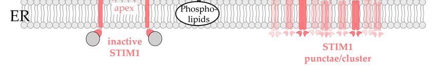
Biomolecules 2022, 12, 352                                                                                                       6 of 37

                                    that can be rescued by STIM1 binding [96,102], and Orai1 R91W is one of the best-known
                                    loss-of-function (LoF) mutations leading to severe combined immune deficiency [66,103]).
                                          However, it is not only the pore unit that is critical for channel activation; rather, a
                                    very large number of positions in all TM domains of the channel complex must be in an
                                    opening-permissive state to ensure an ideal working process of the CRAC channel complex.
                                    Indeed, a single point mutation at more than a dozen different positions in the four TM
                                    domains can result in either gain- or loss-of-function (GoF or LoF), depending on the
                                    properties of the incorporated amino acid [104–111]. Because an LoF mutation always acted
                                    dominant over a GoF mutation in most of the Orai1 mutants tested, it was demonstrated
                                    that Orai1 pore opening requires a global conformational change of the entire channel
                                    complex [108,112–114] in line with recent structural resolutions of an Orai1 mutant in the
                                    open state [95].
                                          STIM1 and Orai1 are sufficient to constitute the CRAC channel [115]. However, due to
                                    the location of STIM1 and Orai1 in the ER membrane and the PM, respectively, and their
                                    assembly at ER-PM junctions upon store-operated activation, it is not surprising that their
                                    function is modulated by lipids. Furthermore, there is increasing evidence that cholesterol-
                                    rich regions define the association and function of the STIM1/Orai1 complex [116,117]. On
                                    the one hand, the function of both STIM1 and Orai1 is modulated by direct interaction
                                    with lipids, including particularly cholesterol and the phospholipid PIP2 [118–123]. On
                                    the other hand, there is plethora of evidence that the STIM1/Orai1 interplay is indirectly
                                    controlled via the spatially separated phospholipid bilayers, the ER and the PM, along with
                                    a variety of lipid- or ER-PM junction-dependent accessory proteins [124]. These forms of
                                    direct and indirect lipid-mediated modulation of CRAC channel function will be reviewed
                                    in the following.

                                    4. Lipids Directly Regulated CRAC Channel Components
                                         The major lipids which regulate STIM1/Orai1 function via direct interaction are PIP2
                                    and cholesterol [25,125–127]. Furthermore, there is evidence for sphingomyelin- [128] and
                                    S-acylation- [123] mediated modulation of CRAC channels (Figure 2). Phospholipids were
                                    hypothesized to contribute to LoF of certain STIM1 mutants [83].

                                    4.1. Phosphoinositides
                                         Shortly after the discovery of STIM proteins, PIP2 was reported to be critical for the
                                    co-regulation of STIM1 and Orai1. In particular, reducing PIP2 levels using an inositol
                                    5-phosphatase decreased store-operated Ca2+ entry. Both the STIM1 C-terminus and Orai1
                                         N-terminus were shown to be sensitive to alterations in PIP2 levels [129,130] (Figure 2,
                                    Table 1).

                                    Table 1. List of lipids and ER-PM junctional proteins that modulate the function of STIM1 and/or
                                    Orai1. The lipids and ER-PM junctional proteins are listed together with their location in the cell
                                    critical for the modulatory effect on STIM1 and/or Orai1, their effects on the functions of the
                                    CRAC channel, STIM1, and Orai1, whether they interact directly with STIM1 or Orai1 and the
                                    corresponding references.

                                                Effect on                                                 Direct
                                                                    Effect on        Effect on
    Lipid Type               Location          Endogenous                                           Interaction with         Ref.
                                                                     STIM1            Orai1
                                                 CRAC                                               STIM1 or Orai1
                                                                                                      STIM1 K-rich
                                                                        ↑
                                                                                                          region
                                                                     STIM1
        PIP2                   PM                    ↑                                   ↑             (aa 672–685)       [129,130]
                                                                  assembly at
                                                                                                    Orai1 N-terminus
                                                                PIP2 rich region
                                                                                                        (aa 28–33)
        PI4P                   PM                    ↑                  ↑               n.d.               n.d.           [130–133]
The Role of Lipids in CRAC Channel Function - MDPI
Biomolecules 2022, 12, 352                                                                                                              7 of 37

                                         Table 1. Cont.

                                                                                                           STIM1 OASF
                                                                                                               (I364)         [119–122,
    cholesterol                   PM                       ↑                ↓                 ↓
                                                                                                         Orai1 N-terminus    129,134,135]
                                                                                                            (L74, Y80)
 sphingo-myelin                   PM                       ↑               n.d.              n.d.              n.d.             [128]
   ER-phospho-                                                                                             STIM1 apex
                                  ER                      n.d.              ↓           not applicable                           [83]
      lipids                                                                                               (F394K/D)
     S-acylation
    (post-transla-
                                  PM                       ↑         not applicable           ↑             Orai1 C143          [123]
  tional tethering
   of fatty acids)
  Protein within                                     Effect on                                                Direct
                                                                        Effect on         Effect on
       ER-PM                    Location            endogenous                                           interaction with
                                                                         STIM1             Orai1
      junction                                        CRAC                                               STIM1 or Orai1
                         ER-PM junction
                          (E-Syt1 located
                                                   no/minor effect
        E-Syt             there in a Ca2+                                  n.d.             n. d.            indirect         [136–142]
                                                     (HeLa cells)
                        dependent manner)
                         embedded in ER
                                  [Ca2+ ]cyt ↑
                            ER →                                         supports
                             ER − PM                                 repletion of ER
       E-Syt1                                      no/minor effect                           n.d.            indirect           [142]
                         Rearrangement of                            stores with Ca2+
                        ER-PM contact sites                           (HEK293 cells)

                                                                       ↑ (E-Syt2S)
       E-Syt2                ER-PM junction          ↑ (E-Syt2S)                             n.d.            indirect           [143]
                                                                       ↓ (E-Syt2L)
                                                      No effect/
                                                    minor (when
       E-Syt3                ER-PM junction          knocked-out           n.d.              n.d.            indirect           [136]
                                                    together with
                                                      E-Syt1/2)
                                                                                                            Indirect
                                                                           ↑
                                                                                                          dependent on        [140,144–
   E-Syt1 + Nir2             ER-PM junction                ↑         Co-localization         n.d.
                                                                                                         DAG levels in the      146]
                                                                      with STIM1
                                                                                                           membrane
                                    FFAT
        VAP                    ER →                       n.d.             n.d.              n.d.            indirect         [147–149]
                               ER − PM
  VAP + ORP3/6               ER-PM junction                ↓                ↓                n.d.              n.d.           [150–152]
                         ER-PM junction,                                                                                      [148,150,
      ORP5/8                                              n.d.             n.d.              n.d.              n.d.
                            ER-Mito                                                                                           152–154]
                                                                            ↑
                                                                                                              Indirect
                                                     not required    Supports STIM1
                                                                                                         Co-localizes with
    GRAMD2A                  ER-PM junction         (more studies     translocation          n.d.                             [155–159]
                                                                                                         E-Syt and STIM1
                                                      required)       STIM1 puncta
                                                                                                          (PIP2 required)
                                                                        formation
                                                                             ↑               ↑
                                                                                                         Indirect (further
                                                                          STIM1        STIM1/Orai1
       ANO8                  ER-PM junction                ↑                                                 studies            [160]
                                                                        oligomeri-      interaction;
                                                                                                            necessary)
                                                                     zation/clustering inactivation
                                                                                             ↑           Direct with Orai1
                                                                     independent of                                           [122,161–
    Caveolin-1                    PM                       ↑                            STIM1/Orai1        (aa 52–60; aa
                                                                         STIM1                                                  165]
                                                                                          coupling           250–258)
Biomolecules 2022, 12, 352                                                                                                 8 of 37

                                   Table 1. Cont.

                                                                                                  Direct with
      Junctate                ER                    ↑                 ↑                 ↑        STIM1 (STIM1          [166]
                                                                                                  N-terminus)
                                                                       ↑
                                                                   supports
                                                                                                Direct interaction
  Juncto-philin 4             ER                    ↑           recruitment of         n.d.                            [167]
                                                                                                  with STIM1
                                                                   STIM1 to
                                                                    ER-PM
                                                                   supports
    Junctate +                                                  recruitment of                  Direct interaction
                             ER-PM                  ↑                                  n.d.                          [166,167]
  juncto-philin 4                                                  STIM1 to                       with STIM1
                                                                    ER-PM
                                                                                                                     [141,168,
      SARAF                   ER                    ↓                 ↓                 ↓         STIM1 CTID
                                                                                                                       169]
       Septin                 PM                        depending on the septin type                 indirect        [170–174]
     STIMATE                  ER                    ↑                 ↑                 ↑         STIM1-CC1            [175]
                                                                                                   indirect
       RASSF                  PM                    ↑                 ↑                 ↑       (modulates PIP2        [176]
                                                                                                    levels)
                                                     ↑
                                             (Regulated by
        AC8                   PM                its direct            ↑                 ↑             Orai1          [177,178]
                                              association
                                             with AKAP79)

                                        At the very end of the C-terminus of STIM proteins is a lysine-rich region that functions
                                   as a PIP2 binding site in both STIM1 and STIM2 [88,179–182] (Figure 2). Mutation of
                                   these positively charged residues or deletion of the polybasic region impairs the stable
                                   association of STIM proteins at the PM after store-depletion. The stable coupling of STIM
                                   to the PM is promoted by the presence of PIP2 and also PIP3 in the PM, which are mainly
                                   enriched in cholesterol-rich regions [117,130,141,179,183,184]. Strikingly, the association
                                   of STIM1 with PIP2 necessitates tetramerization of its lysine-rich region, whereas efficient
                                   binding of STIM2 to PIP2 depends on dimerization of this polybasic domain. This leads
                                   to an enhanced affinity for PIP2 and a reduced activation threshold of STIM2 [185]. After
                                   deletion of this polybasic motif, ER depletion-induced translocation of STIM1 to the PM is
                                   impaired, even though STIM1 retains its ability to form oligomers [179]. In fact, although
                                   the lysine-rich region is dispensable when both STIM1 and Orai1 are overexpressed, it
                                   enhances the efficacy of the STIM1/Orai1 activation process under physiological conditions.
                                   STIM1 first assembles with PM-localized PIP2 and PIP3 pools in cholesterol-rich regions
                                   before interacting directly with Orai1 [186] (Figure 2). This suggests that cholesterol
                                   indirectly drives the accurate STIM1-mediated targeting of SOCE components in lipid rafts
                                   by retaining the necessary phosphoinositide pools.
                                        The N-terminus of Orai1 contains a polybasic region that was reported to be sensitive to
                                   PIP2 levels in the PM and essential for directing Orai1 to membrane areas with distinct PIP2
                                   content [129]. However, the dependence of Orai1 on STIM1 makes it currently challenging
                                   to characterize PIP2 regulation of Orai1 channels alone. One potential way to determine
                                   whether Orai1 is directly regulated by PIP2 is to investigate the effect of PIP2 depletion
                                   on one of the various, currently known, constitutively active Orai1 mutants, which were
                                   reported for instance in Tiffner et al. [108] and Yeung et al. [104].
                                        Interestingly, PI4P, the precursor of PIP2 , was also reported to modulate CRAC
                                   channel function [130–132] (Figure 2). The phosphatidylinositol 3- and phosphatidyl-
                                   inositol 4-kinases inhibitor, Y294002, resulted in inhibition of store-operated Ca2+ entry
Biomolecules 2022, 12, 352                                                                                              9 of 37

                             in a concentration-dependent manner in human platelets [130,131]. The PI4K inhibitor,
                             wortmannin, also led to inhibition of store-operated currents, even though STIM1 puncta
                             formation was preserved [130,132]. Furthermore, knock-down of the PI4K reduced endoge-
                             nous SOCE [130]. To further investigate the role of PI4K, the newly synthesized and more
                             specific PI4P inhibitors might be highly valuable [133].
                                  Overall, PIP2 and PI4P represent modulatory factors of the CRAC channel machinery
                             (Figure 2). While it is clear that STIM1 interacts directly with PIP2 , the effects of PIP2 alone
                             on Orai1 are less explored. Further studies are still required to understand the role of PI4P
                             levels in the CRAC channel choreography.

                             4.2. Cholesterol
                                   Cholesterol was shown to modulate the function of both STIM1 and Orai1, likely via
                             direct interaction [120–122] (Figure 2, Table 1).
                                   Very early studies showed that the removal of cholesterol from membranes reduced
                             SOCE and decreased the ability of a constitutively active STIM1 mutant (D76A) to form
                             punctae [135]. However, there are conflicting studies on the effect of depletion of choles-
                             terol by methyl-β-cyclodextrin (MßCD) on CRAC influx. SOCE in cells with endogenously
                             expressed STIM1 and Orai1 was reduced by the application of MßCD [134]. In another
                             study, it was demonstrated that cholesterol depletion mediated by cyclodextrin led to
                             Orai1 internalization and altered Orai1 diffusion, which was restored by Cav-1 overex-
                             pression. While cholesterol depletion by MßCD reduced STIM1-mediated Orai1 currents,
                             co-expression of Cav-1 maintained or even enhanced current levels observed in the absence
                             of MßCD application as well as Cav-1 overexpression [122]. In contrast, Gwozdz et al. [119]
                             demonstrated that overexpression of STIM1 and Orai1 abolished the inhibitory effect of
                             chemical cholesterol depletion by MßCD on SOCE.
                                   We found that depletion of cholesterol via cholesterol oxidase or filipin enhanced
                             SOCE in both human embryonic kidney 293 (HEK 293) cells and rat basophilic leukemia
                             2H3 (RBL) cells [120]. Similarly, Pacheco et al. [121] demonstrated that cholesterol depletion
                             by MßCD enhanced Orai1 currents only after STIM1- or SOAR-Orai1 coupling [121].
                             Interestingly, MßCD treatment leads to enhanced STIM1-Orai1 coupling [121], whereas
                             the application of cholesterol oxidase left STIM1-Orai1 coupling unaffected [120]. The
                             observed differences are likely due to the gentler manipulation of cholesterol levels by
                             cholesterol oxidase or filipin compared with MßCD, while leaving membrane integrity
                             intact. The differences in membrane composition might also be explained by the distinct
                             effects on STIM1/Orai1 coupling upon the different ways of chemical cholesterol depletion.
                             Furthermore, MßCD treatment was shown to decrease membrane localization of STIM1
                             and disrupts its interaction with Orai1 [129,134]. Alternatively, distinct effects could occur
                             upon overexpression toward endogenous proteins [119].
                                   Observed effects due to chemical cholesterol depletion underlie the direct interplay of
                             cholesterol with STIM1 as well as Orai1 [120,121].
                                   Indeed, we showed direct binding of TopFluor-cholesterol to Orai1 [120] (Figure 2).
                             Searching for a potential cholesterol binding site, we discovered the so-called cholesterol
                             recognition amino acid consensus motif (-L/V-(X)(1-5)-Y-(X)(1-5)-R/K-; X represents from
                             1 to 5 any amino acids before the next conserved residue) [11,187–189] in the ETON region
                             (aa 73–90), which is integral to channel gating by STIM1 [120]. Indeed, mutation of critical
                             sites in this cholesterol binding motif (L74I, Y80S) led to reduced interaction of cholesterol,
                             with both full-length Orai1 mutants as well as an Orai1 N-terminal peptide [120] mutated
                             accordingly. In line with the enhanced currents after chemical cholesterol depletion, Orai1
                             mutants deficient in cholesterol binding also showed an increase in SOCE without affecting
                             the interaction with STIM1. This indicates that STIM1-Orai1 interaction at the C-terminus
                             can still occur, with cholesterol only interfering with STIM1-dependent channel gating
                             through Orai1 N-terminal interactions. However, it is still a matter of debate whether
                             this cytosolic cholesterol binding motif directly interacts with cholesterol or allosterically
                             affects cholesterol binding to Orai1. Based on the Orai channel structures, the proposed
Biomolecules 2022, 12, 352                                                                                           10 of 37

                             N-terminal cholesterol binding site contains the critical positions L74 and Y80. However,
                             they point in opposite directions, which is unfavorable for cholesterol binding. For direct
                             Orai1-N-terminus-cholesterol interaction to take place, the ETON domain would need to be
                             in equilibrium between the α-helix (as in the crystal structure) and random coil structures,
                             as possibly occurring in the isolated Orai1 N-terminal peptide in solution [120]. In this case,
                             cholesterol would most likely favor a random coil structure and make Orai1 unavailable
                             for STIM1 interaction. However, it seems unlikely that a stable α-helix would “unwind”
                             under physiological conditions. Therefore, a more energetically favorable possibility would
                             be a conformational change between an α-helix and the rarer 310 -helix structure (with
                             three residues per turn as opposed to the 3.6 residues per turn in a canonical α-helix) [190].
                             This allows L74 and Y80 to align in the same direction and enable cholesterol binding.
                             Alternatively, it could be hypothesized that the direct cholesterol binding site is located at
                             a distinct site within the Orai1 channel complex which interplays allosterically with the
                             N-terminus, but this requires further study.
                                   Cholesterol associated effects were also linked to a cholesterol binding motif with
                             the consensus sequence L/V-X(1–5)-Y-X(1–5)-R/K in the STIM1 C-terminus [121]. Indeed,
                             a mutation within this binding motif (I364A), located in SOAR, resulted in comparable
                             enhancements of Orai1 currents to chemical cholesterol depletion in both full-length STIM1
                             and a C-terminal STIM1 fragment. Via MD simulations, it was confirmed that cholesterol
                             affects the coupling of SOAR to the membrane, with I364 being the major interface. A
                             combination of Orai1 and STIM1 mutants, both deficient in cholesterol binding, did not
                             further enhance currents compared with ones obtained by mutating either STIM1 or Orai1.
                             This indicates that removal of the cholesterol-binding (CB) domain of either Orai1 or STIM1
                             is sufficient to enhance calcium influx and Orai1 currents, mimicking the effect of cholesterol
                             removal from the PM. Altogether, these results strongly suggest that the CB domain of
                             both, Orai1 and STIM1 coordinates the same cholesterol-mediated mechanism [121].
                                   Regulation of STIM1 or Orai1 by cholesterol may possibly have an underappreciated
                             pathophysiological relevance. We suspected a connection between hypocholesterolemia
                             and enhanced mast cell degranulation. Indeed, patients suffering from hypocholesterolemia
                             tend to have an enhanced allergy response [191], in line with the findings that cholesterol
                             depletion in RBL mast cells enhanced store-operated Ca2+ currents and degranulation [120].
                             However, further studies are still required in this regard. Moreover, the question arises
                             whether other pathophysiological conditions are caused by the interaction of cholesterol
                             and Orai1.
                                   The relationship between cholesterol and Orai remains controversial in terms of
                             its dynamics and physiological significance. Do ER-PM transitions form exclusively at
                             cholesterol-rich regions, with cholesterol inhibition being the standard for “wild-type”
                             SOCE reactions, or is cholesterol binding to Orai turned on and off? What conditions
                             lead to the transport of cholesterol from the PM to its cytosolic binding site at Orai?
                             Moreover, it is currently contentious whether STIM1 couples not only to Orai1 C-terminus,
                             but eventually also to Orai1 loop2 or N-terminus [108,111,112,192–195]. Hence, how do
                             cholesterol and STIM1 compete for the same region of Orai?
                                   It could be hypothesized that when stores are depleted, STIM1 binds to the C-terminus
                             of Orai1 and triggers a conformational rearrangement that removes cholesterol from the
                             N-terminus and instead allows STIM1 to bind, causing the channel to open. This is an
                             intriguing concept of cholesterol-mediated SOCE regulation (Figure 2). However, the
                             physiological ramifications and the mechanism of when and why cholesterol binds to Orai1
                             remain to be elucidated. A detailed characterization of the Orai1 cholesterol binding pocket
                             within the CRAC channel complex, even at the structural level, would be highly valuable
                             for a more detailed understanding of the cholesterol-dependent molecular mechanisms.

                             4.3. Sphingomyelin
                                 Sphingomyelin is very abundant in the PM of mammalian cells, accounting for about
                             20% of the total phospholipids in this bilayer, and is preferentially located in the outer
Biomolecules 2022, 12, 352                                                                                           11 of 37

                             leaflet [9]. The exchange of sphingomyelin with the inner leaflet is less likely due to its
                             polar headgroup. Moreover, it is firmly anchored in the membrane by its hydrophobic part.
                             Physiologically, sphingomyelin in the PM is essential for lateral homogeneity and acts as a
                             source of lipid signaling [44,196,197].
                                  A central position in the sphingomyelin metabolism is occupied by ceramide, which
                             is produced by several enzymatic cycles in the ER. To form sphingomyelin, ceramide is
                             transported to the Golgi. There, ceramides can be endowed with different headgroups
                             resulting in the generation of distinct groups of complex sphingolipids, one of which is
                             sphingomyelin. Sphingomyelin further acts as a substrate for ceramide phosphates and
                             sphingosine, which are produced by specific enzymes. For instance, sphingomyelinase
                             (SMase) D converts sphingomyelin into ceramide-1-phosphatase [196,198,199].
                                  It was reported that sphingomyelin itself directly affects various ion channels [200–202].
                             Among these channels, the CRAC channel was also found to be a target of sphingomyelin
                             (Figure 2, Table 1). SMase D treatment significantly reduced store-operated currents
                             without affecting Ca2+ store-depletion. Although the application of SMase D can lead to the
                             generation of high levels of ceramide phosphates, application of ceramide-1-phosphate did
                             not reduce store-operated currents. These results indicate that CRAC channel modulation
                             occurs directly via sphingomyelin and not via one of its metabolites [128] (Figure 2).
                                  One plausible mechanism for the modulatory role of sphingomyelin on the CRAC
                             channels is their direct interaction. However, further studies resolving their direct interac-
                             tion are still required. Alternatively, SMase D treatment may change PM organization, thus
                             altering CRAC channel activation [128,196].

                             4.4. S-Acylation
                                  S-acylation, also called S-palmitoylation, is the posttranslational tethering of a medium
                             length fatty acid, also called palmitic acid, to a cysteine residue. Importantly, unlike ir-
                             reversible posttranslational lipid modifications such as prenylation and myristoylation,
                             S-acylation of proteins is reversible. Thus, S-acylation allows dynamic and spatiotempo-
                             ral control of protein activity and interaction [203–205]. For more than 50 ion channels,
                             S-acylation was reported to control their gating and trafficking by enhancing the hydropho-
                             bicity of protein segments [205]. The S-palmitoylation reaction is mediated by protein acyl
                             transferases (PATs) in the ER and Golgi containing zinc finger and DHHC domains [206].
                             The process is reversed in the PM by acyl protein thioesterases [207], among which 23 PATs
                             and five thioesterase isoforms were so far identified in humans [208]. It is suggested that the
                             hydrophobic properties of the attached acyl groups may affect the distribution of proteins
                             in the membrane [209].
                                  A recent report revealed the first indication that Orai1 activity is controlled by S-
                             acylation. Orai1 was found to be S-acylated by ZDHHC20 (PAT20) at residue C143 (Figure 2,
                             Table 1), which is critical in controlling its trafficking and function and ensures its local-
                             ization in cholesterol-rich regions. Upon mutation of this cysteine, Orai1 was recruited
                             into cholesterol-poor regions causing STIM1 mediated Orai1 currents to diminish and the
                             signaling at the immunological synapse between T-cell and antigen presenting cell to occur
                             less efficiently. Furthermore, downstream signaling events including long-lasting Ca2+
                             level enhancements, nuclear factor activated T-cell (NFAT) translocation and IL-2 secretion
                             were abolished [123].
                                  At this point, it is unclear whether S-acylation is required for or strengthens Orai1-
                             cholesterol interactions. In this regard, the reduction in STIM1/Orai1 currents with im-
                             paired S-acylation are in line with the effects observed upon chemical cholesterol depletion
                             by MßCD [119,134,135] but are at odds with store-operated current enhancements upon
                             chemical cholesterol depletion by MßCD [120,121]. Therefore, it would be of interest to
                             investigate whether Orai1 C143A is deficient in cholesterol binding. A potential cholesterol
                             binding site was identified in the Orai1 N-terminus at positions located in the same plane
                             such as C143. Thus, if Orai1 C143A is deficient in interaction with cholesterol, one could
Biomolecules 2022, 12, 352                                                                                         12 of 37

                             further test for a potential allosteric interplay of C143 with the N-terminal positions L74
                             and Y80 with respect to cholesterol binding.

                             4.5. Potential Role of Phospholipids in Controlling STIM1 Function
                                  As part of our characterization of the role of a small alpha helical region within
                             the CAD/SOAR apex of the resting STIM1, namely the α2 region, we identified distinct
                             point mutations therein that can impact the STIM1/Orai1 activation cascade in various
                             manners. STIM1 homomerization and consequent Orai1 activation was impaired upon
                             single point mutation to hydrophilic, charged amino acids (STIM1 F394D, STIM1 F394K).
                             Using MD simulations, we found that their loss of homomerization could be due to
                             possible electrostatic interactions with lipid headgroups in the ER membrane and an altered
                             formation of the CC1α1-SOAR/CAD interaction site. Consistent with these results, we
                             demonstrated experimentally that the disruptive effects of F394D depend on the distance of
                             the apex from the ER membrane (Figure 2, Table 1). Collectively, our findings provide first
                             evidence that the CAD/SOAR apex is in the immediate vicinity of the ER membrane in the
                             STIM1 quiescent state [83] which is in line with the recent finding by van Dorp et al. [84].
                             At this point further investigations are required to determine whether the close proximity
                             of STIM1 apex to the ER membrane in the closed state is predominantly stabilized by intra-
                             and intermolecular interactions within the STIM1 proteins or whether ER membrane lipid
                             interactions also contribute. In the latter case, determining which lipids might be involved
                             would be challenging.

                             5. Indirect Control of STIM1/Orai1 Machinery at ER-PM Membrane Contact Sites
                                   In the following parts of the review, the focus is laid on the ER-PM contact sites, as
                             this is the interface for the STIM1/Orai1 interplay.

                             5.1. ER-PM Constact Sites and Methods for Their Characterization
                                   Membrane contact sites (MCSs) are microdomains where the membranes of two or-
                             ganelles get in close proximity without fusion. It is becoming increasingly evident that
                             MCSs are critical to coordinate a variety of physiological events including organelle dy-
                             namics, lipid exchange, Ca2+ signaling and cell survival [210–217]. MCSs of the membrane
                             of intracellular Ca2+ stores such as the sarcoplasmic reticulum or ER membrane and the PM
                             control for instance the excitation-contraction coupling in muscle cells and store-operated
                             Ca2+ entry in non-excitable cells, respectively [213].
                                   Multiple technologies, including electron microscopy and fluorescence microscopy,
                             have significantly advanced our understanding of MCS. In this regard, electron microscopy
                             (EM) is a “gold standard method”, providing a static snapshot of MCS architecture at
                             the nanoscale, albeit under non-native conditions. In contrast, cryo-electron tomography
                             (cryo-ET), which immobilizes non-crystalline samples and allows 3D imaging at a high
                             resolution of 4–10 nm, enables the resolution of MCS in a near-native state [217,218].
                                   To visualize MCS structures in living cells, multispectral fluorescence microscopy uses
                             genetically encoded fluorescent proteins attached to MCS-resident proteins. To overcome
                             the diffraction limit of confocal microscopy at or near the PM [219,220], TIRFM is used to
                             selectively illuminate fluorophores at surface regions with wave penetration of approxi-
                             mately 100 nm into the sample. Therefore, the dynamics and kinetics of proteins residing at
                             the ER-PM contact sites, such as STIM1 puncta formation, can be easily monitored within 1
                             min after Ca2+ store-depletion [73,74,175]. An even better resolution can be achieved with
                             super-resolution microscopy such as photoactivated localization microscopy (PALM) [221]
                             and STochastic Optical Reconstruction Microscopy (STORM) [222] in conjunction with
                             fluorophore-labeled MCS-resident proteins.
                                   To identify unknown binding partners within MCSs, the bimolecular fluorescence
                             complementation (BiFC) method can be used. This attempt is based on fluorescent pro-
                             teins (FP) split into two non-fluorescent parts, each of which can be fused to two proteins
                             residing in opposing membranes. The chromophore fluorescence is recovered only when
Biomolecules 2022, 12, 352                                                                                         13 of 37

                             the FP fragments are close to each other (10–30 nm) [223], thus demonstrating that the two
                             proteins are in close proximity and might interact. As such, BiFC can serve as a great tool
                             for characterizing native MCS distribution and dynamics under different pathophysiologi-
                             cal conditions.
                                   Furthermore, markers that allow remote modulation of intramembrane binding and
                             MCS assembly were developed. These include chemically or light-inducible modules that
                             can modulate the properties of MCS [218], among which we focus on those critical to
                             modulate ER-PM junctions.
                                   An initial tool to gain a better understanding of STIM1-Orai1 communication at ER-
                             PM junctions is the MAPPER (membrane-attached peripheral ER) [140,224]. This marker
                             consists of a signal peptide of STIM1 located in the ER lumen followed by the TM domain
                             of STIM1. Subsequently, an FKBP12-rapamycin binding (FRB) domain followed by the
                             polybasic motif of the small G protein Rit, which binds PIP2 and PIP3 in the PM [182],
                             were added. Several flexible and helical linkers were inserted in the cytosolic portion of
                             MAPPER to guarantee that its expression did not change the gap distance at the ER-PM
                             junctions. MAPPER localized in puncta at the ER-PM contact sites at analogue positions
                             like activated STIM1. A light-sensitive variant of this is the LiMETER (light-inducible
                             membrane tethered peripheral ER tool), based on a STIM1-Rit chimera [175]. Compared to
                             the MAPPER, LiMETER contains the photosensitive LOV2 domain (aa 404–546) instead of
                             the FRB domain. In the inactive state, LiMETER is tightly packed and hides the polybasic
                             region. Upon activation by blue light, this is released, leads to clustering, and interacts
                             with the PM. Remarkably, this process is reversible and therefore could be used to detect
                             protein interactions, as performed with STIM1 and the accessory protein STIMATE [175]
                             (see Section 5.4.5).
                                   Other markers of ER-PM junctions are OptoPB and OptoPBer [225]. Like LiMETER,
                             both contain the LOV2 domain and polybasic regions. The latter could be used for interac-
                             tion studies with specific lipids, such as Rit1 and PIP2 [182]. While OptoPB is a cytosolic
                             protein, OptoPBer is anchored in the ER membrane such as LiMETER. Thus, OptoPBer
                             cannot only be used to study interactions of polybasic domains and lipids, but additionally
                             serves as a spacer between the ER and PM [225]. Upon activation with blue light, OptoPBer
                             forms puncta and reversibly reverts to a uniform distribution upon inactivation in the dark.
                             These tools helped to provide novel insights into the CRAC channel machinery [175,218].
                             For example, to investigate key interaction residues of the polybasic region of STIM1 with
                             the PM, He et al. [225] initially exchanged lysines with alanines within OptoPBer and
                             utilized positive hits on full-length STIM1. They found that the STIM1 double mutant
                             K684A K685A abrogates punctae formation [225] and may also be insufficient to activate
                             Orai1. Furthermore, variation in the linker length between TM and LOV2 domain revealed
                             that the working distance of the CRAC channel is between ~15–30 nm [225].
                                   Taken together, these methods provide the opportunity to obtain a comprehensive
                             picture of the interaction and distance between organelles and the cell membrane. In
                             addition, the optogenetic tools allow precise temporal and spatial control over the formation
                             of contacts between the membrane and organelles. This could also be used to reversibly
                             switch the binding of peptides or proteins to the PM between near and far PM, which is a
                             promising way to study protein–protein interactions. In this context, it should be noted
                             that the genetically encoded photosensitive proteins are still in the development stage and
                             many efforts are still being made to improve them. Their range of applications can be
                             extended immensely by varying, for example, the frequency of the light pulse, the length
                             of the linker between the fusion proteins or the expression ratio of the cellular components
                             under investigation, and will thus be increasingly used as the methodology improves.

                             5.2. Critical Factors Determining STIM1/Orai1 Co-Regulation at the ER-PM Contact Sites
                                  The ER-PM junctions are currently the best understood cellular MCSs. Typically, the
                             ER-PM junctions make up only 0.8–2.5% percent of the cell surface. They are separated
                             by a gap of 10–25 nm and have sizes of 100–200 nm [74,145,226,227]. ER-PM junctions are
under investigation, and will thus be increasingly used as the methodology improves.

                             5.2. Critical Factors Determining STIM1/Orai1 Co-Regulation at the ER-PM Contact Sites
                                   The ER-PM junctions are currently the best understood cellular MCSs. Typically, the
                             ER-PM junctions make up only 0.8–2.5% percent of the cell surface. They are separated by
Biomolecules 2022, 12, 352                                                                                                                  14 of 37
                             a gap of 10–25 nm and have sizes of 100–200 nm [74,145,226,227]. ER-PM junctions are
                             highly dynamic contact sites which depend on a variety of factors, including (i) spatial
                             and temporal fluctuations of the lipid compositions regulated by the PI cycle as described
                             inhighly
                                 Sectiondynamic
                                           2 [13], (ii)contact   sitesCa
                                                          cytosolic    which     depend on [74,213],
                                                                          2+ concentration     a varietyasofwell
                                                                                                               factors,   including
                                                                                                                    as (iii)  a set of (i) spatial
                                                                                                                                        proteins
                               and   temporal      fluctuations
                             located at these contact sites formingof the  lipid   compositions    regulated    by  the  PI  cycle
                                                                                interactions with proteins or lipids [13,118].      as  described
                               in Section
                                   ER-PM2contact[13], (ii)sites
                                                           cytosolic   Ca2+ concentration
                                                                are particularly                [74,213],
                                                                                      specialized           as well
                                                                                                     sites for  Ca2+ as   (iii) a setThis
                                                                                                                      signaling.      of proteins
                                                                                                                                           is not
                               located   at  these   contact   sites  forming     interactions  with
                             surprising since the ER functions as the largest Ca2+ store in the cell.  proteins   or  lipids
                                                                                                                         Thus, [13,118].
                                                                                                                                  Ca 2+ plays an
                                      ER-PM contact sites are particularly specialized sites for Ca2+ signaling. This is not
                             essential role in the formation of MCSs of various organelle                  membranes with the ER mem-
                               surprising since the ER functions as the largest Ca2+ store in the cell. Thus, Ca2+ plays
                             brane [213]. With regard to ER-PM junctions, it was reported that the luminal Ca2+ levels
                               an essential role in the formation of MCSs of various organelle membranes with the ER
                             affect their development and stability. While a certain number of ER-PM contact sites are2+
                               membrane [213]. With regard to ER-PM junctions, it was reported that the luminal Ca
                             already present in the resting state of the cell, Ca2+ store-depletion triggers the formation
                               levels affect their development and stability. While a certain number of ER-PM contact
                             of new contacts [74,140]. These events are often driven by the                     signaling steps of the ele-
                               sites are already present in the resting state of the cell, Ca2+ store-depletion triggers the
                             mentary unit of CRAC channels formed by STIM1 and Orai1, which accumulates in ER-
                               formation of new contacts [74,140]. These events are often driven by the signaling steps of
                             PM contact sites upon store-depletion. The expression of STIM1 alone can already influ-
                               the elementary unit of CRAC channels formed by STIM1 and Orai1, which accumulates
                             ence the formation and size of ER-PM junctions. Indeed, the overexpression of STIM1
                               in ER-PM contact sites upon store-depletion. The expression of STIM1 alone can already
                             greatly increased the dimensions of the ER-PM contact sites, whereas the role of endoge-
                               influence the formation and size of ER-PM junctions. Indeed, the overexpression of STIM1
                             nous STIM1 is still unclear [226–228]. In particular, store-depletion triggers STIM1 puncta
                               greatly increased the dimensions of the ER-PM contact sites, whereas the role of endogenous
                             formation
                               STIM1 is at     theunclear
                                             still    ER-PM[226–228].
                                                                contact sites In by   binding to
                                                                                   particular,      PIP2 in the PM.
                                                                                                store-depletion          As a result,
                                                                                                                     triggers     STIM1STIM1
                                                                                                                                           puncta
                             couples    to Orai1     through    diffusional     trapping.  The   interaction
                               formation at the ER-PM contact sites by binding to PIP2 in the PM. As a result,  between     STIM1     and Orai1
                                                                                                                                            STIM1
                             iscouples
                                the basicto requirement
                                             Orai1 through    fordiffusional
                                                                   CRAC channel        activation
                                                                                trapping.           [73,115,229],
                                                                                            The interaction          but the
                                                                                                                between         localand
                                                                                                                            STIM1     organiza-
                                                                                                                                           Orai1 is
                             tion
                               theand
                                   basicdynamics
                                           requirement  of the
                                                             formembrane
                                                                  CRAC channel architecture   at the
                                                                                      activation      ER-PM junction
                                                                                                  [73,115,229],   but theinfluence       the effi-
                                                                                                                             local organization
                             cacy   of their   interaction     and   gating    [141,170].  The   latter  involves
                               and dynamics of the membrane architecture at the ER-PM junction influence the efficacyseveral    accessory    pro-of
                             teins
                               theirthat  control lipid
                                      interaction      and metabolism       at andThe
                                                            gating [141,170].        thelatter
                                                                                         formation    of ER-PM
                                                                                               involves    several contacts,
                                                                                                                      accessory  some   of which
                                                                                                                                   proteins    that
                             are  also  Ca  2+ sensitive. They can be grouped into proteins either spanning from the ER to
                               control lipid metabolism at and the formation of ER-PM contacts, some of which are also
                             the
                               CaPM    or anchored
                                  2+ sensitive.      Theyin/at
                                                            canone    of the two
                                                                 be grouped         membranes
                                                                                  into             [13], as
                                                                                       proteins either      described
                                                                                                          spanning     fromin the
                                                                                                                              the following.
                                                                                                                                   ER to the PM
                              or anchored in/at one of the two membranes [13], as described in the following.
                             5.3. ER-PM Spanning Proteins Involved in the Modulation of STIM1/Orai1 Function
                              5.3. Proteins
                                   ER-PM Spanning
                                            which areProteins
                                                      locatedInvolved in and
                                                              in the ER  the Modulation of STIM1/Orai1
                                                                             can form direct or indirect Function
                                                                                                         contacts with the
                             PM comprise
                                   Proteinsthe Extended-synaptotagmins
                                            which                          (E-Syts)
                                                   are located in the ER and        with
                                                                             can form     E-Syt
                                                                                       direct or 1-3 [136],contacts
                                                                                                 indirect   VAMP with
                                                                                                                    associ-
                                                                                                                         the
                              PM proteins
                             ated comprise(VAP)
                                            the Extended-synaptotagmins
                                                  [230], GRAMD2A [159],(E-Syts)     with E-Syt81-3
                                                                            and Anoctamine         [136], VAMP
                                                                                                 (ANO8)           associated
                                                                                                           [160] (Figure 3,
                              proteins
                             Table 1). (VAP) [230], GRAMD2A [159], and Anoctamine 8 (ANO8) [160] (Figure 3, Table 1).

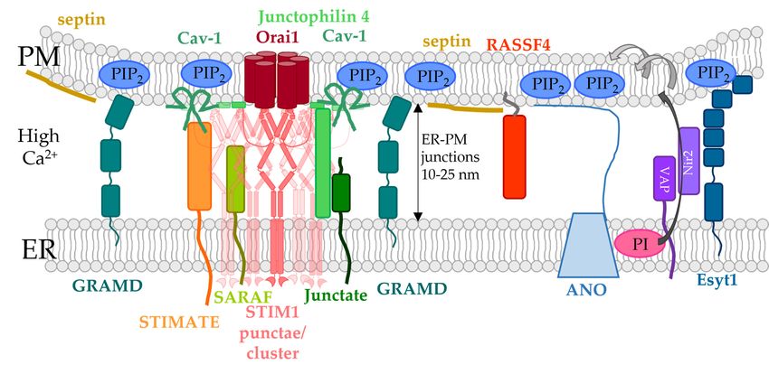
                              Figure 3. ER-PM spanning proteins involved in the modulation of the CRAC channel complex. In
                              addition to the PI cycle, phospholipids can also be transported by ER-PM spanning protein, allowing
                              non-vesicular transport. The localization of ER-PM spanning proteins depends mainly on the lipid
                              composition in the PM, in particular the PIP2 levels. E-Syt proteins are anchored in the ER membrane
                              and are either Ca2+ -dependently (E-Syt1) or -independently (E-Syt2, E-Syt3) bound to PIP2 in the PM.
                              E-Syt proteins can transport DAG from the PM to the ER membrane. Furthermore, E-Syt1 co-localizes
                              with Nir2 to modulate PIP2 levels in the PM. VAP proteins exchange PI and PA between the ER and
                              PM. Moreover, VAP proteins associate with Nir2 or ORP3/6 to span from the ER to the PM. Distinct
You can also read