The Influence of Femoral Morphometric Risk Factors on ACL Injury, a Retrospective Case-Control Study - An
←
→
Page content transcription
If your browser does not render page correctly, please read the page content below
IOSR Journal of Dental and Medical Sciences (IOSR-JDMS)
e-ISSN: 2279-0853, p-ISSN: 2279-0861.Volume 21, Issue 5 Ser.2 (May. 2022), PP 25-29
www.iosrjournals.org

 The Influence of Femoral Morphometric Risk Factors on
  ACL Injury, a Retrospective Case-Control Study – An
                   Indian Perspective.
     Prabu Mounisamy1, Sushma C.2, Duddukunta Vishal Reddy3, Naresh G.4,
                              Sathish Rajaa5
                                   1
                                    (Department of Orthopedic Surgery, JIPMER, India)
                                   2
                                    (Department of Orthopedic Surgery, JIPMER, India)
                                  3
                                    (Department of Orthopedic Surgery, JIPMER, India)
                                  4
                                    (Department of Orthopedic Surgery, JIPMER, India)
                            5
                              (Department of Preventive and Social Medicine, JIPMER, India)

Abstract
Background: ACL injuries lead to significant disability among the injured, often requiring surgical
reconstruction. Anatomical morphometric analysis helps identify individuals who are more susceptible to
incurring an ACL injury, despite activity modifications, thereby improving quality of life and decreasing the
economic burden posed by a potential ACL injury. We explore the relation between femoral morphometric
variations on MRI associated with an ACL tear to facilitate meaningful insights into ACL injury prevention.
Methods: We retrospectively evaluated 269 patients who underwent knee MRI and clinical examination for
suspected ACL pathology. Participants were divided into cases (ACL-injured) and controls (ACL-uninjured)
based on MRI and clinical findings suggestive of a complete ACL tear. In both groups, NW(notch width),
MCW(medial condylar width), LCW(lateral condylar width), BCW(bicondylar width), MLR(medial-lateral
condyle width ratio), NWI(notch width index), IHD(Intercondylar notch height depth ratio), ACL Length, ACL
mid substance width were calculated.
Results: A total of 269 participants were included in the study. The NW, MCW(medial condylar width), LCW,
BCW, NWI, IHD, ACL Length, ACL width between the ACL-uninjured and ACL-injured groups were compared,
and variations were found to be statistically significant. Scatter plots between NWI vs. ACL mid substance
width, NWI vs. IHD, BCW vs. LCW depict a small ACL, a large lateral condyle combined with a stenotic notch
are significant risk factors for ACL injuries.
Conclusion: The distal femur morphometric variations among cases and controls are in accordance with the
existing literature and support the established injury mechanisms leading to ACL injury. It is interesting to note
that these remain the same despite the difference in demographic characteristics of the study population.
Keywords: ACL injury, notch width, notch width index, MRI, femoral morphometrics
---------------------------------------------------------------------------------------------------------------------------------------
Date of Submission: 28-04-2022                                                                 Date of Acceptance: 10-05-2022
---------------------------------------------------------------------------------------------------------------------------------------

                                                     I.      Introduction
         Over the last few decades, research in ACL injury has shifted from finding unique treatments to injury
prevention. A Key part is identifying risk factors and their association with an injury[1][2]. These risk factors are
synergistic in causing an injury and include sex, anatomical variations in the knee, mechanism of injury, and
pre-co conditioning of an athlete apart, among other factors[3]. The single predictable, stable factor in ACL
injury prevention is the anatomical variation of the knee of the individual[4]. Although h this is a widely
researched topic, studies have emphasized the notch width and its interpretation associated with tibial slopes[5].
However, their associations with other distal femur morphometrics, ACL anatomical variation, and mechanism
of injury have been poorly studied in literature. Analysis of femoral morphometrics was limited only to notch
width in view that impingement caused by notch stenosis led to increased chances of ACL injury. We
hypothesize that careful biomechanical consideration would imply the need to analyze other femoral
morphometric risk factors regarding tibiofemoral contact, a valgus moment of loading, and ACL characteristics
regarding the femoral notch to determine any significant association. This study analyzes the anatomical
variation in femoral condyles, femoral notch, and ACL anatomical characteristics to correlate them with
fundamental biomechanical modes of injury resulting in an ACL injury.

DOI: 10.9790/0853-2105022529                                   www.iosrjournal.org                                         25 | Page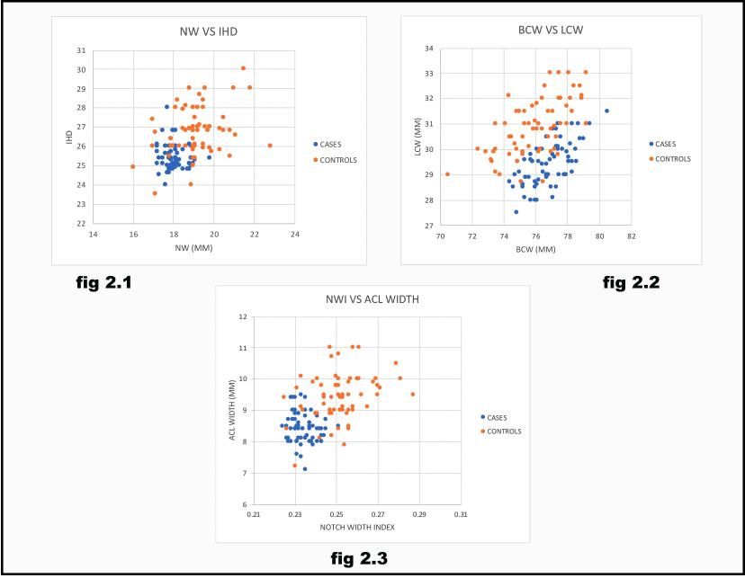
The Influence Of Femoral Morphometric Risk Factors On Acl Injury, A Retrospective Case-..
II. Methodology
This retrospective case-control study was designed by researchers at JIPMER (Jawaharlal Institute of
Postgraduate Medical Education & Research). It was approved with the participant's consent waiver by the
Institutional Ethics Review Board at JIPMER. The medical records of participants who visited the orthopedic
outpatient department (OPD) between January 2017 to December 2019 were reviewed. All participants of either
sex, 18 years or above, with no history of prior knee surgeries with chief complaints of persistent pain and
instability, coupled with h/o of twisting injury to the knee with MRI and clinical evaluation performed one-
month post knee injury were taken as study participants. These participants were then screened for and excluded
from the study if any history/MRI findings suggested multi-ligamentous injury, septic arthritis of the knee,
hemarthrosis of the knee, Skeletal dysplasia, tumours, ligamentous laxity, and malunited intraarticular fractures
of the knee. Thus, the cases (ACL- injured group) represented all Participants meeting the above screening
criteria and having positive findings in clinical tests for an ACL tear (a Positive Lachemann's test and Positive
Pivot shift), indicating the presence of an ACL tear, accompanied by a complete tear of ACL in MRI.
Participants who satisfied the above screening criteria and had knee pain with negative stability tests but
required an MRI for further evaluation and MRI showed an intact ACL were included as controls (ACL -
uninjured group). Clinical assessment and imaging were taken only after a month of the knee injury to avoid
pain during the examination and for hemarthrosis of the injured knee to subside, thereby aiding in more precise
clinical and MRI diagnosis. A third separate researcher, a radiologist, measured the various distal femur
morphometrics on MRI, ensuring consistency in interpretation and measurement technique. Knee MRI
sequences were produced using 1.5 Tesla Magnetom Avanto (Siemens, Germany) with a field of view of
160*160,192*320s, slice thickness 3mm, matrix 256*192 in 3 orthogonal planes. All measurements were
performed using the institutional PACS - centricity universal viewer, GE Health care.
The distal femur anatomical variants measured were Notch width, notch width index, medial femoral
condyle width, lateral femoral condyle, bicondylar width, ACL length, and mid substance ACL width
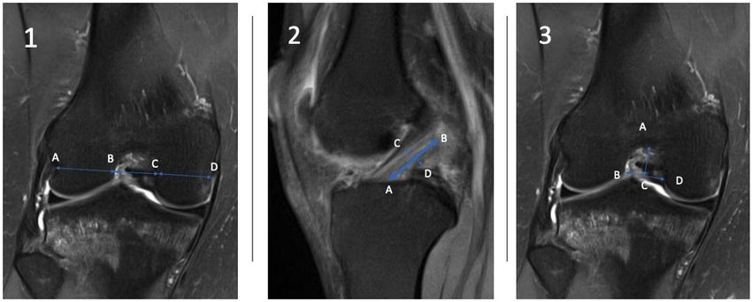
correlating to distal notch entrance. Notch width, NWI, and femoral condyle morphometrics were measured by
using the techniques described by Herzog et al.[6], FIG.1. The ACL morphometrics was measured using the
method described by Ng et al.[7], FIG 1.
Data were analyzed using STATA ver.15.1. Categorical and continuous data were presented as
percentages (%) and mean with standard deviation. Pearson's chi-squared test and Fisher's exact test evaluated
the difference in proportions. A 2-sample t-test was used to determine any significant differences in Distal femur
morphometrics (i.e., NW, MCW, LCW, BCW, MLR, NWI, IHD, ACL Length, ACL width) between the cases
and control groups without any reference to the participant's gender. Further correlation analyses were done
between individual parameters to determine the association.
III. Results
A total of 269 participants were included in the study, of which 202(75%) participants were male.
154(53%) participants belonged between the age of 25 to 50 years, whereas 99(34%) and 36(12%)participants
belonged to age groups 50 years, respectively. A total of 126 participants were included as cases
( ACL-injured group), and 143 participants were included in the control arm (ACL-uninjured group).
The distal femur morphometric means of the cases were NW(18.035 0.85), MCW(28.941 1.21),
LCW(29.329 0.92), BCW(76.305 2.1), MLR(0.98731 0.048), NWI(0.98731 0.008), IHD(26.679 0.737),
ACL Length(30.567 4.19), ACL width(8.350 0.51). The distal femur morphometric means of the controls
were NW(19.100 1.69), MCW(25.685 1.19), LCW(30.726 1.26), BCW(75.575 2.85), MLR(6.11992
65.98), NWI(0.25192 0.016), IHD(26.679 1.18), ACL Length(35.258 4.19), ACL width(9.294 0.758).
Selected summary statistics with the P values based on 2 sample t-tests for NW, MCW, LCW, BCW, MLR,
NWI, IHD, ACL Length, ACL width between the ACL-uninjured and ACL-injured groups are provided in
Table 1.
To provide meaningful insight into the Distribution of anatomical risk factors between injured and
uninjured populations, scatter plots between NWI vs. ACL mid substance width, NWI vs. IHD, BCW vs. LCW
were plotted in figure 2.
IV. Discussion
Our study notes a significant difference in the notch width and NWI among the cases and controls. This
is in concordance with other studies such as Domnick et al.[8], Shelbourne et al.[9], Sonnery et al. [10]. Against
this finding are studied by Lombardo et al.[11], schickendantz et al.[12], and souryal et al. [13]. However, this
finding of significant difference in notch width, NWI adds to the existing literature, which has a divided opinion
on the same quoting bias in measurements, the study subjects, and difference in statistical analysis for the
same[10][14][13]. The difference found in study participants can be attributed to the difference in demographic
DOI: 10.9790/0853-2105022529 www.iosrjournal.org 26 | PageThe Influence Of Femoral Morphometric Risk Factors On Acl Injury, A Retrospective Case-..
characteristics and mode of injury sustained. The participants were not elite sports personalities as in other
studies.
This study provides insight into distal morphometric and ACL injuries by correlating distal femur
morphometrics and their biomechanical implication. Firstly, The cases have significantly smaller ACL lengths
and Mid substance ACL width. When associated with cases having a significantly lower notch width, this
finding correlates with the biomechanics of non-contact ACL injury, wherein a taut ACL, when stretched
against the lateral condyle in a stenosed canal, is more susceptible to an injury[8]. Secondly, It is interesting to
note that bicondylar width and medial condylar width in cases are significantly higher than those in controls.
Axial loading and posterior tibial translation have a unique role in the biomechanics of causing ACL injury,
wherein the above findings suggest a more uniform loading in cases which is probably countered by the larger
surface area of the medial condyle over which the tibia pivots, thereby increasing the stresses in ACL causing
injuries. These findings are concordant with the established injury biomechanics of ACL injury[8][15]. Thirdly,
Considering the femoral notch as a tunnel through which the ACL passes towards its attachment in the femur,
we note that cases have a constricted channel through which ACL passes, i.e., a smaller notch width and a
smaller IHD compared to the controls. Thus any pivoting movement would significantly cause a greater stress
raiser in the tight ACL ligament leading to its injury[3][16].
The graphic depictions of the Distribution of anatomical risk factors among the study participants
between NW vs. IHD, BCW vs. LCW, and NW vs. ACL width are shown in figure 2. we notice that cases are
clustered towards a narrower femoral notch compared to controls. Further, participants with a smaller lateral
condylar notch associated with greater MC, depicted as a greater BCW, are more prone to an ACL injury, again
emphasizing the greater pivoting on the bigger medial condyle base and impingement on the lateral condyle.
Finally, in the scatter plot fig 2.3, cases are seen more clustered towards a smaller NWI and smaller ACL mid
substance width, suggesting that ACL bulk at the site of impingement also plays a role in injury prevention.
These findings are in concordance with the established ACL injury mechanism.
Furthermore, Correlation analysis between the morphometric parameters reveals a direct correlation
between ACL width and IHD, suggesting that a greater femoral notch accommodates a thicker ACL, which
would decrease the chances of incurring an ACL injury.
The study has many limitations. Firstly, the participants were not matched for other risk factors that
would have placed them at a higher risk of suffering an ACL injury. Secondly, participants younger than 18
years were excluded from the study. The reason for exclusion is that bone maturation has yet to occur in these
individuals; thus, evaluating anatomical parameters prematurely can lead to bias in predicting the contribution of
the risk factor in these participants. Thirdly, we acknowledge that MRI measurements can be subjected to inter-
observer inconsistencies. However, with advances in technology from previous studies, with simultaneous
visualization in multiple planes at a single time, we could circumvent this problem for the most part.
V. Conclusions
We note that the distal femur morphometric variations among cases and controls are in accordance with
the existing literature. It is interesting to note that these remain the same despite the difference in demographic
characteristics of the study population. Further, it is seen that the variations in cases that lead to increased
chances of an ACL injury are correlated with the existing ACL injury mechanisms.
References
[1]. Lin C-F, Liu H, Gros MT, Weinhold P, Garrett WE, Yu B. Biomechanical risk factors of non-contact ACL injuries: A stochastic
biomechanical modeling study. Journal of Sport and Health Science 2012;1(1):36–42.
[2]. Barraza LCH, Krishnan. R G, Low J-H, Yeow C-H. The Biomechanics of ACL Injury: Progresses toward Prophylactic Strategies.
Crit Rev Biomed Eng 2013;41(4–5):309–21.
[3]. Domnick C, Raschke MJ, Herbort M. Biomechanics of the anterior cruciate ligament: Physiology, rupture and reconstruction
techniques. WJO 2016;7(2):82.
[4]. Choi WR, Yang J-H, Jeong S-Y, Lee JK. MRI comparison of injury mechanism and anatomical factors between sexes in non-
contact anterior cruciate ligament injuries. PLoS ONE 2019;14(8):e0219586.
[5]. Hashemi J, Chandrashekar N, Gill B, Beynnon BD, Slauterbeck JR, Schutt RC, et al. The Geometry of the Tibial Plateau and Its
Influence on the Biomechanics of the Tibiofemoral Joint: The Journal of Bone and Joint Surgery-American Volume
2008;90(12):2724–34.
[6]. Herzog RJ, Silliman JF, Hutton K, Rodkey WG, Steadman JR. Measurements of the Intercondylar Notch by Plain Film
Radiography and Magnetic Resonance Imaging. Am J Sports Med 1994;22(2):204–10.
[7]. Ng WHA. Imaging of the anterior cruciate ligament. WJO 2011;2(8):75.
[8]. Domnick C, Raschke MJ, Herbort M. Biomechanics of the anterior cruciate ligament: Physiology, rupture and reconstruction
techniques. WJO 2016;7(2):82.
[9]. Shelbourne KD, Davis TJ, Klootwyk TE. The Relationship Between Intercondylar Notch Width of the Femur and the Incidence of
Anterior Cruciate Ligament Tears. Am J Sports Med 1998;26(3):402–8.
[10]. Sonnery-Cottet B, Archbold P, Cucurulo T, Fayard J-M, Bortolletto J, Thaunat M, et al. The influence of the tibial slope and the
size of the intercondylar notch on rupture of the anterior cruciate ligament. The Journal of Bone and Joint Surgery British volume
2011;93-B(11):1475–8.
DOI: 10.9790/0853-2105022529 www.iosrjournal.org 27 | PageThe Influence Of Femoral Morphometric Risk Factors On Acl Injury, A Retrospective Case-..
[11]. Lombardo S, Sethi PM, Starkey C. Intercondylar Notch Stenosis is not a Risk Factor for Anterior Cruciate Ligament Tears in
Professional Male Basketball Players: An 11-Year Prospective Study. Am J Sports Med 2005;33(1):29–34.
[12]. Schickendantz MS, Weiker GG. The predictive value of radiographs in the evaluation of unilateral and bilateral anterior cruciate
ligament injuries. Am J Sports Med 1993;21(1):110–3.
[13]. Souryal TO, Moore HA, Evans JP. Bilaterality in anterior cruciate ligament injuries: Associated intercondylar notch stenosis. Am J
Sports Med 1988;16(5):449–54.
[14]. Khodair S, Elsayed A, Ghieda U. Relationship of distal femoral morphometrics with anterior cruciate ligament injury using MRI.
Tanta Med J 2014;42(2):64.
[15]. Marieswaran M, Jain I, Garg B, Sharma V, Kalyanasundaram D. A Review on Biomechanics of Anterior Cruciate Ligament and
Materials for Reconstruction. Applied Bionics and Biomechanics 2018;2018:1–14.
[16]. Choi WR, Yang J-H, Jeong S-Y, Lee JK. MRI comparison of injury mechanism and anatomical factors between sexes in non-
contact anterior cruciate ligament injuries. PLoS ONE 2019;14(8):e0219586.
Case Control p- value
Age
50 7 29
Sex
Male 106 110
Female 27 46
Notch width 18.035 0.85 19.100 1.69 0.000
Medial condyle width 28.941 1.21 25.685 1.19 0.000
Lateral condyle width 29.329 0.92 30.726 1.26 0.000
Bicondylar width 76.305 2.1 75.575 2.85 0.015
Medial / lateral condyle ratio 0.98731 0.048 0.611992 0.065 0.371
Notch width index 0.23576 0.008 0.25192 0.016 0.000
Intercondylar notch height / depth 26.679 1.18 0.000
25.377 0.737
ACL length 30.567 4.19 35.258 4.19 0.000
ACL width mid substance 8.350 0.51 9.294 0.758 0.000
TABLE: 1 summary statistics for NW, MCW, LCW, BCW, MLR, NWI, IHD, ACL Length, ACL width
between the ACL-uninjured and ACL-injured groups.
DOI: 10.9790/0853-2105022529 www.iosrjournal.org 28 | PageThe Influence Of Femoral Morphometric Risk Factors On Acl Injury, A Retrospective Case-..
FIG 1 . fig 1.1 – AB, BC, CD measure the lateral condylar width, intercondylar notch width, and medial
condylar width. Fig 1.2 – AB, CD measure the ACL length and mid ACL width, Fig 1.3 AC, BD measure the
intercondylar width and height.
Figure 2: scatter plots comparing different anatomical distal femur morphometric variations.
Prabu Mounisamy, et. al. “The Influence of Femoral Morphometric Risk Factors on ACL Injury, a
Retrospective Case-Control Study – An Indian Perspective.” IOSR Journal of Dental and Medical
Sciences (IOSR-JDMS), 21(05), 2022, pp. 25-29.
DOI: 10.9790/0853-2105022529 www.iosrjournal.org 29 | PageYou can also read