The Hunt for Ancient Prions: Archaeal Prion-Like Domains Form Amyloid-Based Epigenetic Elements
←
→
Page content transcription
If your browser does not render page correctly, please read the page content below
The Hunt for Ancient Prions: Archaeal Prion-Like Domains
          Form Amyloid-Based Epigenetic Elements
          Tomasz Zajkowski ,*,1,2,3 Michael D. Lee,†,3 Shamba S. Mondal,†,4 Amanda Carbajal,2,5 Robert Dec,6
          Patrick D. Brennock,2 Radoslaw W. Piast,6 Jessica E. Snyder,2 Nicholas B. Bense,3 Wojciech Dzwolak,6
          Daniel F. Jarosz,7,8 and Lynn J. Rothschild9
          1
           Centre of New Technologies, University of Warsaw, Warsaw, Poland
          2
           University Space Research Association, Mountain View, CA, USA
          3
           Blue Marble Space Institute of Science, Seattle, WA, USA
          4
           Laboratory of Bioinformatics, Nencki Institute of Experimental Biology of Polish Academy of Sciences, Warsaw, Poland
          5
           University of California Santa Cruz, Santa Cruz, CA, USA
          6
           Faculty of Chemistry, Biological and Chemical Research Centre, University of Warsaw, Warsaw, Poland

                                                                                                                                                                     Downloaded from https://academic.oup.com/mbe/article/38/5/2088/6108110 by guest on 18 May 2021
          7
           Department of Chemical and Systems Biology, Stanford University School of Medicine, Stanford, CA, USA
          8
           Department of Developmental Biology, Stanford University School of Medicine, Stanford, CA, USA
          9
           Space Science and Astrobiology Division, NASA Ames Research Center, Moffett Field, CA, USA
          These authors contributed equally to this work.
          *Corresponding author: E-mail: t.zajkowski@cent.uw.edu.pl.
          Associate editor: Tal Pupko

          Abstract
          Prions, proteins that can convert between structurally and functionally distinct states and serve as non-Mendelian
          mechanisms of inheritance, were initially discovered and only known in eukaryotes, and consequently considered to
          likely be a relatively late evolutionary acquisition. However, the recent discovery of prions in bacteria and viruses has
          intimated a potentially more ancient evolutionary origin. Here, we provide evidence that prion-forming domains exist in
          the domain archaea, the last domain of life left unexplored with regard to prions. We searched for archaeal candidate
          prion-forming protein sequences computationally, described their taxonomic distribution and phylogeny, and analyzed
          their associated functional annotations. Using biophysical in vitro assays, cell-based and microscopic approaches, and
          dye-binding analyses, we tested select candidate prion-forming domains for prionogenic characteristics. Out of the 16
          tested, eight formed amyloids, and six acted as protein-based elements of information transfer driving non-Mendelian
          patterns of inheritance. We also identified short peptides from our archaeal prion candidates that can form amyloid
          fibrils independently. Lastly, candidates that tested positively in our assays had significantly higher tyrosine and phe-
Article

          nylalanine content than candidates that tested negatively, an observation that may help future archaeal prion predic-
          tions. Taken together, our discovery of functional prion-forming domains in archaea provides evidence that multiple
          archaeal proteins are capable of acting as prions—thus expanding our knowledge of this epigenetic phenomenon to the
          third and final domain of life and bolstering the possibility that they were present at the time of the last universal
          common ancestor.
          Key words: prion, amyloid, evolution, archaea, LUCA.

              Significance
              This work establishes that amyloid-forming, prion-like domains exist in Archaea and are capable of vertically transmitting
              their prion phenotype—allowing them to function as protein-based elements of inheritance. These observations, coupled
              with prior discoveries in Eukarya and Bacteria, suggest that prion-based self-assembly was potentially present in life’s last
              universal common ancestor, and therefore may be one of the most ancient epigenetic mechanisms.

          Introduction                                                                              baffling patterns of transmission arose because it was caused
          One of the most notable and puzzling disease outbreaks of                                 by a mysterious agent devoid of nucleic acids (Prusiner 1998).
          the last 50 years was bovine spongiform encephalopathy. Its                               The disease left 177 people and over four million cattle dead,

          Published by Oxford University Press on behalf of the Society for Molecular Biology and Evolution 2021.
          This work is written by US Government employees and is in the public domain in the US.
          This article is published and distributed under the terms of the Oxford University Press, Standard Journals Publication Model (https://
          academic.oup.com/journals/pages/open_access/funder_policies/chorus/standard_publication_model)                                            Open Access
          2088                        Mol. Biol. Evol. 38(5):2088–2103 doi:10.1093/molbev/msab010 Advance Access publication January 22, 2021
Hunt for Ancient Prions . doi:10.1093/molbev/msab010                                                                      MBE
harmed the economy of the United Kingdom, and, for a time,           [GARþ] prion to switch between specialist and generalist
left the scientific community with no mechanism to explain           carbon-source utilization strategies, a switch that is heritable
the phenomenon (Murdoch and Murdoch 2015). The cause                 (Brown and Lindquist 2009; Jarosz et al. 2014). The [ESIþ]
was eventually determined to be a misfolded form of an en-           prion drives the emergence and transgenerational inheritance
dogenous protein, designated the Prion Protein (PrP)                 of an activated chromatin state that can result in broad re-
(Prusiner 1998).                                                     sistance to environmental stress, including antifungal drugs
    Although initially named for “proteinaceous infectious par-      (Harvey et al. 2020). The [SMAUGþ] prion allows yeast to
ticles” (Prusiner 1998), ascribing a stable, concise definition to   anticipate nutrient repletion after periods of starvation, pro-
the term “prion” is not trivial. As our scientific understanding     viding a strong selective advantage (Itakura et al. 2020). Other
of prion-like entities continues to develop alongside the con-       nondetrimental prions were found in many distinct species.
cept’s integration into society’s lexicon, the term itself is un-    In Podospora anserina, prion [Het-s] carries out normal cell
der relatively rapid linguistic evolutionary pressure. One           functions in the process of self/nonself recognition (Dalstra
generalizable and useful definition is: “prions are proteins         et al. 2005; Riek and Saupe 2016). In the angiosperm
that convert between structurally and functionally distinct          Arabidopsis thaliana, the Luminidependens protein can

                                                                                                                                         Downloaded from https://academic.oup.com/mbe/article/38/5/2088/6108110 by guest on 18 May 2021
states, at least one of which is self-propagating and self-per-      form a prion and is responsible for signaling flowering as a
petuating” (slightly modified from Alberti et al. [2009] and         result of detecting the temperature change in the environ-
Garcia and Jarosz [2014]). And although certainly useful, this       ment (Chakrabortee, Kayatekin, et al. 2016). In baculoviruses,
does not adequately convey the whole story because it assigns        the prion-forming LEF-10 protein was found to be responsible
the property of prion to the protein itself as an individual unit,   for efficient viral replication and expression (Nan et al. 2019).
whereas some of the key hallmarks of prion activity (e.g., self-     The discovery of functional prions across a broad phyloge-
propagation and non-Mendelian inheritance) only manifest             netic diversity of organisms raises the question of their evo-
through the interaction of multiple copies of a protein under        lutionary origins.
specific conditions. Conceptually this is not entirely dissimilar        The first prions to be discovered form highly ordered
from how quorum sensing can be thought of as an emergent             aggregates called amyloids (Sabate, Rousseau, Schymkowitz,
property, rather than being ascribed to a single unit. It can        Batlle, et al. 2015) and many, but not all, currently known
therefore be useful to also think of the term “prion” as a state
                                                                     prions adopt an amyloid conformation. Amyloids are long,
that emerges when a specific suite of characteristics is met for
                                                                     unbranched protein aggregates characterized by a fibrillar
multiple copies of a protein that is capable of facilitating that
                                                                     morphology and cross b-sheet quaternary protein structure
state. That said, unless noted otherwise, herein the term
                                                                     (Sipe et al. 2016). Just like prions, amyloids were first discov-
“prion” will be utilized in the former, generalized form, refer-
                                                                     ered in association with neurodegeneration. The most well-
ring to an individual protein.
                                                                     known amyloids form protein deposits in the human brain as
    Although prions were being studied in mammals, puzzling
                                                                     in the cases of Ab peptide and Tau protein in Alzheimer’s
nonchromosomal genetic elements were concurrently being
discovered in yeast: [URE3] (Lacroute 1971) and [PSIþ]               disease, huntingtin in Huntington’s disease, a-synuclein in
(Young and Cox 1971). As the prion story unfolded, it soon           Parkinson’s disease, and SOD1 in Amyotrophic lateral sclero-
became clear that [URE3] and [PSIþ] were prions of the               sis (Chiti and Dobson 2006). Similar to prions, amyloids exist
Ure2p and Sup35p proteins, respectively (Chernoff et al.             in myriad organisms, including microbes. Although their pres-
1993; Ter-Avanesyan et al. 1994; Wickner 1994; Lindquist             ence correlates with neurodegeneration, some have argued
et al. 1998; Liebman and Derkatch 1999). All known mam-              that amyloid itself might be overall more beneficial relative to
malian prion diseases are the result of the misfolding of the        other oligomeric forms of aggregation-prone proteins (Walsh
endogenous prion protein (PrP). By contrast, fungal prions are       et al. 2002; Benilova et al. 2012). Amyloids have functional and
more diverse, sparking phenotypes produced by the alterna-           nonpathogenic roles in species as phylogenetically distinct as
tive folds of many different proteins (Wickner 1994; Derkatch        Escherichia coli and humans (Chapman et al. 2002; Biesecker
et al. 1997; Du et al. 2008; Rogoza et al. 2010; Suzuki et al.       et al. 2018). The term “functional amyloids” was created to
2012). Prion conversion of PrP is pathogenic, whereas acqui-         describe nondetrimental amyloids (Fowler et al. 2005). In
sition of many yeast prions is not. As more work has been            microbes, functional amyloids are often found in the extra-
done to elucidate nonpathogenic prion-forming proteins, it           cellular matrix contributing to cellular protection and biofilm
has been shown that prion conversion can serve as a molec-           formation (Chapman et al. 2002; Cegelski et al. 2009). The first
ular switch for multiple biological functions, often exerting a      functional amyloid identified in humans was the PMEL 17
strong influence on survival. They can play a role in adaptive       protein (Fowler et al. 2005; Bissig et al. 2016). Important in
responses to environmental fluctuations (Halfmann and                melanogenesis, amyloids of PMEL 17 create a scaffold for
Lindquist 2010; Rogoza et al. 2010; Itakura et al. 2020), con-       melanin distribution (Fowler et al. 2005; Bissig et al. 2016).
tribute to evolvability by acting as epigenetic elements (True       Amyloid-forming proteins have been detected and experi-
and Lindquist 2000; True et al. 2004; Holmes et al. 2013; Du         mentally verified in all domains of life: Eukaryota, Bacteria,
et al. 2015), and act as evolutionary capacitors (Shorter and        and Archaea (Gebbink et al. 2005; Greenwald and Riek
Lindquist 2005; Masel and Siegal 2009) and bet-hedging devi-         2010; Shanmugam et al. 2019). Prions, on the other hand,
ces (King and Masel 2007; Namy et al. 2008; Halfmann et al.          have to date only been discovered and verified in Eukarya
2010). For example, Saccharomyces cerevisiae employs the             and Bacteria (Prusiner 1998; Yuan and Hochschild 2017).
                                                                                                                                 2089
Zajkowski et al. . doi:10.1093/molbev/msab010 MBE
The propagation of known prions relies on a protein frag- Congo red assay (Fig. 4) Yeast prion reporter assay (Fig. 6)
ment called the prion domain (PrD). These domains are often
enriched in glutamine and asparagine residues and are usually PLAAC prion domain
found in the disordered fragments of proteins. Leveraging
these principles, bioinformatic algorithms can be used to pre- PH domain P3 cPrD12
dict novel PrDs (Li and Lindquist 2000; Michelitsch and
pRANK aggregation domain
Weissman 2000; Alberti et al. 2009). Here, we use computa-
tional prediction methods on 1,262 archaeal proteomes to
ThT assay (Fig. 5A,B) ATR-FTIR (Fig. 5C) TEM (Fig. 5D)
assess the distribution of prion proteins across the archaeal
domain. We identify candidate PrDs (cPrDs) and investigate FIG. 1. Fragments of protein and methods used to analyze them.
the functions associated with the proteins harboring them. Schematic representation of PH domain-containing protein from
We additionally perform a suite of biochemical and genetic Sulfolobus sp. Candidate prion domain identified by PLAAC
experiments on a subset of our identified cPrDs to validate (cPrD12) and aggregation domain identified by pRANK (P3) are
their capacity to form infectious aggregates. Our data reveal marked. Methods applied to analyze the designated fragments are
Downloaded from https://academic.oup.com/mbe/article/38/5/2088/6108110 by guest on 18 May 2021
that multiple archaeal proteins can act as prions, forming listed with references to other figures.
heritable protein aggregates that are capable of acting as
protein-based epigenetic elements. These observations, cou- closely related Nanoarchaeota (GCA_003086475.1 and
pled with prior discoveries in eukarya and bacteria, suggest GCA_003086415.1) and a Euryarchaeota of the class
that prion-based self-assembly is an ancient form of epige- Methanobacteria (GCA_001639265.1; demarcated with red
netics that may have been present in life’s last universal com- plus signs in fig. 2). Other proteomes with relatively high-
mon ancestor (LUCA). densities of identified cPrDs included 10 with 10 cPrDs
per 1,000 proteins (99.2 percentile rank) and 46 with 5
Results (96.4th percentile). Many of these were concentrated within
Computational Identification and Distribution of the class Methanobacteria (across the genera
cPrDs in Archaea Methanobrevibacter, Methanosphaera, and
Leveraging the strong enrichment of glutamine and aspara- Methanobacterium) and within representatives of
gine in prion domains, we utilized the program PLAAC Metallosphaera sedula of the Thermoprotei class (fig. 2).
(Lancaster et al. 2014) to scan for cPrDs in archaeal proteomes
sourced from the UniProt (Bateman 2019) database (see fig. 1 Enriched Functions of Proteins Containing cPrDs
for overview). Of the 1,262 archaeal proteomes scanned, at We utilized Gene Ontology (GO) (Ashburner et al. 2000)
least one cPrD was identified in 873 proteomes (69%). Of annotations (GO “terms”) and goatools (Klopfenstein et al.
the 2,805,234 proteins scanned, 2,797 were identified as con- 2018) to test for enrichment or purification of specific GO
taining cPrDs (0.10%). The distribution of proteomes with terms in our identified cPrD-containing proteins (2,797) as
at least one cPrD identified was largely homogenous across compared with all of the proteins scanned (2,805,234). The
the archaeal domain (fig. 2). There were modestly lower pro- identified cPrD-containing proteins were associated with 706
portions within the Bathyarchaeota (58%) and the Asgard GO terms, 523 of which were found to be significantly
group (62%), and a higher proportion within the enriched or purified in the cPrD group relative to all proteins
Crenarchaeota (79%). We note however that these taxo- (based on Benjamini–Hochberg false-discovery rates 0.05;
nomic summary delineations are somewhat arbitrary, and supplementary data set S2, Supplementary Material online).
as with all databases there can be underlying confounding The vast majority of these (469/523; 90%) were found to be
factors about which lineages may be over- or under- purified in proteins harboring cPrDs relative to all proteins
represented as compared with others. For example, a com- (underrepresented), and 54 were found to be enriched—a
monly sequenced organism will likely be overrepresented, as subset of which are presented in figure 3.
compared with those less commonly sequenced. GO breaks its terms down into three namespaces:
The frequency of identified cPrDs within each proteome Biological Process, Molecular Function, and Cellular
was 2.21 6 3.38 (mean 6 1 SD) with a median of one: 30% Component. Significantly enriched GO terms included pro-
of the scanned proteomes contained 0; 25% had the me- teins involved in: chitin metabolism (GO:0006030), cell wall
dian of 1: 35% had between 2 and 5; and 10% had >5 polysaccharide catabolism (GO:0044347), and cell wall adhe-
(supplementary data set S1, Supplementary Material online). sion (GO:0007155) within the Biological Process namespace;
Normalizing these values to the total number of proteins in transcription coregulator (GO:0003712), tyrosine-based site-
each individual proteome (done as cPrDs per 1,000 proteins) specific recombinase (GO:0009037), and calcium
returns 1.14 6 1.89 (mean 6 1 SD) with a median of 0.6 (GO:0005509) and copper ion binding (GO:0005507) within
(supplementary data set S1, Supplementary Material online; the Molecular Function namespace; and outer membrane
histogram in supplementary fig. S1, Supplementary Material (GO:0019867) within the Cellular Component namespace
online). Based on these normalized values, three proteomes (fig. 3; full results in supplementary data set S2,
were found to have >15 identified cPrDs per 1,000 genes Supplementary Material online). Significantly purified (under-
(placing them in percentile ranks of >99.8) including two represented) GO terms included: cellular amino acid
2090Hunt for Ancient Prions . doi:10.1093/molbev/msab010 MBE
Euryarchaeota
549/777 (71%)
Methanobacteria
Candidate prion domains (cPrDs) Methanobrevibacter spp.
Methanosphaera spp.
per 1,000 proteins Methanobacterium spp.
0.1
>= 5 >= 10 >= 15
Thaumarchaeota
67/98 (68%)
Downloaded from https://academic.oup.com/mbe/article/38/5/2088/6108110 by guest on 18 May 2021
Asgard DPANN
29/47 (62%) 64/91 (70%)
Bathyarchaeota
57/98 (58%) Crenarchaeota
Thermoprotei 92/116 (79%)
Metalosphaera spp.
FIG. 2. A phylogenomic tree of incorporated Archaea with distribution of cPrDs overlain. Tips with at least 1 cPrD are colored blue. Blue diamonds
are places near those with to five cPrDs per 1,000 genes; green circles near those with 10; and red pluses near those with 15. Major taxa
summary values include the number within that group with identified cPrDs out of the total in that group followed by that percentage. The Asgard
group here includes: Thorarchaeota, Odinarchaeota, and Heimdallarchaeota. DPANN group here includes: Woesearchaeota, Pacearchaeota,
Nanoarchaeota, Aenigmarchaeota, Diapherotrites, Micrarchaeota, and Altiarchaeota. The unrooted tree is an estimated maximum-likelihood
of 76 single-copy orthologs forming an alignment of 12,330 amino acids (see Materials and Methods).
(GO:0008652) and nucleotide biosynthesis (GO:0009165) overproduction of only the amyloid-forming portion of the
within Biological Process; nucleotide (GO:0000166) and metal protein is sufficient for prion induction (Ter-Avanesyan et al.
ion binding (GO:0046872) within Molecular Function; and 1994; Masison et al. 1997). This induction is sometimes more
cytoplasm (GO:0005737) and ribosome (GO:0005840) within efficient than with overproduction of the full-length protein
Cellular Component (supplementary data set S2, (Masison and Wickner 1995; Kochneva-Pervukhova et al.
Supplementary Material online). 1998). Another characteristic of known PrDs is their modu-
larity, which allows them to be cloned into other proteins
Multiple Archaeal cPrDs Bind Congo Red thus transferring the ability to form prion aggregates (Li and
Selective binding to dyes, including Congo red, is a diagnostic Lindquist 2000). Preliminary tests also demonstrated that the
characteristic of amyloid. We investigated this property in the use of a cPrD increases the sensitivity of the Congo red assay
cPrDs of the highest scoring (COREscore 30) putative compared with full-length proteins (supplementary fig. S2,
prions from archaeal proteomes. Many cPrDs consisted of Supplementary Material online). For these reasons, and syn-
sequences with long stretches of only Q or N. Because of thesis considerations, we tested cPrDs instead of full-length
the low complexity of these sequences, the synthesis of cod- proteins.
ing DNA in many cases proved impossible. Thus, we chose Bacteria transformed with plasmids encoding the cPrDs
sequences complex enough to allow their synthesis. From this were plated on an induction medium containing Congo
group, we focused on genes with functional annotations, and red. Because in the C-DAG system the expressed cPrDs are
synthesized 16 coding sequences (table 1). We cloned these exported outside of the cell they can interact with the dye in
sequences into a commercially available vector allowing syn- the culture medium. Bacterial colonies that produce cPrDs
thesis and export of investigated cPrDs (Sivanathan and capable of forming amyloids bind Congo red, which then
Hochschild 2013). changes the color of the colony to red. A clear change in
To test if the putative prions were capable of forming color was observed in eight out of 16 colonies (fig. 4 and
amyloid fibrils, we used a bacterial export system for gener- see supplementary fig. S3, Supplementary Material online,
ating extracellular amyloid aggregates (Sivanathan and for results of all 16).
Hochschild 2013). The use of this system enables rapid screen- Microscopy samples were prepared from the colonies that
ing for the ability of the aggregates to bind to the Congo red obtained the strongest color (fig. 4D). Using light microscopy,
dye. A full-length prion protein is not always necessary, nor we confirmed the presence of Congo red binding aggregates
optimal for prion formation (King et al. 1997; Taylor et al. in eight samples as well as in positive controls, but saw no
1999). For example, in most yeast prions proteins, binding to negative controls. We observed that E. coli cells
2091Zajkowski et al. . doi:10.1093/molbev/msab010 MBE
Biological process (GO:0008150) 0 2 4 6 8
chitin metabolism GO:0006030
glucosamine compound metabolism GO:1901071
aminoglycan metabolism GO:0006022
cell wall polysaccharide catabolism GO:0044347
pathogenesis GO:0009405
cell wall modification GO:0042545
cell wall adhesion GO:0007155
Molecular function (GO:0003674)
transcription co-regulator GO:0003712
pectinesterase GO:0030599
tyrosine-based site-specific recombinase GO:0009037
Downloaded from https://academic.oup.com/mbe/article/38/5/2088/6108110 by guest on 18 May 2021
acid phosphatase GO:0003993
cobaltochelatase GO:0051116
dipeptidyl-peptidase GO:0008239
ligase activity, forming N-metal bonds GO:0051002
cysteine-type peptidase GO:0008234
chitin binding GO:0008061
tungstate binding GO:1901359
calcium ion binding GO:0005509
copper ion binding GO:0005507
carbohydrate binding GO:0030246
Cellular component (GO:0005575)
integrin complex GO:0008305
outer membrane GO:0019867
integral component of membrane GO:0016021
0 2 4 6 8
log2-fold enrichment
FIG. 3. Select enriched GO terms of proteins harboring cPrDs as compared with all scanned archaeal proteins. Displayed are log2-fold enrichments
of the given GO term’s frequency among total cPrD-containing proteins (2,797) as compared its frequency among all computationally scanned
archaeal proteins (2,805,234). Considered enrichments were those with Benjamini–Hochberg false-discovery rates of 0.05 (see Materials and
Methods). Full results in supplementary data set S3, Supplementary Material online.
Table 1. Summary of Results from Congo Red and Yeast Prion Reporter Assay Together with the Explanation of Prion Candidates’ IDs Used in the
Text.
Congo Red Yeast Prion Reporter NCBI NCBI NCBI
ID Assay, Results Assay, Results Accession Taxonomy Annotation
cPrD1 CR1 YPR1 AEH61342.1 Methanosalsum zhilinae Transcriptional repressor, CopY family
cPrD2 CR2 YPR2 AGN16545.1 Methanobrevibacter sp. Adhesin-like protein
cPrD3 CR2 YPR1 ARM76690.1 Acidianus manzaensis Zinc ribbon domain-containing protein
cPrD4 CR1 YPR2 KZX15367.1 Methanobrevibacter filiformis tRNA biosynthesis protein MnmC
cPrD5 CR1 YPR2 OQD58440.1 Methanobrevibacter arboriphilus Putative peptidase
cPrD6 CR2 YPR1 WP_009487818.1 Halobacterium sp. DNA starvation/stationary phase
protection Dps
cPrD7 CR2 YPR1 WP_013774943.1 Acidianus hospitalis Zinc ribbon domain-containing protein
cPrD8 CR1 YPR1 WP_014027445.1 Pyrolobus fumarii Tyrosine-type recombinase/integrase
cPrD9 CR1 YPR1 WP_042706453.1 Methanomicrobium mobile YIP1 family protein
cPrD10 CR2 YPR1 WP_054838334.1 Sulfolobus metallicus Zinc ribbon domain-containing protein
cPrD11 CR1 YPR1 WP_066970924.1 Methanobrevibacter filiformis NADAR family protein
cPrD12 CR1 YPR1 WP_066972153.1 Methanobrevibacter filiformis PH domain-containing protein
cPrD13 CR1 YPR1 WP_069282784.1 Sulfolobus sp. Zinc ribbon domain-containing protein
cPrD14 CR2 YPR2 WP_074791546.1 Haloferax larsenii Ribonuclease BN
cPrD15 CR2 YPR2 WP_082223394.1 Halosimplex carsbadense CBS domain-containing protein
cPrD16 CR2 YPR2 WP_084269446.1 Methanobrevibacter curvatus ATP-dependent protease LonB
2092Hunt for Ancient Prions . doi:10.1093/molbev/msab010 MBE
A ylo
id Positive Negative
am ril control control cPrD1 cPrD12
fi b
C
cPrD
E. coli
3 mm
Congo red D
B
100 µm
cPrD E
E. coli
100 µm
Congo red
Downloaded from https://academic.oup.com/mbe/article/38/5/2088/6108110 by guest on 18 May 2021
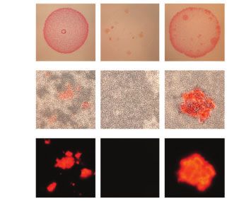
FIG. 4. Comparison of colonies and corresponding protein aggregates produced by bacteria transformed with plasmids encoding cPrDs. (A and B)
Two variants of possible results described in a cartoon. (A) Candidate cPrD forms amyloid aggregates that bind Congo red. (B) cPrD does not form
amyloid aggregates therefore does not bind Congo red. (C) Colonies of Escherichia coli grew on the induction medium containing 0.1% Congo red.
The red color of the colony indicates that the exported protein binds to Congo red. Binding of Congo red is typical for amyloid fibrils. (D) Light
microscopy of preparations made of the same bacterial colonies show aggregates binding Congo red and bacterial cells concentrating around
them. (E) Fluorescent microscopy of protein aggregates from panel (B) is shown. The amyloidogenic fragment of Sup35 (residues 2–253) was used
as a positive control. Fragment of Sup35 not able to form amyloids (residues 125–253) was used as a negative control. Protein names and organisms
from which candidate prion domains (cPrDs) were selected are listed in table 1.
tend to concentrate around the aggregates, consistent with total reflectance Fourier transform infrared spectroscopy
the known role of amyloid aggregates in cell adhesion in (ATRFTIR) (fig. 5C). The major spectral components of
biofilms (Larsen et al. 2007). Detection of Congo red fluores- the vibrational amide I band were below 1,630 cm1 for all
cence in some cases made aggregates more noticeable four peptides we tested, implying the presence of parallel b-
(fig. 4E). In summary, we showed that several cPrDs from sheet structure characteristic of amyloid fibrils
Archaea can bind amyloid specific dye. This is the first step (Zandomeneghi et al. 2009). For P1, the b-sheet signal at
in verification of possible prion nature of the proteins. 1,629 cm1 flanks the dominant spectral component at
1,656 cm1 indicating possible coexistence of tightly packed
Archaeal cPrDs Form Amyloids In Vitro amyloidal b-core with abundant and less ordered conforma-
Congo red staining alone is insufficient for confirmation of tions. A similar observation has been made for amyloid fibrils
the amyloid nature of protein aggregates (Yakupova et al. of Tau protein (Wegmann et al. 2013). We also performed a
2019). Thus, to further evaluate the ability of archaeal prion molecular dynamics simulation of peptide P2 showing that
candidates to form amyloids, we employed an additional set presence of a preformed b-sheet promotes the formation of a
of tests. Following the hypothesis that small regions of a pro- new filament of b-sheet out of unfolded P2 peptide (supple-
tein are often both necessary and sufficient to drive prion mentary fig. S4, Supplementary Material online). Finally, to
behavior (Kochneva-Pervukhova et al. 1998), we analyzed further examine the morphology of the aggregates, we per-
even further shortened segments of the cPrDs. The length formed transmission electron microscopy. These analyses
of cPrDs that yielded positive results in the Congo red assay confirmed the presence of amyloid fibrils in all analyzed prep-
varied from 62 to 179 amino acids. To choose fragments of arations (fig. 5D). Collectively these data establish that amy-
putative archaeal prions most likely to drive aggregation, we loid conversion is promoted by specific sequences within the
used pRANK that assigns 20 amino acid long sequence win- cPrDs.
dow. As a principle, we selected for further analysis only these
peptides which were initially soluble in water. We observed a Archaeal cPrDs Can Promote Protein-Based
very significant increase in thioflavin T (ThT) fluorescence for Inheritance
aggregated forms of peptides P1, P4 and moderate increase of The unusual folding landscapes of prion proteins allow them
ThT emission for P2 and P3 (fig. 5). Binding of ThT to amy- to act as epigenetic mechanisms of inheritance (Alberti et al.
loids increases the quantum yield of fluorescence of the dye 2009). To test whether this was true for our identified archaeal
(Biancalana and Koide 2010). Varying enhancement of ThT cPrDs, we employed an assay based on the well-characterized
fluorescence is often observed for amyloid fibrils, depending prion phenotypes of S. cerevisiae that depend on the prion
on structural features that may reduce ThT binding affinity state of Sup35 protein (Alberti et al. 2009). Normally, the
(Cloe et al. 2011). Furthermore, we found that dissolving protein Sup35 is a part of a translation-termination complex
some peptides in water will trigger a spontaneous increase responsible for recognition of the stop codon and termina-
in ThT emission intensity until a plateau is reached (e.g., P4— tion of translation. The yeast strain that we used in these
see fig. 5B), a hallmark of amyloid formation. To examine the experiments has a premature stop codon in ADE1 gene cod-
secondary structure of the aggregates, we used attenuated ing for SAICAR synthetase. Premature translation termination
2093Zajkowski et al. . doi:10.1093/molbev/msab010 MBE
A B C
ThT fluorescence intensity [a.u.] RC β
ThT fluorescence intensity [a.u.]
P1
8000 1656 1625
5000
P1
6000 P2
P4 4000
P3
4000
3000 P4
P2
400 2000
P3 P4
200 1000 1781
BL
0 0
0 12 24 36 1800 1750 1700 1650 1600
Time [hours] Wavenumber [cm-1]
D
Downloaded from https://academic.oup.com/mbe/article/38/5/2088/6108110 by guest on 18 May 2021
P1 P2 P3 P4
from cPrD5 from cPrD11 from cPrD12 from cPrD13
NNTNNNSNNSIKNINNSNNI KPQNNTNNTTNNNTTNNNTN HNPQNQISRYNNSQYNDSQY MMSMMACNQPIGLGGQQQII
200 nm 200 nm 200 nm 200 nm 200 nm
10 mg/ml 10 mg/ml 10 mg/ml 10 mg/ml 1 mg/ml
FIG. 5. Analysis of aggregates formed by pRANK-peptides. (A) Histogram showing ThT fluorescence intensity levels of samples containing peptides
derived from prion candidates OQD58440.1 (P1); WP_066970924.1 (P2); WP_066972153.1 (P3); WP_069282784.1(P4). Prior to measurement,
samples with the composition: 2.5 mg ml1 peptide, 0.05 M NaCl, 20 mM Th, H2O, pH around 3, were incubated for 24 h at 37 . Three independent
measurements were made for each sample, which was the basis for calculating the mean and error bar. For comparison, the result for the blank is
also presented (BL). (B) ThT fluorescence-monitored reassociation of WP_069282784.1 (P4) peptide. Sample composition and measurement
conditions are the same as in the case of (A). (C) ATRFTIR spectra of the amide I band region of samples containing pRANK-peptides. Before
measurement, small portions of lyophilized peptides were suspended in water, which was then gently evaporated. (D) Micrographs of all four
peptides tested obtained using a transmission electron microscope show the fibrillar morphology of the aggregates. Peptide WP_069282784.1 (P4)
is shown in two different concentrations.
in ADE1 causes accumulation of red pigment—the product bearing the chimeric protein should have a phenotype resem-
of polymerization of aminoimidazole ribotide (AIR) (Smirnov bling [PSIþ]. In contrast, a cPrD that is unable to facilitate the
et al. 1967; Nevzglyadova et al. 2011)—and the ensuing lack of prion-like conversion would have a [psi] phenotype.
adenine production precludes the growth of naive, [psi] cells We substituted the original PrD of Sup35 with 16 different
on adenine-deficient medium (SD-Ade). Occasionally, Sup35 archaeal cPrDs, generating a series of cPrD-Sup35MC chimeric
undergoes conformational conversion to a prion state in proteins. In this yeast-based prion reporter assay (YPR), we
which it forms amyloid fibrils. In this state, the protein is found that 10 chimeric strains grew colonies of a significant
sequestered into aggregates, inhibiting translation termina- size on SD-Ade media. To distinguish quantitatively between
tion—the [PSIþ] phenotype—and promoting readthrough positive (YPRþ) and negative (YPR) results of the assay,
of the premature stop codon in ADE1. Colonies with this images of the Petri dishes were analyzed using ImageJ (see
phenotype can grow on SD-Ade medium, and appear white Materials and Methods for details). Most chimeric strains
on nutrient-rich media like Yeast Peptone Dextrose (YPD) when plated on YPD medium grew colonies much whiter
because accumulation of the red pigment is reduced. than the negative control, suggesting aberrant synthesis of red
Sup35 protein consists of an N-terminal PrD, a middle pigment (fig. 6; see supplementary figs. S5, S7, S8.1, and S8.2,
region (M), and a C-terminal functional domain (C). The Supplementary Material online, for all positive and negative
PrD of Sup35 and other prions are modular and can be trans- results; results are also summarized in table 1 and supplemen-
ferred to nonprion proteins creating new protein-based ele- tary data set S3, Supplementary Material online). The reduc-
ments of inheritance (Li and Lindquist 2000). In our tion of red pigment is characteristic for [PSIþ] phenotype and
experiment, we tested if the original PrD of Sup35 can be indicates the presence of prion form of Sup35. We restreaked
substituted by cPrDs from archaea while preserving the ability the colonies that generated a phenotype resembling [PSIþ]
to undergo prion-dependent phenotypic change. If a cPrD (YPRþ) on YPD media. Some colonies showed reversion to
can function as a bona fide prion domain, the yeast colony [psi] phenotype suggesting that cPrD-Sup35MC is in a
2094Hunt for Ancient Prions . doi:10.1093/molbev/msab010 MBE
A Negative hit (YPR-) Positive hit (YPR+)
do not grow on SD-Ade grow on SD-Ade
red pigment on YPD no red pigment on YPD
incomplete ade1-14 translation complete ade1-14 translation
(accumulation of red pigment) (no red pigment)
start ade1-14 mRNA stop stop start ade1-14 mRNA stop stop
ade1-14 ade1-14
premature
stop-codon
cPrD-SUP35MC recognition cPrD-SUP35MC premature
stop-codon
amyloid read-through
aggregation
functional
repressor non-functional
repressor
cPrD-Sup35MC
cPrD-Sup35MC prion conformation
Downloaded from https://academic.oup.com/mbe/article/38/5/2088/6108110 by guest on 18 May 2021
non-prion conformation
B Negative Positive
cPrD1 cPrD7
SD-Ade
+ + + + + - -
C
YPD
red white white white red/white red/white red/white red red/white
FIG. 6. cPrD-Sup35MC strains display different ability to grow on media lacking adenine and a variety of colony colors. (A) Cartoon explaining the
mechanism of accumulation of red pigment, which depends on the formation of functional repressor. (B and C) Representative images of the
colonies of cPrD-Sup35MC-expressing strains growing on SD-Ade and YPD plates respectively (see supplementary fig. S5, Supplementary Material
online, for all 16 tested and supplementary fig. S7, Supplementary Material online, for images of the whole area of Petri dishes). Colonies
phenotypes [psi] and [PSIþ] are shown for comparison. Positive results are marked with “þ” and are considered positive results of the yeast
prion reporter assay (YPRþ). Negative results are marked with “” and are considered negative results of the yeast prion.
metastable state allowing reversion of prion phenotype (sup- in the sequences of the proteins that tested positive in both
plementary fig. S6, Supplementary Material online). assays, we observed an additional pattern of consecutively
Based on this set of experiments, we conclude that ar- occurring proline and glutamine (P and Q) with nearby aro-
chaeal cPrDs can functionally substitute PrD of Sup35 and matic residues such as tyrosine and phenylalanine (Y and F;
facilitate the formation of prion-based elements of supplementary fig. S10B and C, Supplementary Material on-
inheritance. line)—which was absent in those that tested negative in both
assays (supplementary fig. S10D, Supplementary Material on-
line). Tyrosine content was generally higher in the CRþ/
Tyrosine and/or Phenylalanine Content Distinguishes YPRþ group (Kruskal–Wallis H-test P ¼ 0.036; Wilcoxon
Functional cPrDs rank-sum test P ¼ 0.042; fig. 7A). The only CRþ/YPRþ
In the experiments described above, we found a total of six cPrD that contained no tyrosine, harbored the highest
cPrDs that tested positive both in Congo red and yeast prion amount of phenylalanine (5%) among all cPrDs. Tyrosine
reporter assays (CRþ/YPRþ), and four that tested negative in and phenylalanine content was significantly higher in the
both assays (CR/YPR). We investigated the differences in CRþ/YPRþ group, and also in Q/N-rich PrDs of known prion
sequence composition of cPrDs from these two groups. The proteins in yeast, than in the CR/YPR group (Kruskal–
distribution of glutamine and asparagine content, a key driver Wallis H-test P ¼ 0.01, 0.005; Wilcoxon rank-sum test
of prion behavior (Michelitsch and Weissman 2000), was sim- P ¼ 0.01, 0.005, respectively; fig. 7B). Moreover, the frequency
ilar in each (fig. 7A and supplementary fig. S10A, of tyrosine and phenylalanine in PrDs of the only two cur-
Supplementary Material online). Yet in searching for motifs rently known bacterial prion proteins—transcription
2095Zajkowski et al. . doi:10.1093/molbev/msab010 MBE
A strictly conserved in mammalian PrPs and may function as
an aggregation gatekeeper (Huang and Caflisch 2015a) (sup-
plementary fig. S11, Supplementary Material online). During
self-assembly mediated by this segment, stacks of tyrosine
and phenylalanine stabilize the amyloid core formed by the
stacks of Q/Ns (Gallagher-Jones et al. 2018). In addition, ty-
rosine Y218 which is strictly conserved in mammalian PrPs
(supplementary fig. S11, Supplementary Material online), sta-
bilizes the amyloid fibril formed by full-length human PrP
(Wang et al. 2020). This suggests a potentially generalizable
role of tyrosine/phenylalanine in prion formation, not only
limited to Q/N-rich PrDs.
Discussion
Downloaded from https://academic.oup.com/mbe/article/38/5/2088/6108110 by guest on 18 May 2021
B
Long viewed as enigmatic drivers of disease, prions have
emerged as a form of epigenetics beyond the chromosome
that can be adaptive in eukaryotes ranging from fungi (Namy
et al. 2008; Brown and Lindquist 2009; Li and Kowal 2012;
Jarosz et al. 2014; Harvey et al. 2018) to humans (Hou et al.
2011). Early analyses (Michelitsch and Weissman 2000) sug-
gested prions were absent in other domains of life, consistent
FIG. 7. Tyrosine and/or phenylalanine distinguish positive-testing
with a prevailing view that they are evolutionarily young
cPrDs from negative-testing cPrDs. (A) Comparison of amino acid elements. The recent discovery of prions in bacteria (Yuan
frequency in cPrDs that tested positive in both Congo red and Sup35- and Hochschild 2017) challenged this dogma, yet prions have
based yeast-assay (CRþ/YPRþ), and ones that tested negative in not yet been identified in archaea. In this study, we show that
both experimental assays (CR/YPR). Tyrosine (Y) content of multiple archaeal cPrDs can form amyloids and act as bona
the CRþ/YPRþ cPrDs were higher than the CR/YPR cPrDs in fide elements of information transfer in living cells, fulfilling
general. The CRþ/YPRþ cPrD with no tyrosine (lowest datapoint fundamental tenets of the prion characterization. To the best
in distribution of Y) is the same with highest phenylalanine (F) fre- of our knowledge, neither prions nor intracellular amyloids
quency (highest data point in distribution of F). (B) Frequencies of
have been reported in the domain Archaea to date.
tyrosine and phenylalanine in archaeal cPrDs, yeast, and bacterial
PrDs. CRþ/YPRþ cPrDs as well as yeast PrDs contain significantly
Confirming their presence would indicate their existence in
higher Y/F than CR/YPR cPrDs. The Y/F frequency distribution in all three domains of life, suggesting either that prions were
bacterial PrDs is also similar to that in CRþ/YPRþ cPrDs and yeast present during the time of LUCA, or that they are conver-
PrDs. Since the bacterial group contained PrDs of only the two ex- gently evolved.
perimentally confirmed prions (Rho and SSB), no statistics were per- Though many prions do not form amyloid (Wickner et al.
formed to compare the group with other ones. 2006; Chakrabortee, Byers, et al. 2016; Chakravarty et al. 2020),
in the current study, we focused on amyloid-forming cPrDs
termination factor Rho from Clostridium botulinum and because recently identified bacterial prions have been shown
single-stranded DNA-binding protein SSB from to form amyloids (Yuan and Hochschild 2017), and amyloid
Campylobacter hominis—also revealed elevated levels as fibril formation can also be detected with a relatively high-
compared with our CR/YPR group (fig. 7B). The differ- throughput experimental survey like the Congo red assay
ence between CR/YPR and other groups was enhanced used here. We utilized the computational program PLAAC
further when we compared the content of all amino acids (Lancaster et al. 2014) to identify novel cPrDs. Although
with aromatic side chains (supplementary fig. S10E, PLAAC’s underlying algorithm was trained on yeast PrDs, it
Supplementary Material online). Thus, content of aromatic has recently proven successful in facilitating the identification
residues, especially of tyrosine and phenylalanine, in Q/N-rich of prions in bacteria (Yuan and Hochschild 2017; Fleming
PrDs may play a key role in determining the formation of et al. 2019). Taken with our work here, in which PLAAC
prions across the evolutionary spectrum. also enabled the identification of likely PrDs in Archaea, this
Unlike yeast (or similar prokaryotic) prions which essen- speaks to the chemically conserved nature of at least a subset
tially harbor modular, transferable Q/N-rich PrDs, mamma- of (c)PrDs (amyloid-forming) across all three domains of life.
lian prion protein (PrP) does not have such a region. In We stress, however, that in this initial exploration of potential
addition, PrP and yeast prion proteins do not share significant prions within the domain Archaea, we have performed a
sequence similarity. Interestingly, a segment of the human somewhat limited rather than exhaustive search (being
prion protein (PrP) 169-YSNQNNF-175 containing glutamine, guided by current information largely sourced from Q/N-
asparagine, tyrosine, and phenylalanine, is required for its self- rich, amyloid-forming, yeast-derived prions). Bacteria and ar-
assembly (Huang and Caflisch 2015b). Moreover, the phenyl- chaeal proteomes appear to have fewer Q/N-rich regions in
alanine F175 is highly conserved, and the tyrosine Y169 is general as compared with eukaryotes (Michelitsch and
2096Hunt for Ancient Prions . doi:10.1093/molbev/msab010 MBE
Weissman 2000), but it is as yet still unclear if this translates to nested underneath it such as calcium (GO:0005509) and cop-
them having fewer prions overall, or if it is the case that they per ion binding (GO:0005507) are enriched (fig. 3). This sug-
are encoded with a different chemical signature to which we gests that there is something chemically consistent with Q/N-
currently remain naive. rich amyloids within these specific ion-binding proteins that is
To investigate our computationally identified cPrDs exper- distinct from other metal ion-binding proteins. Although we
imentally, we utilized biochemical and genetic approaches observed an enrichment in outer membrane cellular compo-
(listed in regard to protein fragment, see fig. 1). This included nents and in several cell wall-related biological processes such
staining with amyloid specific dye (fig. 4), infrared spectros- as chitin binding, chitin metabolism, and cell wall adhesion
copy (fig. 5A–C), transmission electron microscopy (fig. 5D), (fig. 3 and supplementary data set S2, Supplementary
and the yeast prion reporter assay (fig. 6). We utilized the Material online), we note that it may simply be the case
model organisms E. coli and S. cerevisiae in our experiments. that cell-wall related proteins like these might tend to harbor
Future work will require working with the source organism of Q/N-rich amyloids. This may place them disproportionately
the cPrD and testing the entire protein. in the category of being detected by PLAAC, but not likely to
Looking across the archaeal domain, many species harbor present prion-like characteristics upon further experimental
Downloaded from https://academic.oup.com/mbe/article/38/5/2088/6108110 by guest on 18 May 2021
at least 1 cPrD. However, Methanobacteria stand out as es- scrutiny. In this case, the only cell-wall related cPrD we tested
pecially rich in cPrDs (fig. 2, right side). Several members of the was found to be negative in both the Congo red and yeast-
genera Methanobrevibacter, Methanosphaera, and prion reporter assays (cPrD2; adhesin-like protein; table 1 and
Methanobacterium have five or more cPrDs per 1,000 pro- supplementary data set S3, Supplementary Material online).
teins (supplementary data set S1, Supplementary Material We observed that tyrosine and phenylalanine content, as
online). Out of 16 candidates tested, six were from the well as total content of aromatic residues, was significantly
Class Methanobacteriaceae, two of which tested positive un- higher in the CRþ/YPRþ group. Tyrosine was also the most
der both experimental assays (table 1 and supplementary abundant hydrophobic residue in PrDs identified in yeast
data set S3, Supplementary Material online). By far the dom- (Alberti et al. 2009; Sabate et al. 2015). MacLea et al. (2015)
inant Gene Ontology (GO) term associated with the cPrDs showed that tyrosine is necessary for stable prion variants of
identified in this group was integral component of membrane Sup35. Later it was proposed that aromatic residues are fa-
from the Cellular Component namespace (GO:0016021; vored in PrDs, because they promote prion formation and
70% of all assigned GO terms; supplementary data set S4, chaperone-dependent prion propagation while avoiding de-
Supplementary Material online), but this was also the case tection by the degradation machinery (Gonzalez Nelson et al.
when looking exclusively outside of the Methanobacteria— 2014; Cascarina et al. 2018). Recently prion formation has
with 58% of all GO terms associated with cPrDs being been associated with phase behavior (Franzmann et al.
GO:0016021, which was enriched overall (fig. 3). The majority 2018). Interestingly it was shown that phase behavior is de-
of the remaining cPrDs from the Methanobacteria were not termined by the number of aromatic residues like tyrosine
annotated (supplementary data set S4, Supplementary and phenylalanine, and their patterning (Martin et al. 2020).
Material online). Possibly contributing to their relatively Our observation adds to the above showing that the role of
higher cPrD frequencies is that they had on average fewer these aromatic residues in prion formation is universal across
proteins overall. The average number of proteins from the domains of life.
incorporated Methanobacteria was 1,915 6 324 (mean 6 1 The first prions discovered in the Domain Eukaryota (yeast
SD), as compared with 2,238 6 1,028 for all others. Likely translation release factor Sup35) (Ter-Avanesyan et al. 1994)
further contributing is that the Q/N-rich regions PLAAC is and the Domain Bacteria (transcription termination factor
identifying inherently tend to be higher in AT-content due to Rho) (Yuan and Hochschild 2017) were both related to cel-
the possible codons encoding them (N ¼ AAT or AAC; lular regulation. Further, amyloids in particular have been
Q ¼ CAA or CAG; supplementary data set S5, noted for their roles in signal transduction such as those
Supplementary Material online), and Methanobacteria have involved with programmed cell death (Daskalov et al.
relatively higher AT-content compared with all others 2015). In the current study, one of our positive-testing prion
(65 6 7.32% vs. 51 6 11.9%; Welch t-test P ¼ 7.64e-23). In candidates was transcriptional repressor CopY (cPrD1; table 1
general, those with higher AT content have higher normalized and supplementary data set S3, Supplementary Material on-
frequencies of cPrDs (supplementary fig. S9, Supplementary line), and one of our most highly enriched GO terms is in-
Material online). volved with transcription regulation (GO:0003712; fig. 3). As
Our GO enrichment analysis revealed that of the annota- amyloid-based prions themselves can orchestrate a regulatory
tions that were significantly enriched or purified in proteins network (Gilks 2004; Hou et al. 2011; Yuan and Hochschild
containing our cPrDs, the vast majority (90%) were purified 2017; Jakobson and Jarosz 2018), this suggests that additional
(e.g., depleted). As such, there is a high level of functional regulatory factors may be controlled by aggregation if they
constraint with regard to which functional domains cPrDs harbor a cPrD. This would provide a scenario in which the
tend to stably coexist with (meaning within the same pro- aggregation state of the protein (as dictated by intracellular
tein). It is notable that overall metal ion binding conditions) might be a mechanism for controlling its activity.
(GO:0046872) is purified in the proteins harboring cPrDs rel- As in the case with the eukaryotic Sup35 (upon which aggre-
ative to all proteins (supplementary data set S2, gation results in less efficient translation termination) (True
Supplementary Material online), yet specific “child” GO terms and Lindquist 2000), and with the bacterial transcriptional
2097Zajkowski et al. . doi:10.1093/molbev/msab010 MBE
terminator Rho (in which aggregation results in transcrip- resulting in 1,262 proteomes holding 2,805,234 proteins as
tional read-through) (Yuan and Hochschild 2017), these types accessed on March 27, 2020.
of “interferences” ultimately may provide a selective benefit The command-line version of PLAAC (Lancaster et al.
by enabling a population under stress to rapidly explore a 2014) (installed from https://github.com/whitehead/plaac in
broader phenotypic landscape. Indeed, it has been demon- April of 2020) was used to identify proteins with putative
strated that prions in S. cerevisiae can confer a fitness advan- prion domains in each proteome individually with default
tage (Halfmann et al. 2010; Suzuki et al. 2012; Itakura et al. settings other than “-a 0” — which tells PLAAC to calculate
2020). The observation that cPrDs and PrDs are commonly and use the current proteome’s background amino acid fre-
found within genes involved in cellular regulation across all quencies entirely, rather than those of S. cerevisiae. From
three domains of life suggests either that: 1) PrDs within these those for which a core length of at least 60 was identified,
gene families are convergently evolved; or 2) PrDs may be a several candidates that also possessed corresponding anno-
relatively early-evolved manifestation of cellular regulation tations were selected for experimental validation.
that was present at the time of LUCA. For now, which of GO enrichment analyses were performed to identify en-
richment/purification of frequencies of GO annotations in
Downloaded from https://academic.oup.com/mbe/article/38/5/2088/6108110 by guest on 18 May 2021
these scenarios unfolded throughout Earth’s biological history
is a question that remains outstanding. the proteins holding identified cPrDs (2,797) as compared
with all scanned proteins (2,805,234) using the goatools
Conclusion v0.6.10 (Klopfenstein et al. 2018) “find_enrichment.py” script
with default settings. Statistical significance was defined as
With this work, we show that archaeal cPrDs can facilitate the
those with Benjamini-Hochberg false-discovery rates of
acquisition of the prion phenotype, allowing them to func-
0.05. Many related GO terms were statistically significant
tion as protein-based elements of inheritance—thus expand- at different depth levels and in those cases the term with the
ing our knowledge of this epigenetic phenomenon to the lowest depth was represented.
third and final domain of life. This adds support to the hy- The phylogenomic tree was produced with GToTree
pothesis that amyloid-based prions were present at the ear- v1.4.11 (Lee and Ponty 2019), within which 76 single-copy
liest stages of life’s evolution. The current study represents an orthologs specific to the archaeal domain (using the
example of a top-down approach that pushes the story of “Archaea.hmm” included with GToTree) were identified
amyloid-based functions further back in evolutionary time. with HMMER3 v3.2.1 (Eddy 2011), individually aligned with
This is complementary to work investigating the “amyloid muscle v3.8.1551 (Edgar 2004), trimmed with trimal (Capella-
world” theory of the origin of life (Chernoff 2004; Maury Gutierrez et al. 2009), and concatenated prior to phylogenetic
2018) that proposes that amyloids may have been forming estimation with FastTree2 v2.1.10 (Price et al. 2010). Tree was
from very short peptides before the emergence of what we initially visualized and edited with the Interactive Tree of Life
might consider the first “living” system (Rode et al. 1999; interface (Letunic and Bork 2007).
Greenwald and Riek 2012; Rufo et al. 2014; Greenwald et al. Aggregation domains (20-amino acid long) from prion
2016; Rout et al. 2018). These two approaches are heading candidates were selected for synthesis with pRANK (http://
toward each other, slowly closing the gap between them, with faculty.pieas.edu.pk/fayyaz/prank.html, last accessed February
many interesting questions remaining to be answered and 2021) (Afsar Minhas et al. 2017) using default settings.
asked. Does a continuity exist between prebiotic amyloids
and modern amyloid-based prions (Lupi et al. 2006)? Are Colony Color Phenotype—Congo Red Assay
extant amyloid-based prions the biochemically evolved Curli-dependent amyloid generator (C-DAG) was used to
descendants of a prebiotic system capable of self-assembly direct the export of heterologous cPrDs to the cell surface
and self-aggregation? Did the self-assembly and self- of E. coli strain VS45 (Sivanathan and Hochschild 2013). Both
aggregation of these short peptides facilitate an expansion bacterial strain and expression vector enabling protein pro-
in range of potential interactions and functionalities? This duction under the control of the arabinose-inducible PBAD
work demonstrating protein-based, epigenetic inheritance promoter was part of commercially available C-DAG amyly-
via PrDs derived from the archaeal domain of life is a valuable loidogenicity kit https://www.kerafast.com/productgroup/
540/c-dag-amyloidogenicity-kit (last accessed February
step toward bridging this gap, but clearly there are many
2021). Export-directed fusion proteins contained the first 42
more to be taken.
residues of CsgA signal sequence at the N terminus as de-
Materials and Methods scribed in the kit protocol. Sec translocon signal sequence is
removed during translocation across the inner membrane,
Computational Identification and Classification of the 22-residue targeting sequence is retained at the N-
Prion Candidates terminus.
Archaeal reference proteomes and annotation information, Lumio tag (CCPGCCGAGG) and His6 tag were added to
including Gene Ontology (GO) annotations (Ashburner et al. the cPrD sequence at the C terminus during gene synthesis
2000), were downloaded from the UniProt database (www.idtdna.com, last accessed February 2021).
(Bateman 2019). To systematically exclude low-quality pro- For assessment of colony color phenotype, overnight cul-
teomes, only those ranked as “standard” based on UniProt’s tures of VS45 were transformed with plasmids containing
“Complete Proteome Detector” algorithm were incorporated, cPrDs. Bacteria were diluted to OD600 0.01 in LB
2098You can also read