AP Biology: Study Guide - EBSCO Connect
←
→
Page content transcription
If your browser does not render page correctly, please read the page content below
AP Biology:
 Study Guide

AP is a registered trademark of the College Board, which was not involved in the production of, and does not endorse, this
product.
Key Exam Details
The AP® Biology exam is a 3-hour, end-of-course test comprised of 60 multiple-choice questions,
for which you will have 1 hour and 30 minutes (this counts for 50% of your score) and 6 free-
response questions, for which you will have 1 hour and 30 minutes (this counts for 50% of your
score).

The exam covers the following course content categories:
 • Chemistry of Life: 8–11% of test questions
 • Cell Structure and Function: 10–13% of test questions
 • Cellular Energetics: 12–16% of test questions
 • Cell Communication and Cell Cycle: 10–15% of test questions
 • Heredity: 8–11% of test questions
 • Gene Expression and Regulation: 12–16% of test questions
 • Natural Selection: 13–20% of test questions
 • Ecology: 10–15% of test questions

This guide will offer an overview of the main tested subjects, along with sample AP multiple-
choice questions that look like the questions you will see on test day.

Chemistry of Life
About 8–11% of the questions on your AP Biology exam will cover the topic Chemistry of Life.

Water and the Elements of Life
Water is made of two hydrogen molecules covalently bonded to an oxygen molecule. The oxygen
atom pulls most of the electrons in the water molecule toward it, giving it a slightly negative
charge and the hydrogen atoms a slightly positive charge. Molecules like water that have distinct
regions of charge based on bond structure are called polar compounds. The charge structure of
water also creates a unique shape, where the hydrogen molecules are concentrated on one side
of the oxygen atom.

The polar nature and shape of water molecules make them ideal for forming hydrogen bonds
between water molecules. Hydrogen bonds are weak bonds that form between a proton in one
molecule and an electronegative atom of another molecule. In the case of water, this is between
the electronegative oxygen of one molecule and the slightly positive hydrogen of another water
molecule. The polar nature of water is important to life for many reasons. For one, it makes water

 1
a solvent to many other molecules. This means that many chemicals that are important to life
are readily dissolved in water and can be distributed throughout an organism due to its
movement.

Water also has properties of cohesion and adhesion. Cohesion occurs when molecules of the
same kind tend to stick together. In water, this is due to hydrogen bond cohesion between water
molecules. Cohesion causes surface tension, which is the tendency of liquid surfaces to shrink to
minimize surface area. This is due to water molecules at the water-air surface interfacing and
forming stronger hydrogen bonds with water molecules below, causing a shrinking of the space
between them. Surface tension causes water droplets to form and allows solid matter to float at
the surface of water.

Adhesion, on the other hand, is the tendency of dissimilar molecules to be attracted to each
other. Adhesive forces can be strong between water and charged molecules and are responsible
for capillary action, which is the movement of liquid through spaces on its own, sometimes in
opposition to gravity. Capillary action is the result of adhesive forces between water and the
surface it is touching, which draws the liquid towards it. Due to cohesive forces, the water also
pulls more water molecules behind it. These properties of water are essential to all life on Earth.
For example, in plants, capillary action is responsible for moving water from the roots up through
the rest of the plant.

Carbon, hydrogen, nitrogen, and oxygen comprise 99% of all living matter. Organic molecules,
which include most molecules with carbon, are the basis of life on Earth.

Carbon has the unique chemical property of being able to form four bonds with other elements,
making it an ideal element to form the backbone of complicated biological molecules. Carbon-
based molecules are able to take on many configurations, as carbon can form single, double, or
triple bonds with other elements. These molecules can take on many shapes: rings, branches, or
long chains. Thus, carbon is the elemental basis of the major biological macromolecules:
carbohydrates, proteins, lipids, and nucleic acids. In addition to carbon, nucleic acids and proteins
rely on nitrogen and phosphorus to build their structure, which we will discuss in more detail
below.

The Makeup and Properties of Macromolecules
Large biological molecules are the building blocks of life. For your AP exam, you should be familiar
with carbohydrates, lipids, proteins, and nucleic acids. Carbohydrates, proteins, and nucleic acids
are usually types of molecules called polymers, which are structures made of repeating smaller
units called monomers. The monomers that make DNA are nucleotides, amino acids make
proteins, and sugars make carbohydrates. The monomer units in each of these cases are not
necessarily identical but are of the same kind of molecule. Large polymers are also called
macromolecules. Lipids, on the other hand, are not generally polymers, thus are not always

 2
considered to be macromolecules. In the formation of biological macromolecules, the
composition and order of monomers affect their function.

Macromolecules form through dehydration synthesis of monomers. In dehydration synthesis, a
covalent bond forms between two monomers, releasing water in the process. The reverse
process breaks down polymers into monomers; this is called hydrolysis, meaning the bond is
lysed by water. Synthesis reactions generally use energy, which is then stored within the covalent
bonds of the macromolecule. When hydrolysis occurs, this energy is released for the cell to use.

Proteins comprise the majority of organic molecules in organisms and have huge diversity in
structures and function. Proteins are made of strings of amino acids connected by covalent
bonds. There are 20 types of amino acids in biological organisms, but they all share similar
structural features.

 3
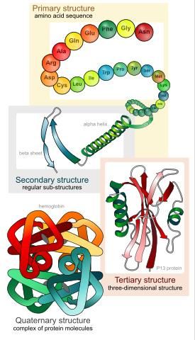
Protein Structure The basic structure of an amino acid is a central carbon atom with an amino group (NH2) on one side, a carboxyl group (COOH) on the opposite side, a hydrogen atom, and an R group that determines the identity of the amino acid. Amino acids are linked together by peptide bonds, which are covalent bonds, formed by a dehydration synthesis reaction between the carboxy terminus of the first amino acid and the amino terminus of the second. This organization gives the protein an order where the beginning of the polypeptide chain has an amine group and the end has a carboxyl group. This directionality is set up when proteins are translated from RNA. The composition and location of amino acids in the polypeptide chain confer their properties to the resulting protein and affect the shape of the protein. Amino acids can be charged, uncharged, hydrophobic, or cause changes in the 3D structure of the protein. The composition and order of amino acids is called the primary structure, which accounts for some of the function of proteins, but not all. Proteins take on very complicated shapes in nature. The secondary structure of proteins arise when proteins fold due to interactions between elements in the amino acid backbone (not including R groups). These folds include helixes, which are helical structures formed by hydrogen bonds between carbonyl groups of one amino acid and the amino group of another that is four amino acids down the line. This structure pushes R groups to the outside of the helix, giving them more opportunity to interact. sheets are another secondary structure formed when sections of the polypeptide chain are parallel to each other. This structure also presents R groups outward on top and bottom, so they can interact. Tertiary structure forms due to interactions between R groups of the same protein. These can include all different types of non-covalent bonds forming between groups or can include strong di-sulfide bonds. Tertiary structures minimize the free energy of a protein by taking the most energetically stable position. Finally, quaternary structure forms between amino acids on different polypeptide chains. Protein structures can be denatured, meaning they lose their higher order structures due to changes in pH or temperature. However, they generally return to their proper structures when conditions return to normal. This means that most of the information needed to form a structure is retained within the polypeptide sequence of a protein. Carbohydrates are an immediate source of energy that most life depends on and form important structural elements of organisms. The formula for carbohydrates is (CH 2O)n, where n refers to the number of times this structure repeats. Monosaccharides contain 3–7 monomers of CH2O connected as a chain or a ring. Common monosaccharides include glucose, fructose, and galactose. Disaccharides form when two monosaccharides undergo dehydration synthesis to form a covalent bond between them. The covalent bond formed between a carbohydrate and another molecule is called a glycosidic bond. Common disaccharides include sucrose, lactose, and maltose. Polysaccharides are long chains of monosaccharides that can be either linear or branched. Common polysaccharides include starch, chitin, cellulose, and glycogen. Disaccharides and polysaccharides can be made of the same or 4
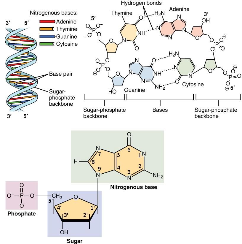
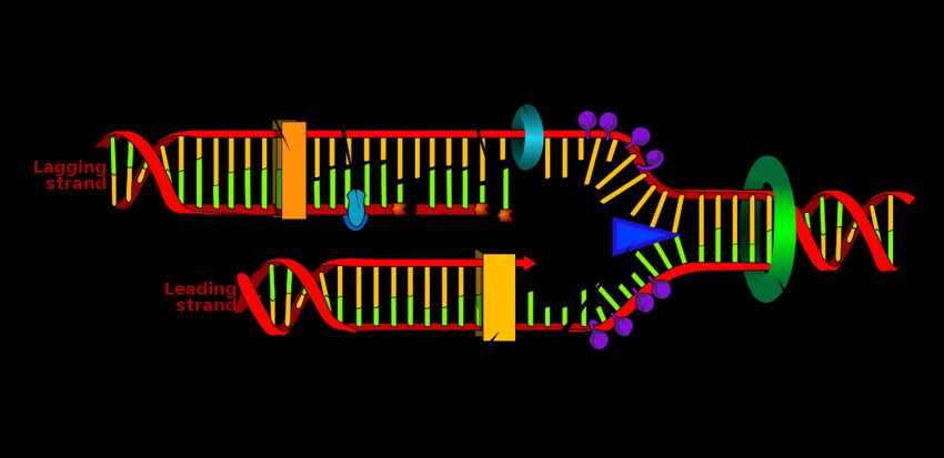
different monosaccharide monomers, the composition of which determines the properties of the
macromolecule.

Lipids are nonpolar macromolecules made of hydrocarbon chains that are generally hydrophobic;
they include fats, waxes, phospholipids, and steroids. Lipids are a structural unit of membranes
and a form of energy storage for organisms. Fats, or triglycerides, are made of a glycerol
backbone and three fatty acid chains. The fatty acid chains of lipids are made of hydrocarbons
bonded to a carboxyl group. The fatty acid chains of triglycerides can be different from each other
in both length and saturation.

If all neighboring bonds in the hydrocarbon chain are single bonds, the fatty acid is called
saturated. Saturated fats have straight fatty acid chains that can pack tightly together and form
solids at room temperature. If there are double bonds in the chain, the fatt y acid is called
unsaturated. If the double bond results in hydrogen atoms on the same side of the acid chain,
this causes a bend in the chain and results in a cis fatty acid. Cis fatty acids tend to form liquid
oils at room temperature, as their shape causes spacing between molecules. If the double bond
results in hydrogen atoms on opposite sides of the fatty acid chain, the molecule retains the
normal shape of the chain, and is called a trans fatty acid. Trans fatty acids behave more like
saturated fats at room temperature but are also difficult for the body to metabolize.

The Structure of DNA and RNA
DNA, deoxyribonucleic acid, is a polymer made of individual nucleotides that carries genetic
information. Nucleotides are made of a sugar (deoxyribose), a phosphate group, and a
nitrogenous base. The deoxyribose group has a ring structure composed of an oxygen and four
carbons. As a convention, carbon atoms of deoxyribose are numbered starting from the
connection between the nucleotide and the first carbon, called the 1’ carbon. A 5th carbon atom
(5’) is bonded to the 4’ carbon of deoxyribose.
The 5’ carbon bonds with the phosphate group. Nucleotides are linked to each other by covalent
bonds between the sugar and phosphate groups to make a single strand of DNA. The connection
between two nucleotides in a strand of DNA always occurs in the same orientation —this is
between the 3’ carbon atom of the first nucleotide and the phosphate group of the second. Since
the phosphate atom is connected to the 5’ carbon, the orientation of the DNA polymer is referred
as 3’ to 5’.

 5
DNA Structure

DNA nucleotides come in four varieties, each with a different base: adenine (A), thymine (T),
guanine (G), or cytosine (C). In RNA, uracil (U) is used instead of thymine. G and A molecules are
called purines and consist of double nitrogenous rings. C, T, and U are called pyrimidines, and
are composed of single nitrogenous rings. Two strands of DNA with complementary base pairs
are linked with hydrogen bonds between base pairs to form the double helix pattern we are
familiar with. Hydrogen bonds occur only between complementary pairs: C-G or A-T (or A-U, in
the case of RNA). You’ll notice that purines bond with their complimentary pyrimidine. Cytosine-
guanine connections pair via three hydrogen bonds, while adenine-thymine pairs with double
hydrogen bonds.
In a DNA polymer, the “backbone” is made of sugar and phosphate groups linked together, with
nucleotides in the center. This bond between the phosphate group and two sugars is a
phosphodiester bond. Two strands of DNA connect together through complementary base
pairing to form a double stranded DNA polymer. Note that the connections between two strands
of DNA are anti-parallel—one strand will have a 3’-5’ directionality, and the second strand will be
oriented in the 5’-3’ direction.

 6
RNA, ribonucleic acid, is similar to DNA but has ribose as the sugar group and has uracil (U)
instead of thymine. Unlike DNA, RNA is usually found in nature as a single strand rather than a
double strand.

 Outside Reading

 • For more on the properties of water:
 https://www.khanacademy.org/science/biology/water-acids-
 and-bases

 • For more on macromolecules:
 https://courses.lumenlearning.com/boundless-
 biology/chapter/synthesis-of-biological-macromolecules/

 • To learn more about nucleotide structure:
 https://www.khanacademy.org/test-
 prep/mcat/biomolecules/dna/a/dna-structure-and-function

 7
Sample Chemistry of Life Questions

The strongest complementary base pair found in DNA is between

A. adenine and guanine.

B. cytosine and thymine.
C. adenine and thymine.

D. cytosine and guanine.

Explanation:
The correct answer is D. Three hydrogen bonds form between cytosine and guanine, making this
a stronger complementary base pair than the adenine/thymine base pair. This is important, for
example, during primer construction for PCR in molecular biology because increased G C
concentration relative to AT concentration leads to strong primer binding to a DNA template.
Adenine and guanine are both purines and do not pair up in the DNA double helix; and cytosine
and thymine are both pyrimidines and do not pair up in the DNA doub le helix. Finally, while
adenine and thymine do pair up in the DNA double helix, there are only two hydrogen bonds that
form between the two. Three hydrogen bonds form between cytosine and guanine, making the
latter base pair stronger.

Water travels up a stem through a process called
A. cohesion-adhesion.

B. cohesion-tension.

C. adhesion-tension.
D. hydrogen bonding.

Explanation:
The correct answer is B. As water evaporates from a leaf, a column of water molecules is pulled
upward through cohesion-tension. The water molecules are bound together through hydrogen
bonding, and the evaporation at the leaf pulls the chain of molecules upward due to the inherent
surface tension of water. Adhesion would mean that the water is sticking to another substance,

 8
which would impede movement upward. Hydrogen bonding joins water molecules together,
aiding the pull of water molecules upward.

A carboxylic acid contains which of the following functional groups?

A. –OH

B. –CHO
C. –COOH

D. –O–

Explanation:
The correct answer is C. A carboxylic acid contains the –COOH (carboxyl) functional group. An
alcohol contains the –OH (hydroxyl) functional group; an aldehyde contains the –CHO (aldehyde)
functional group; and an ether contains the –O– (ether) functional group.

 9
Cell Structure and Function
Around 10–13% of questions on your exam will cover the topic Cell Structure and Function.

Cellular Components and Functions of Those Components
Cells are the smallest functional unit of life. There are two categories of cells: prokaryotes and
eukaryotes. Prokaryotes are unicellular, and do not have cell walls or membrane-bound
organelles, and generally have a single circular chromosome. This group includes bacteria and
archaea. Eukaryotes have multiple chromosomes organized in a membrane-bound nucleus and
also have other organelles. Eukaryotes include single-celled organisms like protozoans and yeast,
as well as multicellular organisms like plants and animals.

Animal and plant cells differ in a several features. Animal cells have a thin plasma membrane
that encloses the cells. The membrane itself is flexible and made of a phospholipid bilayer with
various proteins embedded in the membrane. Proteins in the bilayer regulate which molecules
enter and exit the cell and are important for communication between the cell and the
environment.

Plants, on the other hand, have a cell wall in addition to the plasma membrane made of cellulose
and lignin, giving strength, rigidity, and protection to plant cells. The cell wall also helps the cell
to store water by regulating diffusion and providing the strength to allow high internal pressures
without rupture. Plants, fungi, algae, bacteria, and archaea all have cell walls with slightly
different compositions.

Plants also have a central vacuole, a fluid filled membrane-bound structure that stores water and
nutrients for the cell. Animal cells have vacuoles, but these fulfill other purposes. Plant and
animal cells also differ in how they produce energy; plant cells contain chloroplasts and
mitochondria, while animal cells only have mitochondria. These are described in more detail in
the following table of eukaryotic cell organelles.

 10
Organelle Description
Nucleus The nucleus contains most of the genetic material of a cell in the fo rm of
 chromatin. RNA transcription and processing occur within the nucleus. The
 nucleus also contains the nucleolus, the area where ribosomes are made. The
 nucleus is encased in a plasma membrane with nuclear pores that tightly
 restrict movement of molecules in and out and to the endoplasmic reticulum.
Mitochondria Mitochondria are membrane-bound organelles where cellular respiration,
 the synthesis of ATP from ADP, occurs. ATP is one of the primary molecules
 the animal cell uses to harness energy. Mitochondria have a double
 membrane—the outer one is smooth, while the inner one is rough.
 Mitochondria have their own circular DNA, are able to reproduce within cells,
 and have some of their own protein synthesis machinery.
Chloroplast Chloroplasts are membrane-bound organelles that contain chlorophyll and
 make energy through photosynthesis. Like mitochondria, they also have their
 own DNA and protein synthesis machinery.
Endoplasmic The endoplasmic reticulum (ER) are folded, membrane-bound organelles that
Reticulum are transport hubs from the nucleus to the Golgi apparatus. There are both
 rough and smooth ER. The rough ER is covered with ribosomes and is the site
 where membrane-bound proteins and proteins that are packaged in vesicles
 are made. The smooth ER does not have ribosomes on it, hence the smoother
 appearance, and is the site where many things are synthesized including
 lipids, fats, and steroids.
Golgi Apparatus The Golgi apparatus is made of stacks of membrane sacks. This is the site
 where most protein modification takes place and where proteins are
 packaged and targeted for export from the cell.
Ribosomes Ribosomes perform translation of RNA into protein. Ribosomes are made of
 ribosomal RNA (rRNA) and some associated proteins and can be free in the
 cell, making proteins in the cytoplasm, or can be found on the rough
 endoplasmic reticulum where they synthesize proteins that end up on or in
 cell membranes. Ribosomes are found in every form of life on Earth, providing
 evidence for a common ancestor.
Lysosomes & Lysosomes and peroxisomes are vesicles in the cell where cell waste is
Peroxisomes destroyed and recycled. Lysosomes contain hydrolytic enzymes that destroy
 proteins, cell waste, and damaged organelles. Peroxisomes are where lipids
 and reactive oxygen species are destroyed.
Cytoskeleton The cytoskeleton is made of microtubules, intermediate filaments, and
 microfilaments that collectively give structure to the cell, keep organelles in
 place, help cells move, and provide the framework that proteins move along
 in a cell.
Centrosome The centrosome is the main microtubule organizing center of the cell and
 organizes the mitotic spindle during cell division. A cell has a single
 centrosome unless it is in the cell cycle.

 11
Vesicles Vesicles are membrane sacs that transport molecules in a cell. They can carry
 substances inside for release into the extracellular environment (like
 neurotransmitters), they can carry membrane-bound proteins that end up on
 the cell membrane, and they can also remove pieces of membrane for
 destruction, changing the size and shape of a cell.

 Understanding what happens in
 different organelles as it relates to cell
 function is a common theme in the
 Free Response section of the AP exam.

Cell Size
Cells exchange molecules with their environment to sustain the life of the cell or the life of the
organism, which includes obtaining things like nutrients, thermal heat, and oxygen, as well as
releasing molecules for the organism or waste products. As a cell grows in size, it must maintain
a membrane surface area that allows it to sustain its metabolic needs and functions. Thus, the
cell can only grow as large as the surface area of the cell can support. Mathematically, the volume
of the cell increases faster than the surface area. You can calculate these relationships using the
equations in the following table, with radius (r), length (l), height (h), width (w), and length of one
edge of a cube (l).

 Sphere Cube Rectangular Solid Cylinder
 Volume 4 3 = 3 = ℎ = 2 ℎ
 =
 3
 Surface = 4 2 = 6 2 = 2 ℎ + 2 + 2 ℎ = 2 ℎ + 2 2
 Area

In each case, the volume grows much faster than surface area, limiting the amount a cell can
grow in these shapes. The result of this is that highly metabolically active cells either remain small
in size or take on unique specializations to allow more surface area for exchange. Specializations
can include features like membrane folds that increase surface area of the cell.

 12
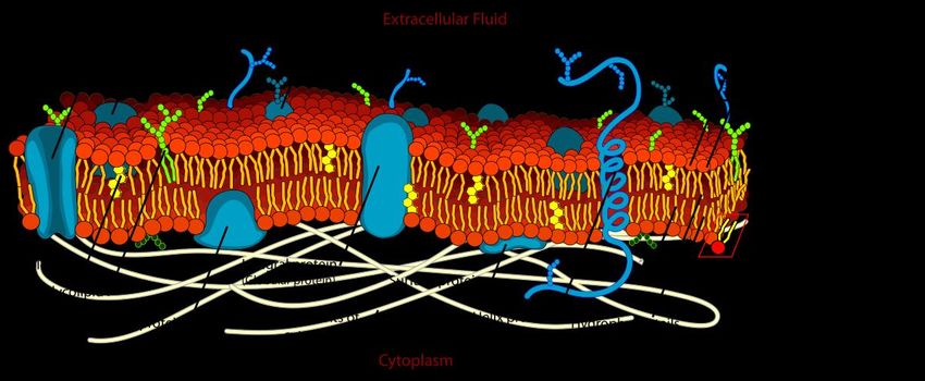
The Cell Membrane Structure and Function
The fluid mosaic model describes the structure of cell membranes. In this model, the plasma
membrane is made of a phospholipid bilayer with cholesterol, proteins, glycolipids, and
glycoproteins embedded within in it. The components of the membrane are in constant motion.

 Cell Membrane

 • Phospholipids comprise the majority of the plasma membrane; these are lipids made of
 a hydrophilic head and hydrophobic tails. The hydrophilic heads are made of a glycerol
 molecule with a phosphate group attached. The hydrophobic tails are made of long, non -
 polar fatty acids. Phospholipids form a bilayer, where the hydrophilic heads face the
 aqueous internal and external environments, and the hydrophobic ends stay in the
 middle.
 • Membrane proteins can traverse the entire membrane, partially extend on one side of it,
 or attach loosely to one side of the membrane depending on its hydrophobicity. Proteins
 that integrate into the membrane are called integral membrane proteins. These have a
 hydrophobic portion that anchors them within the membrane. If they traverse both ends
 of the membrane, they are called transmembrane proteins. Peripheral membrane
 proteins are loosely attached to the membrane either to other integral proteins or to
 phospholipids.
 • Glycolipids and glycoproteins attach to the outer surface of the cell. These are generally
 used as signals for the type of cell that they are. For instance, the immune system uses
 glycoproteins to identify cells that belong to the organism versus invaders.
 • Cholesterol groups are embedded within the hydrophobic bilayer and affect membrane
 fluidity. They act to buffer fluidity at both low and high temperatures, making it so the
 cell can function at a large range of conditions.

 13
The structure of the phospholipid bilayer creates a semipermeable barrier for substances to cross
due to the hydrophobic interface. The cell membrane creates selective permeability, meaning
that only certain molecules can pass through the membrane. Small nonpolar molecules, like N2,
O2, and CO2 pass easily across the membrane. Uncharged polar molecules, like water, can pass
through in small amounts. Hydrophilic substances like large polar molecules and ions cannot pass
freely because the hydrophobic center repels hydrophilic molecules. These require membrane
transporters to move across the membrane.

Plants and bacteria have cell walls made of complex carbohydrates in addition to their
membranes. Cell walls add an additional layer of support by both providing strength as wel l as
protecting cells from both mechanical and osmotic stress.

Cell Regulatory Mechanisms
The cell has several methods to allow transport across the membrane.

 • Passive transport, movement across a membrane without energy being used, occurs due
 to molecule diffusion from areas with high concentration to areas with low concentration
 by taking advantage of their concentration gradients.
 • Active transport, movement across a membrane that requires energy, is used to get
 molecules from areas of low concentration to areas of high concentration.
 • Endocytosis occurs when a vesicle forms in a plasma membrane, taking in molecules from
 the external environment into the cell. This is one way that macromolecules enter the
 cell.
 • Exocytosis occurs when a vesicle fuses with the membrane and dumps contents into the
 extracellular space. This is a method used to take macromolecules out of the cell.

There are three forms of passive transport: simple diffusion, facilitated diffusion, and osmosis.
During simple diffusion, molecules pass across the membrane freely by taking advantage of their
concentration gradients. This is what occurs with oxygen and carbon dioxide; they are permeable
to the membrane, and thus do not need additional transporters to help them get across.

Molecules that are not able to easily pass through the membrane undergo facilitated diffusion.
In facilitated diffusion, channels in the membrane surface or carrier molecules assist molecules
in crossing the cell membrane. For example, aquaporins are channels in the cell’s membrane that
allow water to pass through along a concentration gradient. By limiting the number of aquaporins
in a cell membrane, the amount of water that can flow in or out is regulated, which keeps the
cell from experiencing rapid shifts in water content.

Osmosis is the process by which water moves across a semipermeable membrane from an area
of low concentration of solutes to an area of high concentration of solutes, to equalize

 14
concentrations. Osmolarity is the total concentration of solutes in a solution. Osmotic pressure
refers to the pressure that pulls water from one side of a membrane to the other. Tonicity refers
to the ability of water to move across a membrane by osmosis; this takes into account both the
differences in osmolarity as well as the ability of water to move.

Cell environments can be isotonic, meaning that the concentration of solutes is the same in the
internal and external environments; hypertonic, meaning the external environment has more
solute than the internal environment; or hypotonic, meaning that the external environment has
less solute than the internal environment. Cells in a hypotonic solution take on too much water,
causing them to swell and potentially burst. On the other hand, cells in a hypertonic solution are
at risk of shriveling.

Active transport mechanisms use metabolic energy, often in the form of ATP, to transport
molecules. Active transport is used to maintain concentration gradients across cells. For example,
neurons take advantage of concentration gradients to signal to each other. The internal
environment of a neuron is high potassium and low sodium and external environment is low
potassium and high sodium. These concentration differences create a negative membrane
potential that positions it to respond to stimulation. The cell uses active mechanisms to maintain
this concentration difference and membrane charge using a membrane transporter called the
Na+/K+ ATPase, which uses ATP to move sodium and potassium across the membrane in an
energetically unfavorable direction.

Cellular Compartmentalization
Compartmentalization of processes is an essential feature of cellular environments. Membranes
allow cells to compartmentalize processes that need to be kept separated from the rest of the
environment. For example, the lytic enzymes in lysosomes would be fatal to the cell if released
into the cytosol. Thus, cells developed a way to compartmentalize these enzymes into a
membrane-bound organelle. By having membranes, cells and organelles are able to crea te
internal environments that are different than the external environment.
For example, for neurons to fire action potentials, they take advantage of an electrochemical
gradient where the internal environment is rich in potassium and the external environment is
rich in sodium. Without membranes that actively maintain these different environments,
neurons would not function. Internal membranes also facilitate processes by increasing reaction
surface areas and keeping molecules in the places that they are needed to function. For example,
the internal mitochondrial membrane is an important site of ATP synthesis (discussed later on in
this guide). The mitochondria are formed in a way to maximize internal membrane surface area,
so they can form more ATP.

 15
Membrane bound organelles evolved from prokaryotic cells through a process called
endosymbiosis. This is a form of symbiosis where once cell resides inside of another. The theory
is that organelles like chloroplasts and mitochondria were once free-living prokaryotes. At some
point in history, a eukaryotic cell engulfed a prokaryote—likely a photosynthetic autotroph—but
did not digest it. This generated a mutually beneficial relationship where the prokaryote
generated energy for the eukaryotic cell, and the eukaryotic cell protected the prokaryotic cell
and provided nutrients for it.
Over time, this relationship became permanent as the prokaryotic cell stopped carrying all of the
genes needed for its survival and the eukaryotic cell became dependent on the energy the
prokaryotic cell provided. This is supported through the fact that both share features of
prokaryotic cells, like having their own circular DNA genome. Prokaryotes generally do not have
membrane bound organelles—rather, they have regions with specialized structures and
functions within them.

 Outside Reading

 • To learn more about cell organization:
 https://www.khanacademy.org/test-
 prep/mcat/cells/eukaryotic-cells/a/organelles-article

 • To learn more about cell membrane properties and transport:
 https://opentextbc.ca/anatomyandphysiology/chapter/the-cell-
 membrane/

 • To learn more about membrane transport:
 https://courses.lumenlearning.com/nemcc-ap/chapter/3204/

 16
Sample Cell Structure and Function Questions

A cell that produces and secretes a large number of proteins would probably have

A. extensive rough ER.

B. limited rough ER.
C. few mitochondria.

D. a cell wall.

Explanation:
The correct answer is A. The rough ER (endoplasmic reticulum) is responsible for producing
proteins (in the bound ribosomes) and transporting them within and outside the cell.
Mitochondria are the powerhouses of the cell, responsible for creating energy through
respiration. They are not directly involved in protein synthesis or transport, so you would not
expect to find a remarkably small (or large) number of mitochondria in such a cell. The cell wall
is found in plant cells and is used to protect the cell and provide support. The cell wall is not
directly involved in protein synthesis or transport, so you would not necessarily expect to find a
cell wall in such a cell. Furthermore, animal cells are capable of producing and secretin g large
numbers of proteins, so it is not necessary that this type of cell would have a cell wall.

When a solution has a lower solute concentration than the solution on the other side of a
membrane, that solution is said to be

A. hypotonic.
B. isotonic.

C. hypertonic.

D. undergoing osmosis.

Explanation:
The correct answer is A. A hypotonic solution has a lower solute concentration than the solution
on the other side of a membrane. A hypertonic solution has a higher solute concentration than
the solution on the other side of a membrane. Choice B is incorrect because the term isotonic

 17
indicates that two solutions have equal concentrations. Choice D is incorrect because osmosis
is the term for the diffusion of water across a membrane.

What feature of a plant cell prevents it from bursting when in a highly aqueous solution?

A. a large central vacuole

B. a guard cell
C. a cell wall

D. chloroplasts

Explanation:
The correct answer is C. The rigid cell wall of plant cells prevents lysis of the cell when in an
aqueous environment. The vacuole of the cell expands, creating turgor pressure, but the cell
membrane expands only as much as the cell wall will allow. Choice A is incorrect because the
large central vacuole in plant cells swells as water enters the cell in an aqueous environment. The
size of the vacuole is limited by space within the cell and by the rigid outer cell wall. Choice B is
incorrect because guard cells surround stomata in leaves and regulate transpiration from the
leaf. Choice D is incorrect because chloroplasts are the site of photosynthesis in the cell and are
located within the cytoplasm.

 18
Cellular Energetics
Around 12–16% of the questions on your AP exam will cover Cellular Energetics.

The Structure and Function of Enzymes
For energy releasing reactions to occur, there is usually an associated energy barrier that must
be overcome first. If there were not, then cells would just release energy indefinitely. This energy
hurdle is called the activation energy. This can be overcome by either inputting energy in the
reaction, or through the introduction of a catalyst. Catalysts reduce activation energy and
increase reaction rate without itself undergoing a chemical change. Enzymes are biological
catalysts. Enzymes act by changing the conformation of molecules that they interact with to put
them into a more optimal state for a reaction to proceed. The active site of an enzyme is the
portion that interacts with the molecule. The substrate is the molecule that the enzyme binds
with to facilitate a change.

Enzymes and molecules in a biological reaction are affected by their environment. For example,
increases in heat generally cause reactions to take place faster because increased molecu le
movement increases the frequency of collisions between substrate and enzyme. However,
excessive heat can denature proteins, causing their structures to be altered and inhibiting their
ability to take part in a reaction. Similarly, pH can alter the efficiency of a reaction by altering
hydrogen bonds. These effects can be reversible if the conditions return to baseline. The
concentration of substrates in comparison to enzymes will also affect enzyme activity. Generally,
the more substrate or more enzyme around, the faster the reaction will proceed, as there are
more chances for substrates and enzymes to come into contact.

Other factors that can affect enzyme reaction rates are inhibitors. Competitive inhibitors
compete for the active site of an enzyme, keeping it from binding the substrate and blocking the
reaction. Noncompetitive inhibitors bind to other sites on the enzyme, changing the enzyme’s
conformation and keeping it from efficiently binding the substrate.

The Role of Energy in Living Systems

All living things require energy to sustain life. Bioenergetics is the study of energy transformation
in living organisms. Metabolism is the general term for all energy transformations in living
organisms, including processes like photosynthesis, mitochondrial respiration, and movement. In
biological systems, energy input constantly exceeds loss to keep the systems going. Energy is then
stored in the form of molecules that contain chemical bonds that can be broken down, most
often in the form of sugars or fats. Anabolism is the process where molecules store energy in the
form of chemical bonds. Catabolism is the process by which energy is released by breaking these

 19
bonds. Often, energy release mechanisms are linked to processes, so the energy is immediately
used by the cell.

As mentioned, cells store energy in the form of fats and sugars. When these are broken down,
large amounts of energy are released that may be more than a cell needs to complete a process.
This can lead to wasting precious energy for small tasks. Thus, larger energy forms are generally
first converted to smaller energy molecules that are then used for cells to carry out normal
functions. Adenosine triphosphate (ATP) is the primary source of energy that cells use to
function. ATP can be broken down into ADP (adenosine diphosphate) and AMP (adenosine
monophosphate), through process like fermentation and respiration, releasing energy at both
steps that can be harnessed to power other cellular processes. ATP breakdown is linked to most
cellular processes, providing a streamlined way for cells to harness energy.

The Processes of Photosynthesis
Photosynthesis is the process where light energy is converted to cellular energy in the form of
glucose. During photosynthesis, carbon dioxide, water, and light are converted to glucose and
oxygen:

 6CO2 + 6H2 O + Light → C6 H12 O6 + 6O2
Photosynthesis initially evolved in cyanobacteria, resulting in oxygenation of the Earth’s
atmosphere. The pathways that evolved in prokaryotes are the foundations for eukaryotic
photosynthesis in plants.
Photosynthesis takes place in two main phases: photolysis (the light dependent reaction) and
the Calvin cycle (the light independent reaction). Photolysis takes place in the thylakoid
membranes of chloroplasts. During this step, light energy is absorbed by chlorophylls, which then
generate ATP and NADPH. NADPH is an electron carrier used in biological reactions. Water is used
in this reaction and oxygen is released.
The Calvin cycle takes place in the stroma of chloroplasts. In this step, carbon dioxide from the
environment reacts with ATP and NADPH generated in photolysis to create small 3 carbon sugars,
which are then joined to form glucose. Glucose provides the cell with both energy, as it can be
used to synthesize ATP, and fixed carbon. Fixed carbon refers to organic carbon molecules, like
sugars. Fixed carbon is used by cells to build molecules needed in cells for other processes. The
Calvin cycle both removes carbon from the atmosphere and fixes the energy generated from
photolysis in a form that can be stored in cells for later use.

The light dependent reaction relies on photosystems to harvest light energy. During this process,
called non-cyclic phosphorylation, light is absorbed by two photosystems that harness the

 20
electrons from water to make NADPH and ATP from NADP+ and ADP. There are two
photosystems, photosystem I (PSI) and photosystem II (PSII). Photosystem structures are
embedded in the thylakoid membrane and are made of a light harvesting complex, composed of
proteins and hundreds of chlorophylls and pigments, and a special pair of chlorophyll a at the
core.
During non-cyclic phosphorylation, light energy is first harnessed by PSII through the light
harvesting complex. The energy is passed via PSII’s special pair of chlorophyll a (called P680),
which in turn boost an electron to a higher energy level. The electron is then passed to an
acceptor molecule and replaced with an electron taken from water, releasing oxygen. The high
energy electron is then passed along the membrane by a series of transporters, called the
electron transport chain (ETC). The electron loses a little energy at each step of the ETC; some
of this energy is used to pump H+ ions from the stroma into the thylakoid membrane—this
pumping creates a proton gradient that is linked to formation of ATP from ADP through a process
called chemiosmosis via an enzyme called ATP synthase.
Phosphorylation of ADP to generate ATP in photosynthesis is called photophosphorylation. PSI
receives the excited electron, which is passed along the ETC. Light energy received by PSI is then
used to excite the electron through its special chlorophyll pair (called P700). The electron, now
at a very high energy level, is then passed by an acceptor molecule which passes it along another
electron transport chain that results in the creation of NADPH from NADP+. ATP and NADPH
generated in during the light cycle are then used in the stroma of chloroplasts to generate sugar
from carbon dioxide through the Calvin cycle.

The Processes of Cellular Respiration
All forms of life use respiration and fermentation to break down biological macromolecules, like
sugars and fats, into ATP.
In eukaryotes, cellular respiration breaks down glucose into carbon dioxide and water to
generate ATP. As in photosynthesis, cellular respiration takes place in a series of steps where
energy is passed along an electron transport chain.

 21
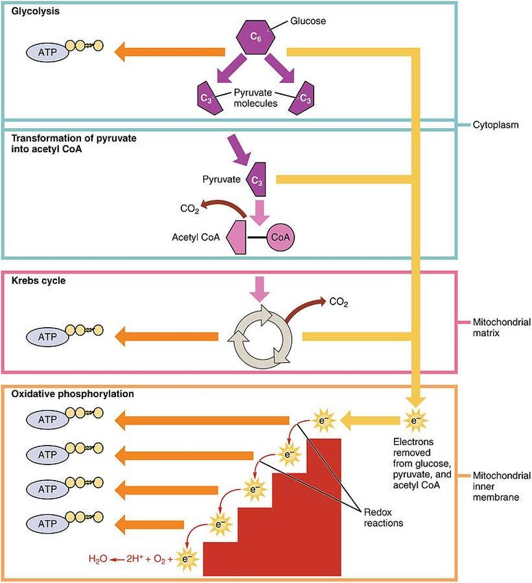
1. In the first step of respiration, called glycolysis, the 6-carbon containing glucose is broken
 down into two 3-carbon pyruvates, two ATP molecules, and two NADH molecules. During
 glycolysis, an ADP is converted to ATP and NAD+ is converted to NADH. Glycolysis takes
 place in the cell’s cytosol.

2. After glycolysis, the pyruvate molecules are transported into mitochondria, where they
 are broken down into two carbon molecules, called acetyl groups, that are bound to
 coenzyme-A, in a molecule called acetyl CoA. This step, called pyruvate oxidation,
 reduces NAD+ to NADH, but does not generate ATP.

3. Acetyl CoA then moves into the citric acid cycle, also called the Krebs cycle. The citric acid
 cycle is a form of aerobic respiration where acetyl co-A, which is a product of oxidized
 pyruvate, is metabolized to produce one NADH, one FADH 2, two carbon dioxides, and
 either one ATP or one GTP.

 22
4. In the final step of cellular respiration, called oxidative phosphorylation, NADH and
 FADH2 are then used to make more ATPs through an electron transport chain in the
 membrane of mitochondria. As in photosynthesis, the electron transport chain uses
 electrons donated by carrier molecules, in this case, NADH and FADH2, which are then
 passed along the chain. As electrons are passed along, they lose some energy, which is
 used to pump H+ ions from the mitochondrial matrix to the intermembrane space. This
 creates a chemical gradient, which is used to convert ADP to ATP through chemiosmosis
 by ATP synthase. The final electron acceptor in oxidative phosphorylation is an O 2
 molecule, which generates water. In prokaryotes, the first three steps of respiration take
 place in the cytosol and oxidative phosphorylation takes place in the cell’s membrane. In
 some anerobic prokaryotes other proton acceptors are used at the end of the electron
 transport chain in a process called anaerobic cellular respiration.

In some cells, the electron transport chains are decoupled from the generation of ATP. This is
used to generate heat to warm the body instead of energy storage.
If oxygen is not available at the end of the electron transport chain to accept electrons, cells take
a slightly different approach to respiration. During fermentation, glycolysis proceeds, but
pyruvates are converted to other organic molecules like lactic acid and alcohol. This regenerates
NAD+ from NADH, which allows glycolysis to continue.

Molecular Diversity and Cellular Response to Environmental Changes
Note that there are a variety of somewhat overlapping mechanisms built in for life to generate
ATP for immediate energy usage. For instance, in the absence of oxygen, cells can convert to
fermentation. Plants have a variety of types of chlorophyll in their cells that can harness energy
from different wavelengths of light. The presence of these different types of molecules help
organisms adapt to changing environments and optimize species survival. This will be discussed
more in later sections on evolution.

 23
Outside Reading

 • To learn more about photosynthesis:
 https://www.khanacademy.org/science/biology/pho
 tosynthesis-in-plants

 • For more on cellular respiration:
 https://www.khanacademy.org/science/biology/cell
 ular-respiration-and-fermentation

Sample Cellular Energetics Questions

Enzymes lower the _______________ of a reaction.

A. temperature
B. activation energy

C. free energy

D. speed

Explanation:
The correct answer is B. Enzymes lower the activation energy of a reaction, or the energy needed
for a reaction to proceed. Enzymes do not affect the temperature or the free energy of a reaction,
and they usually enhance the speed of a reaction by reducing the activation energy.

 24
The light-dependent reactions of photosynthesis transform energy from sunlight into chemical
energy in the form of

A. photons.

B. pyruvic acid.
C. lactic acid.

D. electrons.

Explanation:
The correct answer is D. Electrons generated in photosynthesis are used to produce energy in
the form of ATP or NADPH. Choice A is incorrect because photons are particles of light absorbed
by pigments in the first step of photosynthesis. Choice B is incorrect because pyruvic acid is
formed as an intermediate product in cellular respiration, not photosynthesis. Choi ce C is
incorrect because lactic acid is formed as an intermediate product in anaerobic respiration, not
photosynthesis.

Which of the following is a product of anaerobic respiration?

A. pyruvic acid
B. lactic acid

C. glucose
D. oxygen

Explanation:
The correct answer is B. In anaerobic respiration, pyruvic acid molecules are broken down into
end products. In one type of anaerobic respiration, lactic acid is produced. Choice A is incorrect
because pyruvic acid is broken down, not produced, in aerobic respiration. Choice C is incorrect
because glucose is produced through photosynthesis. Choice D is incorrect because anaerobic
respiration produces carbon end products, ATP, and carbon dioxide.

 25
Cell Communication and Cell Cycle
Around 10–15% of the questions on your AP exam will cover the topic Cell Communication and
Cell Cycle.

The Mechanisms of Cell Communication
Cells communicate to each other through both long-range and short-range signals. These types
of messages often occur through one cell releasing a molecule in the extracellular space, called a
ligand, that is then received by another cell that has a receptor for that ligand. There are four
methods of cell communication: paracrine, autocrine, endocrine, and cell-cell contact.

 • Paracrine signaling occurs when one cell releases ligand into the extracellular space to
 signal to nearby cells. This type of signaling occurs at neuronal synapses when an axon
 terminal releases neurotransmitter on a receiving neuron.
 • Autocrine signaling occurs when a cell releases a signal to itself. This is one way that cells
 regulate their own growth and intracellular processes.
 • Endocrine signaling occurs when a cell releases a ligand, typically into the bloodstream,
 to affect cells a long way away. This is normally how hormones work.
 • Signaling through cell-cell contact occurs when two cells physically contact each other,
 and this causes a signal to be passed on. This can occur through gap junctions on cells,
 which are physical connections between two cells that allow them to exchange small
 signaling molecules, or through binding of ligands and receptors on the surfaces of two
 adjacent cells. Cell surface binding is an important way that the immune system uses to
 recognize pathogens and mount a response against them.

Signal Transduction
When a cell receives a signal, it must have a way to respond to it. Signal transduction pathways
link the signal to the appropriate response. Signal transduction begins when a ligand is recognized
by a receptor. Ligands can be simple molecules like small chemicals, small peptides, or large
proteins. Receptors generally recognize one or a few ligands and have several forms.

 • Intracellular receptors reside within the cell. Hormone receptors are an important
 example of these; when hormones bind a receptor, the receptor changes shape and
 enters the nucleus to induce changes in gene expression.
 • Cell surface receptors reside within the plasma membrane and respond to signals from
 outside. These include ligand-gated ion channels, G-protein coupled receptors, and
 enzyme-linked receptors.

 26
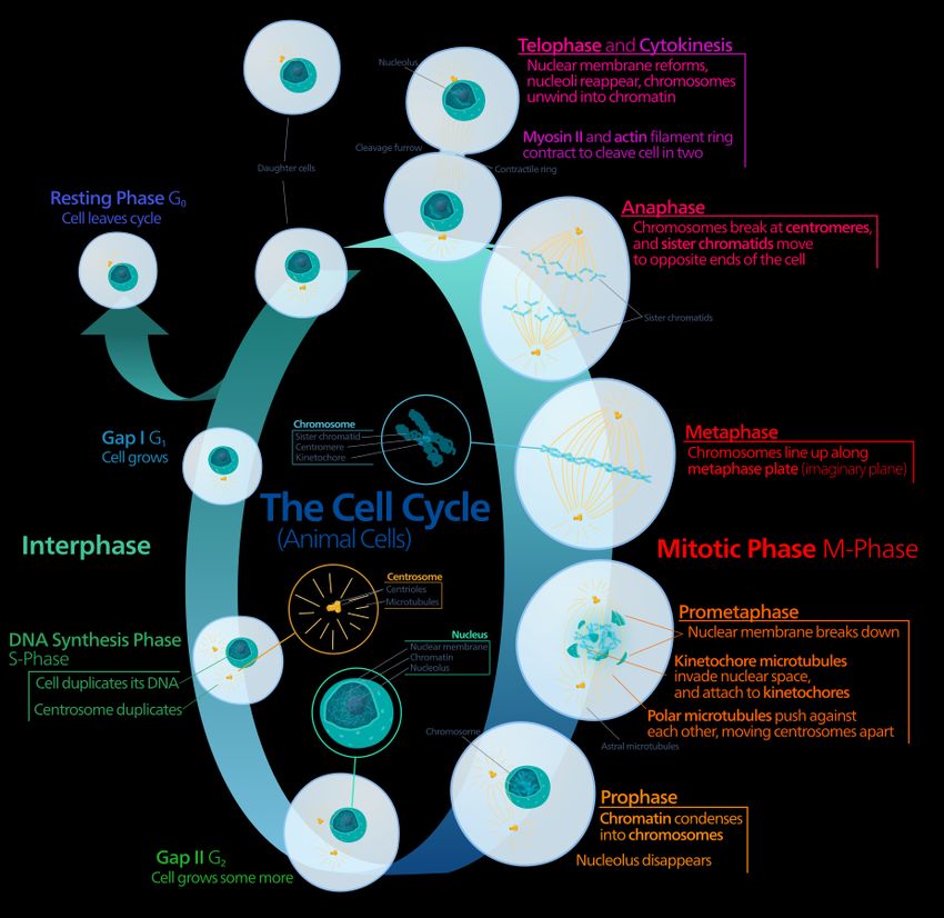
• Ligand-gated ion channels open or close in response to ligand binding, allowing ions to
 either flow through the channel or stopping the flow of ions.
 • G-protein coupled receptors respond to a signal by activating their coupled G-protein.
 This activated G-protein then interacts with other proteins within the cell, causing other
 events to occur. The olfactory system relies on G-protein coupled receptors to transduce
 different odorants into smells that we recognize.
 • Enzyme-linked receptors are a type of receptor that are coupled to an enzyme. Once
 activated, the enzyme is able to induce reactions within the cell.

Once a receptor is activated by a ligand, a signaling cascade is initiated that causes changes within
the cell. This can include things like inducing gene expression, secreting a molecule, changes in
cell growth, or changes in the identity of the cell.

Cellular Responses and Feedback Mechanisms
Feedback mechanisms are used in biological systems to maintain their internal environments and
respond quickly to changes. Negative feedback mechanisms help to return a system to its set
point after a disruption. Negative feedback helps to maintain homeostasis, the relatively
constant state of the internal environment. For example, when your temperature goes up,
several mechanisms occur to bring it back down, like sweating and vasodilation to promote loss
of heat. When your body gets too cold, you shiver to generate more heat, and your blood vessels
constrict to reduce heat loss.
Positive feedback happens when the response to a signal amplifies that response so that there
is a quick change. Positive feedback moves an organism even farther away from equilibrium. This
occurs in situations when there needs to be a rapid response to a signal. An important example
of this occurs in the process of fruit ripening. Fruits release ethylene as they ripen. Ethylene
induces ripening of fruit that it comes into contact with; thus, when one fruit in a bunch begins
to ripen, fruit around it will also ripen. This amplifies the signal as more fruit begin to ripen and
release ethylene.

The Events in a Cell Cycle
The cell cycle is the process that cells undergo to grow, duplicate their DNA, and make two cells
with identical DNA. In eukaryotic cells, the cell cycle includes interphase, mitosis, and cytokinesis.
Interphase is the stage of the cell cycle where cells grow and copy their DNA. During mitosis, the
cell divides to make two cells with identical DNA.

 27
The stages of interphase are as follows:
 1. Gap I (G1): The cell grows in size, copies organelles, and synthesizes the molecules it will
 need to divide.
 2. S phase: The cell synthesizes a duplicate copy of its DNA and makes a second centrosome.
 The two duplicate copies of each strand of DNA are called sister chromatids.
 3. Gap II (G2): The cell continues to grow and prepare for cell division. At the end of G 2,
 mitosis begins.

 28
The stages of mitosis are as follows:
 1. Prophase: The chromatin condenses and the nucleolus disappears. The mitotic spindle,
 a structure made of microtubules and centrosomes that organizes chromosomes during
 cell division, also begins to form.
 2. Prometaphase: The nucleus disappears, chromosomes become very compact and begin
 to attach to the mitotic spindle at their kinetochore. The kinetochore is a specialized
 protein structure that forms at the centromere of chromosomes during mitosis that
 attaches them to spindle microtubules. The centromere is the protein structure that
 attaches two sister chromatids together. In some texts, prometaphase is termed late
 prophase and the two steps are not separated.
 3. Metaphase: Chromosomes line up at the center of the cell at a region called the
 metaphase plate. Each sister chromatid attaches its kinetochore to the microtubules at a
 different centrosome. This ensures that the daughter cells only get one copy of each
 chromatid. The cell will not divide until the chromosomes are properly attached.
 4. Anaphase: Microtubules pull one copy of each chromatid towards their centrosomes.
 5. Telophase: The mitotic spindle breaks down, a nucleus forms around each set of
 chromosomes, the nucleolus disappears, and chromosomes unwind again to form
 chromatin.

Cells split through cytokinesis. Cytokinesis begins around anaphase and ends after telophase is
over. In animal cells, a cleavage furrow forms between the two cells; this actin ring contracts
until it separates the two cells. In plant cells, a stiff cell plate forms between the two cells to split
them. After cell division, daughter cells can either re-enter the cell cycle to form more cells, or
they can go into the resting phase (G0). A cell can remain in G 0 forever, or until it receives a cue
to reenter the cell cycle.
The cell cycle is a tightly regulated process. Remember, this is how a cell passes on genetic
information for an organism to grow. If conditions are not ideal, or if mutations in DNA occur, the
resulting daughter cells could be too damaged to function, which can cause the cells to undergo
regulated cell death (apoptosis) or result in abnormal cell growth (cancer). To keep this from
happening, the cell has positive and negative regulators to guide the cell cycle.
Checkpoints are points within the cell cycle where progression can be stopped until conditions
are deemed favorable to proceed. There are three checkpoints in the cell cycle.

 • The G1 checkpoint occurs during the G 1 phase of the cell cycle. At this point, the cell
 decides whether to irreversibly commit to replicating. At this point, the cell looks for
 growth, cell size, and DNA damage before it decides to enter the S phase. If damage is
 detected or conditions are not favorable, the cell will try to repair or correct problems, or
 wait in G0 until conditions are better.
 • The G2 checkpoint occurs in the G 2 phase of the cell cycle. At this point, the cell checks for
 cell size, protein abundance, and integrity of synthesized chromosomes. If problems are
 found, the cell will try to repair them before advancing.

 29
• The M checkpoint occurs at the end of metaphase. In this step, the cell checks if the
 kinetochores of sister chromatids are attached to the metaphase plate. The cell cycle will
 not proceed until this occurs.

Molecules called cyclins and cyclin dependent kinases (CDKs) are positive regulators of the cell
cycle; that is, they help the cell cycle proceed. Different cyclins increase expression at different
checkpoints in the cell cycle. Without this interaction, the cell cycle will not proceed past a
checkpoint. Cyclins bind their partner CDKs and this interaction causes the CDK to phosphorylate
other target proteins. These phosphorylation events activate proteins that are needed for the
cell cycle to move forward; thus, until a critical level of cyclin/CDKs are present, the cell cycle will
not proceed.
On the other hand, tumor suppressor proteins are responsible for negative regulation of the cell
cycle. These proteins were discovered because mutations in the genes that encode these
proteins cause uncontrolled cell growth in the form of cancer. These proteins have roles like
checking for DNA damage and halting the cell cycle from progressing in case damage is detected.
In some cases, they halt progression through inhibiting cyclin/CDKs.
Understanding the cell cycle and how it contributes to genetic diversity and mutations are
common themes in the free response section of the AP exam.

 Outside Reading
 • To learn more about signaling:
 https://www.khanacademy.org/science/biology/cell-signaling

 • For more information on feedback loops:
 https://www.albert.io/blog/positive-negative-feedback-loops-biology/

 • For more information on cell division:
 https://www.khanacademy.org/science/biology/cellular-molecular-
 biology

 30
Sample Cell Communication and Cell Cycle Questions

Interphase includes which of the following phases of the cell cycle?

A. S phase only

B. G1 and G2 phases only
C. S, G1, and G2 phases only

D. mitosis only

Explanation:
The correct answer is C. Interphase includes the S, G 1, and G2 phases of the cell cycle. Each of
these phases involves growth, protein synthesis, and regular cell function. Mitosis and cytokinesis
are other periods of the cell cycle that compose the cell division phase.

Which of the following occurs first in mitosis?
A. chromatin condenses into chromosomes

B. chromatids separate

C. nuclear membranes form around chromosomes
D. two identical cells are formed

Explanation:
The correct answer is A. One of the first steps in mitosis occurs in prophase, when the genetic
material contained in chromatin condenses into chromosomes. Chromatids separate during the
anaphase stage of mitosis. Before anaphase, chromatin condenses into chromosomes (during
prophase), and these chromosomes align in the center of the cell (during metaphase). Telophase
is the last step of mitosis and is when nuclear membranes form around the two sets of
chromosomes created in the earlier stages of mitosis. Two identical cells are formed during
cytokinesis, after mitosis has concluded.

 31
You can also read


